Embed Size (px)
Citation preview

ANATOMY OF THE PLEURA
Dr Oluwadiya KS www.oluwadiya.sitesled.com
Introduction
• The thoracic cavity is divided mainly into: • Right pleural cavity • Mediastinum • Left Pleural cavity
Pleural cavity
• The pleural cavity is the space lined by a serous membrane called the pleural membrane
• The membrane covers both the lungs and the thoracic wall. The potential space between the two membranes is the pleural cavity
• It contains a thin layer of fluid which helps in lubricating the apposing surfaces of the parietal and viscera layers.

Visceral pleura
• Adheres to and covers the lobes and root of the lung
• It lines the major and minor fissure • The pulmonary ligament extends from hilum to the diaphragm and it consists of two apposed layers of visceral pleura which is continuous with the parietal pleura.

Parietal pleura
• Lines the outer wall of the pleural cavity • Named according to the parts of the wall with which the parietal pleura is associated
• Consequently has four parts: i. Diaphragmatic pleura ii. Coastal iii. Mediastinal and iv. Cervical

Parietal pleura
i. Diaphragmatic
pleura ii. Coastal iii. Mediastinal and iv. Cervical

Parietal pleura
1. Diaphragmatic pleura o This covers the
superior surface of the diaphragm
o It is closely adherent to the diaphragm

Parietal pleura
2. Mediastinal pleura o This covers the structures within the
mediastinum o It is continuous with the visceral pleura
that covers the root of the lung o The part that covers the pericardium also
closely adherent to the pericardium
Parietal pleura
3. Costal pleura o This covers the inner surface of the
thoracic wall o It is loosely attached to the thoracic wall
by endothoracic fascia o The endothoracic fascia is the deep fascia
covering the inner surfaces of the deepest muscle layer of the thoracic wall

Parietal pleura
4. Cervical pleura o Also called the cupola o It loosely covers the apex of the lung that
projects into the neck, superior to the 1st rib o The endothoracic fascia associated with the
cervical pleura is called the suprapleural membrane (Sibson's fascia)
o suprapleural membrane is a thickened, tent‐like structure which extends from the transverse process of the 7th cervical vertebra to the inner border of the 1st rib

Pleural recesses
• Usually, the long does not completely fill the pleural cavity inferiorly .
• Therefore there are places where parietal plural can appose each other
• These are the pleural recesses • They are occupied by the lungs only on (forced) deep inspirations

Pleural recesses
Pleural recesses
• Clinically, the recesses are important because • They provide spaces in which fluid can collect in certain diseases
• They are avenues of aspiration and drainage of such drainages
• They are 2 in number: i. Costodiaphragmatic recess ii. Costomediastinal recess

Pleural recesses
• Costodiaphragmatic recess • This is where the costal pleura reflects onto the superior surface of the diaphragm to become diaphragmatic pleura
• It is the larger and by far, more clinically important of the two recesses
• They are shallow in inspiration and becomes deeper in expiration

Pleural recesses
• Costomediastinal recess o They are located behind the sternum where
costal pleura doubles back to become mediastinal pleura
o They are more conspicuous on the left in the region around the left heart chamber
Pleura reflections
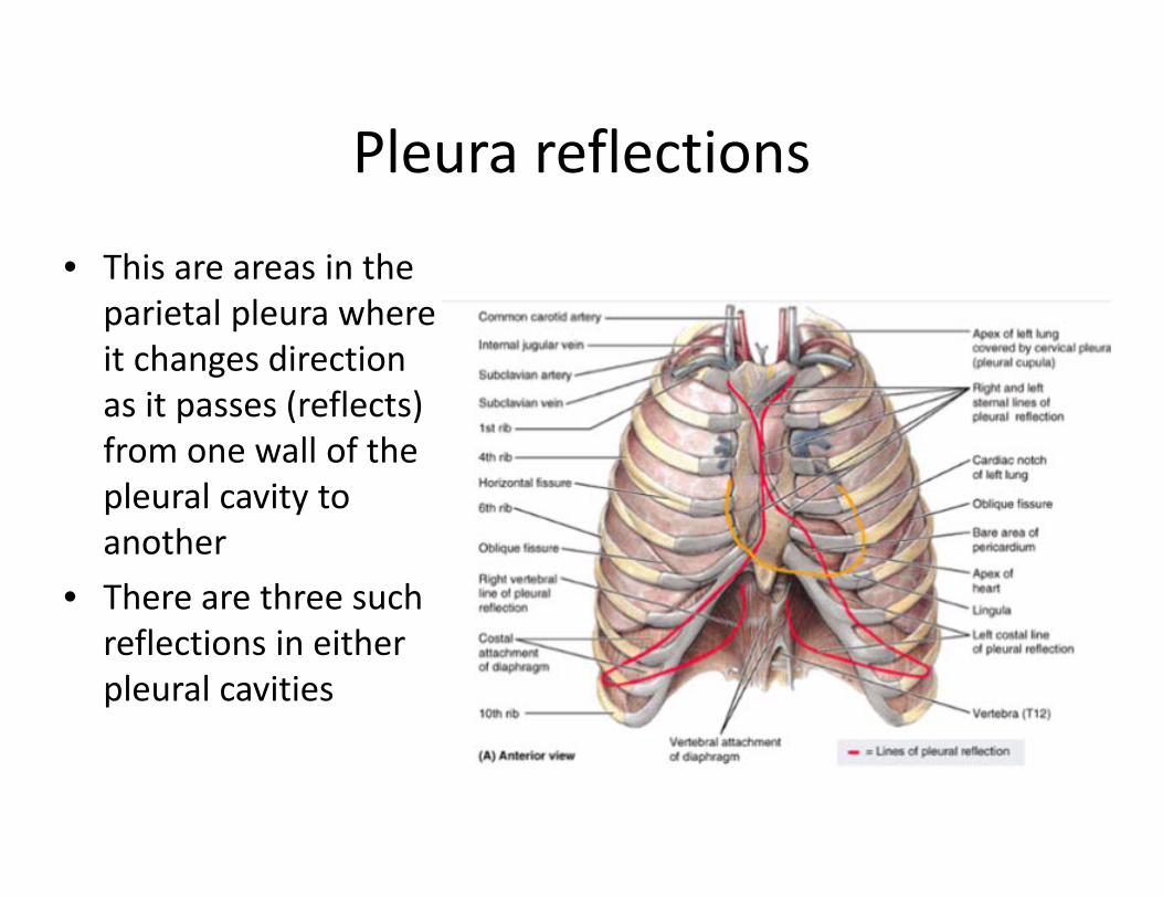
• This are areas in the parietal pleura where it changes direction as it passes (reflects) from one wall of the pleural cavity to another
• There are three such reflections in either pleural cavities
Pleura reflections
• Sternal line of pleural reflection o It is anterior o Occurs where the costal pleura becomes
continuous with the mediastinal pleura o It is sharp and abrupt

Pleura reflections
• Vertebral line of pleural reflection o This is the posterior counterpart of the
sternal line o Occurs where the costal pleura becomes
continuous with the mediastinal pleura posteriorly
o It is a rounder, gradual reflection than the (sharp) anterior reflection
Pleura reflections
• Costal line of pleural reflection • This is also sharp • It occurs where the costal pleura becomes continuous with diaphragmatic pleura inferiorly

Blood supply of the pleura
• Visceral pleura o Arterial supplied is from the branches of the
bronchial and pulmonary arterial systems. o The veins drain to the pulmonary vein.

Blood supply of the pleura • The parietal pleura
o They are supplied by arteries of the structures they cover: ‐ Cervical pleura: intercostal vessels ‐ Costal pleura: intercostal vessels ‐ Diaphragmatic pleura
i. Outer portion: intercostal vessels ii. Inner part : pericardiacophrenic vessels
‐ Mediastinal pleura: pericardiacophrenic vessels

Nerve supply of the pleura • Viscera pleura: No nerve supply • The parietal pleura
o Identical to the vascular supply o They are also supplied by nerves which supply the
structures they cover: ‐ Cervical pleura: intercostal vessel nerves ‐ Costal pleura: intercostal nerves ‐ Diaphragmatic pleura
i. Outer portion: intercostal nerves ii. Inner part : pericardiacophrenic nerves
‐ Mediastinal pleura: pericardiacophrenic nerves
Lymphatic drainage • Viscera
o Drains to the pulmonary plexus located in interlobar and peribronchial spaces.
• Parietal o Coastal, Mediastinal and Cervical pleura: These
drain into the internal thoracic chain anteriorly and intercostal chain posteriorly.
o Diaphragmatic pleura: drains to the retrosternal and mediastinal and (sometimes) the celiac lymph node.