Embed Size (px)
Citation preview

Anatomy, composition and physiology of neuron, dendrite, axon, and synapses


Glial cells/ Astrocyets, Oligodendrocyt , Schwan cells/
Support neuronal cells Produce myelin Act as scavengers Takes up released neurotransmitters Guide migrating neurons and the out growth of axons Form BBB Release growth factor and help nourish nerve cells
Neurons type
Unipolar cells: single process serving as receptor and releasing terminal e.g. In autonomic nervous system
Biplar cells : two process dendrite and axone.g. in retina , olfactory nerves
Pseudo_unipolar cells: e.g. dorsal root ganglia
Multipolar cells : most common typee.g. spinal motor
neurons ,pyramidal cells,pukrinje cells NERONS COULD ALSO BE CLASSIFIED AS :
SENSORY MOTOR
LOCAL INTERNEURONPROJECTION
INTERNEURONNEUROENDOCRINE CELL


Structure of a neuron
Cell body Dendrites:
apical and basal types, are input elements together with the cell body contains nucleus and gives rise to axon
and dendrite
Axons: transmitting element, could be longer than 3m, covered with myelin interrupted by node of Ranvier is the out put element of neuron single axon may form synapses with as many as 100000 neurons
Axon hillock: initial segment of neuron Synapse:
presynaptic terminal, synaptic cleft , postsynaptic membrane
dendrites cell body axon hillock muscle
synaptic connections could be divergent or convergent


Functional organization of neurons
Input component /receptor or synaptic potential/ : signal electrical input signal is graded in amplitude and duration, proportional to amplitude and duration of stimulus
Integrative component : signal electrical action potential is generated only if input signal is greater than
spike threshold stimulus intensity is represented by frequency of action
potential duration of stimulus is represented by number of action
potentials

Functional organization of neurons
Conductile component/action potential/ : Signal is electrical action potentials are all or none every action potentials have same amplitude and duration
information in the signal is represented by frequency and duration Out put component :
signal chemical transmitter total number of action potential determine how much
neurotransmitter should be released


Comparison of local and propagated signals
Input signals
amplitude smallduration brief
summationgraded signal effect depolarizing propagation
passive
Action potential
amplitude largeduration brief
summationall/none signal effect depolarize
propagation
active


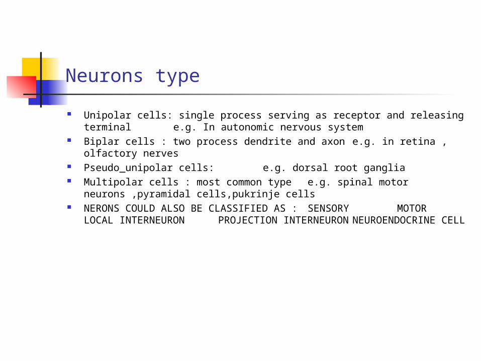
Cytology of Neurons
Nucleus Nuclear envelope Cytoplasm
cytosol including cytoskeletal matrix membranous organelle
Plasmalemma myelin

Membranous organelles
Mitochondria and peroxisomes Rough endoplasmic reticulum/smooth endoplasmic reticulum Golgi complex Secretory vesicles, endosomes, lysosomes Most of these structures are abundant in the cell body and
dendrite and there are no synthetic function differences between cell body and dendrite.
The axon has few mitochondria and smooth endoplasmic reticulum with abundant secratory vesicles

Synthesis and trafficking of neural proteins
Most proteins are synthesized in the cell body The neuron express more the total genetic material than any
other organ Neural cells are engaged in protein synthesis more often than
other cells and hence their chromosomes uncoiled Ribosomal and m RNA are synthesized in the nucleus and
exported through nuclear pore Some genetic information is also contained in the mitochondria Protein synthesis occurs in cytosol where mRNA ribosome and
tRNA form complex Secretory proteins and vacuolar apparatus and plasmalemma
are synthesized and modified in the endoplasmic reticulum Secretory proteins are processed further in the golgi complex
and then transported


cytoskeleton
Microtubules largest diameter fibershelical cylinders
made of protofilaments undergo cycles of
polymerization and depolymerization
Neurofilaments monomers twist to form dimerprotofilamentprotofibrilfilament
Microfilaments polymerized actin monomerssmallest-diameter fibers
undergo cycles of polymerization and depolymerization


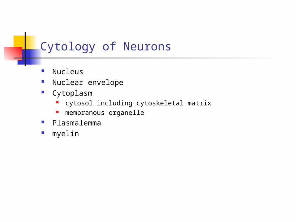
Anterograde Axonal Transport
Fast phase of axonal transport 70-400 mm/d 20-70mm/d 4-20 mm/d Convey mainly plasma membrane proteins such as
acetyl cholinesterase mitochondria multivesicular bodies and secretoty vesicles ATPases
The physiological properties of fast phase axonal transport has been important for tracing connections in the brain
Slow phase of axonal transport 1-4 mm/d 0.2-1.2mm/d Conveys mainly
Neurofilaments and Microtubulins Actin and certain glycolytic enzymes

Retrograde Axonal Transport
Toxins, drugs, heavy metals Neurotropic viruses Nerve Growth Factors and neurotrophines Mitochondria, endosomes

Axonal vs dendrite transport
Axonal Nerofilaments abundant Microtubules are of tau type Microtubules are uniformly
arranged
Dendrite Microtubules abundant MAP-2 type Microtubules are
bidirectionaly arranged These difference in
arrangement explain the
polarization of organelles


myelin
Insulate axons and facilitates speed of action potential transmission
Arranged in concentric bimolecular layers Has a composition similar to plasma membranes Schwan cells form myelin of peripheral nerves and
Oligodendrocyts that of central nerves Schwan cells express their myelin gene in response to contact
with axon while Oligodendrocytes depend also on the presence of Astrocytes


Myelin Proteins
Myelin Basic Protein important for myelin compactionstrongly immunogenicused to produce experimental
allergic encephalomyelitis Myelin Associated glycoprotein supper family of immunoglobulin
involved in cell to cell recognitionis an adhesion molecule that
initiate myelination Protiolipids important for compaction of myelin
mutation in the gene causes hypomyelination and
degeneration Myelin protein zero major protein in peripheral myelin
immunoglobulin familyimportant for myelin compactionmice that lack the protein have poor
motor coordination Peripheral myelin protein 22 encoded by chromosome 17
DNA duplication results in CMT disease

Ion channels
Conduct ions at fast rate Selective for specific ions Ion channels are proteins that span the cell mme Flux of ions through the ion channel is passive Opening and closing of ion channel involves conformational change Open and close in response to specific stimulus
Voltage –gated Ligand –gated Mechanically –gated Gap-junction channels
The binding of exogenous ligands /toxins, poisons and drugs/can make channels open or close
Ion channels are composed of several subunits Channels are also important targets of diseases
myasthenia gravis hyperkalemic periodic paralysis

Synaptic transmission
The average neuron makes 100000 connections Two basic forms of transmission
Chemical Electrical
Electrical transmissions are Short lasting Only excitatory Do not induce long lasting postsynaptic changes Gap junction channels Bidirectional transmission
Chemical transmissions are Variable signaling :inhibitory or excitatory Produce complex behavior Longer lasting / delay in transmission Amplify signals Modify post synaptic receptors both functionally and anatomically Ionotropic receptors :conformational change that opens the channels on binding
transmitter Metabotropic receptors: act by altering intracellular metabolic reaction

Chemical transmitters
Classical Acetylcholine Cathecolamines Glutamates GABA Serotonine histamine
Peptides Substance p Enkephaline Endorphine Prolactin, oxytocine, vasopresin
Soluble gases NO

Cellular basis of connectionist approach
Principle of dynamic polarization : electrical signals within a nerve flow only in one direction
Principle of connectional specificity : nerve cells do not connect indiscriminately with one another to from a network

Specificity and modifiability of neuronal connections
Specific networks Brain has at least two types of neuronal map/ motor and sensory
maps/ which are interconnected with each other by interneuron. The neurons that make up these map do not differ greatly in their electrical properties. Rather, They have different function because of the connections they make.
Parallel processing: deployment of several neuron groups or several pathways to
convey similar information Plasticity :
functional transformation in neurons as a result of appropriate stimulation
How dose nerve cells differ ?
Lack of axon Location of synaptic in puts on the cell
cell body dendrite axon hillock type of target cell
Difference in cell body size and shape, distribution of axon and dendrite tree
Expressing different combination of ion channels providing them with different thresholds ,excitability and firing
patterns. Thus ,neurons with different ion channels encode the same class of
synaptic potential into different firing patterns and thereby convey different signals

Cont.
Chemical transmitter The type of neurotransmitter they use The type of Receptors they have
Myelin content Location in the nervous system –central/peripheral These differences and others may account for different
patterns of disease