Embed Size (px)
Citation preview

191
11Respiratory SystemRhonda M. Jones 11Respiratory SystemRhonda M. Jones
GLOSSARY TERMS
● asthma● bradypnea● bronchitis● bronchophony● chronic obstructive pulmonary disease● crackles● cyanosis● dyspnea● egophony● emphysema● friction rub● hyperpnea● hyperresonance● hypoxemia● orthopnea● pallor● paroxysmal nocturnal dyspnea● pneumonia● resonance● rhonchi● tachypnea● tactile fremitus● wheezes● whispered pectoriloquy
ANATOMY AND PHYSIOLOGY OVERVIEWThe primary function of the respiratory system is to transport airinto and out of the lungs so that oxygen can be exchanged for car-bon dioxide. The upper respiratory system includes the nose,nasal cavity, sinuses, and pharynx. The lower respiratory systemincludes the trachea, bronchi, and lungs (Fig. 11–1). In this chap-ter, only the lower respiratory system is discussed. (For a discus-sion of the upper respiratory system, see Chapter 10.)
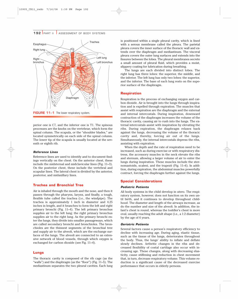
The thoracic cage, or the bones of the chest, consists of 12 tho-racic vertebrae, 12 pairs of ribs, and the sternum (Fig. 11–2). Theribs and the sternum form the rib cage and support the thoraciccavity. The spaces between the ribs are termed the intercostalsspaces and are numbered according to the superior rib above(e.g., the second intercostal space is located below the secondrib). The diaphragm is a muscle that separates the thoracic cavityfrom the abdomen and is used during inspiration.
Surface Landmarks
Surface landmarks of the thorax are useful in identifying the un-derlying internal structures and in describing physical findings.They also facilitate documentation and communication of physi-cal findings to other healthcare professionals.
Anterior Thoracic Landmarks
Primary anterior thoracic landmarks include the suprasternalnotch, sternum, and manubriosternal angle. The suprasternalnotch is the U-shaped depression at the top of the sternum be-tween the clavicles. The sternum, or “breastbone,” consists of themanubrium, the body, and the xiphoid process. The articulationbetween the manubrium and the body of the sternum is themanubriosternal angle, which is commonly referred to as the an-gle of Louis. The angle of Louis is continuous with the second riband is a useful place to start counting the ribs. It is also useful inlocating the underlying structures, because the trachea bifurcatesinto the right and left main bronchi just under the angle of Louis.
Posterior Thoracic Landmarks
Posterior thoracic landmarks include the vertebra prominens,spinous processes, and scapula. The vertebra prominens is theseventh cervical vertebra and is found as the bony spur that pro-trudes from the base of the neck when the neck is flexed anteri-orly. If two vertebra are observed when the neck is flexed, the su-
10905_CH11_redo 7/16/08 1:39 PM Page 191
192 PART I I ASSESSMENT OF BODY SYSTEMS
perior one is C7, and the inferior one is T1. The spinousprocesses are the knobs on the vertebrae, which form thespinal column. The scapula, or the “shoulder blades,” arelocated symmetrically on each side of the spinal column.The lower tip of the scapula is usually located at the sev-enth or eighth rib.
Reference Lines
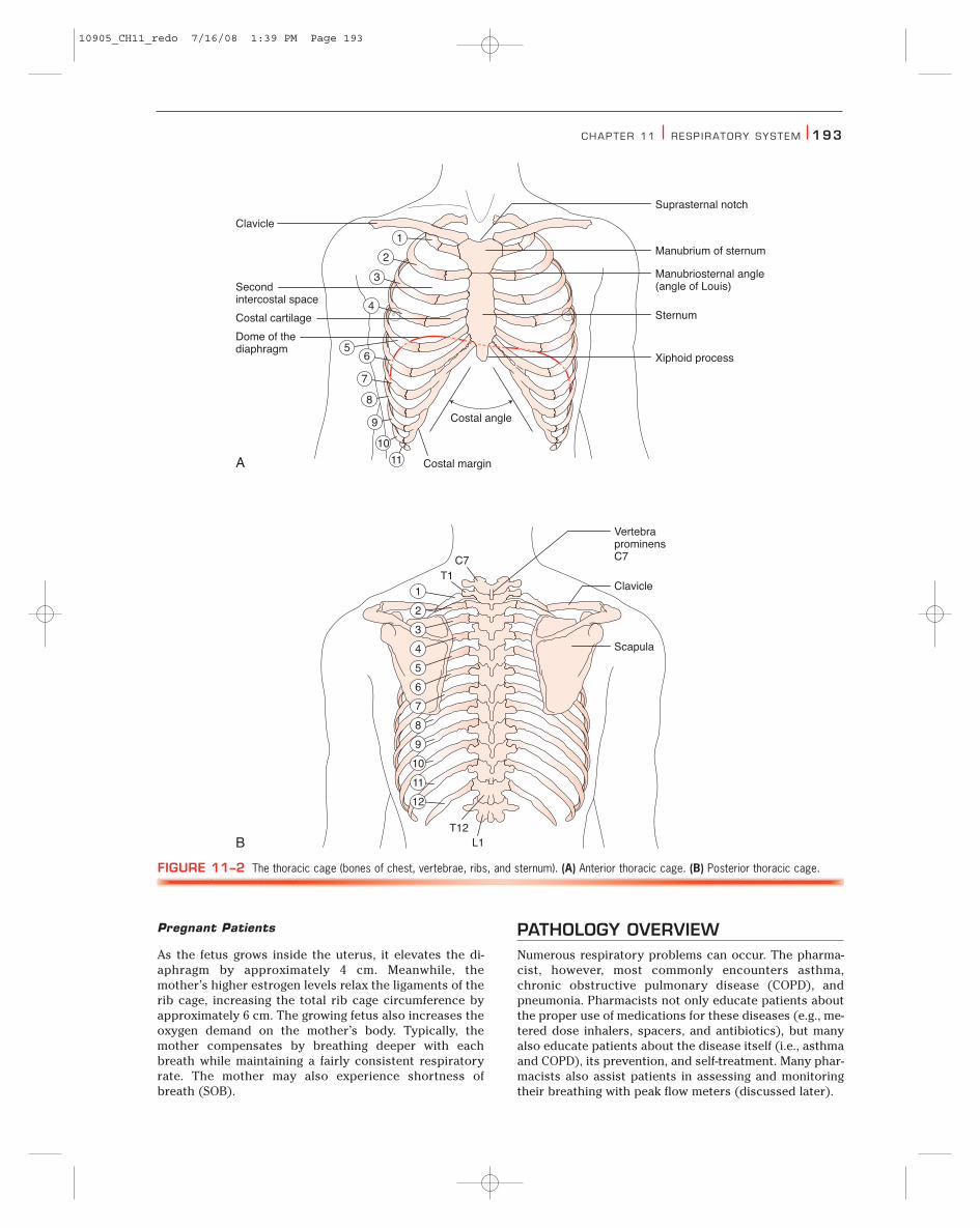
Reference lines are used to identify and to document find-ings vertically on the chest. On the anterior chest, theseinclude the midsternal and midclavicular lines (Fig. 11–3).On the posterior chest, these include the vertebral andscapular lines. The lateral chest is divided by the anterior,posterior, and midaxillary lines.
Trachea and Bronchial Tree
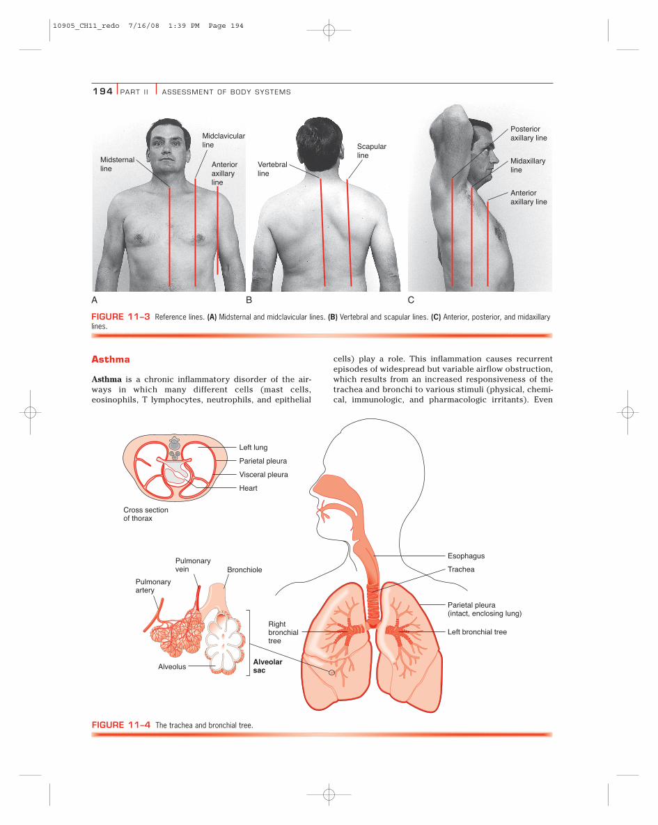
Air is inhaled through the mouth and the nose, and then itpasses through the pharynx, larynx, and finally, a tough,flexible tube called the trachea (i.e., the windpipe). Thetrachea is approximately 1 inch in diameter and 4.25inches in length, and it branches to form the left and rightprimary bronchi (Fig. 11–4). The left primary bronchussupplies air to the left lung; the right primary bronchussupplies air to the right lung. As the primary bronchi en-ter the lungs, they divide into smaller passageways, whichare called secondary bronchi and bronchioles. The bron-chioles are the thinnest segments of the bronchial treeand supply air to the alveoli, which are the exchange sur-faces of the lungs. The alveoli are connected to an exten-sive network of blood vessels, through which oxygen isexchanged for carbon dioxide (see Fig. 11–4).
Lungs
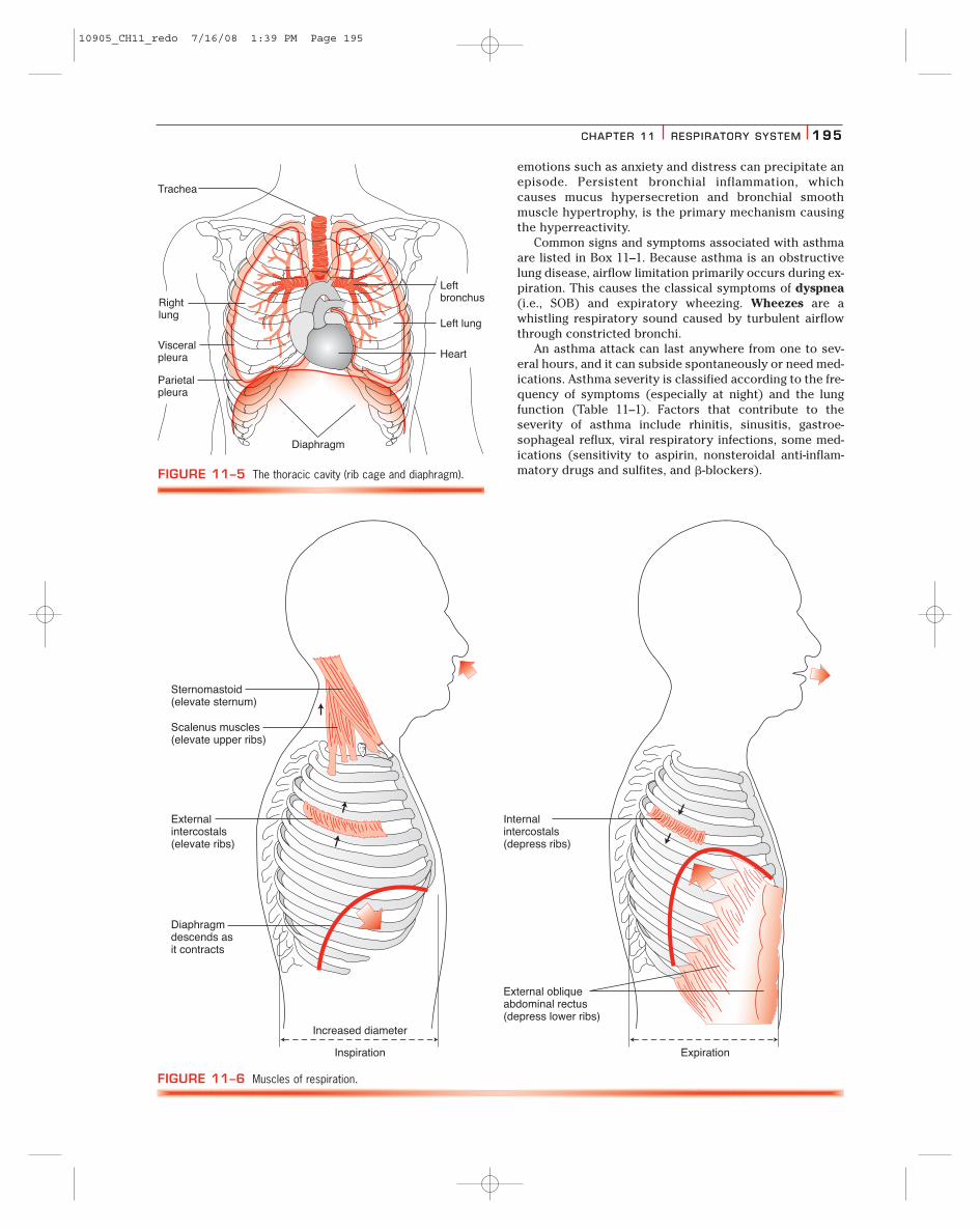
The thoracic cavity is composed of the rib cage (as the“walls”) and the diaphragm (as the “floor”) (Fig. 11–5). Themediastinum separates the two pleural cavities. Each lung
is positioned within a single pleural cavity, which is linedwith a serous membrane called the pleura. The parietalpleura covers the inner surface of the thoracic wall and ex-tends over the diaphragm and mediastinum. The visceralpleura covers the outer lung surfaces and extends into thefissures between the lobes. The pleural membranes secretea small amount of pleural fluid, which provides a moist,slippery coating for lubrication during breathing.
The lungs are each divided into distinct lobes. Theright lung has three lobes: the superior, the middle, andthe inferior. The left lung has only two lobes: the superior,and the inferior. The base of each lung rests on the supe-rior surface of the diaphragm.
Respiration
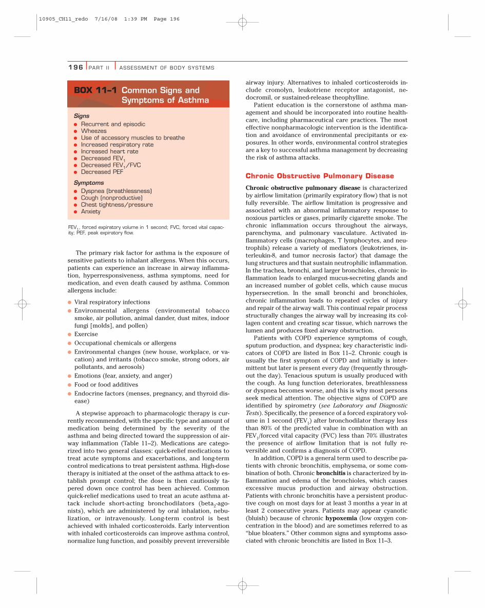
Respiration is the process of exchanging oxygen and car-bon dioxide. Air is brought into the lungs through inspira-tion and is expelled through expiration. The muscles thatassist with respiration are the diaphragm and the externaland internal intercostals. During inspiration, downwardcontraction of the diaphragm increases the volume of thethoracic cavity, causing air to rush into the lungs. The ex-ternal intercostals assist with inspiration by elevating theribs. During expiration, the diaphragm relaxes backagainst the lungs, decreasing the volume of the thoraciccavity and, thereby, forcing air out of the lungs.Simultaneously, the internal intercostals depress the ribs,assisting with expiration.
When the depth and the rate of respiration need to beincreased, such as during exercise or with respiratory dis-tress, the accessory muscles in the neck elevate the ribsand sternum, allowing a larger volume of air to enter thelungs during inspiration. These muscles include the ster-nomastoids, scaleni, and the trapezii (Fig. 11–6). In addi-tion, during expiration, the abdominal muscles powerfullycontract, forcing the diaphragm further against the lungs.
Special Considerations
Pediatric Patients
All body systems in the child develop in utero. The respi-ratory system, however, does not function on its own un-til birth, and it continues to develop throughout child-hood. The diameter and length of the airways increase, asdo the number and size of the alveoli. In addition, the in-fant’s chest is round, whereas the toddler’s chest is moreoval, usually reaching the adult shape (i.e., a 1:2 diameter)by the age of 6 years.
Geriatric Patients
Several factors cause a person’s respiratory efficiency todecline with increasing age. During aging, elastic tissue,such as the tissue of the lungs, deteriorates throughoutthe body. Thus, the lungs’ ability to inflate and deflateslowly declines. Arthritic changes in the ribs and de-creased flexibility of costal cartilage also occur with in-creasing age. These changes, along with decreasing elas-ticity, cause stiffening and reduction in chest movementthat, in turn, decrease respiratory volume. This volume re-duction is a significant cause of the decreased exerciseperformance that occurs in elderly persons.
Right lung
EsophagusTrachea
Left bronchus
Diaphragm
Left lungRightbronchus
FIGURE 11–1 The lower respiratory system.
10905_CH11_redo 7/16/08 1:39 PM Page 192
CHAPTER 11 RESP IRATORY SYSTEM 193
Pregnant Patients
As the fetus grows inside the uterus, it elevates the di-aphragm by approximately 4 cm. Meanwhile, themother’s higher estrogen levels relax the ligaments of therib cage, increasing the total rib cage circumference byapproximately 6 cm. The growing fetus also increases theoxygen demand on the mother’s body. Typically, themother compensates by breathing deeper with eachbreath while maintaining a fairly consistent respiratoryrate. The mother may also experience shortness ofbreath (SOB).
PATHOLOGY OVERVIEWNumerous respiratory problems can occur. The pharma-cist, however, most commonly encounters asthma,chronic obstructive pulmonary disease (COPD), andpneumonia. Pharmacists not only educate patients aboutthe proper use of medications for these diseases (e.g., me-tered dose inhalers, spacers, and antibiotics), but manyalso educate patients about the disease itself (i.e., asthmaand COPD), its prevention, and self-treatment. Many phar-macists also assist patients in assessing and monitoringtheir breathing with peak flow meters (discussed later).
11
C7
VertebraprominensC7
Clavicle
Scapula
T1
T12L1
10
9
8
7
65
4
3
2
1
12
11
10
9
8
7
6
5
4
3
2
1
Clavicle
Suprasternal notch
Manubrium of sternum
Manubriosternal angle(angle of Louis)
Sternum
Xiphoid process
Secondintercostal space
Costal cartilage
Costal margin
Costal angle
Dome of thediaphragm
A
B
FIGURE 11–2 The thoracic cage (bones of chest, vertebrae, ribs, and sternum). (A) Anterior thoracic cage. (B) Posterior thoracic cage.
10905_CH11_redo 7/16/08 1:39 PM Page 193
194 PART I I ASSESSMENT OF BODY SYSTEMS
Asthma
Asthma is a chronic inflammatory disorder of the air-ways in which many different cells (mast cells,eosinophils, T lymphocytes, neutrophils, and epithelial
cells) play a role. This inflammation causes recurrentepisodes of widespread but variable airflow obstruction,which results from an increased responsiveness of thetrachea and bronchi to various stimuli (physical, chemi-cal, immunologic, and pharmacologic irritants). Even
A B C
Midsternalline
Midclavicularline
Anterioraxillaryline
Anterioraxillary line
Posterioraxillary line
Midaxillaryline
Vertebralline
Scapularline
FIGURE 11–3 Reference lines. (A) Midsternal and midclavicular lines. (B) Vertebral and scapular lines. (C) Anterior, posterior, and midaxillarylines.
Esophagus
Trachea
Rightbronchialtree
Left bronchial tree
Parietal pleura(intact, enclosing lung)
AlveolusAlveolarsac
Pulmonaryvein
Heart
Visceral pleura
Cross sectionof thorax
Parietal pleura
Left lung
Bronchiole
Pulmonaryartery
FIGURE 11–4 The trachea and bronchial tree.
10905_CH11_redo 7/16/08 1:39 PM Page 194
CHAPTER 11 RESP IRATORY SYSTEM 195
emotions such as anxiety and distress can precipitate anepisode. Persistent bronchial inflammation, whichcauses mucus hypersecretion and bronchial smoothmuscle hypertrophy, is the primary mechanism causingthe hyperreactivity.
Common signs and symptoms associated with asthmaare listed in Box 11–1. Because asthma is an obstructivelung disease, airflow limitation primarily occurs during ex-piration. This causes the classical symptoms of dyspnea(i.e., SOB) and expiratory wheezing. Wheezes are awhistling respiratory sound caused by turbulent airflowthrough constricted bronchi.
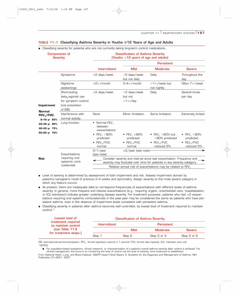
An asthma attack can last anywhere from one to sev-eral hours, and it can subside spontaneously or need med-ications. Asthma severity is classified according to the fre-quency of symptoms (especially at night) and the lungfunction (Table 11–1). Factors that contribute to theseverity of asthma include rhinitis, sinusitis, gastroe-sophageal reflux, viral respiratory infections, some med-ications (sensitivity to aspirin, nonsteroidal anti-inflam-matory drugs and sulfites, and �-blockers).
CHAPTER 11 RESP IRATORY SYSTEM 195
Rightlung
Trachea
Leftbronchus
Left lung
Heart
Parietalpleura
Visceralpleura
Diaphragm
FIGURE 11–5 The thoracic cavity (rib cage and diaphragm).
Increased diameter
Inspiration Expiration
Sternomastoid(elevate sternum)
Scalenus muscles(elevate upper ribs)
Externalintercostals(elevate ribs)
Internalintercostals(depress ribs)
External obliqueabdominal rectus(depress lower ribs)
Diaphragmdescends asit contracts
FIGURE 11–6 Muscles of respiration.
10905_CH11_redo 7/16/08 1:39 PM Page 195
196 PART I I ASSESSMENT OF BODY SYSTEMS
The primary risk factor for asthma is the exposure ofsensitive patients to inhalant allergens. When this occurs,patients can experience an increase in airway inflamma-tion, hyperresponsiveness, asthma symptoms, need formedication, and even death caused by asthma. Commonallergens include:
● Viral respiratory infections● Environmental allergens (environmental tobacco
smoke, air pollution, animal dander, dust mites, indoorfungi [molds], and pollen)
● Exercise● Occupational chemicals or allergens● Environmental changes (new house, workplace, or va-
cation) and irritants (tobacco smoke, strong odors, airpollutants, and aerosols)
● Emotions (fear, anxiety, and anger)● Food or food additives● Endocrine factors (menses, pregnancy, and thyroid dis-
ease)
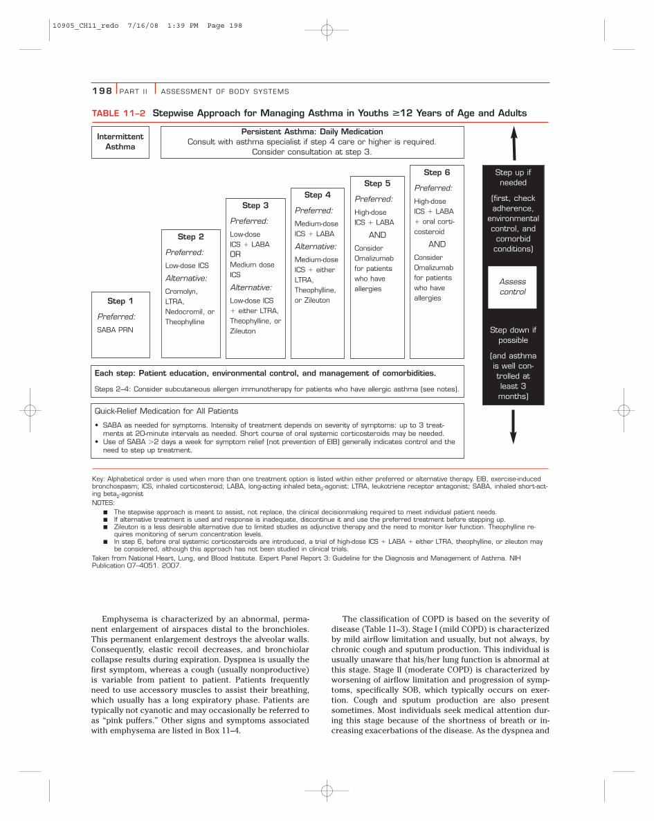
A stepwise approach to pharmacologic therapy is cur-rently recommended, with the specific type and amount ofmedication being determined by the severity of theasthma and being directed toward the suppression of air-way inflammation (Table 11–2). Medications are catego-rized into two general classes: quick-relief medications totreat acute symptoms and exacerbations, and long-termcontrol medications to treat persistent asthma. High-dosetherapy is initiated at the onset of the asthma attack to es-tablish prompt control; the dose is then cautiously ta-pered down once control has been achieved. Commonquick-relief medications used to treat an acute asthma at-tack include short-acting bronchodilators (beta2-ago-nists), which are administered by oral inhalation, nebu-lization, or intravenously. Long-term control is bestachieved with inhaled corticosteroids. Early interventionwith inhaled corticosteroids can improve asthma control,normalize lung function, and possibly prevent irreversible
airway injury. Alternatives to inhaled corticosteroids in-clude cromolyn, leukotriene receptor antagonist, ne-docromil, or sustained-release theophylline.
Patient education is the cornerstone of asthma man-agement and should be incorporated into routine health-care, including pharmaceutical care practices. The mosteffective nonpharmacologic intervention is the identifica-tion and avoidance of environmental precipitants or ex-posures. In other words, environmental control strategiesare a key to successful asthma management by decreasingthe risk of asthma attacks.
Chronic Obstructive Pulmonary Disease
Chronic obstructive pulmonary disease is characterizedby airflow limitation (primarily expiratory flow) that is notfully reversible. The airflow limitation is progressive andassociated with an abnormal inflammatory response tonoxious particles or gases, primarily cigarette smoke. Thechronic inflammation occurs throughout the airways,parenchyma, and pulmonary vasculature. Activated in-flammatory cells (macrophages, T lymphocytes, and neu-trophils) release a variety of mediators (leukotrienes, in-terleukin-8, and tumor necrosis factor) that damage thelung structures and that sustain neutrophilic inflammation.In the trachea, bronchi, and larger bronchioles, chronic in-flammation leads to enlarged mucus-secreting glands andan increased number of goblet cells, which cause mucushypersecretion. In the small bronchi and bronchioles,chronic inflammation leads to repeated cycles of injuryand repair of the airway wall. This continual repair processstructurally changes the airway wall by increasing its col-lagen content and creating scar tissue, which narrows thelumen and produces fixed airway obstruction.
Patients with COPD experience symptoms of cough,sputum production, and dyspnea; key characteristic indi-cators of COPD are listed in Box 11–2. Chronic cough isusually the first symptom of COPD and initially is inter-mittent but later is present every day (frequently through-out the day). Tenacious sputum is usually produced withthe cough. As lung function deteriorates, breathlessnessor dyspnea becomes worse, and this is why most personsseek medical attention. The objective signs of COPD areidentified by spirometry (see Laboratory and DiagnosticTests). Specifically, the presence of a forced expiratory vol-ume in 1 second (FEV1) after bronchodilator therapy lessthan 80% of the predicted value in combination with anFEV1/forced vital capacity (FVC) less than 70% illustratesthe presence of airflow limitation that is not fully re-versible and confirms a diagnosis of COPD.
In addition, COPD is a general term used to describe pa-tients with chronic bronchitis, emphysema, or some com-bination of both. Chronic bronchitis is characterized by in-flammation and edema of the bronchioles, which causesexcessive mucus production and airway obstruction.Patients with chronic bronchitis have a persistent produc-tive cough on most days for at least 3 months a year in atleast 2 consecutive years. Patients may appear cyanotic(bluish) because of chronic hypoxemia (low oxygen con-centration in the blood) and are sometimes referred to as“blue bloaters.” Other common signs and symptoms asso-ciated with chronic bronchitis are listed in Box 11–3.
Signs● Recurrent and episodic● Wheezes● Use of accessory muscles to breathe● Increased respiratory rate● Increased heart rate● Decreased FEV1● Decreased FEV1/FVC● Decreased PEF
Symptoms● Dyspnea (breathlessness)● Cough (nonproductive)● Chest tightness/pressure● Anxiety
FEV1, forced expiratory volume in 1 second; FVC, forced vital capac-ity; PEF, peak expiratory flow.
BOX 11–1 Common Signs andSymptoms of Asthma
10905_CH11_redo 7/16/08 1:39 PM Page 196
CHAPTER 11 RESP IRATORY SYSTEM 197
TABLE 11–1 Classifying Asthma Severity in Youths ��12 Years of Age and Adults
� Classifying severity for patients who are not currently taking long-term control medications.
Components of Classification of Asthma SeveritySeverity (Youths �12 years of age and adults)
Persistent
Intermittent Mild Moderate Severe
Symptoms �2 days/week �2 days/week Daily Throughout the
but not daily day
Nighttime �2�/month 3–4�/month �1�/week but Often 7�/week
awakenings not nightly
Short-acting �2 days/week �2 days/week Daily Several times
beta2-agonist use but not per day
for symptom control �1�/day
Impairment (not prevention
of EIB)
Interference with None Minor limitation Some limitation Extremely limited
normal activity
Lung function • Normal FEV1betweenexacerbations
• FEV1 �80% • FEV1 �80% • FEV1 �60% but • FEV1 �60%predicted predicted �80% predicted predicted
• FEV1/FVC • FEV1/FVC • FEV1/FVC • FEV1/FVCnormal normal reduced 5% reduced 5%
0–1/year �2/year (see note)(see note)
Risk Consider severity and interval since last exacerbation. Frequency andseverity may fluctuate over time for patients in any severity category.
Relative annual risk of exacerbations may be related to FEV1
� Level of severity is determined by assessment of both impairment and risk. Assess impairment domain bypatient’s/caregiver’s recall of previous 2–4 weeks and spirometry. Assign severity to the most severe category inwhich any feature occurs.
� At present, there are inadequate data to correspond frequencies of exacerbations with different levels of asthmaseverity. In general, more frequent and intense exacerbations (e.g., requiring urgent, unscheduled care, hospitalization,or ICU admission) indicate greater underlying disease severity. For treatment purposes, patients who had �2 exacer-bations requiring oral systemic corticosteroids in the past year may be considered the same as patients who have per-sistent asthma, even in the absence of impairment levels consistent with persistent asthma.
� Classifying severity in patients after asthma becomes well controlled, by lowest level of treatment required to maintaincontrol.*
Classification of Asthma Severity
Intermittent Persistent
Mild Moderate Severe
Step 1 Step 2 Step 3 or 4 Step 5 or 6
EIB, exercise-induced bronchospasm; FEV1, forced expiratory volume in 1 second; FVC, forced vital capacity; ICU, intensive care unit*NOTES:
� For population-based evaluations, clinical research, or characterization of a patient’s overall asthma severity after control is achieved. Forclinical management, the focus is on monitoring the level of control not the level of severity, once treatment is established.
From National Heart, Lung, and Blood Institute. NAEPP Expert Panel Report 3: Guideline for the Diagnosis and Management of Asthma. NIHPublication 07–4051. 2007.
NormalFEV1/FVC:8–19 yr 85%
20–39 yr 80%
40–59 yr 75%
60–80 yr 70%
Exacerbationsrequiring oralsystemic corti-costeroids
Lowest level of treatment required to maintain control
(see Table 11-2 for treatment steps.)
10905_CH11_redo 7/16/08 1:39 PM Page 197
198 PART I I ASSESSMENT OF BODY SYSTEMS
Emphysema is characterized by an abnormal, perma-nent enlargement of airspaces distal to the bronchioles.This permanent enlargement destroys the alveolar walls.Consequently, elastic recoil decreases, and bronchiolarcollapse results during expiration. Dyspnea is usually thefirst symptom, whereas a cough (usually nonproductive)is variable from patient to patient. Patients frequentlyneed to use accessory muscles to assist their breathing,which usually has a long expiratory phase. Patients aretypically not cyanotic and may occasionally be referred toas “pink puffers.” Other signs and symptoms associatedwith emphysema are listed in Box 11–4.
The classification of COPD is based on the severity ofdisease (Table 11–3). Stage I (mild COPD) is characterizedby mild airflow limitation and usually, but not always, bychronic cough and sputum production. This individual isusually unaware that his/her lung function is abnormal atthis stage. Stage II (moderate COPD) is characterized byworsening of airflow limitation and progression of symp-toms, specifically SOB, which typically occurs on exer-tion. Cough and sputum production are also presentsometimes. Most individuals seek medical attention dur-ing this stage because of the shortness of breath or in-creasing exacerbations of the disease. As the dyspnea and
TABLE 11–2 Stepwise Approach for Managing Asthma in Youths ��12 Years of Age and Adults
Key: Alphabetical order is used when more than one treatment option is listed within either preferred or alternative therapy. EIB, exercise-inducedbronchospasm; ICS, inhaled corticosteroid; LABA, long-acting inhaled beta2-agonist; LTRA, leukotriene receptor antagonist; SABA, inhaled short-act-ing beta2-agonistNOTES:
� The stepwise approach is meant to assist, not replace, the clinical decisionmaking required to meet individual patient needs.� If alternative treatment is used and response is inadequate, discontinue it and use the preferred treatment before stepping up.� Zileuton is a less desirable alternative due to limited studies as adjunctive therapy and the need to monitor liver function. Theophylline re-
quires monitoring of serum concentration levels.� In step 6, before oral systemic corticosteroids are introduced, a trial of high-dose ICS � LABA � either LTRA, theophylline, or zileuton may
be considered, although this approach has not been studied in clinical trials.Taken from National Heart, Lung, and Blood Institute. Expert Panel Report 3: Guideline for the Diagnosis and Management of Asthma. NIHPublication 07–4051. 2007.
IntermittentAsthma
Persistent Asthma: Daily MedicationConsult with asthma specialist if step 4 care or higher is required.
Consider consultation at step 3.
Each step: Patient education, environmental control, and management of comorbidities.
Steps 2–4: Consider subcutaneous allergen immunotherapy for patients who have allergic asthma (see notes).
Quick-Relief Medication for All Patients
• SABA as needed for symptoms. Intensity of treatment depends on severity of symptoms: up to 3 treat-ments at 20-minute intervals as needed. Short course of oral systemic corticosteroids may be needed.
• Use of SABA �2 days a week for symptom relief (not prevention of EIB) generally indicates control and theneed to step up treatment.
Step 2
Preferred:
Low-dose ICS
Alternative:
Cromolyn,LTRA,Nedocromil, orTheophylline
Step 3
Preferred:
Low-doseICS � LABAORMedium doseICS
Alternative:
Low-dose ICS� either LTRA,Theophylline, orZileuton
Step 4
Preferred:
Medium-doseICS � LABA
Alternative:
Medium-doseICS � eitherLTRA,Theophylline,or Zileuton
Step 5
Preferred:
High-doseICS � LABA
AND
ConsiderOmalizumabfor patientswho have allergies
Step 6
Preferred:
High-doseICS � LABA� oral corti-costeroid
AND
ConsiderOmalizumabfor patientswho have allergies
Step up ifneeded
(first, checkadherence,
environmentalcontrol, andcomorbid conditions)
Step down ifpossible
(and asthmais well con-trolled atleast 3months)
Step 1
Preferred:
SABA PRN
Assesscontrol
10905_CH11_redo 7/16/08 1:39 PM Page 198
CHAPTER 11 RESP IRATORY SYSTEM 199
exacerbations increase, the patient’s quality of life beginsto be affected. Stage III (severe COPD) is characterized bysevere airflow limitation, greater shortness of breath, re-duced exercise capacity, fatigue, and repeated exacerba-tions that almost always have an impact on the patient’squality of life. Stage IV (very severe COPD) is character-
ized by severe airflow limitation and respiratory failure.Patients may also have clinical signs of cor pulmonale(right heart failure) including elevation of the jugular ve-nous pressure and pitting ankle edema. At this stage, qual-ity of life is significantly impaired and exacerbations mayby life-threatening.
Risk factors for COPD include both genetic factors (de-ficiency of a1-antitrypsin and airway hyperresponsive-ness) and environmental exposures. By far, cigarettesmoking is the most significant environmental exposurefor the development of COPD. Other environmental riskfactors include air pollution and heavy exposure to occu-
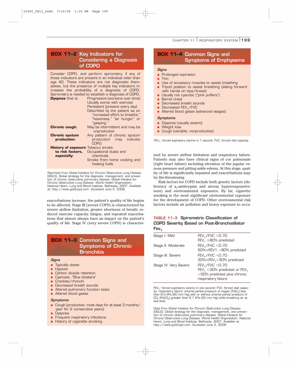
TABLE 11–3 Spirometric Classification ofCOPD Severity Based on Post-BronchodilatorFev1
Stage I: Mild FEV1/FVC �0.70FEV1 �80% predicted
Stage II: Moderate FEV1/FVC �0.7050%�FEV1 �80% predicted
Stage III: Severe FEV1/FVC �0.7030%�FEV1�50% predicted
Stage IV: Very Severe FEV1/FVC �0.70FEV1 �30% predicted or FEV1�50% predicted plus chronicrespiratory failure
FEV1: forced expiratory volume in one second; FVC: forced vital capac-ity; respiratory failure: arterial partial pressure of oxygen (PaO2) lessthan 8.0 kPa (60 mm Hg) with or without arterial partial pressure ofCO2 (PaCO2) greater than 6.7 kPa (50 mm Hg) while breathing air atsea level.
Data from Global Initiative for Chronic Obstructive Lung Disease(GOLD). Global strategy for the diagnosis, management, and preven-tion of chronic obstructive pulmonary disease. Global Initiative forChronic Obstructive Lung Disease, World Health Organization, NationalHeart, Lung and Blood Institute. Bethesda, 2007. Available at:http://www.goldcopd.com. Accessed June 2, 2008.
Consider COPD, and perform spirometry, if any ofthese indicators are present in an individual older thanage 40. These indicators are not diagnostic them-selves, but the presence of multiple key indicators in-creases the probability of a diagnosis of COPD.Spirometry is needed to establish a diagnosis of COPD.Dyspnea that is: Progressive (worsens over time)
Usually worse with exercisePersistent (present every day)Described by the patient as an
“increased effort to breathe,”“heaviness,” “air hunger,” or“gasping.”
Chronic cough: May be intermittent and may beunproductive
Chronic sputum Any pattern of chronic sputumproduction: production may indicate
COPDHistory of exposure Tobacco smoke
to risk factors, Occupational dusts and especially: chemicals
Smoke from home cooking andheating fuels
BOX 11–2 Key Indicators forConsidering a Diagnosisof COPD
Reprinted from Global Initiative for Chronic Obstructive Lung Disease(GOLD). Global strategy for the diagnosis, management, and preven-tion of chronic obstructive pulmonary disease. Global Initiative forChronic Obstructive Lung Disease, World Health Organization,National Heart, Lung and Blood Institute. Bethesda, 2007. Availableat: http://www.goldcopd.com. Accessed June 2, 2008.
Signs● Typically obese● Hypoxia● Carbon dioxide retention● Cyanosis; “Blue bloaters”● Crackles/rhonchi● Decreased breath sounds● Altered pulmonary function tests● Altered blood gases
Symptoms● Cough (productive; most days for at least 3 months/
year for 2 consecutive years)● Dyspnea● Frequent respiratory infections● History of cigarette smoking
BOX 11–3 Common Signs andSymptoms of ChronicBronchitis
Signs● Prolonged expiration● Thin● Use of accessory muscles to assist breathing● Tripod position to assist breathing (sitting forward
with hands on hips/knees)● Usually not cyanotic (“pink puffers”)● Barrel chest● Decreased breath sounds● Decreased FEV1/FVC● Altered blood gases (advanced stages)
Symptoms● Dyspnea (usually severe)● Weight loss● Cough (variable; nonproductive)
FEV1, forced expiratory volume in 1 second; FVC, forced vital capacity.
BOX 11–4 Common Signs andSymptoms of Emphysema
10905_CH11_redo 7/16/08 1:39 PM Page 199
200 PART I I ASSESSMENT OF BODY SYSTEMS
pational dusts and chemicals (e.g., grain, coal, and as-bestos).
The overall approach to managing stable COPD is indi-vidualization of therapy to address symptoms and im-prove quality of life. Treatment is usually a stepwise in-crease in pharmacologic therapy based on the severity ofdisease (Table 11–4). Individualized assessment of diseaseseverity as well as response to various therapies is a keymanagement strategy. Pharmacologic therapy is used toprevent and to control symptoms, to reduce the fre-quency of exacerbations, and to improve exercise/activitytolerance. Unfortunately, no existing medication has beenshown to modify the long-term decline in lung function.Bronchodilator medications are central to the sympto-matic management of COPD. These include beta2-agonists,anticholinergics and methylxanthines used singly or incombination and are used on an as-needed or scheduledbasis depending on the severity of the COPD. Regulartreatment with long-acting bronchodilators is more effec-tive and convenient than treatment with short-actingbronchodilators. The addition of regular treatment withinhaled glucocorticosteroids to bronchodilator treatmentis appropriate for symptomatic Stage III or IV COPD pa-tients. Scheduled treatments with inhaled steroids are re-served for symptomatic patients with a documentedspirometric response to their use or for those with anFEV1 less than 50% of the predicted value and repeated ex-acerbations that require treatment with antibiotics, oralglucocorticosteroids, or both. Chronic treatment with oralglucocorticosteroids is not recommended because of un-favorable side effects and no evidence of long-term bene-fit from their use. Other pharmacologic agents used for
symptom control include antibiotics for infectious exacer-bations as well as influenza and pneumococcal vaccines.
Nonpharmacologic prevention and treatment includespatient education, smoking cessation, avoidance of envi-ronmental factors, exercise training, and oxygen therapy.Patient education is a key component in the managementof COPD. Smoking cessation is the single most effective in-tervention to reduce the risk of developing COPD and tostop its progression. The numerous products availableover the counter (OTC) present pharmacists with an idealopportunity to have a positive impact on patient care byplaying an integral part in smoking cessation.
Pneumonia
Pneumonia is an inflammation of the lungs that is mostcommonly caused by a community-acquired bacterial in-fection, Streptococcus pneumoniae, which is also generallyreferred to as pneumococcal pneumonia. Other bacterialpathogens of community- and hospital-acquired pneumo-nia are listed in Box 11–5. The infection causes interalveo-lar exudation (slow release of fluid containing proteinsand white blood cells) that results in consolidation or so-lidification of the lungs. Typically, the consolidation isconfined to one lobe (e.g., right lower lobe pneumonia).Risk factors for developing pneumonia include:
● Age (elderly and infants)● Smoking● Chronic bronchitis● Chronic illness (e.g., congestive heart failure [CHF], di-
abetes, and COPD)
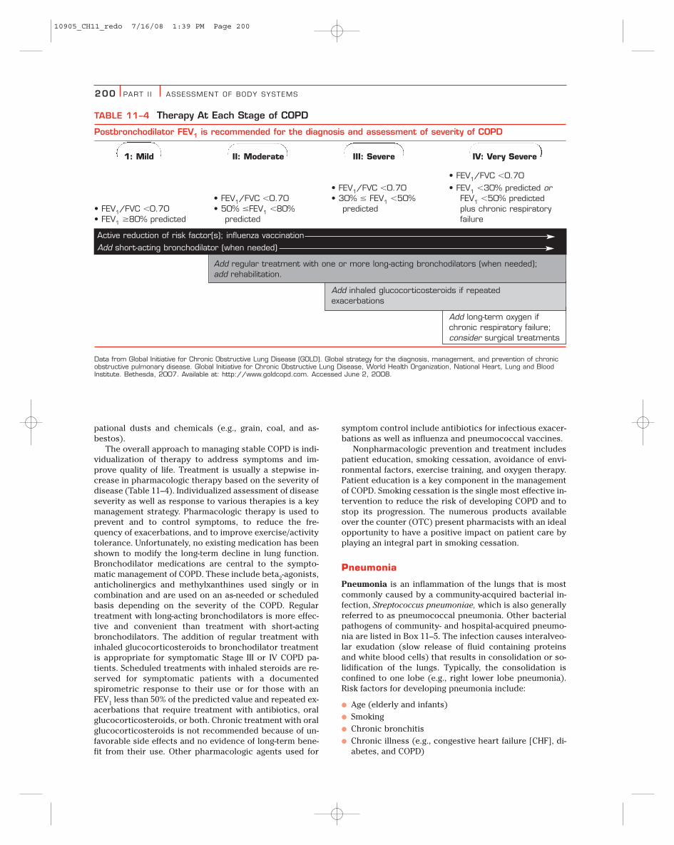
TABLE 11–4 Therapy At Each Stage of COPD
Postbronchodilator FEV1 is recommended for the diagnosis and assessment of severity of COPD
1: Mild II: Moderate III: Severe IV: Very Severe
• FEV1/FVC �0.70
• FEV1/FVC �0.70 • FEV1 �30% predicted or• FEV1/FVC �0.70 • 30% � FEV1 �50% FEV1 �50% predicted
• FEV1/FVC �0.70 • 50% �FEV1 �80% predicted plus chronic respiratory• FEV1 �80% predicted predicted failure
Active reduction of risk factor(s); influenza vaccination
Add short-acting bronchodilator (when needed)
Add regular treatment with one or more long-acting bronchodilators (when needed);add rehabilitation.
Add inhaled glucocorticosteroids if repeatedexacerbations
Add long-term oxygen ifchronic respiratory failure;consider surgical treatments
Data from Global Initiative for Chronic Obstructive Lung Disease (GOLD). Global strategy for the diagnosis, management, and prevention of chronicobstructive pulmonary disease. Global Initiative for Chronic Obstructive Lung Disease, World Health Organization, National Heart, Lung and BloodInstitute. Bethesda, 2007. Available at: http://www.goldcopd.com. Accessed June 2, 2008.
10905_CH11_redo 7/16/08 1:39 PM Page 200
CHAPTER 11 RESP IRATORY SYSTEM 201
● Stroke
● Critical illness
● Alcoholism
● Surgery (ineffective coughing and deep breathing aftersurgery)
Typically, pneumonia follows a viral upper respiratorytract infection, with patients abruptly experiencing highfever; “chills”; productive cough with rust-colored, puru-lent sputum; and sharp chest pain. Other signs and symp-toms associated with pneumonia are listed in Box 11–6.The treatment of bacterial pneumonia initially involvesthe empirical use of a relatively broad-spectrum antibioticthat is effective against probable pathogens after appro-priate cultures and specimens for laboratory evaluationshave been obtained. Factors that help to determine thepotential pathogens involved include patient age, previ-ous and current medication history, underlying dis-
ease(s), major organ function, and present clinical status.Community-acquired pneumonia is commonly treatedwith a macrolide/azalide (clarithromycin, erythromycin,azithromycin), fluoroquinolone (gatifloxacin, levofloxacin,ciprofloxacin), extended spectrum cephalosporin (ceftri-axone, ceftazidime, cefepime), or doxycycline.
SYSTEM ASSESSMENTSubjective Information
Patients frequently present to the pharmacist with vari-ous subjective respiratory complaints. These patients typ-ically request advice concerning OTC “cough and cold”products. To determine the most probable cause of therespiratory symptoms and the need for a specific OTCproduct or physician referral, the pharmacist must ask ap-propriate questions to elicit specific patient data.
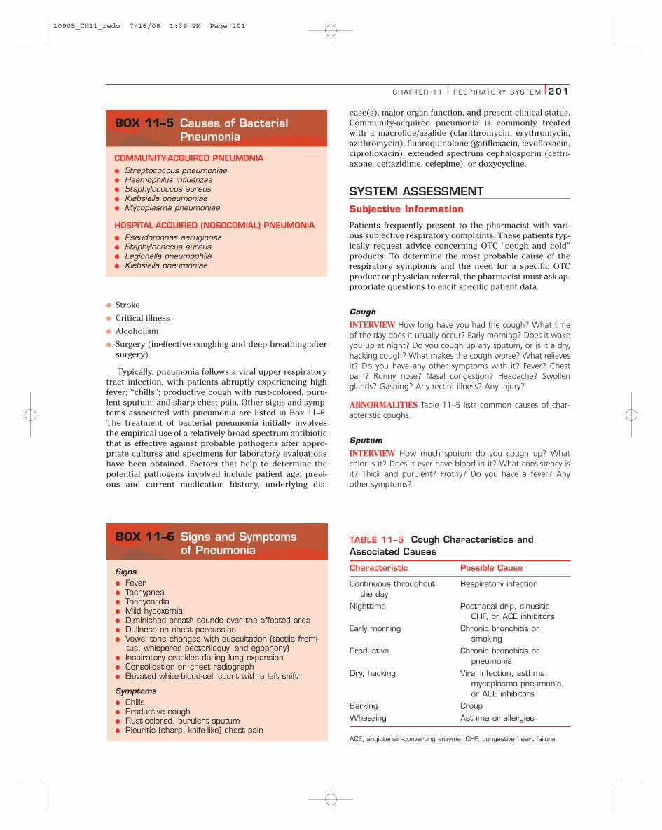
Cough
INTERVIEW How long have you had the cough? What timeof the day does it usually occur? Early morning? Does it wakeyou up at night? Do you cough up any sputum, or is it a dry,hacking cough? What makes the cough worse? What relievesit? Do you have any other symptoms with it? Fever? Chestpain? Runny nose? Nasal congestion? Headache? Swollenglands? Gasping? Any recent illness? Any injury?
ABNORMALITIES Table 11–5 lists common causes of char-acteristic coughs.
Sputum
INTERVIEW How much sputum do you cough up? Whatcolor is it? Does it ever have blood in it? What consistency isit? Thick and purulent? Frothy? Do you have a fever? Anyother symptoms?
COMMUNITY-ACQUIRED PNEUMONIA● Streptococcus pneumoniae● Haemophilus influenzae● Staphylococcus aureus● Klebsiella pneumoniae● Mycoplasma pneumoniae
HOSPITAL-ACQUIRED (NOSOCOMIAL) PNEUMONIA● Pseudomonas aeruginosa● Staphylococcus aureus● Legionella pneumophila● Klebsiella pneumoniae
BOX 11–5 Causes of BacterialPneumonia
Signs● Fever● Tachypnea● Tachycardia● Mild hypoxemia● Diminished breath sounds over the affected area● Dullness on chest percussion● Vowel tone changes with auscultation (tactile fremi-
tus, whispered pectoriloquy, and egophony)● Inspiratory crackles during lung expansion● Consolidation on chest radiograph● Elevated white-blood-cell count with a left shift
Symptoms● Chills● Productive cough● Rust-colored, purulent sputum● Pleuritic (sharp, knife-like) chest pain
BOX 11–6 Signs and Symptoms of Pneumonia
TABLE 11–5 Cough Characteristics andAssociated Causes
Characteristic Possible Cause
Continuous throughout Respiratory infectionthe day
Nighttime Postnasal drip, sinusitis, CHF, or ACE inhibitors
Early morning Chronic bronchitis or smoking
Productive Chronic bronchitis or pneumonia
Dry, hacking Viral infection, asthma, mycoplasma pneumonia,or ACE inhibitors
Barking Croup
Wheezing Asthma or allergies
ACE, angiotensin-converting enzyme; CHF, congestive heart failure.
10905_CH11_redo 7/16/08 1:39 PM Page 201
202 PART I I ASSESSMENT OF BODY SYSTEMS
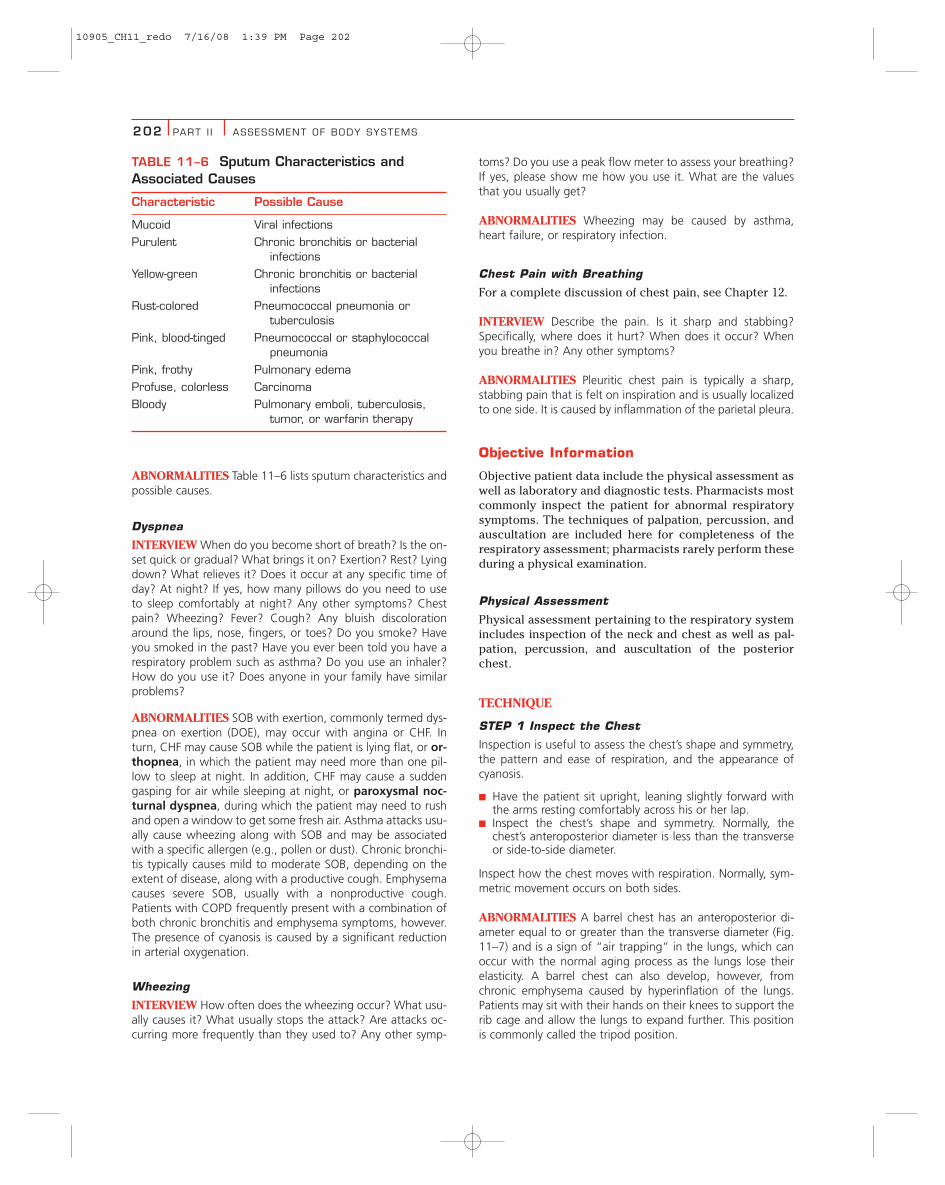
ABNORMALITIES Table 11–6 lists sputum characteristics andpossible causes.
Dyspnea
INTERVIEW When do you become short of breath? Is the on-set quick or gradual? What brings it on? Exertion? Rest? Lyingdown? What relieves it? Does it occur at any specific time ofday? At night? If yes, how many pillows do you need to useto sleep comfortably at night? Any other symptoms? Chestpain? Wheezing? Fever? Cough? Any bluish discolorationaround the lips, nose, fingers, or toes? Do you smoke? Haveyou smoked in the past? Have you ever been told you have arespiratory problem such as asthma? Do you use an inhaler?How do you use it? Does anyone in your family have similarproblems?
ABNORMALITIES SOB with exertion, commonly termed dys-pnea on exertion (DOE), may occur with angina or CHF. Inturn, CHF may cause SOB while the patient is lying flat, or or-thopnea, in which the patient may need more than one pil-low to sleep at night. In addition, CHF may cause a suddengasping for air while sleeping at night, or paroxysmal noc-turnal dyspnea, during which the patient may need to rushand open a window to get some fresh air. Asthma attacks usu-ally cause wheezing along with SOB and may be associatedwith a specific allergen (e.g., pollen or dust). Chronic bronchi-tis typically causes mild to moderate SOB, depending on theextent of disease, along with a productive cough. Emphysemacauses severe SOB, usually with a nonproductive cough.Patients with COPD frequently present with a combination ofboth chronic bronchitis and emphysema symptoms, however.The presence of cyanosis is caused by a significant reductionin arterial oxygenation.
Wheezing
INTERVIEW How often does the wheezing occur? What usu-ally causes it? What usually stops the attack? Are attacks oc-curring more frequently than they used to? Any other symp-
toms? Do you use a peak flow meter to assess your breathing?If yes, please show me how you use it. What are the valuesthat you usually get?
ABNORMALITIES Wheezing may be caused by asthma,heart failure, or respiratory infection.
Chest Pain with Breathing
For a complete discussion of chest pain, see Chapter 12.
INTERVIEW Describe the pain. Is it sharp and stabbing?Specifically, where does it hurt? When does it occur? Whenyou breathe in? Any other symptoms?
ABNORMALITIES Pleuritic chest pain is typically a sharp,stabbing pain that is felt on inspiration and is usually localizedto one side. It is caused by inflammation of the parietal pleura.
Objective Information
Objective patient data include the physical assessment aswell as laboratory and diagnostic tests. Pharmacists mostcommonly inspect the patient for abnormal respiratorysymptoms. The techniques of palpation, percussion, andauscultation are included here for completeness of therespiratory assessment; pharmacists rarely perform theseduring a physical examination.
Physical Assessment
Physical assessment pertaining to the respiratory systemincludes inspection of the neck and chest as well as pal-pation, percussion, and auscultation of the posteriorchest.
TECHNIQUE
STEP 1 Inspect the Chest
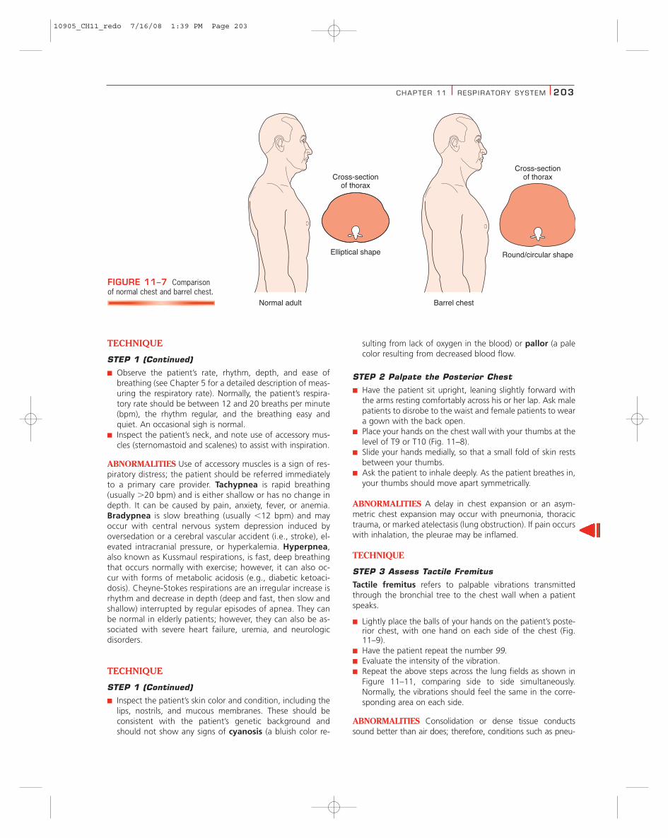
Inspection is useful to assess the chest’s shape and symmetry,the pattern and ease of respiration, and the appearance ofcyanosis.
■ Have the patient sit upright, leaning slightly forward withthe arms resting comfortably across his or her lap.
■ Inspect the chest’s shape and symmetry. Normally, thechest’s anteroposterior diameter is less than the transverseor side-to-side diameter.
Inspect how the chest moves with respiration. Normally, sym-metric movement occurs on both sides.
ABNORMALITIES A barrel chest has an anteroposterior di-ameter equal to or greater than the transverse diameter (Fig.11–7) and is a sign of “air trapping” in the lungs, which canoccur with the normal aging process as the lungs lose theirelasticity. A barrel chest can also develop, however, fromchronic emphysema caused by hyperinflation of the lungs.Patients may sit with their hands on their knees to support therib cage and allow the lungs to expand further. This positionis commonly called the tripod position.
TABLE 11–6 Sputum Characteristics andAssociated Causes
Characteristic Possible Cause
Mucoid Viral infections
Purulent Chronic bronchitis or bacterial infections
Yellow-green Chronic bronchitis or bacterial infections
Rust-colored Pneumococcal pneumonia or tuberculosis
Pink, blood-tinged Pneumococcal or staphylococcal pneumonia
Pink, frothy Pulmonary edema
Profuse, colorless Carcinoma
Bloody Pulmonary emboli, tuberculosis, tumor, or warfarin therapy
10905_CH11_redo 7/16/08 1:39 PM Page 202
CHAPTER 11 RESP IRATORY SYSTEM 203
TECHNIQUE
STEP 1 (Continued)
■ Observe the patient’s rate, rhythm, depth, and ease ofbreathing (see Chapter 5 for a detailed description of meas-uring the respiratory rate). Normally, the patient’s respira-tory rate should be between 12 and 20 breaths per minute(bpm), the rhythm regular, and the breathing easy andquiet. An occasional sigh is normal.
■ Inspect the patient’s neck, and note use of accessory mus-cles (sternomastoid and scalenes) to assist with inspiration.
ABNORMALITIES Use of accessory muscles is a sign of res-piratory distress; the patient should be referred immediatelyto a primary care provider. Tachypnea is rapid breathing(usually �20 bpm) and is either shallow or has no change indepth. It can be caused by pain, anxiety, fever, or anemia.Bradypnea is slow breathing (usually �12 bpm) and mayoccur with central nervous system depression induced byoversedation or a cerebral vascular accident (i.e., stroke), el-evated intracranial pressure, or hyperkalemia. Hyperpnea,also known as Kussmaul respirations, is fast, deep breathingthat occurs normally with exercise; however, it can also oc-cur with forms of metabolic acidosis (e.g., diabetic ketoaci-dosis). Cheyne-Stokes respirations are an irregular increase isrhythm and decrease in depth (deep and fast, then slow andshallow) interrupted by regular episodes of apnea. They canbe normal in elderly patients; however, they can also be as-sociated with severe heart failure, uremia, and neurologicdisorders.
TECHNIQUE
STEP 1 (Continued)
■ Inspect the patient’s skin color and condition, including thelips, nostrils, and mucous membranes. These should beconsistent with the patient’s genetic background andshould not show any signs of cyanosis (a bluish color re-
sulting from lack of oxygen in the blood) or pallor (a palecolor resulting from decreased blood flow.
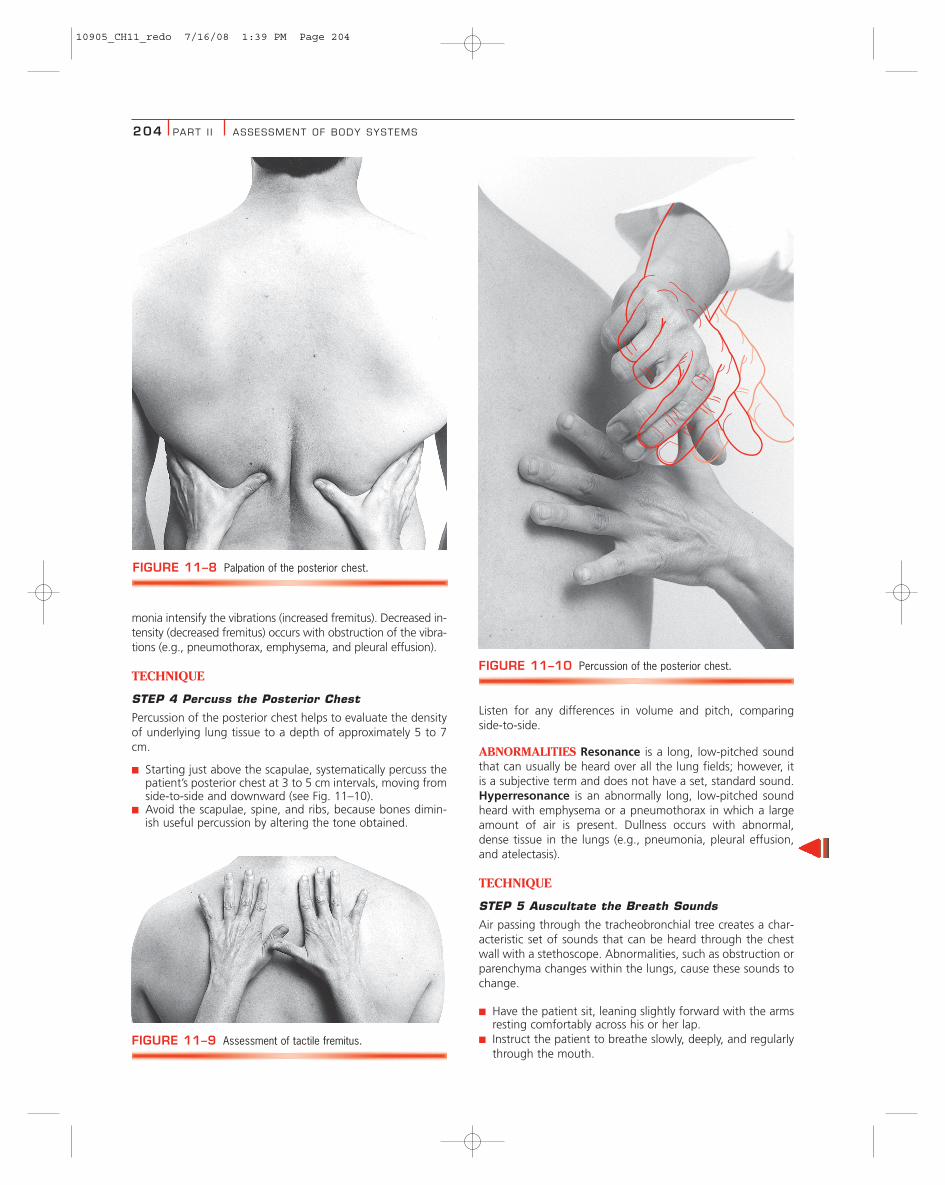
STEP 2 Palpate the Posterior Chest
■ Have the patient sit upright, leaning slightly forward withthe arms resting comfortably across his or her lap. Ask malepatients to disrobe to the waist and female patients to weara gown with the back open.
■ Place your hands on the chest wall with your thumbs at thelevel of T9 or T10 (Fig. 11–8).
■ Slide your hands medially, so that a small fold of skin restsbetween your thumbs.
■ Ask the patient to inhale deeply. As the patient breathes in,your thumbs should move apart symmetrically.
ABNORMALITIES A delay in chest expansion or an asym-metric chest expansion may occur with pneumonia, thoracictrauma, or marked atelectasis (lung obstruction). If pain occurswith inhalation, the pleurae may be inflamed.
TECHNIQUE
STEP 3 Assess Tactile Fremitus
Tactile fremitus refers to palpable vibrations transmittedthrough the bronchial tree to the chest wall when a patientspeaks.
■ Lightly place the balls of your hands on the patient’s poste-rior chest, with one hand on each side of the chest (Fig.11–9).
■ Have the patient repeat the number 99.■ Evaluate the intensity of the vibration.■ Repeat the above steps across the lung fields as shown in
Figure 11–11, comparing side to side simultaneously.Normally, the vibrations should feel the same in the corre-sponding area on each side.
ABNORMALITIES Consolidation or dense tissue conductssound better than air does; therefore, conditions such as pneu-
Cross-sectionof thorax
Elliptical shape
Normal adult Barrel chest
Round/circular shape
Cross-sectionof thorax
FIGURE 11–7 Comparisonof normal chest and barrel chest.
10905_CH11_redo 7/16/08 1:39 PM Page 203
204 PART I I ASSESSMENT OF BODY SYSTEMS
monia intensify the vibrations (increased fremitus). Decreased in-tensity (decreased fremitus) occurs with obstruction of the vibra-tions (e.g., pneumothorax, emphysema, and pleural effusion).
TECHNIQUE
STEP 4 Percuss the Posterior Chest
Percussion of the posterior chest helps to evaluate the densityof underlying lung tissue to a depth of approximately 5 to 7cm.
■ Starting just above the scapulae, systematically percuss thepatient’s posterior chest at 3 to 5 cm intervals, moving fromside-to-side and downward (see Fig. 11–10).
■ Avoid the scapulae, spine, and ribs, because bones dimin-ish useful percussion by altering the tone obtained.
Listen for any differences in volume and pitch, comparingside-to-side.
ABNORMALITIES Resonance is a long, low-pitched soundthat can usually be heard over all the lung fields; however, itis a subjective term and does not have a set, standard sound.Hyperresonance is an abnormally long, low-pitched soundheard with emphysema or a pneumothorax in which a largeamount of air is present. Dullness occurs with abnormal,dense tissue in the lungs (e.g., pneumonia, pleural effusion,and atelectasis).
TECHNIQUE
STEP 5 Auscultate the Breath Sounds
Air passing through the tracheobronchial tree creates a char-acteristic set of sounds that can be heard through the chestwall with a stethoscope. Abnormalities, such as obstruction orparenchyma changes within the lungs, cause these sounds tochange.
■ Have the patient sit, leaning slightly forward with the armsresting comfortably across his or her lap.
■ Instruct the patient to breathe slowly, deeply, and regularlythrough the mouth.
FIGURE 11–9 Assessment of tactile fremitus.
FIGURE 11–10 Percussion of the posterior chest.
FIGURE 11–8 Palpation of the posterior chest.
10905_CH11_redo 7/16/08 1:39 PM Page 204
CHAPTER 11 RESP IRATORY SYSTEM 205
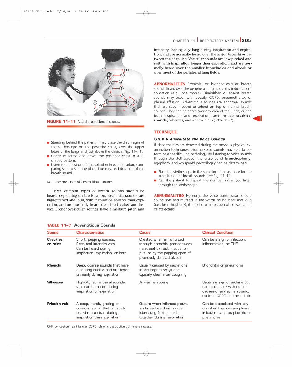
■ Standing behind the patient, firmly place the diaphragm ofthe stethoscope on the posterior chest, over the upperlobes of the lungs and just above the clavicle (Fig. 11–11).
■ Continue across and down the posterior chest in a Z-shaped pattern.
■ Listen to at least one full respiration in each location, com-paring side-to-side the pitch, intensity, and duration of thebreath sound.
Note the presence of adventitious sounds.
Three different types of breath sounds should beheard, depending on the location. Bronchial sounds arehigh-pitched and loud, with inspiration shorter than expi-ration, and are normally heard over the trachea and lar-ynx. Bronchovesicular sounds have a medium pitch and
intensity, last equally long during inspiration and expira-tion, and are normally heard over the major bronchi or be-tween the scapulae. Vesicular sounds are low-pitched andsoft, with inspiration longer than expiration, and are nor-mally heard over the smaller bronchioles and alveoli orover most of the peripheral lung fields.
ABNORMALITIES Bronchial or bronchovesicular breathsounds heard over the peripheral lung fields may indicate con-solidation (e.g., pneumonia). Diminished or absent breathsounds may occur with obesity, COPD, pneumothorax, orpleural effusion. Adventitious sounds are abnormal soundsthat are superimposed or added on top of normal breathsounds. They can be heard over any area of the lungs, duringboth inspiration and expiration, and include crackles,rhonchi, wheezes, and a friction rub (Table 11–7).
TECHNIQUE
STEP 6 Auscultate the Voice Sounds
If abnormalities are detected during the previous physical ex-amination techniques, eliciting voice sounds may help to de-termine a specific lung pathology. By listening to voice soundsthrough the stethoscope, the presence of bronchophony,egophony, and whispered pectoriloquy can be determined.
■ Place the stethoscope in the same locations as those for theauscultation of breath sounds (see Fig. 11–11).
■ Ask the patient to repeat the number 99 as you listenthrough the stethoscope.
ABNORMALITIES Normally, the voice transmission shouldsound soft and muffled. If the words sound clear and loud(i.e., bronchophony), it may be an indication of consolidationor atelectasis.
1 1
2
3 3
4 4
5 5
6 6
7 79 9
8 8
FIGURE 11–11 Auscultation of breath sounds.
TABLE 11–7 Adventitious Sounds
Sound Characteristics Cause Clinical Condition
Crackles Short, popping sounds. Created when air is forced Can be a sign of infection, or rales Pitch and intensity vary. through bronchial passageways inflammation, or CHF
Can be heard during narrowed by fluid, mucus, or inspiration, expiration, or both pus, or by the popping open of
previously deflated alveoli
Rhonchi Deep, coarse sounds that have Usually caused by secretions Bronchitis or pneumoniaa snoring quality, and are heard in the large airways and primarily during expiration typically clear after coughing
Wheezes High-pitched, musical sounds Airway narrowing Usually a sign of asthma but that can be heard during can also occur with other inspiration or expiration causes of airway narrowing,
such as COPD and bronchitis
Friction rub A deep, harsh, grating or Occurs when inflamed pleural Can be associated with any creaking sound that is usually surfaces lose their normal condition that causes pleural heard more often during lubricating fluid and rub irritation, such as pleuritis or inspiration than expiration together during respiration pneumonia
CHF, congestive heart failure; COPD, chronic obstructive pulmonary disease.
10905_CH11_redo 7/16/08 1:39 PM Page 205
206 PART I I ASSESSMENT OF BODY SYSTEMS
■ Ask the patient to repeat ee as you listen through thestethoscope.
ABNORMALITIES Normally, it should sound like ee. If con-solidation is present, the word will sound like ay, which istermed egophony.
■ Ask the patient to whisper one-two-three as you listenthrough the stethoscope.
ABNORMALITIES Normally, the words should sound veryfaint and muffled. Consolidation and pleural effusions cancause these sounds to be more distinctive and clear. This iscalled whispered pectoriloquy.
Laboratory and Diagnostic Tests
Pulmonary function tests include blood gas measure-ments, oxygen saturation (O2 sat), and spirometry.Arterial blood gas measurements are the best indicatorsof overall lung function and include PaO2, PaCO2, and pH.The adequacy of gas exchange in the lungs determines thevalues of these gases. Normal values for arterial bloodgases are listed in Table 11–8. Oxygen saturation is the ra-tio between the actual amount of oxygen bound to hemo-globin and the potential amount of oxygen that could bebound to hemoglobin at a given pressure. Normally, the O2sat of arterial blood is 97.5% at a PaO2 of 100 mm Hg. TheO2 sat is very useful in determining the need for supple-mental oxygen therapy.
Spirometry includes tests that measure various lungvolumes with a spirometer. The tidal volume is the volumeof air that is inhaled or exhaled during normal breathing.The vital capacity is the maximum volume of air that aperson can exhale after maximum inhalation. The volumeof air that remains in the lungs after maximum exhalationis the residual volume. The total lung capacity is the vitalcapacity plus the residual volume. Because patients withobstructive lung diseases (e.g., asthma or COPD) have dif-ficulty exhaling, they usually have decreased vital capac-ity, increased residual volume, and normal total lung ca-pacity. In addition to measuring lung volumes, thespirometer can also be used to assess the patient’s abilityto move air into and out of the lungs. The forced expira-tory volume is the maximal volume of air that is exhaledas forcefully and as completely as possible after maximalinhalation. This volume curve is plotted against time. TheFEV1 of the FVC is commonly used to evaluate the lung’sability to move air; it is usually documented as the per-centage of the total volume of air exhaled, or theFEV1/FVC. Normally, FEV1 is 80% of the FVC.
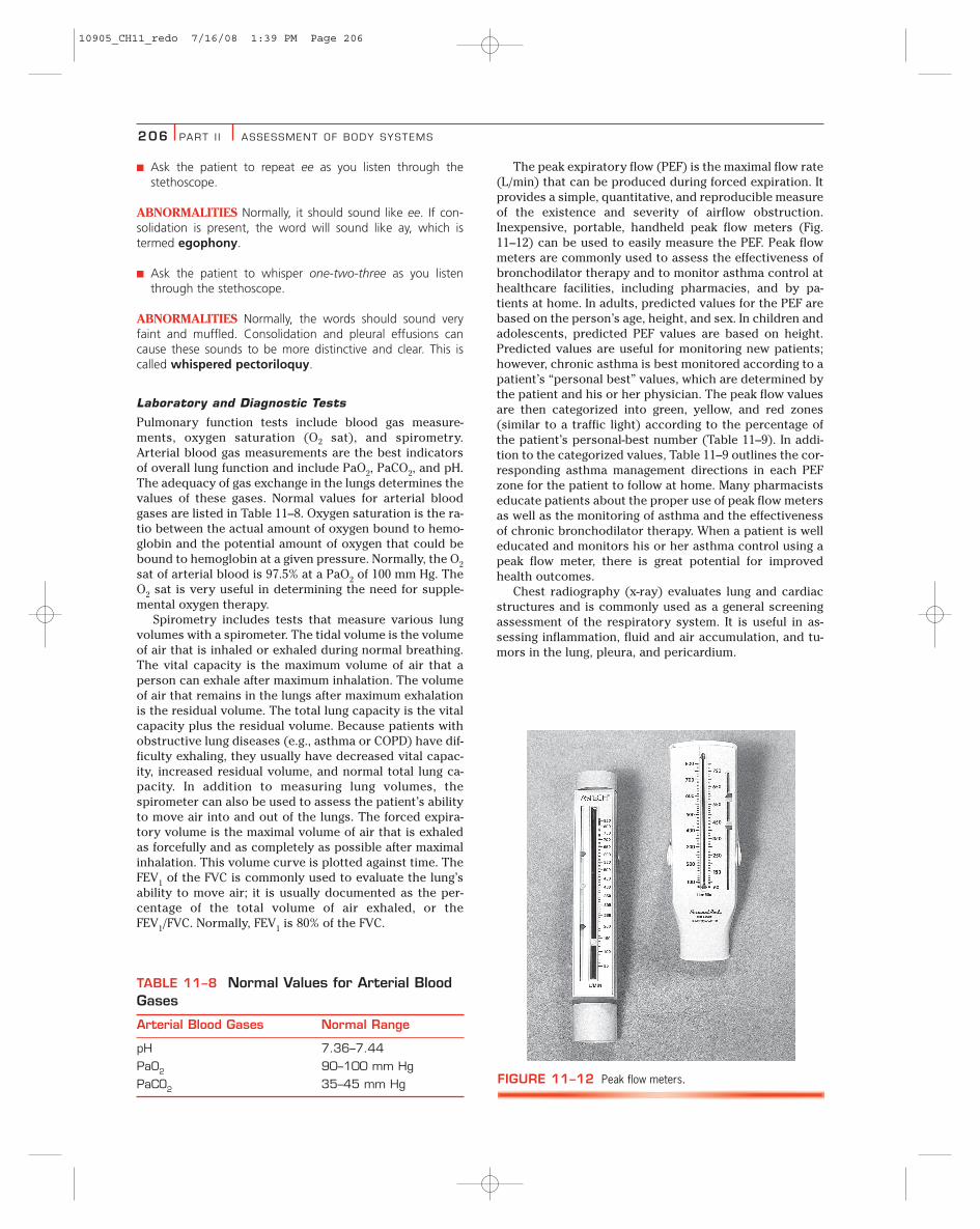
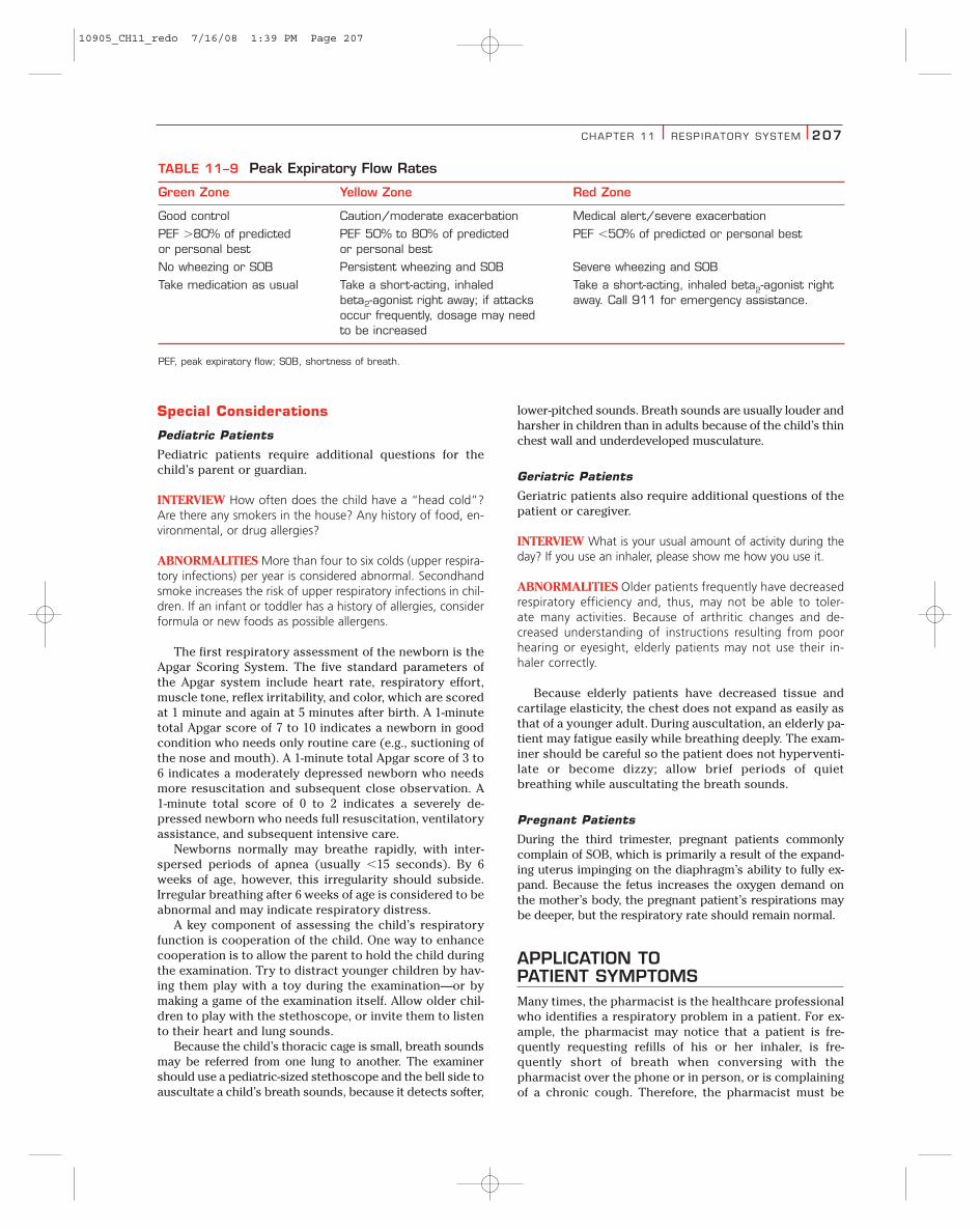
The peak expiratory flow (PEF) is the maximal flow rate(L/min) that can be produced during forced expiration. Itprovides a simple, quantitative, and reproducible measureof the existence and severity of airflow obstruction.Inexpensive, portable, handheld peak flow meters (Fig.11–12) can be used to easily measure the PEF. Peak flowmeters are commonly used to assess the effectiveness ofbronchodilator therapy and to monitor asthma control athealthcare facilities, including pharmacies, and by pa-tients at home. In adults, predicted values for the PEF arebased on the person’s age, height, and sex. In children andadolescents, predicted PEF values are based on height.Predicted values are useful for monitoring new patients;however, chronic asthma is best monitored according to apatient’s “personal best” values, which are determined bythe patient and his or her physician. The peak flow valuesare then categorized into green, yellow, and red zones(similar to a traffic light) according to the percentage ofthe patient’s personal-best number (Table 11–9). In addi-tion to the categorized values, Table 11–9 outlines the cor-responding asthma management directions in each PEFzone for the patient to follow at home. Many pharmacistseducate patients about the proper use of peak flow metersas well as the monitoring of asthma and the effectivenessof chronic bronchodilator therapy. When a patient is welleducated and monitors his or her asthma control using apeak flow meter, there is great potential for improvedhealth outcomes.
Chest radiography (x-ray) evaluates lung and cardiacstructures and is commonly used as a general screeningassessment of the respiratory system. It is useful in as-sessing inflammation, fluid and air accumulation, and tu-mors in the lung, pleura, and pericardium.
TABLE 11–8 Normal Values for Arterial BloodGases
Arterial Blood Gases Normal Range
pH 7.36–7.44
PaO2 90–100 mm Hg
PaCO2 35–45 mm Hg FIGURE 11–12 Peak flow meters.
10905_CH11_redo 7/16/08 1:39 PM Page 206
CHAPTER 11 RESP IRATORY SYSTEM 207
Special Considerations
Pediatric Patients
Pediatric patients require additional questions for thechild’s parent or guardian.
INTERVIEW How often does the child have a “head cold”?Are there any smokers in the house? Any history of food, en-vironmental, or drug allergies?
ABNORMALITIES More than four to six colds (upper respira-tory infections) per year is considered abnormal. Secondhandsmoke increases the risk of upper respiratory infections in chil-dren. If an infant or toddler has a history of allergies, considerformula or new foods as possible allergens.
The first respiratory assessment of the newborn is theApgar Scoring System. The five standard parameters ofthe Apgar system include heart rate, respiratory effort,muscle tone, reflex irritability, and color, which are scoredat 1 minute and again at 5 minutes after birth. A 1-minutetotal Apgar score of 7 to 10 indicates a newborn in goodcondition who needs only routine care (e.g., suctioning ofthe nose and mouth). A 1-minute total Apgar score of 3 to6 indicates a moderately depressed newborn who needsmore resuscitation and subsequent close observation. A1-minute total score of 0 to 2 indicates a severely de-pressed newborn who needs full resuscitation, ventilatoryassistance, and subsequent intensive care.
Newborns normally may breathe rapidly, with inter-spersed periods of apnea (usually �15 seconds). By 6weeks of age, however, this irregularity should subside.Irregular breathing after 6 weeks of age is considered to beabnormal and may indicate respiratory distress.
A key component of assessing the child’s respiratoryfunction is cooperation of the child. One way to enhancecooperation is to allow the parent to hold the child duringthe examination. Try to distract younger children by hav-ing them play with a toy during the examination—or bymaking a game of the examination itself. Allow older chil-dren to play with the stethoscope, or invite them to listento their heart and lung sounds.
Because the child’s thoracic cage is small, breath soundsmay be referred from one lung to another. The examinershould use a pediatric-sized stethoscope and the bell side toauscultate a child’s breath sounds, because it detects softer,
lower-pitched sounds. Breath sounds are usually louder andharsher in children than in adults because of the child’s thinchest wall and underdeveloped musculature.
Geriatric Patients
Geriatric patients also require additional questions of thepatient or caregiver.
INTERVIEW What is your usual amount of activity during theday? If you use an inhaler, please show me how you use it.
ABNORMALITIES Older patients frequently have decreasedrespiratory efficiency and, thus, may not be able to toler-ate many activities. Because of arthritic changes and de-creased understanding of instructions resulting from poorhearing or eyesight, elderly patients may not use their in-haler correctly.
Because elderly patients have decreased tissue andcartilage elasticity, the chest does not expand as easily asthat of a younger adult. During auscultation, an elderly pa-tient may fatigue easily while breathing deeply. The exam-iner should be careful so the patient does not hyperventi-late or become dizzy; allow brief periods of quietbreathing while auscultating the breath sounds.
Pregnant Patients
During the third trimester, pregnant patients commonlycomplain of SOB, which is primarily a result of the expand-ing uterus impinging on the diaphragm’s ability to fully ex-pand. Because the fetus increases the oxygen demand onthe mother’s body, the pregnant patient’s respirations maybe deeper, but the respiratory rate should remain normal.
APPLICATION TO PATIENT SYMPTOMSMany times, the pharmacist is the healthcare professionalwho identifies a respiratory problem in a patient. For ex-ample, the pharmacist may notice that a patient is fre-quently requesting refills of his or her inhaler, is fre-quently short of breath when conversing with thepharmacist over the phone or in person, or is complainingof a chronic cough. Therefore, the pharmacist must be
TABLE 11–9 Peak Expiratory Flow Rates
Green Zone Yellow Zone Red Zone
Good control Caution/moderate exacerbation Medical alert/severe exacerbation
PEF �80% of predicted PEF 50% to 80% of predicted PEF �50% of predicted or personal bestor personal best or personal best
No wheezing or SOB Persistent wheezing and SOB Severe wheezing and SOB
Take medication as usual Take a short-acting, inhaled Take a short-acting, inhaled beta2-agonist right beta2-agonist right away; if attacks away. Call 911 for emergency assistance.occur frequently, dosage may need to be increased
PEF, peak expiratory flow; SOB, shortness of breath.
10905_CH11_redo 7/16/08 1:39 PM Page 207
220088 PART I I ASSESSMENT OF BODY SYSTEMS
ronmental irritants, and CHF. The pharmacist shouldalso keep in mind that angiotensin-converting enzyme(ACE) inhibitors may also cause a cough. Patients usu-ally complain of a persistent (not episodic), dry, nonpro-ductive cough that is usually worse at night. Patientsmay also describe it as a tickling sensation. In addition,ACE inhibitor–induced coughs are more common inwomen than in men.
208 PART I I ASSESSMENT OF BODY SYSTEMS
able to evaluate common respiratory symptoms, deter-mine possible causes of these symptoms, and take appro-priate action to either further assess the symptom or tocorrect the problem identified. Common respiratorysymptoms include dyspnea, wheezing, and cough.
Dyspnea (Case Study 11-1)
Patients with dyspnea may report that they “can’t getenough air” or complain of “breathlessness.” Variouscauses of dyspnea include:
● Pulmonary: COPD, asthma, and emphysema● Cardiac: CHF and coronary artery disease● Emotional: anxiety
Wheezing (Case Study 11-2)
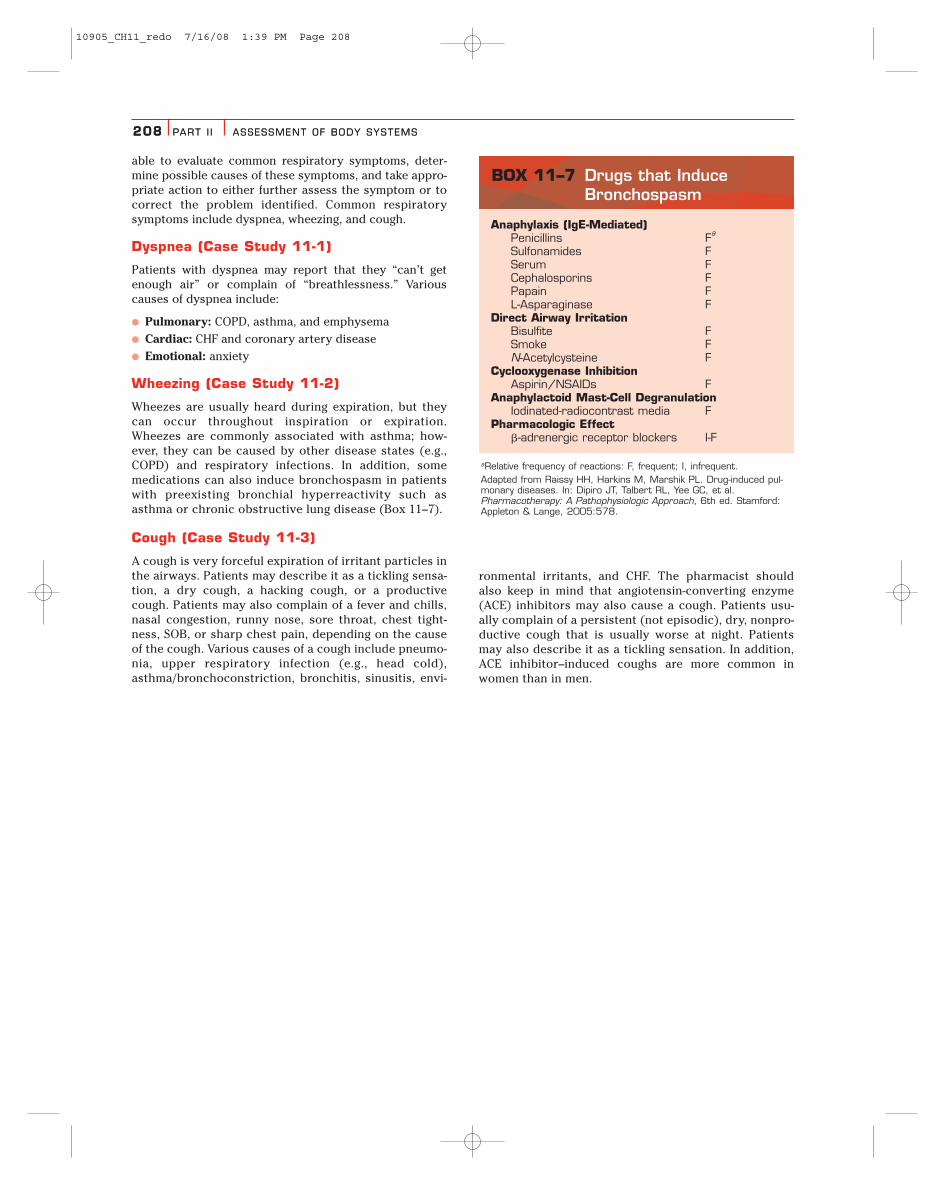
Wheezes are usually heard during expiration, but theycan occur throughout inspiration or expiration.Wheezes are commonly associated with asthma; how-ever, they can be caused by other disease states (e.g.,COPD) and respiratory infections. In addition, somemedications can also induce bronchospasm in patientswith preexisting bronchial hyperreactivity such asasthma or chronic obstructive lung disease (Box 11–7).
Cough (Case Study 11-3)
A cough is very forceful expiration of irritant particles inthe airways. Patients may describe it as a tickling sensa-tion, a dry cough, a hacking cough, or a productivecough. Patients may also complain of a fever and chills,nasal congestion, runny nose, sore throat, chest tight-ness, SOB, or sharp chest pain, depending on the causeof the cough. Various causes of a cough include pneumo-nia, upper respiratory infection (e.g., head cold),asthma/bronchoconstriction, bronchitis, sinusitis, envi-
Anaphylaxis (IgE-Mediated)Penicillins Fa
Sulfonamides FSerum FCephalosporins FPapain FL-Asparaginase F
Direct Airway IrritationBisulfite FSmoke FN-Acetylcysteine F
Cyclooxygenase InhibitionAspirin/NSAIDs F
Anaphylactoid Mast-Cell DegranulationIodinated-radiocontrast media F
Pharmacologic Effect�-adrenergic receptor blockers I-F
BOX 11–7 Drugs that InduceBronchospasm
aRelative frequency of reactions: F, frequent; I, infrequent.Adapted from Raissy HH, Harkins M, Marshik PL. Drug-induced pul-monary diseases. In: Dipiro JT, Talbert RL, Yee GC, et al.Pharmacotherapy: A Pathophysiologic Approach, 6th ed. Stamford:Appleton & Lange, 2005:578.
10905_CH11_redo 7/16/08 1:39 PM Page 208
CHAPTER 11 RESP IRATORY SYSTEM 209
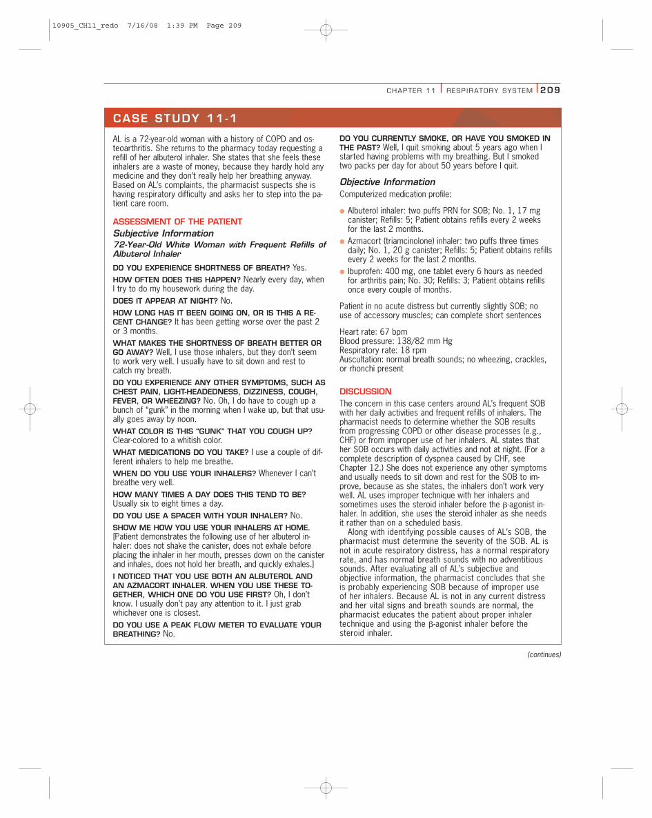
AL is a 72-year-old woman with a history of COPD and os-teoarthritis. She returns to the pharmacy today requesting arefill of her albuterol inhaler. She states that she feels theseinhalers are a waste of money, because they hardly hold anymedicine and they don’t really help her breathing anyway.Based on AL’s complaints, the pharmacist suspects she ishaving respiratory difficulty and asks her to step into the pa-tient care room.
ASSESSMENT OF THE PATIENTSubjective Information72-Year-Old White Woman with Frequent Refills ofAlbuterol Inhaler
DO YOU EXPERIENCE SHORTNESS OF BREATH? Yes.HOW OFTEN DOES THIS HAPPEN? Nearly every day, whenI try to do my housework during the day.DOES IT APPEAR AT NIGHT? No.HOW LONG HAS IT BEEN GOING ON, OR IS THIS A RE-CENT CHANGE? It has been getting worse over the past 2or 3 months.WHAT MAKES THE SHORTNESS OF BREATH BETTER ORGO AWAY? Well, I use those inhalers, but they don’t seemto work very well. I usually have to sit down and rest tocatch my breath.DO YOU EXPERIENCE ANY OTHER SYMPTOMS, SUCH ASCHEST PAIN, LIGHT-HEADEDNESS, DIZZINESS, COUGH,FEVER, OR WHEEZING? No. Oh, I do have to cough up abunch of “gunk” in the morning when I wake up, but that usu-ally goes away by noon.WHAT COLOR IS THIS “GUNK” THAT YOU COUGH UP?Clear-colored to a whitish color.WHAT MEDICATIONS DO YOU TAKE? I use a couple of dif-ferent inhalers to help me breathe.WHEN DO YOU USE YOUR INHALERS? Whenever I can’tbreathe very well.HOW MANY TIMES A DAY DOES THIS TEND TO BE?Usually six to eight times a day.DO YOU USE A SPACER WITH YOUR INHALER? No.SHOW ME HOW YOU USE YOUR INHALERS AT HOME.[Patient demonstrates the following use of her albuterol in-haler: does not shake the canister, does not exhale beforeplacing the inhaler in her mouth, presses down on the canisterand inhales, does not hold her breath, and quickly exhales.]I NOTICED THAT YOU USE BOTH AN ALBUTEROL ANDAN AZMACORT INHALER. WHEN YOU USE THESE TO-GETHER, WHICH ONE DO YOU USE FIRST? Oh, I don’tknow. I usually don’t pay any attention to it. I just grabwhichever one is closest.DO YOU USE A PEAK FLOW METER TO EVALUATE YOURBREATHING? No.
DO YOU CURRENTLY SMOKE, OR HAVE YOU SMOKED INTHE PAST? Well, I quit smoking about 5 years ago when Istarted having problems with my breathing. But I smokedtwo packs per day for about 50 years before I quit.
Objective InformationComputerized medication profile:
● Albuterol inhaler: two puffs PRN for SOB; No. 1, 17 mgcanister; Refills: 5; Patient obtains refills every 2 weeksfor the last 2 months.
● Azmacort (triamcinolone) inhaler: two puffs three timesdaily; No. 1, 20 g canister; Refills: 5; Patient obtains refillsevery 2 weeks for the last 2 months.
● Ibuprofen: 400 mg, one tablet every 6 hours as neededfor arthritis pain; No. 30; Refills: 3; Patient obtains refillsonce every couple of months.
Patient in no acute distress but currently slightly SOB; nouse of accessory muscles; can complete short sentences
Heart rate: 67 bpmBlood pressure: 138/82 mm HgRespiratory rate: 18 rpmAuscultation: normal breath sounds; no wheezing, crackles,or rhonchi present
DISCUSSIONThe concern in this case centers around AL’s frequent SOBwith her daily activities and frequent refills of inhalers. Thepharmacist needs to determine whether the SOB resultsfrom progressing COPD or other disease processes (e.g.,CHF) or from improper use of her inhalers. AL states thather SOB occurs with daily activities and not at night. (For acomplete description of dyspnea caused by CHF, seeChapter 12.) She does not experience any other symptomsand usually needs to sit down and rest for the SOB to im-prove, because as she states, the inhalers don’t work verywell. AL uses improper technique with her inhalers andsometimes uses the steroid inhaler before the �-agonist in-haler. In addition, she uses the steroid inhaler as she needsit rather than on a scheduled basis.
Along with identifying possible causes of AL’s SOB, thepharmacist must determine the severity of the SOB. AL isnot in acute respiratory distress, has a normal respiratoryrate, and has normal breath sounds with no adventitioussounds. After evaluating all of AL’s subjective and objective information, the pharmacist concludes that sheis probably experiencing SOB because of improper use of her inhalers. Because AL is not in any current distressand her vital signs and breath sounds are normal, thepharmacist educates the patient about proper inhaler technique and using the �-agonist inhaler before thesteroid inhaler.
CASE STUDY 11 -1
(continues)
10905_CH11_redo 7/16/08 1:39 PM Page 209
210 PART I I ASSESSMENT OF BODY SYSTEMS
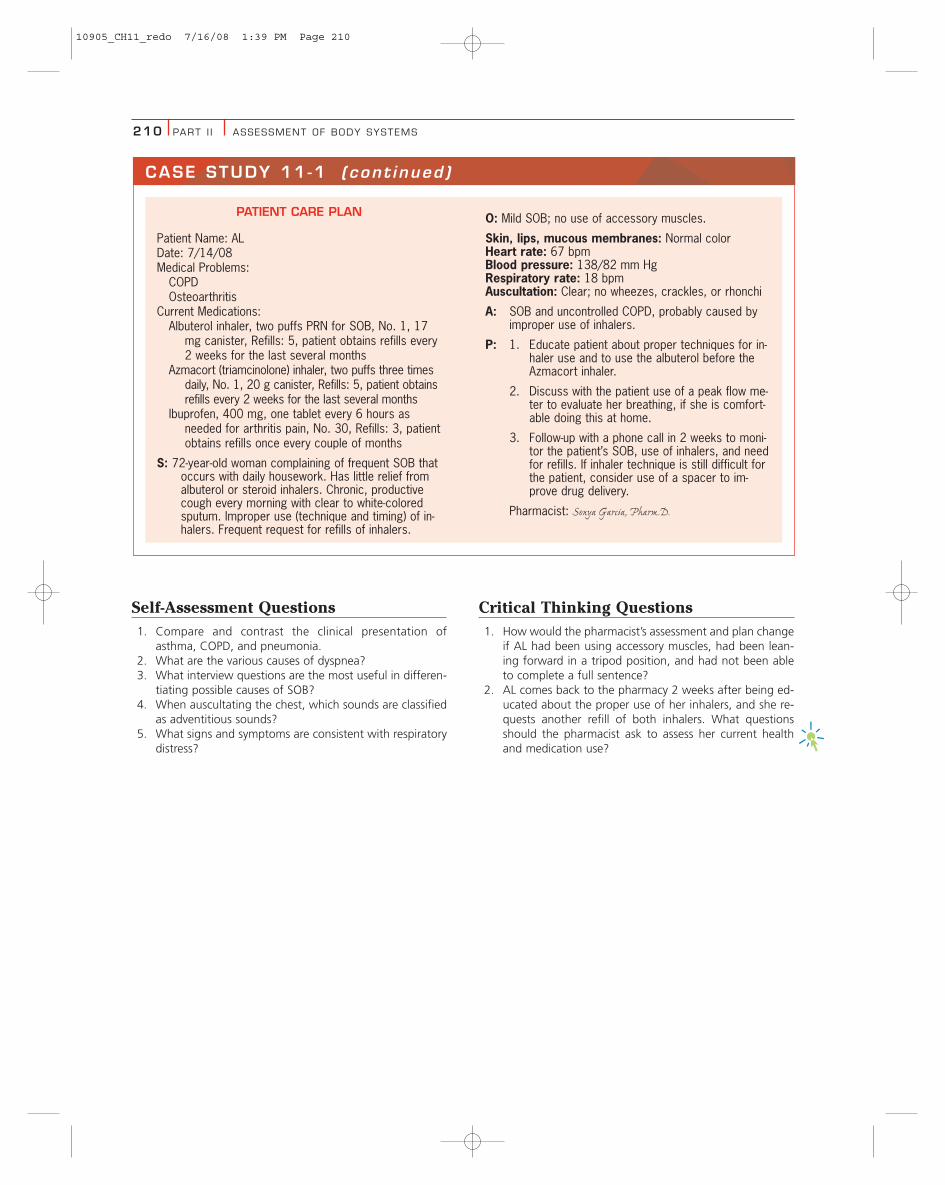
PATIENT CARE PLAN
Patient Name: ALDate: 7/14/08Medical Problems:
COPDOsteoarthritis
Current Medications:Albuterol inhaler, two puffs PRN for SOB, No. 1, 17
mg canister, Refills: 5, patient obtains refills every2 weeks for the last several months
Azmacort (triamcinolone) inhaler, two puffs three timesdaily, No. 1, 20 g canister, Refills: 5, patient obtainsrefills every 2 weeks for the last several months
Ibuprofen, 400 mg, one tablet every 6 hours asneeded for arthritis pain, No. 30, Refills: 3, patientobtains refills once every couple of months
S: 72-year-old woman complaining of frequent SOB thatoccurs with daily housework. Has little relief fromalbuterol or steroid inhalers. Chronic, productivecough every morning with clear to white-coloredsputum. Improper use (technique and timing) of in-halers. Frequent request for refills of inhalers.
O: Mild SOB; no use of accessory muscles.
Skin, lips, mucous membranes: Normal colorHeart rate: 67 bpmBlood pressure: 138/82 mm HgRespiratory rate: 18 bpmAuscultation: Clear; no wheezes, crackles, or rhonchi
A: SOB and uncontrolled COPD, probably caused byimproper use of inhalers.
P: 1. Educate patient about proper techniques for in-haler use and to use the albuterol before theAzmacort inhaler.
2. Discuss with the patient use of a peak flow me-ter to evaluate her breathing, if she is comfort-able doing this at home.
3. Follow-up with a phone call in 2 weeks to moni-tor the patient’s SOB, use of inhalers, and needfor refills. If inhaler technique is still difficult forthe patient, consider use of a spacer to im-prove drug delivery.
Pharmacist: Sonya Garcia, Pharm.D.
CASE STUDY 11 -1 (cont inued )
Self-Assessment Questions1. Compare and contrast the clinical presentation of
asthma, COPD, and pneumonia.2. What are the various causes of dyspnea?3. What interview questions are the most useful in differen-
tiating possible causes of SOB?4. When auscultating the chest, which sounds are classified
as adventitious sounds?5. What signs and symptoms are consistent with respiratory
distress?
Critical Thinking Questions1. How would the pharmacist’s assessment and plan change
if AL had been using accessory muscles, had been lean-ing forward in a tripod position, and had not been ableto complete a full sentence?
2. AL comes back to the pharmacy 2 weeks after being ed-ucated about the proper use of her inhalers, and she re-quests another refill of both inhalers. What questionsshould the pharmacist ask to assess her current healthand medication use?
10905_CH11_redo 7/16/08 1:39 PM Page 210
CHAPTER 11 RESP IRATORY SYSTEM 211
JB is a 10-year-old boy with a lifelong history of asthma. Heand his mother come into the pharmacy with a new prescrip-tion for a steroid inhaler. The pharmacist asks JB and hismother to step into the patient care room to discuss thenew medication.
ASSESSMENT OF THE PATIENTSubjective Information10-Year-Old Boy with a New Prescription for aSteroid InhalerSINCE YOU HAVE A NEW PRESCRIPTION TODAY, I AS-SUME THAT YOU JUST CAME FROM THE DOCTOR’S OF-FICE? Yes, we did.HAS JB BEEN HAVING PROBLEMS CONTROLLING HISASTHMA? Yes. Lately, he has been experiencing wheezing,coughing, and shortness of breath almost every day.WHAT USUALLY BRINGS ON AN ASTHMA ATTACK?Usually exerting himself, like when he goes outside to play.WHAT MEDICATIONS HAS JB BEEN USING? Albuterol in-haler, two puffs every 4 to 6 hours when he needs it to helphim breathe. Over the past couple of months, he has beenusing it nearly every day, and it seems to stop the asthmaattack.DOES JB TAKE ANY OTHER PRESCRIPTION OR NONPRE-SCRIPTION MEDICATIONS? No. Oh, I do give him Tylenolonce in awhile for a headache.JB, SHOW ME HOW YOU USE YOUR INHALER. [JBdemonstrates proper technique for using the albuterol in-haler.]
Objective InformationComputerized medication profile:
● Albuterol inhaler: two puffs every 4 to 6 hours as neededfor wheezing; No. 1; Refills: 11; Patient obtains refillsevery 3 to 4 weeks
● AeroBid (flunisolide): two puffs twice a day; No. 1; Refills:11; new prescription today
Patient in no acute distressSkin, lips, and mucous membranes: Normal colorHeart rate: 60 bpmRespiratory rate: 20 rpmBlood pressure: 112/70 mm HgLung auscultation: Bilateral expiratory wheezesPeak flow meter: 60% of predicted best
DISCUSSIONJB is a young boy with a long-standing history of asthma.Recently, his asthma has been uncontrolled, with frequent at-tacks occurring at home when he goes outside to play. JBuses the albuterol inhaler appropriately, which usually re-lieves the asthma attack, and he is not taking any medica-tions that may induce an attack. Today, he visited his physi-cian, who prescribed a steroid (AeroBid) inhaler. JB’s vitalsigns are within normal limits. JB is not in acute distress butdoes have expiratory wheezes on lung auscultation and is at60% of his predicted ability with a peak flow meter.
The pharmacist concludes that JB’s asthma attacks proba-bly result from worsening of his asthma, not from improperuse of his inhaler or from other medications. The pharmacistalso agrees that a scheduled steroid inhaler is the most ap-propriate therapy for JB at this time. The pharmacist edu-cates JB and his mother about the proper use of the newAeroBid inhaler and continued use of the albuterol inhaler. Tomonitor JB’s asthma at home, the pharmacist also educatesJB and his mother about the appropriate use of a peak flowmeter and initiates a home asthma management plan ac-cording to what JB’s peak flow meter readings are at home.The pharmacist also schedules a follow-up assessment withJB and his mom in 1 month to evaluate the frequency ofasthma attacks, the effectiveness of the new inhaler, anyside effects, and the readings from the peak flow meter.
PATIENT CARE PLAN
Patient Name: JBDate: 10/17/08Medical Problems:
AsthmaCurrent Medications:
Albuterol inhaler, two puffs every 4 to 6 hours asneeded for wheezing, No. 1, Refills: 11
AeroBid (flunisolide), two puffs twice a day, new pre-scription today
S: 10-year-old boy with frequent wheezing, SOB, andcoughing when playing outside. Relieved with al-buterol inhaler. Uses inhaler appropriately. Sawphysician today; new prescription: AeroBid inhaler,two puffs BID.
O: Patient in no acute distress.
Heart rate: 60 bpmRespiratory rate: 20 rpmBlood pressure: 112/70 mm HgLungs: Bilateral expiratory wheezesPeak flow meter: 60% of predicted best (yellow zone)
A: Progressive worsening of asthma.
P: 1. Educate patient and mother about proper useof AeroBid inhaler with continued use of al-buterol inhaler.
2. Educate patient and mother about proper useof peak flow meter.
3. Institute a home asthma management programto monitor and treat JB’s asthma.
4. Follow-up assessment in 1 month to checkasthma symptoms, frequency of attacks, effi-cacy of steroid inhaler, peak flow meter read-ings, and use of inhalers.
Pharmacist: Joshua Jones, Pharm.D.
CASE STUDY 11 -2
10905_CH11_redo 7/16/08 1:39 PM Page 211
212 PART I I ASSESSMENT OF BODY SYSTEMS
(continues)
CASE STUDY 11 -3
BD is a 67-year-old female who comes into the pharmacyand asks the pharmacist to recommend a product for acough that she has been having. Keeping in mind that therecould be several different causes of BD’s complaint, thepharmacist asks BD to step into the patient care room sothat he can further assess her cough.
ASSESSMENT OF THE PATIENTSubjective Information67-Year-Old Woman Complaining of CoughHOW LONG HAVE YOU HAD THE COUGH? The past weekor so. It came on fairly suddenly.WHAT TYPE OF COUGH IS IT? DRY AND HACKING? PRO-DUCTIVE? It is productive. I usually cough up a lot of “gunk”from my lungs.WHAT COLOR IS THE “GUNK” THAT YOU COUGH UP?Sort of rust-colored.DOES IT OCCUR AT ANY PARTICULAR TIME OF DAY? No.It is all day long.DO YOU ALSO HAVE THE COUGH DURING THE NIGHT?Once in awhile, but usually not.WHAT MAKES IT WORSE? Nothing really.WHAT MAKES IT BETTER? HAVE YOU TRIED ANY MED-ICATION TO HELP WITH IT? I haven’t tried anything yet.That’s why I came here today.ANY OTHER SYMPTOMS? FEVER? CHILLS? RUNNYNOSE? SHORTNESS OF BREATH? CHEST PAIN? I haven’ttaken my temperature, so I don’t know if I have a fever. Ihave had the chills the past day or so, but I’ve been able tobreathe okay and I haven’t had any chest pain or runny nose.HAVE YOU BEEN ILL RECENTLY? Yes. With this cough, Ijust don’t feel good.WHAT MEDICATIONS ARE YOU TAKING? Lisinopril 20 mgonce a day, for high blood pressure.WHEN DID YOU START TAKING THE LISINOPRIL? A cou-ple of years ago.WHAT NONPRESCRIPTION MEDICATIONS ARE YOU TAK-ING? None. I don’t like taking pills if I don’t need to.
Objective InformationComputerized medication profile:
● Lisinopril: 20 mg, one tablet once a day for blood pres-sure; No. 60; Refills: 11; Patient obtains refills every 25to 35 days.
Patient frequently coughs (productive, with rust-colored sputum)Skin, lips, and mucous membranes: Normal colorNo use of accessory musclesTemperature: 102°FHeart rate: 104 bpmRespiratory rate: 22 rpmBlood pressure: 124/78 mm HgLung auscultation: Decreased breath sounds and crackles in
right lower lobe;
DISCUSSIONWhen a patient complains of a cough, the pharmacist mustask several questions to determine a possible cause. In thecase of BD, the pharmacist needs to determine if the coughresults from a common “cold,” a respiratory infection (e.g.,pneumonia), a respiratory disease (e.g., asthma, COPD), orthe lisinopril, an ACE inhibitor. ACE inhibitor–induced coughhas a prevalence of approximately 19% to 25%, occurringpredominantly in females. The cough is typically dry, nonpro-ductive, persistent and not paroxysmal. The severity of thecough varies from a tickle to a debilitating cough with insom-nia and vomiting. The cough can begin within 3 days or havea delayed onset of up to 12 months following initiation ofACE–inhibitor therapy. The cough usually subsides within 1to 4 days of discontinuing therapy. BD complains of a pro-ductive cough with rust-colored sputum that occurs all daylong but usually not at night. She has had chills the last dayor two and does not feel well but has not experienced anyother symptoms. The only medication BD is taking is lisino-pril, once a day. The pharmacist witnesses BD coughing andnotices that the sputum is slightly rust-colored. On physicalexamination, the patient has a fever, tachypnea, and tachy-cardia. Her breath sounds are decreased in the right lowerlobe of her lungs and she has crackles in the right lowerlobe.
Self-Assessment Questions1. What signs and symptoms are commonly associated with
asthma?2. What factors, including specific medications, may induce
wheezing or an acute asthma attack?3. Besides lung auscultation, what other tests are useful in
assessing or monitoring lung function in patients withasthma?
4. Describe the green, yellow, and red zones of a peak flowmeter.
Critical Thinking Questions1. JB comes back 1 month later for his follow-up appoint-
ment and states that his PEF values have frequently beenin the yellow zone. What does this mean? What ques-tions should the pharmacist ask JB to further assess hisasthma management?
2. A 23-year-old college student enters the pharmacy andstates that he has been having chest tightness. He is notsure what wheezing is, but he thinks that is what he hasbeen experiencing. He wants to try an OTC product hesaw on television that is supposed to help you breathebetter, and he asks the pharmacist if it works very well.How should the pharmacist respond to this patient?What questions should the pharmacist ask to further as-sess his health problem?
10905_CH11_redo 7/16/08 1:39 PM Page 212
CHAPTER 11 RESP IRATORY SYSTEM 213
CASE STUDY 11 -3 (cont inued )
After evaluating BD’s subjective and objective information,the pharmacist concludes that all her signs and symptomsare more consistent with pneumonia rather than as a side ef-fect of the lisinopril. The pharmacist recommends that thepatient see her physician today for antibiotic therapy. Thepharmacist calls BD’s doctor and makes an appointment forher later that morning.
PATIENT CARE PLAN
Patient Name: BDDate: 2/28/08Medical Problems:
HypertensionCurrent Medication:
Lisinopril 20 mg, once a day, No. 60, Refills: 11
S: 67-year-old woman complaining of a productivecough with rust-colored sputum that occurs all daylong and does not feel well. Came on suddenlyabout 1 week ago. Has had chills the last day ortwo. No SOB or chest pain. Has not tried anythingto relieve the cough.
O: Patient frequently coughs (productive, with rust-col-ored sputum).
Temperature: 102°FHeart rate: 104 bpmRespiratory rate: 22 rpmBlood pressure: 124/78 mm HgAuscultation: Decreased breath sounds in right lower
lobe.
A: 1. Productive cough, probably caused by a bacte-rial infection.
2. Hypertension: controlled.
P: 1. Refer patient to physician for antibiotic therapy.
2. Call physician’s office, and schedule an appoint-ment for later this morning
3. Follow-up assessment in 2 weeks to monitorpatient signs and symptoms of pneumonia.
Pharmacist: John Davis, Pharm.D.
Self-Assessment Questions1. What are useful interview questions to differentiate pos-
sible causes of a cough?2. Differentiate the common characteristics and various
causes of a cough and sputum production.3. What do the terms bronchophony, egophony, and whis-
pered pectoriloquy mean?
Critical Thinking Questions1. In the case of BD, how would the pharmacist’s assess-
ment and plan change if he had complained of a dry, tick-ling cough that usually occurred at night and did nothave a fever or chills?
2. A 56-year-old woman is taking warfarin, an anticoagu-lant, and aspirin, a blood “thinner,” for her heart andcomplains that she has been coughing up a lot of sputumevery morning. She also has been smoking two packs perday for the last 40 years. What questions should thepharmacist ask to assess this patient’s cough and sputumproduction?
10905_CH11_redo 7/17/08 3:11 PM Page 213
221144 PART I I ASSESSMENT OF BODY SYSTEMS
BIBLIOGRAPHY
Finesilver C. Respiratory assessment. RN 1992;55(2):22–30.Global Initiative for Chronic Obstructive Lung Disease (GOLD).
Global strategy for the diagnosis, management, and preventionof chronic obstructive pulmonary disease. Global Initiative forChronic Obstructive Lung Disease, World Health Organization,National Heart, Lung, and Blood Institute. Bethesda, 2007.Available at: http://www.goldcopd.com. Accessed June 2,2008.
Glover ML, Reed MD. Lower Respiratory Tract Infections. In:Dipiro JT, Talbert RL, Yee GC, et al. Pharmacotherapy: A Patho-physiologic Approach, 6th ed. Stamford: Appleton & Lange,2005:1943–1962.
Kuhn JK, McGovern M. Respiratory assessment of the elderly. JGerontol Nurs 1992;18(5):40–43.
National Heart, Lung, and Blood Institute.
NAEPP Expert Panel Report 3. Guideline for the Diagnosis andManagement of Asthma. NIH Publication 07-4051, 2007.
Raissy HH, Harkins M, Marshik PL. Drug-Induced PulmonaryDiseases. In: Dipiro JT, Talbert RL, Yee GC, et al. Pharmacother-apy: A Pathophysiologic Approach, 6th ed. Stamford: Appleton& Lange, 2005:577–590.
Self TH. Asthma. In: Applied Therapeutics: The Clinical Use ofDrugs, 8th ed. Baltimore: Lippincott Williams & Wilkins, 2005;23:1–43.
Striesmeyer JK. A four-step approach to pulmonary assessment.Am J Nurs 1993;93(8):22–31.
William DM, Kradjan WA. Chronic obstructive pulmonary disease.In: Applied Therapeutics: The Clinical Use of Drugs, 8th ed.Baltimore: Lippincott Williams & Wilkins, 2005;24:1–28.
Zerngast WW. Drug-induced pulmonary disorders. In: AppliedTherapeutics: The Clinical Use of Drugs, 8th ed. Baltimore:Lippincott Williams & Wilkins, 2005;16:1–15.
10905_CH11_redo 7/17/08 3:11 PM Page 214


![[PPT]Weber: Health Assessment in Nursing - Lippincott …downloads.lww.com/wolterskluwer_vitalstream_com/sample... · Web viewWeber: Health Assessment in Nursing Subject Chapter 13:](https://img.dokumen.tips/doc/110x75/5b389c917f8b9a4b0a8be181/pptweber-health-assessment-in-nursing-lippincott-web-viewweber-health.jpg)