Embed Size (px)
Citation preview

REVIEW
Anatomical Models: a Digital Revolution
John R. Fredieu1& Jennifer Kerbo1 & Mark Herron2
& Ryan Klatte3 & Malcolm Cooke4
Published online: 28 March 2015# The Author(s) 2015. This article is published with open access at Springerlink.com
Abstract The use of three-dimensional (3D) anatomicalmodels is ubiquitous in medical education. Medical educatorsrely on models to depict anatomical structures in a more effi-cient format than the cadaver; to move away from the clutter,discomfort, and complexity of a cadaveric dissection; and toclarify characteristics or functions of an anatomical structurethat are not readily apparent in situ. Here, we review the use ofphysical anatomical models in teaching anatomical sciences inmedical education. In addition, we examine the production ofdigital 3D models for interactive media and the production ofphysical models of anatomical structures using additivemanufacturing (3D printing) methods. Finally, we examinemethods of implementation of these visual and tactile re-sources in medical curricula. This review is intended as aprimer for educators contemplating on the use of these learn-ing objects in medical education.
Keywords 3Dmodeling . Additivemanufacturing . 3Dprinting . Anatomy .Medical education
Introduction
The use of three-dimensional (3D) anatomical models is ubiq-uitous inmedical education.Medical educators rely onmodelsto depict anatomical structures in a more efficient format thanthe cadaver [1–3] or when conforming to institutional con-straints or social mores [4–6]. Anatomical models allow theuser to move away from the clutter, discomfort, and complex-ity of a cadaveric dissection and can clarify characteristics orfunctions of an anatomical structure that are not readily appar-ent in situ. Models are very useful to explain anatomical rela-tionships and function in structures that may be too small todiscern adequately in a cadaver or that are constrained byother structures. Thus, anatomy education is enhanced andfacilitated through the use of accurate anatomical models.
The use of anatomical models inmedical curricula has beenreported as effective in teaching and learning anatomy, al-though the form of the model and its presentation may impactefficacy in learning [3, 7–9]. Models can focus perspective onspecific characteristics of an anatomical structure that are im-portant in the educational objectives of the curriculum. Formedical or dental students, accurate models are helpful inguiding cadaver dissection by providing an ideal view to assistin an approach to a structure or region. Anatomical models areimportant educational tools in institutions or settings that areunable to support the space, costs, or regulatory requirementsrequired for cadaveric dissection or specimen storage. Forthese reasons, anatomy education will always benefit from afinely constructed 3D model.
The objective of the application of anatomical models in acurriculum is to either enable or enhance student learning. Themodel can be presented in a medical curriculum as a stand-alone learning asset or as part of a learning object, a collectionof materials that help the student meet a specific learningobjective [10, 11]. However, studies have suggested that the
* John R. [email protected]
1 Department of Anatomy, School of Medicine, CaseWestern ReserveUniversity, 10900 Euclid Ave., Cleveland, OH 44106, USA
2 Case Western Reserve University School of Medicine,Cleveland, OH, USA
3 Lerner Research Institute, Cleveland Clinic, Cleveland, OH, USA4 Department of Mechanical and Aerospace Engineering, Case
Western Reserve University School of Engineering, Cleveland, OH,USA
Med.Sci.Educ. (2015) 25:183–194DOI 10.1007/s40670-015-0115-9

impact of such materials varies by factors such as topic exam-ined, visual and interactive modalities used, and student de-mographics and learning characteristics, and that individualcomputer-based educational materials must be assessed ag-gressively to ensure effective learning [12–15].
The anatomical model does not have to be physical. Digital3D anatomical models have been reported to be effective inenhancing learning and retention in medical and dental stu-dents [16–20]. However, not all studies have supported thisefficacy and the impact of digital 3D models on learning re-quires further examination [21, 22]. Similar to physicalmodels, the impact on student learning of using digital 3Dmodels in medical education is likely dependent on topic,presentation, and student learning styles.
In this review, we discuss the history of 3D models and thecurrent state in the design and construction of digital andphysical 3D models. In addition, we discuss the impact ofthese digital media and advances in additive manufacturingtechnology on the role of physical and digital 3D anatomicalmodels in medical education. Finally, we discuss possiblemethods of implementing digital and printed 3D models inthe medical school curriculum.
History of the Anatomical Model
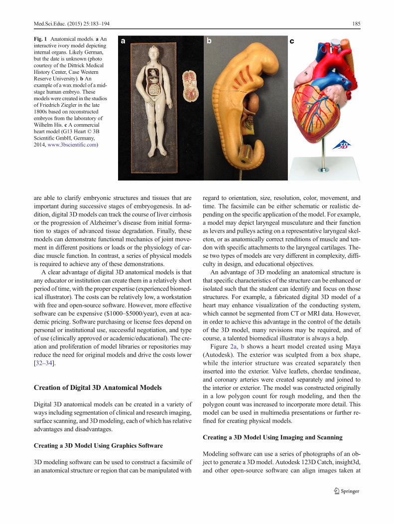
The best Bmodel^ for investigating human anatomy has al-ways been the human cadaver itself, because, in most cases,all the parts are there in the correct arrangement, the finemembranous and facial elements are intact, and the presenta-tion of structures (soft, hard, smooth, rough, dry, moist) isaccurate. It is safe to say that, from the beginning of curiosity,early man investigated wounds and organs of their dead breth-ren. However, in today’s regulated and socially conscious in-stitutions, access to a cadaver may be limited through budget-ary or social issues, or, even if a cadaver is available, presen-tation of the desired cadaveric anatomy may be confusing,such as that of the pelvic spaces and fascia. Finally, bodydonation programs, storage, chemical and biological hazardcompliance, and proper disposition of the cadaveric speci-mens may be daunting financial and logistical burdens forsome institutions. These issues can be addressed through theuse of fabricated anatomical models. Ancient and contempo-rary anatomical models range greatly in the detail and material(Fig. 1). They have advanced from simple wood or ivoryrepresentations of anatomical structures (Fig. 1a) to more de-tailed papier-mache or plaster models, to the intricate waxmodels of Susini, Towne, or Ziegler (Fig. 1b), and finally, tothe present-day commercial plastic models (Fig. 1c) [1, 2, 6,23–25]. The anatomical models also ranged in applicationincluding use by physicians tomaintain discretion with femalepatients, training surgeons in lieu of cadavers, disrupting
medico-political authorities, and of course, teaching anatomy[4, 5].
Physical anatomical models possess inherent limitations intheir use, including costs, storage requirements, security, andmaintenance. The costs of commercial models can reach thou-sands of dollars depending onmaterial, size, detail, resolution,and interactivity. In addition, particularly with large studentpopulations, physical anatomical models can be damaged,misplaced/stolen, or become just plain filthy from constanthandling by students over decades. Finally, purchasing andmaintaining the range of anatomical models required to depictall the variations and anomalies observed in human anatomyis not feasible. Thus, the use of physical anatomical modelspresents challenges in selectivity and cost.
With the development of plastination by Gunther vonHagens and the Bedutainment^ of anatomy [26–28], somefaculty speculated as to the demise of fabricated models inpreference for actual anatomical specimens that are renderedresistant to degradation and damage. Plastinized specimenshave been shown to be effective teaching tools [29, 30]. How-ever, legal/ethical liabilities in donation and waste, as well ascosts of acquisition (can be thousands of dollars for regionalanatomical specimens) and proper storage facilities impacttheir use in anatomy education [31]. In addition, most of thelimitations in the use of physical anatomical models describedabove are also applicable to plastinized specimens.
Digital 3D Models
Speculation as to the demise of physical models has also beena response to the development of advanced digital 3D render-ings of structures and specimens through medical imagery ordigital 3D modeling. Digital 3D models of anatomical struc-tures can be accessed on a computer, through mobile apps, orthrough stand-alone interactive workstations (i.e.,Anatomage, Touch of Life). The availability of these materialsin the gross anatomy laboratory and at study workstations mayreduce the need for physical anatomical models or evenprinted atlases and may facilitate teaching by bringing thematerial to the dissecting table. However, costs and spacerequirements for a dedicated interactive 3D imaging systemare significant (∼$100,000 for an Anatomage table and sup-port media), and as always, the advanced technology is likelyto become dated and unsupported over time. The use of iPadsand mobile devices reduces the costs, but they depend on thedevelopment of apps that have been assessed as to their impacton learning.
An advantage of digital 3D models is that they can bemanipulated temporally to depict changes in a structure orspecimen with regard to age or developmental stage, the im-pact of disease or injury, or functional mechanics. Digital 3Dmodels can depict morphogenesis of the heart or inner ear and
184 Med.Sci.Educ. (2015) 25:183–194
are able to clarify embryonic structures and tissues that areimportant during successive stages of embryogenesis. In ad-dition, digital 3Dmodels can track the course of liver cirrhosisor the progression of Alzheimer’s disease from initial forma-tion to stages of advanced tissue degradation. Finally, thesemodels can demonstrate functional mechanics of joint move-ment in different positions or loads or the physiology of car-diac muscle function. In contrast, a series of physical modelsis required to achieve any of these demonstrations.
A clear advantage of digital 3D anatomical models is thatany educator or institution can create them in a relatively shortperiod of time, with the proper expertise (experienced biomed-ical illustrator). The costs can be relatively low, a workstationwith free and open-source software. However, more effectivesoftware can be expensive ($1000–$5000/year), even at aca-demic pricing. Software purchasing or license fees depend onpersonal or institutional use, successful negotiation, and typeof use (clinically approved or academic/educational). The cre-ation and proliferation of model libraries or repositories mayreduce the need for original models and drive the costs lower[32–34].
Creation of Digital 3D Anatomical Models
Digital 3D anatomical models can be created in a variety ofways including segmentation of clinical and research imaging,surface scanning, and 3Dmodeling, each of which has relativeadvantages and disadvantages.
Creating a 3D Model Using Graphics Software
3D modeling software can be used to construct a facsimile ofan anatomical structure or region that can bemanipulated with
regard to orientation, size, resolution, color, movement, andtime. The facsimile can be either schematic or realistic de-pending on the specific application of the model. For example,a model may depict laryngeal musculature and their functionas levers and pulleys acting on a representative laryngeal skel-eton, or as anatomically correct renditions of muscle and ten-don with specific attachments to the laryngeal cartilages. The-se two types of models are very different in complexity, diffi-culty in design, and educational objectives.
An advantage of 3D modeling an anatomical structure isthat specific characteristics of the structure can be enhanced orisolated such that the student can identify and focus on thosestructures. For example, a fabricated digital 3D model of aheart may enhance visualization of the conducting system,which cannot be segmented from CT or MRI data. However,in order to achieve this advantage in the control of the detailsof the 3D model, many revisions may be required, and ofcourse, a talented biomedical illustrator is always a help.
Figure 2a, b shows a heart model created using Maya(Autodesk). The exterior was sculpted from a box shape,while the interior structure was created separately theninserted into the exterior. Valve leaflets, chordae tendineae,and coronary arteries were created separately and joined tothe interior or exterior. The model was constructed originallyin a low polygon count for rough modeling, and then thepolygon count was increased to incorporate more detail. Thismodel can be used in multimedia presentations or further re-fined for creating physical models.
Creating a 3D Model Using Imaging and Scanning
Modeling software can use a series of photographs of an ob-ject to generate a 3Dmodel. Autodesk 123D Catch, insight3d,and other open-source software can align images taken at
Fig. 1 Anatomical models. a Aninteractive ivory model depictinginternal organs. Likely German,but the date is unknown (photocourtesy of the Dittrick MedicalHistory Center, Case WesternReserve University). b Anexample of a wax model of a mid-stage human embryo. Thesemodels were created in the studiosof Friedrich Ziegler in the late1800s based on reconstructedembryos from the laboratory ofWilhelm His. c A commercialheart model (G13 Heart © 3BScientific GmbH, Germany,2014, www.3bscientific.com)
Med.Sci.Educ. (2015) 25:183–194 185

different angles from an object to create a digital 3D model ofthe object. The resolution and accuracy depend on the photo-graphic parameters and the software capabilities and may bequite limited. Thus, a model of a liver or femur produced usingthis method may be more instructive than a model of an iso-lated sphenoid bone (Fig. 3a).
Digital 3D anatomical models may be created using laser-based 3D scanners or micro-CT, clinical CT, and MRI [35].Laser-based 3D scanners range in price from a few hundred tomany thousands of dollars and differ in resolution, specimensize, and ease of use. These devices can create sophisticatedwatertight 3D models that can be used as content in anima-tions or in 3D printing. Laser-based scanners can be handheld,desktop, or industrial instruments that interpret the surface ofan object either through laser triangulation, which usesreflected light from the surface of the object to calculate dis-tance, or laser pulse-based/phase shift to measure time-of-flight of reflected light to calculate distance. Models createdfrom these methods depict surface structure only and mayhave resolution and accuracy concerns. Thus, the use of sur-face scanning in the creation of 3D models has limitations.
Micro-CT and clinical CT imaging use X-ray emissions,while MRI uses radio frequency-based emissions to generatecross-sectional images of an object that can be used to create3D reconstructions of the object. Micro-CT imaging can pro-duce precise digital models at a resolution of 0.5 μm (Fig. 3b),while clinical CTand MRI can resolve structures on a submil-limeter scale. In many cases, a clinical scan is sufficient toreconstruct anatomical structures such as organs and cavities[36] (Fig. 3c), but micro-CT may be required for detailedosteological analysis or the generation of high-resolutionmodels. In both micro-CT and clinical CT, parameters may
be adjusted to visualize internal structures of soft tissue andbone. These structures may be isolated through segmentation,as described below, to extract specific structures from withinlarger scans. Most digital 3D anatomical models are createdfrom clinical CT imagery simply because patient data is col-lected using this modality and a large collection of these dataexist. In addition, the chamber size of most micro-CT units isquite small and unable to scan long bones or large specimens.
MRI data can be used to generate digital 3D models ofanatomical structures and readily detects soft tissue structure,which makes it more useful than CT in segmentation andextraction of soft structures. The advantages of MRI data arethe detection of soft tissues, the large array of patient data thatalready exists, and the lack of exposure of the subject toradiation.
Disadvantages of these imaging methods are cost (a micro-CTscan of a sphenoid bone in an institutional core facility maycost $200–300), scan size, and consent issues for patient data.Hospitals and clinics have large repositories of clinical CT andMRI patient data that, if organized properly, would offer atreasure trove of possible anatomical models for use in educa-tion. However, both Internal Review Board approval and closecooperation between radiologists, medical physicists, and ed-ucators are required. Public repositories of scanned materialand segmented structures exist and are expanding (i.e., Nation-al Biomedical Imaging Archive (NBIA); Cornell Visualizationand Image Analysis (VIA) group).
Creating a 3D Model Using Segmentation
While digital 3Dmodels created by CTandMRI scanning canbe quite detailed and can be modified to visualize specific
Fig. 2 Surface (a) and meshrenders (b) of a heart modelcreated using Maya and 3D Max(Autodesk, San Rafael, CA,USA)
186 Med.Sci.Educ. (2015) 25:183–194

tissues, creating models of specific organs or tissues containedwithin the scan requires the use of segmentation. Segmenta-tion assigns a label to pixels within an image that correspondto a specific structure. For example, the isolation and recon-struction of the hyoid bone in a head and neck CTorMRI scanseries may be accomplished by the selection of pixels basedon contrast differences between the bone and surroundingtissue in the appropriate sections. The selection of pixels canoccur by either assigning a mask to select for pixels corre-sponding to the hyoid bone or tracing the hyoid bone fieldand excluding the rest of the data. Segmentation is relativelyeasy when isolating bone or the tracheal lumen because con-trast thresholds can be a range that is sufficiently narrow toexclude unwanted pixels. However, soft tissues are more dif-ficult to segment due to similar grayscale values on the image.For example, if the desired structure is the peritoneal
membrane, contrast differences between the peritoneal mem-brane and the surrounding fascial and muscular layers may notbe sufficient to automatically or semiautomatically segmentinto different structures. In this case, the structure must bemanually segmented in each image, followed by digital recon-struction. Thus, substantial effort may be required to isolateand reconstruct some tissues.
There is an array of software (free ware and commercial)that enables segmentation and reconstruction of Digital Imag-ing and Communications in Medicine (DICOM) medical im-age stacks. The DICOM format is the international standardformat for medical images (ISO 12052) and has replaced theuse of X-ray film. Osirix, 3D-Slicer, and InVesalius are exam-ples of free-ware or low-cost DICOM viewers that enablesegmentation. A combination of Osirix free ware and the Vir-tual Human enables anyone to create 3Dmodels of anatomicalstructures, but the resolution may be below that suitable formedical education. More powerful segmentation software so-lutions exist, such as Mimics (Materialise), but their costs canbe substantial (can be $1–5000/year depending on version,capabilities, and negotiation). The use of more powerful seg-mentation software cannot offset the issues of resolution of theimage, but they can simplify the process through automationand creative segmentation [37–39].
The DICOM image reconstruction typically converts thesegmented anatomy to a 3D mesh model, such as astereolithography or standard tessellation language (STL) file.The STL file can be further processed into a nonuniform ra-tional B-spline (NURBS) model as needed using appropriatesoftware, including Geomagic (3D Systems) and Rhino(Robert McNeel & Associates). Either the mesh or NURBSmodel can be edited in 3D modeling software to enhancefeatures or correct problems encountered in segmentation.Mesh-based 3D modelers include Maya (Autodesk), Blender(The Blender Foundation), andMagics (Materialise); NURBS3D modelers include Rhino and mainstream CAD softwaresuch as Autodesk Creative Suite (Autodesk) and Solidworks(Dassault Systèmes SolidWorks Corp.).
Application of the Digital 3D Model
Once the desired digital 3D anatomical model is obtained, itcan be used in animations or as an object in an interactiveformat. Advantages that these models have over the cadaveror physical 3D models include their application in many edu-cational formats (lectures, online material, and print) and por-tability (downloadable to any PC or mobile device). Thesemodels can be altered to enhance desired learning objectivesor to conform to learner characteristics. These models offer agreat advantage over static 2D images in terms of orientationand exploration of internal structure. For example, a 2D imageof a heart may show extreme detail, but a student may be ableto Bfly through^ a digital 3D model of the heart to obtain a
Fig. 3 Digital 3D models created using different methods. aModel of asphenoid bone created using Autodesk 123D catch software (Autodesk,San Rafael, CA, USA). Resolution is poor and below that needed for usein education. b Model of a sphenoid bone obtained through micro-CTimaging and reconstruction using 3D Slicer (www.slicer.org). Highresolution is obtained and the model can be used at size or scaledwithout loss of structure and resolution. c A 3D volume render of aheart from clinical CT imaging. Gross structure of the heart chambers isclear, while the epicardial surface and myocardial layers are missing
Med.Sci.Educ. (2015) 25:183–194 187

clear view of the size and location of specific structures, aswell as their relationship with surrounding anatomy. In addi-tion, digital 3D models can be interactive or can depict aspecific function (joint movement, ovulation).
Using Digital 3D Anatomical Models to CreatePhysical Models
A technological advance that may reduce the need for pur-chasing or maintaining a large library of physical 3D anatom-ical models is additive manufacturing. Additive manufactur-ing describes a field of fabrication technologies that build apart by the joining of materials layer by layer to produce aspecific object [40]. Additive manufacturing technology (re-ferred to here as 3D printing, a generic term) has advancedtremendously over the past two decades and has become func-tional in the development and construction of physical 3Danatomical models.
In general, 3D printers use the STL file format as the inputfor the build geometry. In order to successfully 3D print amodel, the STL file needs to be watertight and free of othererrors. For example, the model shown in Fig. 2a requiredpreprocessing by propriety software that is vendor and printerspecific. In this case, Insight (Stratasys), which is the prepro-cessing software used to analyze and prepare models for 3Dprinting on the Stratasys range of 3D printers, was used. In-sight provides manipulation of a range of printer build param-eters that determine the build quality and resolution of thefinished model. This process was followed by a virtual buildof the model at the individual layer level to assess the previ-ously set parameters. This is typically an iterative processwhere some compromises are often made between speed,quality, and cost. For the model in Fig. 2a, this analysis ini-tially indicated that some model features were too small toprint appropriately. These errors were mainly a result of dataconversion from the native 3D modeling format to a .stl rep-resentation, which is the required data format for the prepro-cessing software. Thus, an otherwise well-constructed modelmay require several adjustments in order to print using thedesired method or material.
Methods of 3D Printing
Several different methods of 3D printing have been devel-oped, and each has specific benefits and limitations in thecreation of anatomical models (Table 1). Differences betweenthe methods and the printers themselves include materialsavailable, resolution, accuracy, repeatability, stability, costs,safety, size limitations, speed, and the number of materialsper build. The importance of each of these parameters differsdepending on the specific application of the printed object. T
able1
Resolutiondatawas
obtained
from
themanufacturer’sspecifications
atwww.stratasys.com
,www.3Dsystem
s.com,and
www.Zcorp.com
Additive
manufacturing
technology
Materials
Material/build
Approximate
resolution
(mm)
Stability
Safety
Advantages
Disadvantages
Stereolithography
(SLA)
Proprietaryresins
invariouscolorsand
opacities
10.05
Changes
inflexibility
andopacity
with
time
Resin
off-gassing
Relativelyfastbuild
time
Printerandmaterialcosts($1000s
to$100,000s),requiressupport
materialand
postprocessing
Selectivelaser
sintering(SLS)
Powderedglass,
ceramic,m
etal,
plastic
10.1
Stable
Laser
andfumes
Noseparatesupport
materialrequired
Printerandmaterialcosts
($100,000s),very
limitedoptio
nsforflexiblematerials
Fuseddeposition
modeling(FDM)
Therm
oplastics
1/nozzle
0.18
Stable
Fumes
andultrafine
particles
Low
-cost(hobby)
machines
(aslowas
$300)and
DIY
kitsavailable.
Solublesupports
Productio
nprintersandmaterialcosts
($100,000s),requires
supportand
very
limitedoptio
nsforflexible
materials
Powder-binder
printin
g(PBP)
Plaster,cornstarch
1,butm
ultip
lecolors
0.1
Stable
Skin
sensitivity
tomaterials,particles
Fastbuild
time,no
separate
supportm
aterialrequired
Extremelyfragile
modelsthatrequire
infiltrationto
increase
strength.
Cost$
1000sto
$100,000s
Polyjet
UV-cured
proprietary
resins
inflexible
andrigidform
s
>1material
and>1color
0.016
Changes
inflexibility
andopacity
with
time
Skin
sensitivity
touncuredresin
bestavailablesurface
finish
andresolutio
n,custom
izablematerial
properties
Productio
nprintersandmaterial
costs($100,000s)requires
supports
188 Med.Sci.Educ. (2015) 25:183–194
For the creation of anatomical models for education, resolu-tion, stability, and cost are important parameters.
SLA uses an ultraviolet laser to cure photosensitive resin insequential, thin, horizontally oriented layers that eventuallyconstruct the desired object [41]. Detailed models can be cre-ated in hours. However, the disadvantages of this methodinclude costs of the printer ($1000 to >$100,000, althoughprices are dropping), costs of the materials, postprocessing(removal of support material and curing), and long-term sta-bility of the object.
Selective laser sintering (SLS) uses lasers to fuse powderedmaterials into a desired shape [42]. This method allows 3Dprinting in a variety of materials including powders of plastic,metal, glass, and ceramic. SLS does not require support ma-terials since the unsintered material acts as support. Stabilitydepends upon the material used.
Fused deposition modeling (FDM) uses thermoplastics ex-truded by a heated nozzle in a semiliquid state to producelayers of the desired object that then harden immediately afterextrusion [43]. Different types of thermoplastic materials canbe used that differ in hardness, flexibility, color, and translu-cency. Removable support material is required for smaller orprotruding parts.
Powder-binder printers lay down a layer of powdered mate-rial followed by a binding agent. Colors can be applied selec-tively in the bindermaterial. Distinct advantages are the printingspeed and ability to print an object using multiple colors.
Polyjet technology builds 3D models by laying down pho-topolymer that is cured by UV. Speed and accuracy are advan-tages, but support material is required. In addition, the long-term stability of some materials can be a significant issue.
Although much improvement in costs, resolution, objectscanning, image segmentation, and printing materials is need-ed for 3D printing to usurp the business model of commercialanatomical suppliers, these improvements may not be faraway. The printer itself has been a costly investment, rangingfrom a few to hundreds of thousands of dollars. However, thecost is dropping rapidly with the development of desktop 3Dprinters (<$300) and more inexpensive materials (grass trim-mer filament, recycled plastic bottles). The resolution of theprinted object is also a consideration. Some higher end printersclaim <0.01 mm resolution, but the costs of such a printer canbe a substantial burden to most institutions. The resolution ofmost desktop printers may be insufficient for printing a faithfulreplica of a structure as delicate as a sphenoid bone or anatrioventricular valve leaflet, especially if scaled to a smallersize. However, a nice articulated hand skeleton can be printedwith attached musculature at these resolutions.
Materials
Many different materials can be used to construct a 3D printedobject. In addition, several different materials or colors can be
used. This ability allows the construction of complex modelswith hard, soft, opaque, and transparent components. Thermo-plastics, including acrylonitrile butadiene styrene (ABS) andpolylactic acid (PLA), ferrous and nonferrous metals, ceramic,elastic polymers, and many opaque, colored, and transparentproprietary materials can be used in the construction of anobject, along with support materials that can be dissolved orwashed away in postproduction processing. The choice ofmaterial must be made on esthetic, structural, and practicalparameters. Strength, color, flexibility, opacity, stability, andcosts must be considered. In addition, the printing processmay require postprocessing of the object to ensure strength,durability, or transparency.
Examples of materials are shown in Fig. 4a, b. The heartmodel described above was printed on a Stratasys Fortus 400in ABS with PLA as support material (Fig. 4a) and on aStratasys Connex 350 in TangoPlus (Stratasys) with a separateremovable material as support (Fig. 4b). Internal chambers,valve leaflets, chordae tendineae, and coronary artery lumenare demonstrated in both the ABS model and the flexibleTangoPlus print (Fig. 4c, d). The TangoPlus model requiredprinting in a split model in order to remove support material(nonpolymerized material) efficiently without damage to thefine structures. The removal of support material should beconsidered in model design, in particular in printing fine struc-tures or using fragile materials.
Resolution and Accuracy
The resolution of a printed object is dependent upon the res-olution of the digital 3D model, the resolution that can beachieved by the printer itself, and the material used in printing.Using a poorly constructed or low-resolution digital 3Dmodelon an expensive, high-resolution printer will produce poorlyconstructed and low-resolution physical 3D models. Con-versely, well-constructed 3D models may not be able to over-come the limitations of a low-resolution less expensive printer.Finally, the choice of material can affect resolution if the ma-terial cannot be extruded or cured in a sufficiently smallamount or pattern or if the material cannot be supported prop-erly during object construction.
The accuracy of a printed model in the representation of thedigital model is dependent on the printing method, the capa-bilities of the specific printer, and the material used [44]. Ac-curacy with ±0.0–1.0 % seems to be the range of commercialprinters; however, maintaining those tolerances requires cali-bration and maintenance. Different materials have a differentimpact on accuracy both at the time of printing and over timeafter printing. ABS, once cooled, is very stabile in configura-tion and appearance over time. However, polyjet and SLAresins may show alterations in dimension, opacity, and flexi-bility. In anatomical model construction for medical educa-tion, dimensional accuracy may not be as important as
Med.Sci.Educ. (2015) 25:183–194 189

resolution and stability, since there is much variation in ana-tomical structure.
Desired resolution and accuracy as well as required stabil-ity affect the choice of printer and material. These parametersmay also affect the speed of the printing process and theamount of materials used. These parameters also affect thecost of the printed object. Trade-offs between resolution andcosts must be assessed individually to avoid a negative impacton accuracy or usefulness of the model.
Costs
The costs of a printed object are dependent upon the size andcomplexity of the 3D model, the choice of material used, andthe cost to operate the printer. Commercial models offer someguarantees of quality and accuracy that can be assured throughthe inspection of the model prior to purchase. The creation of3D models by faculty for use in local curricula may requireseveral versions to achieve a suitable digital 3D model andprinted 3D object. At this time, these costs may indicate that acommercial model is a better choice. Creating and printing afabricated digital 3D heart model with accurate internal struc-ture may cost thousands of dollars in artist and engineer time,print costs, and assessment of educational impact. Of course,
many models can be printed after achieving a good design,which would decrease the costs overall. However, if 3D print-ing is to be a significant educational technology in medicaland dental education, costs need to be reduced drastically.
Safety Concerns
There are safety concerns with the use of 3D printers. Someprinters use high voltage and can have extremely hot surfaces,which presents obvious concerns. The results of a recent studysuggest that some 3D printers emit ultrafine particles [45].Particles of similar size have been shown to find their wayinto pulmonary airways, and even the brain, resulting in respi-ratory and neurological symptoms [46–49]. In addition to par-ticles, heating some plastics, as in FDM, releases toxic com-pounds including benzene, carbon monoxide, hydrogen cya-nide, and hydrogen chloride, which may be harmful in suffi-cient concentrations [50, 51]. These are not trivial concernssince the detrimental health effects of ultrafine particle inha-lation or toxic compound exposure may not become apparentfor many years. Most commercial printers operate in a closedcompartment or environment that can contain or filter theemitted particles or fumes. However, unless specific ventila-tion systems are in place, 3D printing should be performed
Fig. 4 Physical 3D modelsprinted using FDM and polyjetmethods and the digital modeldepicted in Fig. 2. a Completemodel printed in ABS on aStratasys Fortus 400 printer(Stratasys, Rehovot, Israel). bInternal structure of the modeldepicted in a (valve leaflets,chordae tendinae, and papillarymuscles) is visible. c Completemodel printed in TangoPlus on aStratasys Connex 350 (Stratasys,Rehovot, Israel). d Internalstructure of the model depicted inc (valve leaflets, chordaetendinae, and papillary muscles)is visible
190 Med.Sci.Educ. (2015) 25:183–194

cautiously. This is especially true when using open DIYprinters. In addition, postprocessing may require causticchemicals, sanding, or coating, which present safety hazardsas well.
Evidence of Efficacy in Learning
A digital 3Dmodel offers advantages over digital 2D or phys-ical models in interactivity, perspective, access, and cost, ei-ther as a stand-alone learning asset or as part of a larger digitallearning object. Studies have examined the use of digitallearning objects in education [52, 53], and several have evenconsidered these materials in medical education [10, 13, 18,19, 22]. Evidence has been presented for positive, negative,and neutral impacts of digital models on student learning [18,53–58]. Thus, questions remain as to whether digital 3Dmodels or learning objects are more useful and effective instudent learning than text or physical models.
Physical 3D anatomical models offer some advantages tostudents over simple illustrations, text, or even virtual models[3, 8]. One mechanism that models may apply to learning isthat of offloading cognition or the freeing of cognitive re-sources during learning [59]. Although this specific articleexamined learning in children, the hypothesis may have somesignificance in adults. As an example, a detailed physicalmodel of the isolated sphenoid bone provides a student visualand tactile data concerning the size, regions, foramina, spines,and processes that are important in understanding the role ofthis bone in cranial organization. Thus, valuable cognitiveresources may be redirected to understanding the correlationof cranial nerves and muscles to specific points on the model,rather than trying to understand the relationships between thegreater and lesser wings of the sphenoid on a 2D illustration.
Implementation of 3D Modeling and Printingin Medical and Dental Curricula
Digital 3D Anatomical Models
Digital 3D anatomical models can applied in many ways in amedical or dental curriculum. Onlinematerials, lecture presen-tations, instructor notes, and assessment can include 3Dmodels of organs or tissues that are interactive (interactivePDFs) and enhanced in attention to specific structures. Char-acteristics of the models, such as resolution, color, scale, opac-ity, interactivity, time, and the inclusion of adjunct images ortext, can be manipulated to enhance specific learning objec-tives or to target specific student populations. As describedabove, these models can be used as individual learning assetsor as part of a larger learning object.
Printed Anatomical Models
The simplest implementation of printed 3D anatomicalmodels into medical and dental curricula is as current anatom-ical models are used. The value of 3D printing lies in thechoice of anatomical perspective, resolution, and scale. In-structors can now create models from specific perspectives,such as disarticulated skull bones, expanded models of thetemporal bone, and pulmonary or vascular structures.
A limitation to the application of 3D printed models is thedepiction of fascia. Anatomical education is incomplete with-out the student understanding fascia, fascial compartments,and connective tissue [60]. Physical anatomical models, eithercommercial, plastinized, or 3D printed, cannot depict fine fas-cial elements and connective tissue compartmentalization oforgans and vessels. In curricula that do not engage in cadaverdissection or prosection, the students are at a disadvantage andmust learn these concepts in postgraduate education.
Costs of printed 3D models have been cited as impedi-ments to their implementation into the medical curriculum[61]. However, after the model is created and verified and withoptimization of the printing parameters, subsequent prints aremuch less expensive than the first print or a quality commer-cial model. If costs go downwith time, as is anticipated, it maybe reasonable to print small models for all students to have andreview themselves. There may come a time where ideal ca-davers can be printed in their entirety in material suitable forBdissection,^ with incorporation of fascial elements, no con-straints in variation or size, and in reasonable time.
Clinical Case Studies
Recently, Zein and colleagues reported that the 3D printing ofthe livers of living donors may improve preoperative surgicalplanning, reduce unnecessary surgeries, and decrease thecomplications of liver transplant surgery [62]. Similar preop-erative organ studies have been performed by other groups[60, 63–67]. The 3D printed liver models, when linked topatient data, can be an effective learning object to teach anat-omy, pathology, and radiology to medical students and resi-dents. This learning object provides the student an opportunityto confirm imaging findings and to understand the relationshipof the image with the 3D model or actual liver. While theseconcepts seem trivial, they are the basis of understanding thevisual models created by CT or MRI and the application ofthose models to clinical cases. The difficult question is how toimplement such an object into the medical curriculum.
Many medical schools have incorporated active learningmethods in their medical curriculum, including small-groupproblem-based learning (PBL). In its usual form, PBLmethods introduce basic science or clinical concepts to themedical student through specific clinical case presentations.This format fits well with the concept of using a comparison of
Med.Sci.Educ. (2015) 25:183–194 191
digital imaging and 3D model construction to teach anatomyand radiology. This implementation can also be applied toother organ systems and clinical cases to include cardiopul-monary, renal, neural, and skeletal concepts. However, broad-ening of the implementation of this method would require amethod of obtaining both usable 3D models and correspond-ing patient data. The collaboration of clinical radiologists toidentify possible cases, 3D modelers to segment the patientdata and create the physical 3D models, and medical schooleducators to design a complete learning object around theseassets is required. In addition, there must be assurance that thepatient data is handled according to Internal Review Boardand Health Insurance Portability and Accountability Act of1996 (HIPAA) guidelines. Further, the costs involved in thecreation of printable models and the printing of these modelsin sufficient size, quantity, and resolution are substantial. The-se obstacles will likely be reduced with time by the creation ofinstitutional and commercial case databases that can beaccessed by association or license and the anticipated signifi-cant reduction in cost of 3D printers and materials.
Assessment of the impact of these new teaching methodson student learning is required to determine if they are worththe effort in full implementation. For schools with a fairlystable curriculum, simple comparisons of the student perfor-mance before and after implementation may be sufficient toconsider continuing implementation. Other schools that maybe transitioning between curricular structure and conceptswould need to be more creative in their assessment protocolto separate outcomes from the influence of other significantcurricular changes. In this case, extracurricular assessmentmay be required, such as random assignment of students todifferent learning groups that are exposed to different teachingmethods or large focus group data. In either case, validation ofthe teaching methodology is critical to proceed with advancesin implementation of these technologies in the medicalcurriculum.
A Convergence of Digital 3D Modeling and 3D Printing
The concept of Btangible multimedia^ has been described asthe linking of online educational content with a tangible ob-ject, which, together, represents the learning object [68]. Stud-ies have examined the impact of this concept on learning inpreschool children, whom they describe as having a Blargelearning gap^ in the multimedia environment since they lacklogical thinking and abstract reasoning. Medical and dentalstudents do not lack logical thinking or abstract reasoning;however, they may have difficulty reconstructing a clear viewof an anatomical structure or embryological event from a dig-ital rendering, then applying that view to clinical concepts. Atangible object may enable the student to overcome this diffi-culty and enhance learning by reducing extraneous cognitive
load. Thus, learning may be enhanced by linking multimediaeducational material with a tangible object.
A direct application of tangible multimedia could involvethe implementation of a digital simulation of human embry-onic cardiovascular development in the medical curriculum.The material may consist of animations of primitive heart tubeformation and heart looping, as well as atrial, ventricular, andoutflow tract septation, all of which have been indicated bymedical students in focus group data as conceptually difficult.Digital 3Dmodels of normal hearts and hearts with congenitalanomalies allow the user to view andmanipulate renderings ofspecific anomalies and to confirm the information presented inthe simulation. As the students complete the material and em-bedded assessment tools, they would be provided a 3D printedheart (tangible object) produced from the same digital 3Dmodels observed in the animations and interactive materials.Themodels are printed in a form that provides repetition of thelearning objectives and reinforcement of embryological con-cepts. The built-in assessment in the form of quizzes or pos-sibly small games will allow tracking of student performance,as well as short-term goals to advance through the material.This format allows implementation of the material withoutdisruption of the current curriculum. It can be treated as ad-junct material or a principal teaching tool. What is unique hereis that the students end the material with a tangible model thatthey can manipulate. This teaching tool can also be applied topatient and family education to strengthen informed consentand explore treatment choices.
Future Considerations
The prospects for application of digital 3D models and 3Dprinting in medical education are extensive. As the technolo-gies advance, it is likely that current issues such as cost andsafety will be addressed and become less significant.
A limitation in the application of 3D printing in medicaleducation and a significant factor in cost is the availability ofdigital 3D models. A great idea for a printed model waits onthe creation of a usable digital 3D model in a format recog-nized by a 3D printer. If the digital 3D model requires seg-mentation from clinical imaging or creation using 3D model-ing software, it may take some time to complete. Time andeffort spent by an engineer, technician, radiologist, or biomed-ical illustrator to segment or create a digital 3D model add tothe costs. One solution is the creation of peer-reviewed digital3Dmodel libraries or repositories. Authors submit models thatare reviewed and revised. If accepted, they are placed in thelibrary for use in education. A small fee could be charged tosupport the operation of the library. While a basic set ofmodels would be similar to a basic anatomy atlas, the numberof models required to depict difficult anatomy, anatomicalvariations, and anomalies is vast. Searching a library and
192 Med.Sci.Educ. (2015) 25:183–194
obtaining an appropriate model though licensing is muchmore cost effective than creating a model from scratch.
The implementation of 3D printing, as well as any newteaching tool into the curriculum, must be assessed for effica-cy in learning. A positive assessment adds to the cost effec-tiveness of the material and the technology. Assessment in thecase of digital learning objects can be imbedded in the mate-rial as quizzes, case studies, and even games. The embeddedassessment along with tracking of user performance enablesthe entire learning object to be a stand-alone package that,when accessed, provides performance outcomes. Combinethis type of assessment with evaluation of the Bdissection^of a 3D printed organ or anatomical region and a completerecord of student engagement in the material and learningoutcome can be obtained, all without disruption of the currentcurriculum.
Safety in the process of 3D printing, the materials used, andassurances of good environmental stewardship are importantto the advancement of 3D printing technologies. Informationas to safe exposure rates to ultrafine particles and compoundsemitted during 3D printing using specific materials should beprovided to users, and users should be cognizant of the need toprotect themselves and others from excessive exposure.
Acknowledgments The authors thank Dr. James Edmonson and LauraTravis of the DittrickMuseum at CaseWestern Reserve University for theivory model image; Drs. Raymond Muzic, Joseph Molter, and PrabhakarRajiah from University Hospitals for their help understanding medicalimaging and segmentation; Kellie Pasini and staff at Thinkbox CWRUfor their help with modeling and printing; and 3B Scientific for commer-cial heart model images. Some aspects of this study and review werefunded by a Nord Grant through the University Center for Innovation inTeaching and Education at Case Western Reserve University.
Conflict of Interest The authors declare no conflict of interest.
Open Access This article is distributed under the terms of the CreativeCommons Attribution License which permits any use, distribution, andreproduction in any medium, provided the original author(s) and thesource are credited.
References
1. Marković D, Marković ŽB. Development of anatomical models—chronology. Acta Med Medianae. 2010;49:56–62.
2. Riva A, Conti G, Solinas P, Loy F. The evolution of anatomicalillustration and wax modelling in Italy from the 16th to early 19thcenturies. J Anat. 2010;216:209–22.
3. PawlinaW, Drake RL. Anatomical models: don’t banish them fromthe anatomy laboratory yet. Anat Sci Educ. 2013;6:209–210.22.
4. Russell KF. Ivory anatomical manikins. Med Hist. 1972;16:131–42.
5. Maerker A. Florentine anatomical models and the challenge ofmedical authority in late-eighteenth-century Vienna. Stud HistPhil Biol Biomed Sci. 2012;43:730–40.
6. Hopwood N. The art of medicine. Lancet. 2008;372:1946–7.
7. Khot Z, Quinlan K, Norman GR, Wainman B. The relative effec-tiveness of computer-based and traditional resources for educationin anatomy. Anat Sci Educ. 2013;6:211–5.
8. Preece D, Williams SB, Lam R, Weller R. BLet’s get physical^:advantages of a physical model over 3D computer models andtextbooks in learning imaging anatomy. Anat Sci Educ. 2013;6:216–24.
9. Lombardi SA, Hicks RE, Thompson KV, Marbach-Ad G. Are allhands-on activities equally effective? Effect of using plasticmodels,organ dissections, and virtual dissections on student learning andperceptions. Adv Physiol Educ. 2014;38:80–6.
10. Ruiz JG, Mintzer MJ, Issenberg SB. Learning objects in medicaleducation. Med Teach. 2006;28:599–605.
11. Sugand K, Abrahams P, Khurana A. The anatomy of anatomy: areview for its modernization. Anat Sci Educ. 2010;3:83–93.
12. Ayres P, Paas F. Can the cognitive load approach make instructionalanimations more effective? Appl Cogn Psychol. 2007;21:811–20.
13. Mayer RE. Applying the science of learning to medical education.Med Educ. 2010;44:543–9.
14. Moreno R. Optimising learning from animations by minimisingcognitive load: cognitive and affective consequences of signallingand segmentation methods. Appl Cogn Psychol. 2007;21:765–81.
15. Tabbers HK, Martens RL, van Merriënboer JJG. Multimedia in-structions and cognitive load theory: effects of modality and cueing.Br J Educ Psychol. 2004;74:71–81.
16. Fredieu J, Watson J, Hughart C, Nikiforova T. Human develop-ment: development of the face and palate. MedEdPORTAL.2011;8334.
17. Hu A,Wilson T, Ladak H, Haase P, Doyle P, Fung K. Evaluation ofa three-dimensional educational computer model of the larynx:voicing a new direction. J Otolaryngol Head Neck Surg. 2010;39:315–22.
18. Marsh KR, Giffin BF, Lowrie Jr DJ. Medical student retention ofembryonic development: impact of the dimensions added by mul-timedia tutorials. Anat Sci Educ. 2008;1:252–7.
19. Tan S, Hu A, Wilson T, Ladak H, Haase P, Fung K. Role of acomputer-generated three-dimensional laryngeal model in anatomyteaching for advanced learners. J Laryngol Otol. 2012;126:395–401.
20. Yue C, Kim J, Ogawa R, Stark E, Kim S. Applying the cognitivetheory of multimedia learning: an analysis of medical animations.Med Educ. 2013;47:375–87.
21. Garg AX, Norman GR, Eva KW, Spero L, Sharan S. Is there anyreal virtue of virtual reality?: the minor role of multiple orientationsin learning anatomy from computers. AcadMed. 2002;77(10):S97–9.
22. Ruiz JG, Cook DA, Levinson AJ. Computer animations in medicaleducation: a critical literature review. Med Educ. 2009;43:838–46.
23. Ballestriero R. Anatomical models and wax Venuses: art master-pieces or scientific craft works? J Anat. 2010;216:223–34.
24. Hopwood N (2002) Embryos in wax: models from the ZieglerStudio. Cambridge and Bern: Whipple Museum of the History ofScience, University of Cambridge, and Institute of the History ofMedicine, University of Bern.
25. Maerker A. Anatomizing the trade. Technol Cult. 2013;54:531–62.26. Guyer RL. Metamorphosis: beautiful education to smarmy edutain-
ment. Am J Bioeth. 2007;7:30.27. Valdecasas A, Correas A, Guerrero C, Juez J. Understanding com-
plex systems: lessons from Auzoux’s and von Hagens’s anatomicalmodels. J Biosci. 2009;34:835–43.
28. Von Hagens G. Impregnation of soft biological specimens withthermosetting resins and elastomers. Anat Rec. 1979;194:247–55.
29. Douglass C, Glover R. Plastination: preservation technology en-hances biology teaching. Am Biol Teach (Natl Assoc Biol Teach).2003;65:503.
Med.Sci.Educ. (2015) 25:183–194 193

30. Latorre RM, García-Sanz MP, Moreno M, Hernández F, Gil F,López O, et al. How useful is plastination in learning anatomy?Vet Med Educ. 2007;34:172–6.
31. Jones DG. Re-inventing anatomy: the impact of plastination onhow we see the human body. Clin Anat. 2002;15:436–40.
32. Becker BW. Digital learning object repositories. Behav Soc SciLibr. 2010;29:86–8.
33. Kerfoot E, Lamata P, Niederer S, Hose R, Spaan J, Smith N. Shareand enjoy: anatomical models database—generating and sharingcardiovascular model data using web services. Med Biol EngComput. 2013;51:1181–90.
34. Richards G, McGreal R, Hatala M, Friesen N. The evolution oflearning object repository technologies: portals for on-line objectsfor learning. J Dist Educ. 2002;17:67–79.
35. Wulf J, Rohde I, Koppe T, Winder RJ. Three-dimensional micro-imaging (μCT) based physical anatomic teaching models: imple-mentation of a new learning aid for routine use in anatomy lectures.Stud Health Technol Inform. 2012;173:549–51.
36. McMenamin PG, Quayle MR, McHenry CR, Adams JW. The pro-duction of anatomical teaching resources using three-dimensionalD3D] printing technology. Anat Sci Educ. 2014. doi:10.1002/ase.1475. Article first published online.
37. Heimann T, Meinzer H. Statistical shape models for 3D medicalimage segmentation: a review. Med Image Anal. 2009;13:543–63.
38. van Rikxoort EM, van Ginneken B. Automated segmentation ofpulmonary structures in thoracic computed tomography scans: areview. Phys Med Biol. 2013;58:R187–220.
39. Zhuang X. Challenges and methodologies of fully automatic wholeheart segmentation: a review. J Healthc Eng. 2013;4:371–408.
40. Huang S, Liu P, Mokasdar A, Hou L. Additive manufacturing andits societal impact: a literature review. Int J Adv Manuf Technol.2013;67:1191–203.
41. Hull CW. Apparatus for production of three-dimensional objects bystereolithography. Patent. 1986; 4575330, Mar 11.
42. Deckard C, Beaman J. Recent advances in selective laser sintering,14th Conference on Production Research in Technology(University of Michigan). 1987; pp. 447–451.
43. Crump S. Fused deposition modeling (FDM): putting rapid back inprototyping, 2nd Int. Conf. on Rapid Prototyping. 1991; p. 358–361
44. Salmi M, Paloheimo K, Tuomi J, Wolff J, Mäkitie A. Accuracy ofmedical models made by additive manufacturing (rapidmanufacturing). J Craniomaxillofac Surg. 2013;41:603–9.
45. Stephens B, Azimi P, El Orch Z, Ramos T. Ultrafine particle emis-sions from desktop 3D printers. Atmos Environ. 2013;79:334–9.
46. Andersen ZJ, Olsen TS, Andersen KK, Loft S, Ketzel M,Raaschou-Nielsen O. Association between short-term exposure toultrafine particles and hospital admissions for stroke inCopenhagen, Denmark. Eur Heart J. 2010;31:2034–40.
47. Peters A,Wichmann HE, Tuch T, Heinrich J, Heyder J. Respiratoryeffects are associated with the number of ultrafine particles. Am JRespir Crit Care Med. 1997;155:1376–83.
48. Schmid O, Möller W, Semmler-Behnke M, Ferron GA, Karg E,Lipka J, et al. Dosimetry and toxicology of inhaled ultrafine parti-cles. Biomarkers. 2009;14 Suppl 1:67–73.
49. Shannahan JH, Kodavanti UP, Brown JM. Manufactured and air-borne nanoparticle cardiopulmonary interactions: a review ofmech-anisms and the possible contribution of mast cells. Inhal Toxicol.2012;24:320–39.
50. Schaper MM, Thompson RD, Detwiler-Okabayashi K. Respiratoryresponses of mice exposed to thermal decomposition products from
polymers heated at and above workplace processing temperatures.Am Ind Hyg Assoc J. 1994;55:924–34.
51. Shapi MM, Hesso A. Gas chromatographic-mass spectrometricanalysis of some potential toxicants amongst volatile compoundsemitted during large-scale thermal degradation of poly (acryloni-trile-butadiene-styrene) plastic. J Chromatogr. 1991;562:681–96.
52. Falloon G, Janson R, Janson A. Digital learning objects: a need foreducational leadership. Ágora. 2009;44:48–53.
53. Farha NW. An exploratory study into the efficacy of learning ob-jects. J Educ Online. 2009;6:1–32.
54. Jenkinson J.Measuring the effectiveness of educational technology.Proc Int Conf e-Learn. 2009;222–227.
55. Keedy AW, Durack JC, Sandhu P, Chen EM, O’Sullivan PS,Breiman RS. Comparison of traditional methods with 3D computermodels in the instruction of hepatobiliary anatomy. Anat Sci Educ.2011;4:84–91.
56. McNulty JA, Sonntag B, Sinacore JM. Evaluation of computer-aided instruction in a gross anatomy course: a six-year study.Anat Sci Educ. 2009;2:2–8.
57. Nguyen N, Nelson AJ, Wilson TD. Computer visualizations: fac-tors that influence spatial anatomy comprehension. Anat Sci Educ.2012;5:98–108.
58. Sergovich A, Johnson M, Wilson TD. Explorable three-dimensional digital model of the female pelvis, pelvic contents,and perineum for anatomical education. Anat Sci Educ. 2010;3:127–33.
59. Manches A, O’Malley C. Tangibles for learning: a representationalanalysis of physical manipulation. Pers Ubiquit Comput. 2012;16:405–19.
60. Korf H, Wicht H, Snipes RL, Timmermans J, Paulsen F, Rune G,et al. The dissection course—necessary and indispensable for teach-ing anatomy to medical students. Ann Anat. 2008;190:16–22.
61. Rengier F, Mehndiratta A, von Tengg-Kobligk H, Zechmann CM,Unterhinninghofen R, Kauczor H, et al. 3D printing based on im-aging data: review of medical applications. Int J Comput AssistRadiol Surg. 2010;5:335–41.
62. Zein NN, Hanouneh IA, Bishop PD, Samaan M, Eghtesad B,Quintini C, et al. Three-dimensional print of a liver for preoperativeplanning in living donor liver transplantation. Liver Transpl.2013;19:1304–10.
63. Beermann J, Tetzlaff R, Bruckner T, Schöebinger M, Müller-StichBP, Gutt CN, et al. Three-dimensional visualisation improves un-derstanding of surgical liver anatomy. Med Educ. 2010;44:936–40.
64. Cohen A, Laviv A, Berman P, Nashef R, Abu-Tair J. Mandibularreconstruction using stereolithographic 3-dimensional printingmodeling technology. Oral Surg Oral Med Oral Pathol OralRadiol Endod. 2009;108:661–6.
65. Ikegami T, Maehara Y. Transplantation: 3D printing of the liver inliving donor liver transplantation. Nat Rev Gastroenterol Hepatol.2013;10:697–8.
66. Thomas D, Azmi M, Tehrani Z. 3D additive manufacture of oraland maxillofacial surgical models for preoperative planning. Int JAdv Manuf Technol. 2014;71:1643–51.
67. XiaoK, Zardawi F, Noort R, Yates J. Developing a 3D colour imagereproduction system for additive manufacturing of facial prosthe-ses. Int J Adv Manuf Technol. 2014;70:2043–9.
68. Tsong CK, Toh SC, Samsudin Z. Tangible multimedia: a case studyfor bringing tangibility into multimedia learning. Turkish Online JEduc Technol. 2012;11:442–50.
194 Med.Sci.Educ. (2015) 25:183–194