Embed Size (px)
Citation preview

285Zhai et al. – Fibrovascular bundles in palms
© International Association of Wood Anatomists, 2013 DOI 10.1163/22941932-00000024 Published by Koninklijke Brill NV, Leiden
AnAtomicAl And mechAnicAl chArActeristics of leAf-sheAth fibrovAsculAr bundles in pAlms
shengcheng Zhai*, Yoshiki horikawa, tomoya imai and Junji sugiyamaResearch Institute for Sustainable Humanosphere (RISH), Kyoto University,
Uji, Kyoto 611-0011, Japan*Corresponding author; e-mail: [email protected]
ABStRACt
this study presents anatomical characteristics, mechanical properties, micro-fibril angles (MFAs) and Klason lignin contents of leaf-sheath fibrovascular bundles from 14 palm genera (18 species). Observed by light microscopy, all fibrovascular bundles consisted equally of thick-walled sclerenchyma fibers and vascular tissue, while the shape and localization of vascular tissues on the transverse sections varied among species. It was possible to group these fibrovascular bundles into 3 types based on the vascular tissue’s differences: type A – rounded in the central region; type B – angular in the marginal region; and type C – aliform in the central region. these three anatomical types of fibrovascular bundles showed some correlation with a current phylogenetic clas-sification of palm species. Through mechanical tests, this research confirmed the correlation between diameter and mechanical properties of the fibrovascular bundles of palms; tensile strength and Young’s modulus showed a decreasing trend with increasing diameter. We clarified that this trend was due to a marked increase in the proportion of transverse sectional area comprised by vascular tissue with increasing diameter of fibrovascular bundles. The MFAs of fibro-vascular bundles ranged from 10.3º to 47.1º, which were generally larger than those of non-woody plants, conifers, and broad-leaved trees. The Klason lignin contents of palm species were also high, ranging from 18.3% to 37.8%, with a mean value of 29.6 %. These large MFAs and high lignin contents could lead to the long-term plastic deformation and relatively low tensile strength of palm fibrovascular bundles.Keywords: Arecaceae, fibrovascular bundle, vascular tissue, tensile strength, MFA, Klason lignin.
INtRODUCtION
the palm family (Arecaceae) consists of approximately 184 genera and about 2400 species. Most palm species are distributed in tropical and subtropical areas, particularly in tropical Asia and America, with some species in Africa (Pei et al. 1991; Dransfield et al. 2008). Some of the species have been cultivated as economically important agri- cultural crops. the anatomy of numerous palm species has been described in the
IAWA Journal 34 (3), 2013: 285–300
Downloaded from Brill.com01/22/2022 02:57:31AMvia free access

286 IAWA Journal 34 (3), 2013
literature, including coconut palm (Cocos nucifera), oil palm (Elaeis guineensis), nipa palm (Nypa fruticans), and others (Law et al. 2007; Munawar et al. 2007; Khalil et al. 2008; tamunaidu & Saka 2011; Shinoj et al. 2011). However, many other common palm species are distributed throughout Asia and America, and play important roles in local areas. For instance, windmill palm (Trachycarpus fortunei) growing in Asia provides a rich source of fibrovascular bundles from the leaf-sheath parts and is known for its use in thatched roofs, sofas, mats, mattresses, marine ropes, and traditional work-ing tools (Zhai et al. 2012, 2013). the Kitul palm (Caryota urens), growing in humid tropical Asia, is commonly cultivated in villages and is best known for the production of jaggery (a crude brown sugar) from the inflorescence in Sri Lanka. The Kitul palm provides a significant source of income for local economies (Ratnayake et al. 1990; de Zoysa 1992; Ashton et al. 1998). Furthermore, the Corypha palms provide raw material for palm-leaf manuscripts. Large collections of palm-leaf manuscripts are available in archives, museums, libraries, and Buddhist Gompas in India and Southeast Asia (Swarnakamal 1965; Dhawan 1995; Anupam 2002). The widespread use of palms by man has received much attention. The palm family is known to have been important to past civilizations; palm remains are often excavated at archeological sites (Morcote-Ríos & Bernal 2001; Li 2008; Tengberg 2012; Thomas et al. 2012). tomlinson et al. (1961, 1990, 2011) did comprehensive comparative re- search on palm anatomy, especially on palm stem anatomy. Thomas and De Franceschi (2013) provided new descriptors for standardized accounts of palm stems. And some selected features of stem anatomy showed relation with the phylogenetic classification which allows specialists in paleontology to better exploit palm fossils. Except palm stems, the fibrovascular bundles in leaf-sheaths also have been widely used and many palm fiber-based products are excavated from archeological sites. For instance, the palm species mentioned before, windmill palm, Kitul palm and the Corypha palms, produce abundant natural fiber resources. However, few publications have reported on the anatomical, chemical and mechanical characteristics of the fibrovascular bundles containing the fibers. This paper presents the anatomical characteristics, mechanical properties, microfibril angles (MFAs), and Klason lignin contents of leaf-sheath fibro-vascular bundles from 14 palm genera. This whole set of knowledge in palm leaf-sheath fibrovascular bundles could facilitate further understanding of archeological palm fiber-based products and utilization of these widespread natural fiber resources in future.
MATERIALS AND METHODS
Materials In general, after the soft ground tissues of leaf-sheaths break down, a reticulate mass of fibrovascular bundles sheathing the whole palm stem is left behind (Tomlinson 1961, 1990). These remaining fibrovascular bundles are easily hand-collected from the palm stem surface. Eighteen species belonging to 14 palm genera, which produced fibrovascular leaf-sheath bundles, were selected for study: Butia capitata (Mart.) Becc. (bca), Cocos nucifera L. (cnu), Syagrus romanzoffiana (Cham.) Glassman (sro), Elaeis guineensis Jacq. (egu), Medemia nobilis (Hildebrandt & H.Wendl.) Gall. (mno),
Downloaded from Brill.com01/22/2022 02:57:31AMvia free access

287Zhai et al. – Fibrovascular bundles in palms
Phoenix dactylifera L. (pda), Phoenix roebelenii O’Brien (pro), Arenga engleri Becc. (aen), Arenga sp. (asp), Caryota maxima Blume ex Mart. (cma), Caryota monostachya Becc. (cmo), Caryota urens L. (cur), Corypha umbraculifera L. (cum), Sabal umbra-culifera Mart. (sum), Washingtonia filifera (Linden ex André) H.Wendl. ex de Bary (wfi), Livistona chinensis (Jacq.) R. Br. ex Mart. (lch), Trachycarpus fortunei (Hook.) H.Wendl. (tfo), and Rhapis excelsa (thunb.) A. Henry (rex). The fibrovascular bun- dles of these species were collected from the surfaces of palm trees at Beijing Botanical Garden, Kunming Botanical Garden, Shenzhen Botanical Garden, Nanjing Botanical Garden, and Taipei Botanical Garden in China. The fibrovascular bundles were rinsed in running tap water to remove dust from their surface. These samples were air-dried prior to use in further experiments.
Anatomical observation of fibrovascular bundles At least 10 fibrovascular bundles of each palm species were embedded in Epoxy resin (Epon812 Resin Embedding Kit, TAAB®, TAAB Laboratories Equipment Ltd., UK). Transverse sections (3 μm thick) were cut from the embedded specimens using a semi-thin microtome (Leica RM2145, Germany) with a glass knife, and stained by safranin to observe lignified tissue. The sectional images were created using a light microscope (Olympus BX51, Japan) equipped with a digital camera (Olympus DP73, Japan). These images were used for quantitative analysis of the anatomical character-istics using standard shareware software ImageJ v.1.46r (Rasband 1997–2012). Ana-tomical characteristics of the fibrovascular bundles examined included 1) the component cell types, 2) the localization of sclerenchyma fibers and vascular tissue (xylem and phloem), and 3) the amount of transverse sectional area occupied by sclerenchyma fibers (SF) and by vascular tissue (SV). the ratio of vascular tissue area to total transverse sectional area (SV / SF+SV) was also calculated.
Measurement of fiber dimensions and derived values The dimensions of fibers include length, diameter, cell-wall thickness, and lumen diameter. Thirty fibrovascular bundles from each palm species were macerated at 60 °C for 24 h in a solution of acetic acid and hydrogen peroxide (1:1 ratio) (Franklin 1954). After maceration, all specimens were washed with distilled water for neutralization, and were shaken gently in the distilled water until individual fibers were separated. The fibers were then stained with 1% safranin solution. The dimensions of 30 fibers were measured, and average values with standard deviations were calculated. An ANOVA was performed to test for significant differences (p < 0.05) in fiber dimensions among palm species. Three derived values were also calculated:
1. Slenderness ratio = Fiber length / Fiber diameter 2. Flexibility coefficient = (Fiber lumen diameter / Fiber diameter) × 100% 3. Runkel ratio = (Fiber cell-wall thickness / Fiber lumen diameter) × 2
The fiber dimensions and derived values were compared with published data to assess the suitability of the palms for pulping and other utilizations.
Downloaded from Brill.com01/22/2022 02:57:31AMvia free access

288 IAWA Journal 34 (3), 2013
X-ray diffraction analysis Typical fibrovascular bundles were taken from the 14 palm genera (18 species) for X-ray diffraction analysis. The X-ray diffraction diagrams were obtained using a Bruker Hi-Star detector using CuKα radiation (= 1.5418 Å) produced by a rotating anode X-ray generator at tube voltage 45 kV and tube current 90 mA (MAC Science M18XHF). The distance between the specimen and the detector was 15 cm. The data were pro- cessed, merged, and scaled using the SAINt program (Bruker) (Watanabe et al. 2002). All measurements were performed in triplicate. The files were converted into 16-bit image files by FIT2D (European Synchrotron Radiation Facility, France). Using im-age analysis software ImageJ (v.1.46r), the MFA was determined based on azimuthal intensity distribution of 200 reflections of cellulose Iβ.
Measurement of Klason lignin content The Klason lignin (acid-insoluble lignin) content of palm fibrovascular bundles were determined using the tAPPI standard T-222 (1998). The bundles were extracted using sulfuric acid (72 % w/w) as a reagent, and the residue was used to determine Klason lignin. The fibrovascular bundles were not pretreated with ethanol-benzene, because of limited amounts of sample. All measurements were performed in triplicate. It has been suggested that the ash content of acid-insoluble residue should be measured to obtain accurate results for Klason lignin (Sluiter et al. 2011). The residues were transferred to crucibles and placed in a muffle furnace at 575 ± 2 °C for 5 h to obtain ash content.
Tensile strength tests of fibrovascular bundles All fibrovascular bundles were air-dried to a moisture content ranging from 8% to 10% by weight. After cutting the fibrovascular bundles into 20–25 mm lengths, the bundles were fixed on paper frames with 10 mm gauge length (Zhai et al. 2012) by medium-viscosity epoxy adhesives (Aron Alpha EX2020, Toagosei America, Inc., Japan), according to the preparation procedure mentioned in the ASTM D-3379-75 standard (1978). The diameter of each fibrovascular bundle was measured using a digital optical microscope (Micro Square, DS-3USV, RAS Machine Tool Technolo-gies, Inc., USA) at 10 randomly selected points. the transverse sectional area of each fibrovascular bundle was determined using the circle equation based on the mean value of measured diameter. Prior to mechanical testing, specimens were conditioned at 60% relative humidity and 20 °C for 1 month. Following the ASTM D-882 standard, the mechanical properties of fibrovascular bundles were determined using a universal testing machine (Instron 4411) with a crosshead speed of 1 mm /min. Before testing, the middle part of the supporting paper was cut. Thirty fibrovascular bundles from each palm species were tested in order to perform statistical analyses. The data were assessed by ANOVA to test for significant differences (p < 0.05).
Downloaded from Brill.com01/22/2022 02:57:31AMvia free access
289Zhai et al. – Fibrovascular bundles in palms
RESULTS AND DISCUSSION
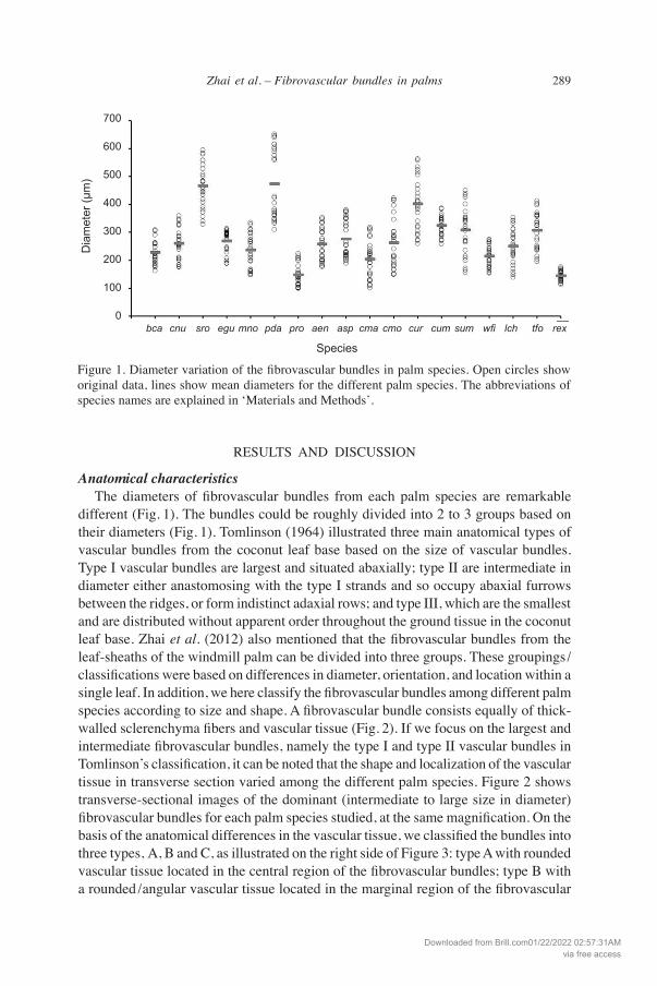
Anatomical characteristics The diameters of fibrovascular bundles from each palm species are remarkable different (Fig. 1). The bundles could be roughly divided into 2 to 3 groups based on their diameters (Fig. 1). Tomlinson (1964) illustrated three main anatomical types of vascular bundles from the coconut leaf base based on the size of vascular bundles. type I vascular bundles are largest and situated abaxially; type II are intermediate in diameter either anastomosing with the type I strands and so occupy abaxial furrows between the ridges, or form indistinct adaxial rows; and type III, which are the smallest and are distributed without apparent order throughout the ground tissue in the coconut leaf base. Zhai et al. (2012) also mentioned that the fibrovascular bundles from the leaf-sheaths of the windmill palm can be divided into three groups. These groupings /classifications were based on differences in diameter, orientation, and location within a single leaf. In addition, we here classify the fibrovascular bundles among different palm species according to size and shape. A fibrovascular bundle consists equally of thick-walled sclerenchyma fibers and vascular tissue (Fig. 2). If we focus on the largest and intermediate fibrovascular bundles, namely the type I and type II vascular bundles in Tomlinson’s classification, it can be noted that the shape and localization of the vascular tissue in transverse section varied among the different palm species. Figure 2 shows transverse-sectional images of the dominant (intermediate to large size in diameter) fibrovascular bundles for each palm species studied, at the same magnification. On the basis of the anatomical differences in the vascular tissue, we classified the bundles into three types, A, B and C, as illustrated on the right side of Figure 3: type A with rounded vascular tissue located in the central region of the fibrovascular bundles; type B with a rounded /angular vascular tissue located in the marginal region of the fibrovascular
700
600
500
400
300
200
100
0 bca cnu sro egumno pda pro aen asp cmacmo cur cumsum wfi lch tfo rex
Species
Dia
met
er (µ
m)
Figure 1. Diameter variation of the fibrovascular bundles in palm species. Open circles show original data, lines show mean diameters for the different palm species. The abbreviations of species names are explained in ‘Materials and Methods’.
Downloaded from Brill.com01/22/2022 02:57:31AMvia free access

290 IAWA Journal 34 (3), 2013
bundles (Arenga and Caryota); type C with aliform vascular tissue in the central region of the fibrovascular bundle (remaining species) (Fig. 3). These three types of fibrovascular bundles also showed correlation with the phylogenetic classification of palms (Fig. 3). The fibrovascular bundles from the tribes Cocoseae, Borasseae, and Phoeniceae belonged to type A, and those from the tribe Caryoteae to type B. With the exception of Rhapis excelsa, the fibrovascular bundles from the tribes Trachycarpeae, Corypheae, and Sabaleae were classified as type C. In general, the anatomical differences between different palm species within a genus are quantitative or so small as to be obscured by the variation exhibited by a single leaf sheath (Tomlinson 1961, 1964). The observations presented here provide the first
Figure 2. Transverse sectional images of leaf-sheath fibrovascular bundles of different palm species at the same magnification. The dotted area/circles in pda, cma, cmo and cur indicate thin-walled fibers. — Scale bars: 100 µm.
bca cnu sro egu mno
pda pro aen asp cma
cmo cur cum sum
wfilchtforex
Downloaded from Brill.com01/22/2022 02:57:31AMvia free access

291Zhai et al. – Fibrovascular bundles in palms
Figure 3. A diagram to show the phylogenetic classification of 14 genera of palms (Arecaceae), redrawn from Dransfield et al. (2008) and tomlinson et al. (2011). A, B, C indicate the three types of vascular tissues in fibrovascular bundles, where the gray area is occupied by scleren-chyma fibers and the white area by a vascular tissue.
40%
30%
20%
10%
0%
SV
/(SF
+S
V)
0 100 200 300 400 500 Diameter (µm)
Cocosnucifera
Elaeisguineensis
Phoenixroebelenii
Sabalumbraculifera
Trachycarpusfortunei
Figure 4. The ratio of vascular tissue area to whole transverse sectional area (= SV /(SF +SV) plotted against diameter of fibrovascular bundles in five palm species.
Downloaded from Brill.com01/22/2022 02:57:31AMvia free access
292 IAWA Journal 34 (3), 2013
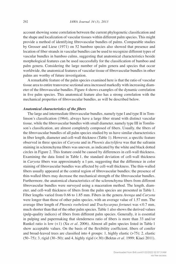
account showing some correlation between the current phylogenetic classification and the shape and localization of vascular tissues within different palm species. This might provide a method of identifying fibrovascular bundles of palms. Comparable studies by Grosser and Liese (1971) on 52 bamboo species also showed that presence and location of fiber strands in vascular bundles can be used to recognize different types of vascular bundles in bamboo culms, suggesting that anatomical characteristics beside morphological features can be used successfully for the classification of bamboo and palm genera. Considering the large number of palm genera and species that occur worldwide, the anatomical features of vascular tissue of fibrovascular bundles in other palms are worthy of future investigation. A remarkable feature of the palm species examined here is that the ratio of vascular tissue area to entire transverse sectional area increased markedly with increasing diam-eter of the fibrovascular bundles. Figure 4 shows examples of the dynamic correlation in five palm species. This anatomical feature also has a strong correlation with the mechanical properties of fibrovascular bundles, as will be described below.
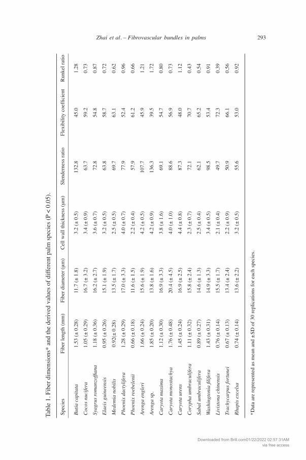
Anatomical characteristics of the fibers The large and intermediate fibrovascular bundles, namely type I and type II in Tom-linson’s classification (1964), always have a large fiber strand with distinct vascular tissue, while the fibrovascular bundles with small diameter, namely type III in Tomlin-son’s classification, are almost completely composed of fibers. Usually, the fibers of the fibrovascular bundles of all palm species studied by us have similar characteristics in fiber length, diameter and cell-wall thickness (Table 1). However, a specific feature observed in three species of Caryota and in Phoenix dactylifera was that the safranin staining in sclerenchyma fibers was uneven, as indicated by the white and black dotted circles in Figure 2. This feature could be caused by differences in cell-wall thickness. Examining the data listed in Table 1, the standard deviation of cell-wall thickness in Caryota fibers was approximately ± 1 µm, suggesting that the difference in color staining of fibrovascular bundles was affected by cell-wall thickness. The thin-walled fibers usually appeared at the central region of fibrovascular bundles; the presence of thin-walled fibers may decrease the mechanical strength of the fibrovascular bundles. Furthermore, the anatomical characteristics of the sclerenchyma fibers from different fibrovascular bundles were surveyed using a maceration method. The length, diam-eter, and cell-wall thickness of fibers from the palm species are presented in Table 1. Fiber lengths varied from 0.66 to 1.85 mm. Fibers in the genera Arenga and Caryota were longer than those of other palm species, with an average value of 1.57 mm. The average fiber length of Phoenix roebelenii and Trachycarpus fortunei was < 0.7 mm, much shorter than that of the other palm species. Table 1 also shows the derived values (pulp-quality indices) of fibers from different palm species. Generally, it is essential in pulping and papermaking that slenderness ratio of fibers is more than 33 and/or Runkel ratio is low (<1) (Xu et al. 2006). Almost all palm species listed in table 1 show acceptable values. On the basis of the flexibility coefficient, fibers of conifer and broad-leaved trees are classified into 4 groups: 1, highly elastic (>75); 2, elastic (50–75); 3, rigid (30–50); and 4, highly rigid (< 30) (Bektas et al. 1999; Kiaei 2011).
Downloaded from Brill.com01/22/2022 02:57:31AMvia free access
293Zhai et al. – Fibrovascular bundles in palms
Tabl
e 1.
Fib
er d
imen
sion
s* a
nd th
e de
rived
val
ues o
f diff
eren
t pal
m sp
ecie
s (P
< 0.
05).
Spe
cies
Fi
ber l
engt
h (m
m)
Fibe
r dia
met
er (µ
m)
Cel
l wal
l thi
ckne
ss (µ
m)
Slen
dern
ess r
atio
Fl
exib
ility
coe
ffici
ent
Run
kel r
atio
But
ia c
apita
ta
1.53
(± 0
.28)
11
.7 (±
1.8
) 3.
2 (±
0.5
) 13
2.8
45.0
1.
28
Coc
os n
ucife
ra
1.05
(± 0
.29)
16
.7 (±
3.2
) 3.
4 (±
0.9
) 63
.7
59.2
0.
73
Sya
grus
rom
anzo
ffian
a 1.
18 (±
0.3
6)
16.2
(± 2
.7)
3.6
(± 0
.7)
72.8
54
.8
0.87
Ela
eis g
uine
ensi
s 0.
95 (±
0.2
6)
15.1
(± 1
.9)
3.2
(± 0
.5)
63.8
58
.7
0.72
Med
emia
nob
ilis
0.92
(± 0
.28)
13
.5 (±
1.7
) 2.
5 (±
0.5
) 69
.7
63.1
0.
62
Pho
enix
dac
tylif
era
1.28
(± 0
.29)
17
.0 (±
3.3
) 4.
0 (±
0.7
) 77
.9
52.4
0.
96
Pho
enix
roeb
elen
ii 0.
66 (±
0.1
8)
11.6
(± 1
.5)
2.2
(± 0
.4)
57.9
61
.2
0.66
Are
nga
engl
eri
1.66
(± 0
.24)
15
.6 (±
1.9
) 4.
2 (±
0.5
) 10
7.7
45.9
1.
21
Are
nga
sp.
1.85
(± 0
.20)
13
.8 (±
1.6
) 4.
2 (±
0.9
) 13
6.3
39.5
1.
72
Car
yota
max
ima
1.12
(± 0
.30)
16
.9 (±
3.3
) 3.
8 (±
1.6
) 69
.1
54.7
0.
80
Car
yota
mon
osta
chya
1.
76 (±
0.4
8)
20.4
(± 4
.5)
4.0
(± 1
.0)
88.6
56
.9
0.73
Car
yota
ure
ns
1.45
(± 0
.24)
16
.9 (±
2.5
) 4.
4 (±
0.8
) 87
.3
48.0
1.
12
Cor
ypha
um
brac
ulife
ra
1.11
(± 0
.32)
15
.8 (±
2.4
) 2.
3 (±
0.7
) 72
.1
70.7
0.
43
Sab
al u
mbr
acul
ifera
0.
89 (±
0.2
7)
14.6
(± 1
.3)
2.5
(± 0
.4)
62.1
65
.2
0.54
Was
hing
toni
a fil
ifera
1.
43 (±
0.3
1)
14.9
(± 3
.3)
3.4
(± 0
.5)
98.5
53
.4
0.91
Liv
isto
na c
hine
nsis
0.
76 (±
0.1
4)
15.5
(± 1
.7)
2.1
(± 0
.4)
49.7
72
.3
0.39
Tra
chyc
arpu
s for
tune
i 0.
67 (±
0.1
3)
13.4
(± 2
.4)
2.2
(± 0
.9)
50.9
66
.1
0.56
Rha
pis e
xcel
sa
0.74
(± 0
.14)
13
.6 (±
2.2
) 3.
2 (±
0.5
) 55
.6
53.0
0.
92
*D
ata
are
repr
esen
ted
as m
ean
and
±SD
of 3
0 re
plic
atio
ns fo
r eac
h sp
ecie
s.
Downloaded from Brill.com01/22/2022 02:57:31AMvia free access
294 IAWA Journal 34 (3), 2013 Ta
ble
2. M
echa
nica
l pro
perti
es*,
MFA
s an
d K
laso
n lig
nin
cont
ents
of fi
brov
ascu
lar b
undl
es fr
om d
iffer
ent p
alm
spec
ies (
P <
0.05
).
Spe
cies
D
iam
eter
S V
/(S V
+S F
) Te
nsile
stre
ngth
Yo
ung’
s mod
ulus
B
RK
Stra
in
MFA
K
laso
n lig
nin
(µ
m)
(%)
(MPa
) (G
Pa)
(%)
(°)
(%)
But
ia c
apita
ta
228
(± 4
2)
12
170
(± 6
3)
2.5
(± 1
.6)
11 (±
7)
17.6
(± 3
.9)
18.7
(± 0
.8)
Coc
os n
ucife
ra
260
(± 5
7)
15
63 (±
32)
1.
8 (±
0.7
) 9
(± 6
) 29
.3 (±
1.5
) 25
.6 (±
1.1
)
Sya
grus
rom
anzo
ffian
a 46
6 (±
79)
12
13
4 (±
39)
1.
3 (±
0.8
) 25
(± 1
6)
13.4
(± 1
.8)
33.0
(± 0
.6)
Ela
eis g
uine
ensi
s 26
9 (±
42)
25
22
8 (±
74)
2.
2 (±
1.1
) 17
(± 5
) 21
.1 (±
13.
1)
26.2
(± 2
.0)
Med
emia
nob
ilis
236
(± 6
0)
8 83
(± 3
1)
1.7
(± 0
.7)
24 (±
12)
29
.7 (±
3.5
) 34
.7 (±
2.2
)***
Pho
enix
dac
tylif
era
473
(± 1
25)
10
147
(± 7
6)
1.5
(± 1
.0)
25 (±
15)
15
.6 (-
**)
18.3
(± 1
.3)
Pho
enix
roeb
elen
ii 14
7 (±
39)
3
162(
± 52
) 2.
1 (±
0.9
) 21
(± 8
) 23
.5 (±
1.4
) 33
.0 (±
0.3
)***
Are
nga
engl
eri
259
(± 5
7)
7 20
2 (±
78)
2.
9 (±
1.7
) 13
(± 8
) 10
.3 (±
1.4
) 29
.6 (±
3.2
)
Are
nga
sp.
276
(± 6
6)
5 76
(± 2
7)
1.7
(± 0
.6)
8 (±
4)
21.0
(± 1
2.8)
37
.8 (±
0.7
)
Car
yota
max
ima
202
(± 6
5)
5 12
8 (±
48)
2.
3 (±
1.5
) 40
(± 2
5)
30.1
(± 3
.3)
38.2
(± 0
.3) *
**
Car
yota
mon
osta
chya
26
2 (±
90)
4
99 (±
38)
1.
2 (±
0.6
) 40
(± 1
7)
47.1
(± 6
.6)
34.8
(± 0
.3)
Car
yota
ure
ns
402
(± 9
4)
6 78
(± 2
6)
1.2
(± 0
.6)
62 (±
23)
34
.8 (±
2.2
) 28
.1 (±
2.6
)
Cor
ypha
um
brac
ulife
ra
323
(± 3
7)
5 57
(± 2
9)
1.2
(± 0
.5)
10 (±
6)
28.1
(± 2
.6)
30.4
(± 1
.9)*
**
Sab
al u
mbr
acul
ifera
30
8 (±
87)
27
11
1 (±
36)
2.
3 (±
1.3
) 13
(± 8
) 20
.7 (±
5.9
) 25
.8 (±
0.7
)
Was
hing
toni
a fil
ifera
21
4 (±
38)
9
170
(± 5
8)
1.7
(± 0
.7)
21 (±
7)
22.9
(± 3
.5)
35.2
(± 0
.9)
Liv
isto
na c
hine
nsis
25
0 (±
63)
30
–
– –
24.2
(± 2
.6)
31.6
(± 0
.8)
Tra
chyc
arpu
s for
tune
i 30
7 (±
65)
12
54
(± 2
3)
1.2
(± 0
.5)
12 (±
8)
38.9
(± 4
.5)
35.9
(± 1
.2)
Rha
pis e
xcel
sa
144
(± 1
7)
4 10
9 (±
36)
2.
9 (±
0.9
) 11
(± 4
) 29
.5 (±
4.2
) 33
.9 (±
1.0
)
*
Dat
a of
mec
hani
cal p
rope
rties
are
repr
esen
ted
as m
ean
and
± SD
of 3
0 re
plic
atio
ns fo
r eac
h sp
ecie
s. *
* O
nly
one
fibro
vasc
ular
bun
dle
was
test
ed fo
r obt
aini
ng m
ean
MFA
. The
mea
sure
men
ts w
ere
perf
orm
ed in
trip
licat
e fo
r oth
er sp
ecie
s. *
** T
he K
laso
n lig
nin
cont
ents
of t
hese
pal
m sp
ecie
s inc
lude
ash
con
tent
s. Th
ese
data
wer
e no
t use
d fo
r dis
cuss
ion.
Downloaded from Brill.com01/22/2022 02:57:31AMvia free access
295Zhai et al. – Fibrovascular bundles in palms
The flexibility coefficients in Arenga spp. and Butia capitata were < 50; these fibers can be considered as rigid. The flexibility coefficients of the fibers from the other 15 palm specimens ranged between 50 and 75; these fibers are considered elastic.
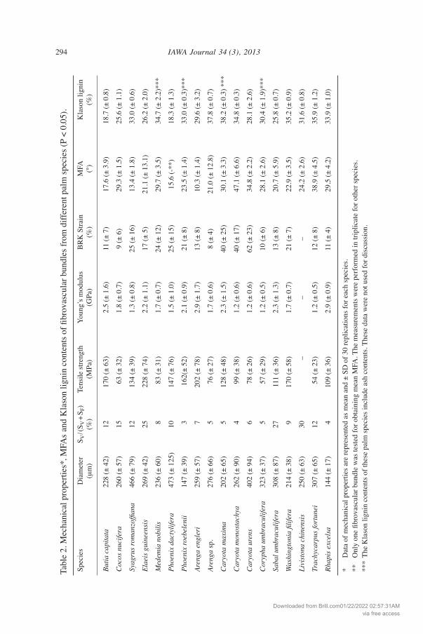
Microfibril angles and Klason lignin contents The MFAs of fibrovascular bundles among the palm species, as analyzed by X-ray diffraction, varied from 10.3º to 47.1º (mean = 25.4º) (Table 2). The MFAs were larger than those reported for other non-woody plant fibers including flax (Linum sp., 11º), jute (Corchorus sp., 8.1º), sisal (Agave sisalana, 10–22º), pine apple (Anannus comosus, 8–14º), and banana (Musa sepientum, 11º) (Satyanarayana et al. 1982; Baley 2002). The MFAs of palm species were also larger than those reported for wood fibers and tracheids (El-Osta et al. 1973; Yamamoto et al. 1993; Lichtenegger et al. 1999; Bonham & Barnett 2001). The Klason lignin contents of fibrovascular bundles from the different palm species ranged from 18.4% to 37.8%, with a mean value of 29.6% (Table 2). These values are much higher than those reported for other non-woody plants, including flax (2.0%), jute (15.9%), sisal (12%), and banana (12%) (Baley 2002; Razera & Frollini 2003; Cordeiro et al. 2004; Megiatto et al. 2007). Lignin contents of the palm species were similar to those reported for conifers, including noble fir (Abies procera, 29.3%), west-ern white pine (Pinus monoticola, 19.3%), and Douglas fir (Pseudotsuga menziesii, 27.2%). Palm fibrovascular bundles had relatively high lignin contents compared to those reported for broad-leaved trees, including yellow birch (Betula alleghaniensis, 22.7%), quaking aspen (Populus tremuloides, 19.3%), and basswood (Tilia americana, 20.0%) (Panshin et al. 1964).
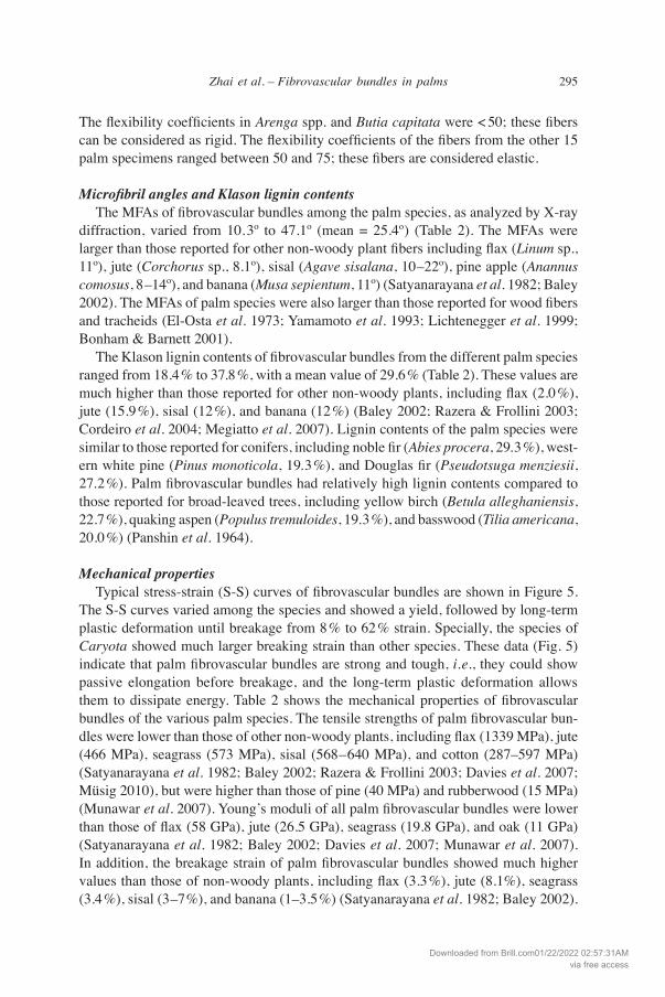
Mechanical properties Typical stress-strain (S-S) curves of fibrovascular bundles are shown in Figure 5. The S-S curves varied among the species and showed a yield, followed by long-term plastic deformation until breakage from 8% to 62% strain. Specially, the species of Caryota showed much larger breaking strain than other species. These data (Fig. 5) indicate that palm fibrovascular bundles are strong and tough, i.e., they could show passive elongation before breakage, and the long-term plastic deformation allows them to dissipate energy. Table 2 shows the mechanical properties of fibrovascular bundles of the various palm species. The tensile strengths of palm fibrovascular bun-dles were lower than those of other non-woody plants, including flax (1339 MPa), jute (466 MPa), seagrass (573 MPa), sisal (568–640 MPa), and cotton (287–597 MPa) (Satyanarayana et al. 1982; Baley 2002; Razera & Frollini 2003; Davies et al. 2007; Müsig 2010), but were higher than those of pine (40 MPa) and rubberwood (15 MPa) (Munawar et al. 2007). Young’s moduli of all palm fibrovascular bundles were lower than those of flax (58 GPa), jute (26.5 GPa), seagrass (19.8 GPa), and oak (11 GPa) (Satyanarayana et al. 1982; Baley 2002; Davies et al. 2007; Munawar et al. 2007). In addition, the breakage strain of palm fibrovascular bundles showed much higher values than those of non-woody plants, including flax (3.3%), jute (8.1%), seagrass (3.4%), sisal (3–7%), and banana (1–3.5%) (Satyanarayana et al. 1982; Baley 2002).
Downloaded from Brill.com01/22/2022 02:57:31AMvia free access
296 IAWA Journal 34 (3), 2013
The high lignin contents in combination with large MFAs account for the relatively low tensile strength, limited elastic deformation, and long-term plastic deformation of palm fibrovascular bundles. We found that the diameter of fibrovascular bundles influenced the tensile strength and Young’s modulus for all palm species. The general relationships between diameter and mechanical properties of fibrovascular bundles from 8 selected palm species are illustrated in Figure 6. It was striking that as diameter increased, tensile strength and Young’s modulus decreased. A similar phenomenon was found in fiber bundles from flax (Baley 2002), ramie, abaca leaf, and pineapple leaf (Munawar et al. 2007). this phenomenon cannot be easily explained, because if the characteristics of fiber bundles are the same, the mechanical properties of fiber bundles of different diameters should also be the same. Our previous publication pointed out that the thick-walled scleren-chyma fibers predominantly contribute to the mechanical properties of fibrovascular bundles in windmill palm, while vascular tissues tend to reduce mechanical strength (Zhai et al. 2012). Considering the similar structural and mechanical properties of the individual fiber cells in a fibrovascular bundle, the ratio of vascular tissue to the entire transverse sectional area (sclerenchyma + vascular tissue) would be a key factor affecting mechanical properties. We found that this ratio increased markedly with increasing diameter of the fibrovascular bundles (Fig. 4), indicating that the percentage of sclerenchyma fibers that affected mechanical properties decreased. In other words, the larger the diameter of fibrovascular bundles, the lower the mechanical strength, in all palm species examined.
300
250
200
150
100
50
0 0 100 200 300 400 500 Strain (%)
Stre
ss (M
Pa)
Figure 5. Typical stress-strain curves of fibrovascular bundles of different palm species, obtain- ed at a crosshead speed of 1 mm min-1 and using a gauge length of 10 mm. Dashed-dotted line, dashed line and dotted lines show stress-strain curves of species of Arenga (aen, asp), Caryota (cma, cmo, cur) and Phoenix (pda, pro), respectively. Other species are in solid line. the abbre-viations of species names are explained in ‘Materials and Methods’.
Downloaded from Brill.com01/22/2022 02:57:31AMvia free access

297Zhai et al. – Fibrovascular bundles in palms
CONCLUSIONS
This study on the anatomical characteristics and mechanical properties of fibrovascular bundles in leaf sheaths of different palm species showed three types of fibrovascular bundles, based on the shape and localization of vascular tissues. these three types of fibrovascular bundles showed correlation with the phylogenetic classification of palm species.
400
300
200
100
0 0 100 200 300 400 500 600 700 Diameter (µm)
Tens
ile s
treng
th (
MP
a)ButiacapitataCocosnuciferaSyagrusromansoffianaElaeisguineensisMedemianobilisPhoenixdactyliferaPhoenixroebeleniiRhapisexelsa
5.0
4.0
3.0
2.0
1.0
0.0 0 100 200 300 400 500 600 700 Diameter (µm)
Youn
g’s
mod
ulus
(G
Pa)
ButiacapitataCocosnuciferaSyagrusromansoffianaElaeisguineensisMedemianobilisPhoenixdactyliferaPhoenixroebeleniiRhapisexelsa
a
b
Figure 6. Relationships between diameter and mechanical properties for the fibrovascular bundles in different palm species. – a: Tensile strength; b: Young’s modulus, both plotted as a function of diameter of the fibrovascular bundles.
Downloaded from Brill.com01/22/2022 02:57:31AMvia free access

298 IAWA Journal 34 (3), 2013
The correlation between diameter and mechanical properties of fibrovascular bundles was clarified. By observing the area occupied by sclerenchyma fibers and vascular tissue, we found that the proportion of the transverse sectional area comprised by vascular tissue increased markedly with increasing diameter of the fibrovascular bundles, explaining why tensile strength and Young’s modulus decreased with increasing diameter of the bundles. The large MFAs of palm fibers in combination with high lignin content, result in limited elastic deformation, long-term plastic deformation, and relatively low tensile strength of palm fibrovascular bundles. The mechanical properties are presumably af-fected by fiber dimensions (fiber length, fiber diameter and cell wall thickness), vascular tissue area, fibrovascular bundle diameter, MFA, and lignin content.
ACKNOWLEDGEMENTS
The authors thank Prof. Bunzo Mikami, from the Department of Agronomy and Horticultural Science, Graduate School of Agriculture, Kyoto University, for X-ray diffraction analysis. the authors are also grateful to Prof. Kenji Umemura in the Research Institute for Sustainable Humanosphere, Kyoto University, for testing mechanical properties. One of the authors, Shengcheng Zhai, thanks the G30 Program of the Faculty of Agriculture, Kyoto University, and the Kambayashi Scholarship Foundation for their generous financial support. The authors specially thank Prof. Biao Pan, from the College of Material Science and Engineering, Nanjing Forestry University, for collecting samples from different botanical gardens.
REFERENCES
American Society for Testing and Materials. 1978. ASTM D 3379-75 standard test method for tensile strength and Young’s modulus for high-modulus single-filament materials; pp. 847–852. ASTM, Philadelphia.
Anupam S. 2002. Palm leaf manuscripts of the world: material, technology and conservation. Studies in Conservation, Suppl. 1: 15–24. Maney Publishing.
Ashton PMS, Gamage S, Gunatilleke IAUN & Gunatilleke CVS. 1998. Using Caribbean pine to establish a mixed plantation: testing effects of pine canopy removal on plantings of rain forest tree species. Forest Ecol. Manag. 106: 211–222.
Baley C. 2002. Analysis of the flax fibres tensile behaviour and analysis of the tensile stiffness increase. Compos. Part A. 33: 939–948.
Bektas I, Tutus A & Eroglu H. 1999. A study of the suitability of Calabrian pine (Pinus brutia) for pulp and paper manufacture. Turkish J. Agric. Forest. 23: 589–599.
Bonham VA & Barnett JR. 2001. Fibre length and microfibril angle in silver birch (Betula pen-dula Roth). Holzforschung 55: 159–162.
Cordeiro N, Belgacem MN, Torres IC & Moura JCVP. 2004. Chemical composition and pulp- ing of banana pseudo-stems. Ind. Crops Prod. 19: 147–154.
Davies P, Morvan C, Sire O & Baley C. 2007. Structure and properties of fibres from sea-grass (Zostera marina). J Mater. Sci. 42 (13): 4850–4857.
de Zoysa ND. 1992. tapping patterns of the Kitul palm (Caryota urens) in the Sinharaja area, Sri Lanka. Principes 36: 28–33.
Dhawan S. 1995. Essential oil for prevention of mould growth on palm leaf manuscripts. In: Proceeding of the third International Conference on Biodeterioration of Cultural Property. The Conservation Science Division, The Fine Arts Department, Bangkok; pp. 272–282.
Downloaded from Brill.com01/22/2022 02:57:31AMvia free access
299Zhai et al. – Fibrovascular bundles in palms
Dransfield J, Uhl NW, Asmussen CB, Baker WJ, Harley MM & Lewis CE. 2008. Genera Palma- rum. The evolution and classification of palms. Kew Publ., Royal Botanic Gardens, Kew.
El-Osta ML, Kellogg RM, Foschi RO & Butters RG. 1973. A direct X-ray technique for meas-uring microfibril angle. Wood and Fiber 5: 118–128.
Franklin GL. 1954. A rapid method for softening wood for anatomical analysis. Trop. Woods 88: 35–36.
Grosser D & Liese W. 1971. On the anatomy of Asian bamboos, with special reference to their vascular bundles. Wood Sci. technol. 5: 290–312.
Khalil HPSA, Azura MN, Issam AM, Said MR & Adawi TOM. 2008. Oil palm empty fruit bunches (OPEFB) reinforced in new unsaturated polyester composites. J. Reinf. Plast. Compos. 27: 1817–1826.
Kiaei M. 2011. Basic density and fiber biometry properties of hornbeam wood in three different altitudes at age 12. Middle-east J. Sci. Res. 8: 663–668.
Law KN, Daud WRW & Ghazali A. 2007. Morphological and chemical nature of fiber stands of oil palm empty-fruit bunch (OPEFB). BioResources 2: 351–362.
Li S. 2008. Bencao gangmu (1578) – compendium of materia medica. Science and technology Publishing, Shanghai [in Chinese].
Lichtenegger H, Retterer A, Stanzl-Tschegg S & Fratzl P. 1999. Variation of cellulose micro-fibril angles in softwoods and hardwoods – a possible strategy of mechanical optimization. J. Struct. Biol. 128: 257–269.
Megiatto J, Hoareau W, Gardrat C, Frollini E & Castellan A. 2007. Sisal fibers: Surface chemi- cal modification using reagent obtained from a renewable source; characterization of hemi-cellulose and lignin as model study. J. Agric. Food Chem. 55: 8576–8584.
Morcote-Ríos G & Bernal R. 2001. Remains of palms (Palmae) at archaeological sites in the New World: A review. Bot. Rev. 67: 309–350.
Munawar SS, Umemura K & Kawai S. 2007. Characterization of the morphological, physical, and mechanical properties of seven nonwood plant fiber bundles. J. Wood Sci. 53: 108–113.
Müsig J. 2010. Industrial applications of natural fibers: Structure, properties and technical ap-plications. John Wiley & Sons Ltd, United Kingdom.
Panshin AJ, de Zeeuw C & Brown HP. 1964. Textbook of wood technology. Vol. 1: Structure, identification, uses, and properties of the commercial woods of the United States (Ed. 2). McGraw-Hill, Inc., USA.
Pei S, Chen S & Tong S. 1991. Angiospermae-Monocotyledoneae, Palmae. Vol. 13. Flora Rei-publicae Popularis Sinicae. Science Press, Beijing (in Chinese).
Rasband WS. 1997–2012. ImageJ. U.S. National Institutes of Health, Bethesda, Maryland, USA, http://imagej.nih.gov/ij/.
Ratnayake PDKC, Gunatilleke CVS & Gunatilleke IAUN. 1990. Caryota urens L. (Palmae): An indigenous multiple purpose tree species in the wet lowlands of Sri Lanka. Regional Workshop on Multipurpose Tree Species, IFS/Winrock Int., Los Baños, Philippines.
Razera IAT & Frollini E. 2003. Composites based on jute fibers and phenolic matrices: proper-ties of fibers and composites. J. Appl. Polymer Sci. 91: 1077–1085.
Satyanarayana KG, Pillai CKS, Sukumaran K, Pillai SGK, Rohatgi PK & Kalyani Vijayan. 1982. Structure property studies of fibres from various parts of the coconut tree. J. Mater. Sci. 17: 2453–2462.
Shinoj S, Visvanathan R, Panigrahi S & Kochubabu M. 2011. Oil palm fiber (OPF) and its com-posites: A review. Ind. Crops Prod. 33: 7–22.
Sluiter A, Hames B, Ruiz R, Scarlata C, Sluiter J, templeton D & Crocker D. 2011. Determina- tion of structural carbohydrates and lignin in biomass. National Renewable Energy Labora-tory, Golden, CO, USA. http://www.nrel.gov/biomass/analytical_procedures.html.
Downloaded from Brill.com01/22/2022 02:57:31AMvia free access

300 IAWA Journal 34 (3), 2013
Swarnakamal B. 1965. Conservation of palm-leaf manuscripts. Baroda Museum and Picture Gallery, Bull. 19: 59–65.
tamunaidu P & Saka S. 2011. Chemical characterization of various parts of nipa palm (Nypa fruticans). Ind. Crops Prod. 34: 1423–1428.
TAPPI Standard T 222. 1998. Acid insoluble lignin in wood and pulp.Tengberg M. 2012. Beginnings and early history of date palm garden cultivation in the Middle
East. J. Arid Environ. 86: 139–147.Thomas R & De Franceschi D. 2013. Palm stem anatomy and computer-aided identification: the
Coryphoideae (Arecaceae). Amer. J. Bot. 100: 289–313.Thomas R, Tengberg M, Moulhérat C, Marcon V & Besenval R. 2012. Analysis of a proto-
historic net from Shahi Yump, Baluchistan (Pakistan). Archaeol. Anthropol. Sci. 4: 15–23.tomlinson PB. 1961. Anatomy of the monocotyledons. II. Palmae. Oxford University Press,
London.tomlinson PB. 1964. the vascular skeleton of coconut leaf base. Phytomorphology 14:
218–230.tomlinson PB. 1990. the structural biology of palms. Clarendon Press, Oxford.Tomlinson PB, Horn JW & Fisher JB. 2011. The anatomy of palms. Oxford University Press,
New York.Watanabe A, Yoshimura T, Mikami B, Hayashi H, Kagamiyama H & Esaki N. 2002. Reaction
mechanism of alanine racemase from Bacillus stearothermophilus. J. Biol. Chem. 277 (21): 19166–19172.
Xu F, Zhong XC, Sun RC & Lu Q. 2006. Anatomy, ultrastructure and lignin distribution in cell wall of Caragana korshinskii. Ind. Crops Prod. 24: 186–193.
Yamamoto H, Okuyama T & Yoshida M. 1993. Method of determining the mean microfibril angle of wood over a wide range by the improved Cave’s method. Mokuzai Gakkaishi 39: 375–381.
Zhai S, Horikawa Y, Imai T & Sugiyama J. 2013. Cell wall characterization of windmill palm (Trachycarpus fortunei) fibers and its functional implications. IAWA J. 34: 20–33.
Zhai S, Li D, Pan B, Sugiyama J & Itoh T. 2012. Tensile strength of windmill palm (Trachycar-pus fortunei) fiber bundles and its structural impactions. J. Mater. Sci. 47: 949–959.
Accepted: 1 May 2013
Downloaded from Brill.com01/22/2022 02:57:31AMvia free access