Embed Size (px)
Citation preview
184 | march 2012 | volume 42 | number 3 | journal of orthopaedic & sports physical therapy
[ clinical commentary ]
Rupture of the anterior cruciate ligament (ACL) is one of the most common knee ligament injuries, with an annual incidence of 35 per 100 000 people.26,82 This event occurs primarily in active individuals, and female athletes are 2
to 3 times more likely to have an ACL injury than male athletes.26,82
Consequently, ACL reconstruction is one of the most commonly performed or-thopaedic surgeries in the United States. Traditional ACL reconstruction, in which a single graft is used to reconstruct the ACL, has been shown to result in normal International Knee Documentation Com-mittee Subjective Knee Form scores in only 61% to 67% of patients after surgery and rehabilitation.12 Of more concern, however, is the finding that 40% to 90%
TT SYNOPSIS: The goal of every orthopaedic surgeon should be to restore anatomy as close as possible to normal. Intense research on recon-struction of the anterior cruciate ligament (ACL) and an advancing knowledge of the anatomy and function of the 2 primary bundles of the ACL have led to techniques of ACL reconstruction that more closely restore normal anatomy. Restoring the ACL footprint is one of the most important goals of the surgery, and the choice between anatomic single-bundle and double-bundle ACL reconstruction is determined by the anatomical features of each
patient. After reconstruction, the graft undergoes a complex, lengthy process of remodeling; therefore, inappropriate (early), aggressive rehabilitation can lead to graft failure and compromise the patient’s outcome. The purpose of this article is to provide an overview of the anatomy and function of the ACL, the methods for anatomic single-bundle and double-bundle ACL reconstruction, and our recommendations for postoperative rehabilitation. J Orthop Sports Phys Ther 2012;42(3):184-195. doi:10.2519/jospt.2012.3783
TT KEY WORDS: ACL, knee, surgery
1Post-Doctoral Research Associate, Department of Orthopaedic Surgery, University of Pittsburgh, Pittsburgh, PA. 2Post-Doctoral Research Associate, Department of Orthopaedic Surgery, University of Pittsburgh, Pittsburgh, PA. 3Distinguished Service Professor, David Silver Professor and Chairman, Department of Orthopaedic Surgery, University of Pittsburgh, Pittsburgh, PA. 4Director of Clinical Research, Department of Orthopaedic Surgery, University of Pittsburgh, Pittsburgh, PA. The authors received funding from Smith & Nephew, Inc to support research related to reconstruction of the anterior cruciate ligament. Additionally, the authors are supported by research funding from the National Institute of Arthritis and Musculoskeletal and Skin Diseases (grant number AR056630-01A2), and the first author was Research Fellow of the German Speaking Association of Arthroscopy (AGA) at the Department of Orthopaedic Surgery, University of Pittsburgh. Address correspondence to Dr Freddie H. Fu, Kaufman Medical Building, Suite 1011, 3471 Fifth Avenue, University of Pittsburgh, Pittsburgh, PA 15213. E-mail: [email protected]
DANIEL HENSLER, MD1 • CAROLA F. VAN ECK, MD, PhD2 • FREDDIE H. FU, MD, DSc, DPs3 • JAMES J. IRRGANG, PT, PhD, ATC, FAPTA4
Anatomic Anterior Cruciate Ligament Reconstruction Utilizing
the Double-Bundle Technique
of patients who undergo ACL reconstruc-tion have radiographic knee osteoarthri-tis 7 to 12 years after surgery.52,60 In the last decade, anatomic double-bundle reconstruction of the ACL has gained popularity and become a widely accepted and used method to reconstruct the ACL. Though differences in the outcomes of single-bundle and double-bundle ACL reconstruction comprise a topic of ongo-ing discussion, it is generally agreed that
both methods need to be anatom-ically performed.23,38,55 Anatomic ACL reconstruction techniques aim to better restore the normal anatomy and biomechanics of the
knee, and are hypothesized to potentially decrease the incidence of osteoarthritis after ACL reconstruction.
In this paper, the different aspects of anatomic ACL reconstruction will be dis-cussed. We will focus on the anatomy, bio-mechanics, and kinematics of the ACL, methods for anatomic single-bundle and double-bundle reconstruction, and impli-cations for postoperative rehabilitation.
Anatomy of the ACLSurgeons in all specialties need to have an in-depth knowledge of anatomy to maximize outcomes for their patients. Based on recent research, knowledge of the anatomy of the ACL is advancing, and this has led to new and different ap-proaches to restore the anatomical struc-ture and physiological function of the ACL.
The ACL consists of 2 functional bun-dles—the anteromedial (AM) and pos-terolateral (PL) bundles4,7,28,59—named for their position on the tibia (FIGURE 1). Recent research has indicated that 2 dis-
SUPPLEMENTAL VIDEO ONLINE
42-03 Hensler.indd 184 2/22/2012 6:15:09 PM
Jour
nal o
f O
rtho
paed
ic &
Spo
rts
Phys
ical
The
rapy
®
Dow
nloa
ded
from
ww
w.jo
spt.o
rg a
t Uni
vers
ity o
f M
ichi
gan
on O
ctob
er 1
0, 2
014.
For
per
sona
l use
onl
y. N
o ot
her
uses
with
out p
erm
issi
on.
Cop
yrig
ht ©
201
2 Jo
urna
l of
Ort
hopa
edic
& S
port
s Ph
ysic
al T
hera
py®
. All
righ
ts r
eser
ved.
journal of orthopaedic & sports physical therapy | volume 42 | number 3 | march 2012 | 185
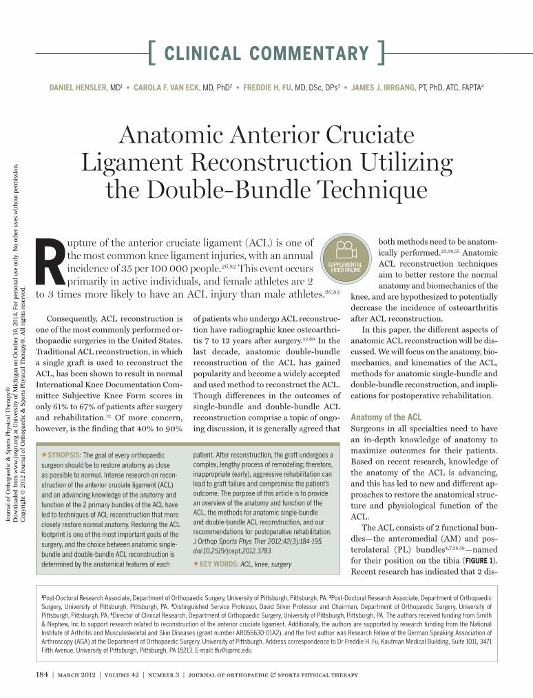
tinct bundles, separated by a septum of vascularized connective tissue,19 are al-ready in existence in a fetus after approxi-mately 20 weeks of development, which leads one to assume that the 2-bundle anatomy of the ACL is hereditary.
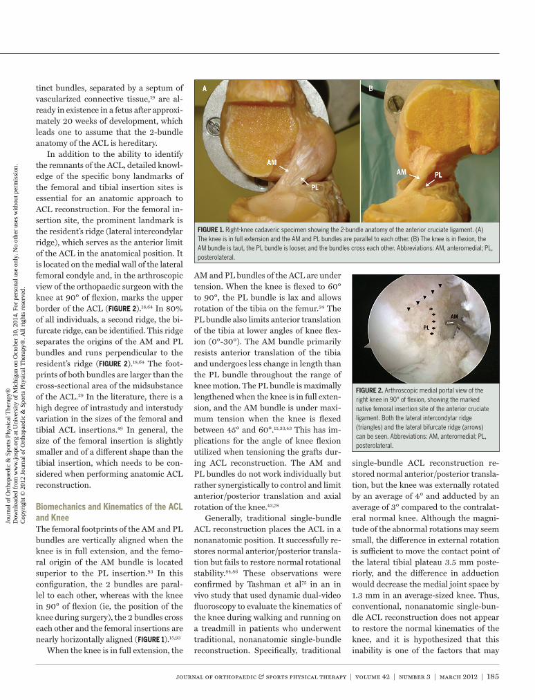
In addition to the ability to identify the remnants of the ACL, detailed knowl-edge of the specific bony landmarks of the femoral and tibial insertion sites is essential for an anatomic approach to ACL reconstruction. For the femoral in-sertion site, the prominent landmark is the resident’s ridge (lateral intercondylar ridge), which serves as the anterior limit of the ACL in the anatomical position. It is located on the medial wall of the lateral femoral condyle and, in the arthroscopic view of the orthopaedic surgeon with the knee at 90° of flexion, marks the upper border of the ACL (FIGURE 2).18,64 In 80% of all individuals, a second ridge, the bi-furcate ridge, can be identified. This ridge separates the origins of the AM and PL bundles and runs perpendicular to the resident’s ridge (FIGURE 2).18,64 The foot-prints of both bundles are larger than the cross-sectional area of the midsubstance of the ACL.29 In the literature, there is a high degree of intrastudy and interstudy variation in the sizes of the femoral and tibial ACL insertions.49 In general, the size of the femoral insertion is slightly smaller and of a different shape than the tibial insertion, which needs to be con-sidered when performing anatomic ACL reconstruction.
Biomechanics and Kinematics of the ACL and KneeThe femoral footprints of the AM and PL bundles are vertically aligned when the knee is in full extension, and the femo-ral origin of the AM bundle is located superior to the PL insertion.93 In this configuration, the 2 bundles are paral-lel to each other, whereas with the knee in 90° of flexion (ie, the position of the knee during surgery), the 2 bundles cross each other and the femoral insertions are nearly horizontally aligned (FIGURE 1).15,93
When the knee is in full extension, the
AM and PL bundles of the ACL are under tension. When the knee is flexed to 60° to 90°, the PL bundle is lax and allows rotation of the tibia on the femur.24 The PL bundle also limits anterior translation of the tibia at lower angles of knee flex-ion (0°-30°). The AM bundle primarily resists anterior translation of the tibia and undergoes less change in length than the PL bundle throughout the range of knee motion. The PL bundle is maximally lengthened when the knee is in full exten-sion, and the AM bundle is under maxi-mum tension when the knee is flexed between 45° and 60°.15,33,43 This has im-plications for the angle of knee flexion utilized when tensioning the grafts dur-ing ACL reconstruction. The AM and PL bundles do not work individually but rather synergistically to control and limit anterior/posterior translation and axial rotation of the knee.43,78
Generally, traditional single-bundle ACL reconstruction places the ACL in a nonanatomic position. It successfully re-stores normal anterior/posterior transla-tion but fails to restore normal rotational stability.84,86 These observations were confirmed by Tashman et al75 in an in vivo study that used dynamic dual-video fluoroscopy to evaluate the kinematics of the knee during walking and running on a treadmill in patients who underwent traditional, nonanatomic single-bundle reconstruction. Specifically, traditional
single-bundle ACL reconstruction re-stored normal anterior/posterior transla-tion, but the knee was externally rotated by an average of 4° and adducted by an average of 3° compared to the contralat-eral normal knee. Although the magni-tude of the abnormal rotations may seem small, the difference in external rotation is sufficient to move the contact point of the lateral tibial plateau 3.5 mm poste-riorly, and the difference in adduction would decrease the medial joint space by 1.3 mm in an average-sized knee. Thus, conventional, nonanatomic single-bun-dle ACL reconstruction does not appear to restore the normal kinematics of the knee, and it is hypothesized that this inability is one of the factors that may
FIGURE 1. Right-knee cadaveric specimen showing the 2-bundle anatomy of the anterior cruciate ligament. (A) The knee is in full extension and the AM and PL bundles are parallel to each other. (B) The knee is in flexion, the AM bundle is taut, the PL bundle is looser, and the bundles cross each other. Abbreviations: AM, anteromedial; PL, posterolateral.
FIGURE 2. Arthroscopic medial portal view of the right knee in 90° of flexion, showing the marked native femoral insertion site of the anterior cruciate ligament. Both the lateral intercondylar ridge (triangles) and the lateral bifurcate ridge (arrows) can be seen. Abbreviations: AM, anteromedial; PL, posterolateral.
42-03 Hensler.indd 185 2/22/2012 6:15:10 PM
Jour
nal o
f O
rtho
paed
ic &
Spo
rts
Phys
ical
The
rapy
®
Dow
nloa
ded
from
ww
w.jo
spt.o
rg a
t Uni
vers
ity o
f M
ichi
gan
on O
ctob
er 1
0, 2
014.
For
per
sona
l use
onl
y. N
o ot
her
uses
with
out p
erm
issi
on.
Cop
yrig
ht ©
201
2 Jo
urna
l of
Ort
hopa
edic
& S
port
s Ph
ysic
al T
hera
py®
. All
righ
ts r
eser
ved.

186 | march 2012 | volume 42 | number 3 | journal of orthopaedic & sports physical therapy
[ clinical commentary ]contribute to posttraumatic knee osteo-arthritis after ACL injury and surgery.
In contrast, anatomic double-bundle ACL reconstruction appears to better restore rotational stability compared to single-bundle reconstruction.86,87 In a ca-daveric model, Yagi et al86 demonstrated that reconstructing both bundles of the ACL resulted in more normal restoration of knee kinematics, particularly internal and external rotation of the tibia. Howev-er, these better results may be due to the anatomic placement of the ACL and not necessarily the double-bundle technique. Single-bundle ACL reconstruction can also be performed in an anatomic fash-ion. Yamamoto et al87 showed that ana-tomic single-bundle reconstruction with a laterally placed femoral tunnel can re-store knee kinematics to a level similar to that achieved by anatomic double-bundle reconstruction when the knee is near full extension; however, double-bundle re-construction resulted in more normal kinematics when the knee was at higher angles of flexion. The true benefits of anatomic double-bundle reconstruction compared to anatomic single-bundle re-construction should be the focus of future studies.
The clinical evidence for double-bundle ACL reconstruction is mounting but is still inconclusive. There have been 16 prospective clinical outcome studies that have compared double-bundle ACL reconstruction to single-bundle ACL re-construction,1,3,8,35,40,41,47,57,58,70,74,77,83,85,88,90 of which 10 were randomized clinical trials.1,3,35,40,41,57,70,74,83,90 A meta-analysis of 4 randomized clinical trials by Meredick et al55 revealed that double-bundle ACL reconstruction resulted in a signifi-cantly smaller side-to-side difference in tibial translation, as measured with the KT1000 Knee Ligament Arthrometer (MEDmetric Corporation, San Diego, CA); there was no difference in the pro-portion of individuals who had a normal or nearly normal pivot shift test. Howev-er, a closer analysis of the data reported by Meredick et al55 revealed that 88% of patients who underwent double-bundle
ACL reconstruction had a normal pivot shift test after surgery, compared to 62% of those who underwent single-bundle reconstruction. This result indicates that a normal pivot shift was more common following double-bundle ACL recon-struction (pooled odds ratio, 3.8; 95% confidence interval: 1.8, 7.8).38
Since the meta-analysis by Meredick et al,55 there have been 6 additional randomized clinical trials comparing double-bundle ACL reconstruction to sin-gle-bundle ACL reconstruction.3,35,70,74,83,90 Three of the trials3,35,70 demonstrated that double-bundle ACL reconstruction re-sulted in significantly better side-to-side differences in anterior translation and a significantly higher proportion of normal pivot shift tests. To date, however, none of the studies have demonstrated that double-bundle ACL reconstruction re-
sults in better patient-reported outcomes. Most importantly, long-term trials to compare the development and progres-sion of posttraumatic knee osteoarthritis after single-bundle and double-bundle ACL reconstruction are needed to dem-onstrate the true benefits of anatomic double-bundle ACL reconstruction.
ANATOMIC ACL RECONSTRUCTION
In the opinion of the authors, there are 4 fundamental principles of anatomic ACL reconstruction. The
first 2 principles are to appreciate the native anatomy of the ACL and to indi-vidualize surgery to the patient’s specific anatomy and functional needs. Because of the high degree of variation in the sizes of the tibial and femoral insertion
FIGURE 3. Arthroscopic view of a right knee in 90° of flexion. (A) Lateral portal view of a 14-mm insertion site. (B) Lateral portal view of a 22-mm insertion site. (C) Central portal view of a 12-mm notch. (D) Central portal view of a 20-mm notch. This figure shows the large variations in tibial insertion site and femoral intercondylar notch size. Online video available at www.jospt.org.
42-03 Hensler.indd 186 2/22/2012 6:15:12 PM
Jour
nal o
f O
rtho
paed
ic &
Spo
rts
Phys
ical
The
rapy
®
Dow
nloa
ded
from
ww
w.jo
spt.o
rg a
t Uni
vers
ity o
f M
ichi
gan
on O
ctob
er 1
0, 2
014.
For
per
sona
l use
onl
y. N
o ot
her
uses
with
out p
erm
issi
on.
Cop
yrig
ht ©
201
2 Jo
urna
l of
Ort
hopa
edic
& S
port
s Ph
ysic
al T
hera
py®
. All
righ
ts r
eser
ved.
journal of orthopaedic & sports physical therapy | volume 42 | number 3 | march 2012 | 187
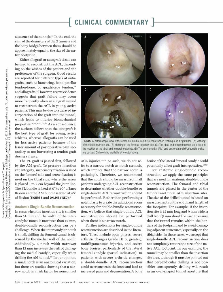
sites, as well as the sizes of the femo-ral intercondylar notch, the insertion sites and notch need to be measured to determine whether single-bundle or double-bundle ACL reconstruction best suits the needs of the individual patient (FIGURE 3 and ONLINE VIDEO).78 A tibial inser-tion site shorter than 14 mm in length and a notch narrower than 12 mm in width are too small to accommodate double-bundle ACL reconstruction.79 The third principle is to restore native anatomy by placing the graft in the center of the foot-print. The fourth principle is to restore the physiological function of the graft by applying appropriate tension to mimic the native ACL as closely as possible.5 As such, when anatomic double-bundle ACL reconstruction is performed, the graft for the PL bundle is tensioned with the knee at 0° of flexion and the graft for the AM
bundle is tensioned with the knee flexed to 45° to 60°. This is consistent with bio-mechanical evidence that the PL bundle is under maximal tension with the knee in full extension and that the AM bundle is under maximal tension with the knee in 45° to 60° of flexion.89 For anatomic single-bundle ACL reconstruction, the graft is tensioned with the knee in 10° to 20° of flexion. However, in clinical prac-tice, there is currently no consensus on the optimal knee flexion angles during graft tensioning in both single-bundle and double-bundle approaches.44,66
It is our belief that for anatomic single-bundle and double-bundle ACL reconstruction, 3 arthroscopic portals (central, anterolateral, and accessory me-dial) should be created (FIGURE 4).16 Cre-ation of 3 portals has several advantages compared to the traditional 2-incision technique described in the literature.6 The creation of a third portal allows for better visualization of the femoral ACL insertion site location, making a notch-plasty unnecessary.6 The anterolateral portal is placed laterally, adjacent to the patellar tendon and the inferior border of the patella. The central portal is lo-cated slightly above the medial menis-cus and directly adjacent to the medial border of the patellar tendon. With the arthroscope in the central portal, a view along the ACL directly to the femoral ACL footprint is possible. The accessory medial portal is located 2 cm medial to
the central portal and is created under arthroscopic visualization, with sufficient space from the medial condyle to avoid damaging the condyle. Placement of the arthroscope in the central portal helps to visualize the lateral wall of the femoral notch and the ACL footprint; therefore, with the accessory portal as a working portal, this technique eliminates the need for notchplasty.
Anatomic Double-Bundle ReconstructionAfter the portals are established and di-agnostic arthroscopy of the medial and lateral tibiofemoral and patellofemoral compartments is performed to inspect the menisci and chondral surfaces, focus is turned toward the intercondylar notch to determine the rupture pattern of the ACL. The ACL is most likely to be torn at the femoral site, but there can also be midsubstance tears as well as ruptures at the tibial site. By visualizing and probing the remnants of the ACL, possible single-bundle tears can be diagnosed where 1 bundle remains intact.91 In addition to the aforementioned bony landmarks, the remnants of the torn ACL can help the surgeon to locate the native tibial and femoral insertion sites. The anterior/posterior and medial/lateral dimensions on the tibia, as well as the proximal/dis-tal and anterior/posterior dimensions on the femur, are measured. Along with measurement of the intercondylar notch width, the measurements are used to de-termine whether single-bundle or dou-ble-bundle reconstruction is preferred for the patient.
After the origins of the 2 bundles on the tibia and femur are marked, 2 tun-nels in the tibia and femur are drilled (FIGURES 2 and 5). The size of the tun-nels is determined by the size of the ACL footprints. To restore the normal size relationship between the AM and PL bundles, the sizes of the graft and tunnel for the AM bundle should be larger than the graft and tunnel for the PL bundle. When drilling the femoral and tibial tun-nels, a bony bridge of approximately 2 mm needs to be preserved to prevent co-
FIGURE 4. Three-portal technique marked on a right knee in an operating position of 90° of flexion. The lateral portal (LP), central portal (CP), and accessory medial portal (AMP) are shown.
FIGURE 5. Arthroscopic lateral portal view of the right knee in 90° of flexion, showing the marked native tibial anterior cruciate ligament insertion site. Abbreviations: AM, anteromedial; PL, posterolateral.
42-03 Hensler.indd 187 2/22/2012 6:15:13 PM
Jour
nal o
f O
rtho
paed
ic &
Spo
rts
Phys
ical
The
rapy
®
Dow
nloa
ded
from
ww
w.jo
spt.o
rg a
t Uni
vers
ity o
f M
ichi
gan
on O
ctob
er 1
0, 2
014.
For
per
sona
l use
onl
y. N
o ot
her
uses
with
out p
erm
issi
on.
Cop
yrig
ht ©
201
2 Jo
urna
l of
Ort
hopa
edic
& S
port
s Ph
ysic
al T
hera
py®
. All
righ
ts r
eser
ved.
188 | march 2012 | volume 42 | number 3 | journal of orthopaedic & sports physical therapy
[ clinical commentary ]alescence of the tunnels.81 In the end, the sum of the diameters of the 2 tunnels and the bony bridge between them should be approximately equal to the size of the na-tive footprint.
Either allograft or autograft tissue can be used to reconstruct the ACL, depend-ing on the wishes of the patient and the preferences of the surgeon. Good results are reported for different types of auto-grafts, such as hamstring, bone-patellar tendon-bone, or quadriceps tendon,66 and allografts.9 However, recent evidence suggests that graft failure may occur more frequently when an allograft is used to reconstruct the ACL in young, active patients. This may be due to a delayed in-corporation of the graft into the tunnel, which leads to inferior biomechanical properties.14,39,51,53,63,67 As a consequence, the authors believe that the autograft is the best type of graft for young, active athletes, whereas allografts can be used for less active patients because of the lesser amount of postoperative pain sec-ondary to not harvesting a tendon graft during surgery.
The PL graft is passed first, followed by the AM graft. To preserve insertion site integrity, suspensory fixation is used on the femoral side and screw fixation is used on the tibial side, where the screw is placed 1 to 2 cm beyond the joint line. The PL bundle is fixed at 0° to 10° of knee flexion and the AM bundle is fixed at 45° of flexion (FIGURE 6 and ONLINE VIDEO).5
Anatomic Single-Bundle ReconstructionIn cases when the insertion site is smaller than 14 mm and the width of the inter-condylar notch is narrower than 12 mm, double-bundle reconstruction can be a challenge. When the intercondylar notch is small, drilling the femoral tunnel is ob-scured by the medial wall of the notch. Additionally, a notch width narrower than 12 mm increases the risk of damag-ing the medial condyle, especially when drilling the AM tunnel.80 In our opinion, a small notch is an anatomical variation, but there are studies showing that a nar-row notch is a risk factor for noncontact
ACL injuries.34,36 As such, we do not re-fer to a narrow notch as notch stenosis, which implies that the narrow notch is pathologic. Therefore, we recommend that the notch should be measured in all patients undergoing ACL reconstruction to determine whether double-bundle or single-bundle ACL reconstruction should be performed. Rather than performing a notchplasty to create the additional room necessary for double-bundle reconstruc-tion, we believe that single-bundle ACL reconstruction should be performed when there is a narrow notch.
Further indications for single-bundle reconstruction are described in the litera-ture. These include open physes, severe arthritic changes (grade III or greater), multiligamentous injuries, and severe bone bruises, particularly of the lateral femoral condyle (partial indication). In patients with severe arthritic changes, a double-bundle ACL reconstruction could overconstrain the knee and lead to increased pain and degeneration. A bone
bruise of the lateral femoral condyle could potentially affect graft incorporation.61,69
For anatomic single-bundle recon-struction, we apply the same principles that are used for anatomic double-bundle reconstruction. The femoral and tibial tunnels are placed in the center of the femoral and tibial ACL insertion sites. The size of the drilled tunnel is based on measurements of the width and length of the footprint. For example, if the inser-tion site is 12 mm long and 9 mm wide, a drill bit of 9 mm should be used to ensure that the tunnel remains within the bor-ders of the footprint and to avoid damag-ing adjacent structures, especially on the tibial side. In these cases, we accept that single-bundle ACL reconstruction does not completely restore the size of the na-tive ACL footprint. In our example, the tunnel may be smaller than the insertion site area, although it must be pointed out that perpendicular drilling is not pos-sible; consequently, drilling will result in an oval-shaped tunnel aperture that
FIGURE 6. Arthroscopic view of the anatomic double-bundle reconstruction technique in a right knee. (A) Marking of the tibial insertion site. (B) Marking of the femoral insertion site. (C) The tibial and femoral tunnels are drilled in the location of the tibial and femoral footprints. (D) The anteromedial (AM) and posterolateral (PL) bundle grafts are passed. Online video available at www.jospt.org.
42-03 Hensler.indd 188 2/22/2012 6:15:15 PM
Jour
nal o
f O
rtho
paed
ic &
Spo
rts
Phys
ical
The
rapy
®
Dow
nloa
ded
from
ww
w.jo
spt.o
rg a
t Uni
vers
ity o
f M
ichi
gan
on O
ctob
er 1
0, 2
014.
For
per
sona
l use
onl
y. N
o ot
her
uses
with
out p
erm
issi
on.
Cop
yrig
ht ©
201
2 Jo
urna
l of
Ort
hopa
edic
& S
port
s Ph
ysic
al T
hera
py®
. All
righ
ts r
eser
ved.
journal of orthopaedic & sports physical therapy | volume 42 | number 3 | march 2012 | 189
may actually restore the length of the footprint.
Failure After ACL ReconstructionGraft failure is an ongoing topic of dis-cussion in the literature, as well as at meetings and conferences. Rates of func-tional graft failure are reported to be be-tween 0% and 27.3%.65 The main cause of graft failure is related to malposition of the tunnel, for example, placing the tibial tunnel too anteriorly or placing the graft too vertically.17,42 Poor biological incorpo-ration of the graft,54 recurrent trauma, or early return to sport27 may also lead to graft failure. Most studies that reported graft failure after ACL reconstruction included patients who underwent non-anatomic ACL reconstruction. However, after anatomic ACL reconstruction, a higher graft failure rate may be expected because, as demonstrated by Kato et al,45 the forces in an anatomically placed graft will be greater (comparable to the native ACL) than those in a nonanatomically placed graft (less force than the native ACL due to the nonanatomic position of the graft). Therefore, rehabilitation and return to sport after anatomic ACL re-construction may need to be progressed slower than after a traditional, nonana-tomic ACL reconstruction.
REHABILITATION
Except for a slower return to functional activities, rehabilitation after anatomic ACL reconstruction
follows rehabilitation guidelines simi-lar to those of traditional, nonanatomic single-bundle ACL reconstruction. Ini-tially, we were concerned that anatomic double-bundle ACL reconstruction might interfere with the restoration of range of motion; however, our clinical experience indicates that this has not been the case. In fact, we have observed an earlier and better return of the full range of knee ex-tension and flexion after anatomic ACL reconstruction. Another concern is that, based on biomechanical studies, graft forces are greater when the graft is ana-
tomically positioned.45 For this reason, functional activities that place a high load on the graft, such as jumping, cutting, pivoting, and return to sport, are more gradually initiated and progressed after anatomic ACL reconstruction.
Below is a description of the reha-bilitation program followed at our in-stitution. The rehabilitation programs after anatomic single-bundle and dou-ble-bundle ACL reconstruction are the same. Immediately after surgery, the fo-cus is to minimize pain and swelling, re-store full passive extension symmetrical to the noninvolved knee, achieve 90° to 100° of knee flexion, restore the ability to perform a straight leg raise (SLR) with-out a quadriceps lag, and progress to full weight bearing so the individual can walk without assistive devices or a gait devia-tion. The day after surgery, patients be-gin to perform ankle pumps, quadriceps sets, SLRs, gastrocnemius and hamstring stretches, and heel slides. The patient is encouraged to make frequent use of cold to control postoperative pain and swell-ing. The patient ambulates with axillary crutches, using weight bearing as toler-ated, with the knee brace locked in full extension. Unless the patient had a con-comitant meniscus repair, the brace can be unlocked for ambulation at the end of the first week after surgery. If the patient had a concomitant meniscus repair, use of the brace locked in full extension is continued for 4 to 6 weeks to minimize shear stresses on the healing meniscus during ambulation.72
During the first 4 to 6 weeks after surgery, the rehabilitation program is gradually progressed. Active and active-assisted range-of-motion exercises are used to restore range of motion as toler-ated. If the patient had a concomitant meniscus repair, knee flexion is limited to 90° for 4 weeks after surgery. Patel-lar mobilization is used to maintain or increase patellar mobility, especially su-perior glide. Emphasis is placed on being able to perform a full, sustained isometric contraction of the quadriceps that results in superior migration of the patella and
the ability to perform a SLR with the knee at the end range of full extension. High-intensity electrical stimulation that is sufficient to produce a full, sustained contraction of the quadriceps is used to improve quadriceps strength. Several randomized clinical trials have dem-onstrated the benefits of high-intensity electrical stimulation to improve quad-riceps strength,21,71 gait,71 and patient-reported outcomes21,46 following ACL reconstruction. As range of motion im-proves, quadriceps strengthening can be progressed to include limited-arc (from 90° to 60°) non–weight-bearing (open-chain) knee extension exercises and low-level weight-bearing (closed-chain) exercises, with weight equally distributed on both extremities (eg, minisquats, wall slides). Standing weight shifts progress-ing to unilateral balance exercises can be used to improve the ability to tolerate full weight bearing and to begin to improve balance and postural control. Gait train-ing is performed as necessary to ensure that the individual uses a normal heel-toe gait and does not walk with a flexed knee during the midstance of gait. Progressive resisted exercises are also initiated for the hamstrings and hip muscles; however, to allow for healing of the harvest site, we delay resisted hamstring exercises for 4 to 6 weeks following harvest of the hamstring. If available, pool exercises can be used to improve range of motion, strength, and gait.
If the patient fails to progress with range of motion and/or has difficulty ini-tiating a quadriceps contraction for more than 1 to 2 weeks after surgery, the post-operative rehabilitation program may need to be altered and the surgeon should be alerted. Joint mobilization and cyclic or static stretching of the joint may be needed to restore extension or flexion of the knee. If extension and flexion are both limited, we believe that emphasis should first be placed on restoring extension. If stretching contributes to increased pain and inflammation, it may be necessary to temporarily limit or discontinue stretch-ing exercises until irritability of the joint
42-03 Hensler.indd 189 2/22/2012 6:15:16 PM
Jour
nal o
f O
rtho
paed
ic &
Spo
rts
Phys
ical
The
rapy
®
Dow
nloa
ded
from
ww
w.jo
spt.o
rg a
t Uni
vers
ity o
f M
ichi
gan
on O
ctob
er 1
0, 2
014.
For
per
sona
l use
onl
y. N
o ot
her
uses
with
out p
erm
issi
on.
Cop
yrig
ht ©
201
2 Jo
urna
l of
Ort
hopa
edic
& S
port
s Ph
ysic
al T
hera
py®
. All
righ
ts r
eser
ved.
190 | march 2012 | volume 42 | number 3 | journal of orthopaedic & sports physical therapy
[ clinical commentary ]
is reduced. Biofeedback may be consid-ered if the patient has difficulty recruiting the quadriceps muscle. Active-assisted, terminal, non–weight-bearing knee ex-tension in the range of 20° of flexion to full end-range extension can be used to re-educate and strengthen the quadriceps if a quadriceps lag is present.
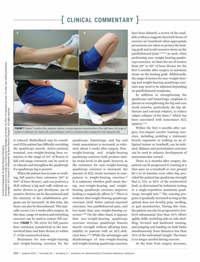
When the patient has no pain or swell-ing, full passive knee extension (90° to 100° of knee flexion), and can perform a SLR without a lag and walk without as-sistive devices or gait deviations, use of assistive devices can be discontinued and the intensity of the rehabilitation pro-gram can be increased. At this time, the brace can also be discontinued. This typi-cally occurs 3 to 4 weeks after surgery. At this time, range-of-motion and stretching exercises can be used to restore full mo-tion (FIGURE 7). We strive for full passive knee extension symmetrical to the non-involved knee and knee flexion to within 5° of the noninvolved knee.
Resistance for non–weight-bearing and weight-bearing exercises for the
quadriceps, hamstrings, and hip and trunk musculature is increased, as toler-ated, about 4 weeks after surgery. Non–weight-bearing and weight-bearing quadriceps exercises both produce simi-lar strain levels in the graft; however, as the resistance for non–weight-bearing quadriceps exercises is increased, the amount of ACL strain increases in com-parison to weight-bearing exercises.22 It is unknown whether graft strain dur-ing non–weight-bearing and weight-bearing quadriceps exercises improves healing or negatively affects it.10 There is evidence that weight-bearing quadriceps exercises yield better patient-reported outcomes, less patellofemoral pain, and less laxity than non–weight-bearing ex-ercises.13,68 On the other hand, it appears that non–weight-bearing quadriceps exercises increase quadriceps femoris muscle strength without affecting knee stability in patients with an ACL-defi-cient knee.50,66 While the advantages and disadvantages of non–weight-bearing and weight-bearing quadriceps exercises
have been debated, a review of the avail-able evidence suggests that both forms of exercise are beneficial when appropriate precautions are taken to protect the heal-ing graft and avoid excessive stress on the patellofemoral joint.11,20,56 As such, when performing non–weight-bearing quadri-ceps exercises, we limit the arc of motion from 90° to 60° of knee flexion for the first 3 months after surgery to minimize strain on the healing graft. Additionally, the range of motion for non–weight-bear-ing and weight-bearing quadriceps exer-cises may need to be adjusted depending on patellofemoral symptoms.
In addition to strengthening the quadriceps and hamstrings, emphasis is placed on strengthening the hip and core trunk muscles, particularly the hip ab-ductors and external rotators, to reduce valgus collapse of the knee,62 which has been associated with noncontact ACL injuries.31,32
Within the first 3 months after sur-gery, low-impact aerobic training exer-cises, including pedaling a stationary bicycle ergometer or walking on an el-liptical trainer or treadmill, can be initi-ated. Balance and perturbation exercises can be used to enhance development of neuromuscular control.
Three to 4 months after surgery, the patient can be progressed to running at a slow pace on a treadmill or over ground for 5 to 10 minutes every other day, pro-vided the patient has quadriceps strength that is 75% to 80% of the noninvolved limb, as determined by isokinetic testing or a single-repetition maximum quad-riceps strength test.37 The running pro-gram is gradually increased as long as the patient does not develop pain, swelling, or gait asymmetries. During this time, the patient can also be progressed to low-level submaximal (less than 50% effort) agility drills, including side-to-side shuf-fling, forward and backward running, and jumping and landing on both limbs simultaneously from distances less than 50% of the individual’s height. The brace is no longer needed during exercise.
As the time from surgery increases,
FIGURE 7. Patient 3 months after anatomic anterior cruciate ligament reconstruction of the right knee. Full range of motion in extension (A), flexion (B), and kneeling (C and D) is achieved when compared to the uninjured side.
42-03 Hensler.indd 190 2/22/2012 6:15:17 PM
Jour
nal o
f O
rtho
paed
ic &
Spo
rts
Phys
ical
The
rapy
®
Dow
nloa
ded
from
ww
w.jo
spt.o
rg a
t Uni
vers
ity o
f M
ichi
gan
on O
ctob
er 1
0, 2
014.
For
per
sona
l use
onl
y. N
o ot
her
uses
with
out p
erm
issi
on.
Cop
yrig
ht ©
201
2 Jo
urna
l of
Ort
hopa
edic
& S
port
s Ph
ysic
al T
hera
py®
. All
righ
ts r
eser
ved.

journal of orthopaedic & sports physical therapy | volume 42 | number 3 | march 2012 | 191
the progression of the patient to higher-level functional activities becomes more variable and difficult to predict. This is due to variations in the surgical proce-dure, surgeon preferences, and individ-ual factors. Therefore, the initiation of higher-level functional activities, such as running, jumping and landing, cutting and pivoting, and return to sport, may deviate from the time periods listed in the postoperative rehabilitation guidelines. Because of variations between patients, we progress the functional training and return-to-sport phases based on the pa-tient’s ability to perform the activities without deviations or symptoms (pain, swelling, sense of instability).
During the functional training and return-to-sport phases of rehabilitation after ACL reconstruction, emphasis is placed on strengthening through the full range of motion, improving neuromus-cular control, and ensuring a gradual increase in function that culminates in return to sport.
Once the patient is able to tolerate running 2.4 to 3.2 km without pain or swelling, the patient can be progressed to a higher order of agility and plyometric drills. Typically, these activities begin ap-proximately 6 months after surgery. Ini-tial agility drills can include side-to-side shuffling, forward and backward run-ning, and ladder drills. More challeng-ing agility drills include carioca and cone drills that involve changing directions at various angles. Initially, these activi-ties should be performed at 50% effort, progressing to 75% and eventually 100% effort, as tolerated.
During this time, the patient can also be progressed to plyometric jumping and landing drills. Initially, these activities should focus on landing and appropriate attenuation of force through the lower extremity. Such activities include dou-ble-limb jumping, single-limb jumping, and dropping and landing from a plyo-metric box. As the patient becomes pro-ficient with correct jumping and landing mechanics, plyometric exercises can be made more challenging by increasing the
height or distance of the jump, increas-ing the duration of the drills, incorporat-ing changes in direction, and combining multiple tasks.
Once the patient is able to tolerate full-effort running, jumping, and agility drills, return to sport can be considered and a functional brace can be readjusted for at least 6 months. The time frame for return to sport following anatomic ACL reconstruction is variable, but generally occurs 9 to 12 months after surgery and is dependent on concomitant surgical pro-cedures, individual patient tolerance for the activities, surgeon preferences, and the physical demands of the sport. Ini-tially, training for return to sport should begin with unopposed components of the individual’s athletic activity. As the patient becomes proficient and can per-form these activities safely, the speed and complexity of the activities can be increased. Training with opposition from other players should be gradually intro-duced. To return to full participation in sports, the patient should be progressed from partial return to practice to full re-turn to practice, followed by return to competition.
DISCUSSION
In recent years, traditional ap-proaches and methods to reconstruct the ACL have been critically evaluated,
and it has been shown that the femoral and tibial tunnels are often placed in a nonanatomic position.30 Nonanatomic placement of tunnels is most likely due to the surgeon’s efforts to avoid roof im-pingement and abrasion of the graft, which occurs when the tibial tunnel is placed too anteriorly. As a result, the sur-geon may place the tibial tunnels more posteriorly. Use of a transtibial method to create the femoral tunnel also contributes to nonanatomic placement of the graft.48 For example, to place the femoral tunnel close to the native location of the femoral ACL insertion site, it is often necessary to position the tibial tunnel within the tibial insertion site for the PL bundle.
Despite this posterior placement of the tibial tunnel, use of a transtibial method to drill the femoral tunnel still results in a femoral tunnel that is too high in the in-tercondylar notch (the high-AM position of the femoral tunnel). To avoid this, we recommend using a 3-portal technique that allows for use of the medial portal to create the AM femoral tunnel indepen-dent of the tibial tunnel, allowing one to achieve a more anatomic reconstruction.
Misplaced grafts are one of the most important causes of graft failure.25,73 Fur-thermore, a misplaced graft can result in worse clinical outcomes, with limited range of motion and nonphysiological knee kinematics,75,92 especially when roof impingement occurs. If the graft is mis-placed, revision ACL reconstruction to place the graft more anatomically may need to be considered. A misplaced graft may also adversely affect biological heal-ing of the graft within the tunnel and compromise healing of the bone-tendon interface.61 It is our opinion that a delay or failure to regain full range of motion during rehabilitation is often an indicator of nonanatomic placement of the graft.
The concerns related to nonanatom-ic graft placement have prompted us to use more anatomic and individual-ized ACL reconstruction. Single-bundle and double-bundle ACL reconstruction can be performed in an anatomic man-ner.69 Data from recent studies that have compared the clinical outcomes after single-bundle and double-bundle ACL reconstruction must be carefully inter-preted. For example, one study com-pared single-bundle ACL reconstruction, which was performed using a transtibial method to create the femoral tunnel, to anatomic double-bundle reconstruction.2 When comparing the clinical outcomes of single-bundle and double-bundle ACL reconstruction, both procedures should have been performed anatomically. Dif-ficulty in conducting a randomized clini-cal trial to compare single-bundle ACL reconstruction to double-bundle ACL reconstruction may arise if the individ-ual’s anatomy precludes double-bundle
42-03 Hensler.indd 191 2/22/2012 6:15:18 PM
Jour
nal o
f O
rtho
paed
ic &
Spo
rts
Phys
ical
The
rapy
®
Dow
nloa
ded
from
ww
w.jo
spt.o
rg a
t Uni
vers
ity o
f M
ichi
gan
on O
ctob
er 1
0, 2
014.
For
per
sona
l use
onl
y. N
o ot
her
uses
with
out p
erm
issi
on.
Cop
yrig
ht ©
201
2 Jo
urna
l of
Ort
hopa
edic
& S
port
s Ph
ysic
al T
hera
py®
. All
righ
ts r
eser
ved.

192 | march 2012 | volume 42 | number 3 | journal of orthopaedic & sports physical therapy
[ clinical commentary ]
REFERENCES
1. Adachi N, Ochi M, Uchio Y, Iwasa J, Kuriwaka M, Ito Y. Reconstruction of the anterior cruciate ligament. Single- versus double-bundle multi-stranded hamstring tendons. J Bone Joint Surg Br. 2004;86:515-520.
2. Aglietti P, Giron F, Cuomo P, Losco M, Mondanelli N. Single- and double-incision double-bundle ACL reconstruction. Clin Orthop Relat Res. 2007;454:108-113. http://dx.doi.org/10.1097/BLO.0b013e31802baaf4
3. Aglietti P, Giron F, Losco M, Cuomo P, Ciardullo A, Mondanelli N. Comparison between single- and double-bundle anterior cruci-ate ligament reconstruction: a prospective, randomized, single-blinded clinical trial. Am J Sports Med. 2010;38:25-34. http://dx.doi.org/10.1177/0363546509347096
4. Amis AA, Dawkins GP. Functional anatomy of the anterior cruciate ligament. Fibre bundle actions related to ligament replacements and injuries. J Bone Joint Surg Br. 1991;73:260-267.
5. Anderson CJ, Westerhaus BD, Pietrini SD, et al. Kinematic impact of anterome-dial and posterolateral bundle graft fixation angles on double-bundle anterior cruci-ate ligament reconstructions. Am J Sports Med. 2010;38:1575-1583. http://dx.doi.org/10.1177/0363546510364841
6. Araujo PH, van Eck CF, Macalena JA, Fu FH. Advances in the three-portal technique for anatomical single- or double-bundle ACL recon-struction. Knee Surg Sports Traumatol Arthrosc. 2011;19:1239-1242. http://dx.doi.org/10.1007/s00167-011-1426-z
7. Arnoczky SP. Anatomy of the anterior cruciate ligament. Clin Orthop Relat Res. 1983;172:19-25.
8. Asagumo H, Kimura M, Kobayashi Y, Taki M, Takagishi K. Anatomic reconstruction of the anterior cruciate ligament using double-bundle
ACL reconstruction. This can occur if the insertion sites are too small or the inter-condylar notch is too narrow to permit double-bundle ACL reconstruction. For this reason, a prospective randomized controlled trial that compares single-bundle ACL reconstruction to double-bundle ACL reconstruction may need to exclude patients who have insertion sites that are too small or a notch that is too narrow. Furthermore, a study that com-pares single-bundle ACL reconstruction to double-bundle ACL reconstruction should exclude individuals with associ-ated injuries, such as meniscal tears, chondral injuries, or multiple ligament injuries, to achieve homogeneous groups.
The limitations of currently available clinical outcome measures must be con-sidered when comparing single-bundle to double-bundle ACL reconstruction. It is hypothesized that the addition of the PL bundle during anatomic double-bundle ACL reconstruction will result in improved rotational stability of the knee. As such, a measure of rotational laxity of the knee is an important out-come to include in a trial comparing single-bundle to double-bundle ACL re-construction. Clinical studies comparing single-bundle and double-bundle ACL reconstruction have relied on clinical measures of laxity such as the KT1000 Knee Ligament Arthrometer and pivot shift test. These clinical measures may not be sensitive enough to detect differ-ences in laxity between single-bundle and double-bundle ACL reconstruction. Reliance on the pivot shift test as an end point for a study comparing single-bun-dle to double-bundle ACL reconstruction is a concern, particularly when the test is performed on a patient who is awake. Alternate methods to quantify rotation are needed. High-technology methods to precisely measure knee kinematics, such as dynamic stereoradiography, have shown promising results in 6 degrees of freedom75,76 but are not feasible at most centers. A simple, clinically applicable tool, similar to the KT1000 Knee Liga-ment Arthrometer, that could be used to
reliably quantify rotational laxity of the knee needs to be developed.
To date, evaluating the clinical out-comes of anatomic double-bundle ACL reconstruction has focused on the abil-ity of the procedure to restore normal anteroposterior and rotational laxity of the knee. In the future, researchers should also consider the effects of ana-tomic double-bundle ACL reconstruc-tion on the sense of instability and the ability to participate in strenuous sports. Long-term follow-up studies are needed to determine the effects of double-bundle ACL reconstruction on preventing or re-ducing the risk of knee osteoarthritis and its associated pain and disability. In the interim, high-field magnetic resonance imaging could be used to detect early evidence of cartilage changes.
In comparison to nonanatomic grafts, an anatomically placed graft will experi-ence greater in situ forces.45 Although in-creased loading of the graft may protect other structures in the knee from pro-gressive degeneration, the higher load on the graft must be considered during postoperative recovery and rehabilitation while the graft is still healing and matur-ing. Because of the higher graft loads after anatomic ACL reconstruction, we recommend that rehabilitation be pro-gressed more carefully. As a result, we do not recommend return to sport until 9 to 12 months after surgery, which is slower than the more commonly used acceler-ated rehabilitation approach described by Shelbourne and Nitz,68 which advocates return-to-sport activities 3 to 6 months after surgery.
CONCLUSION
In our opinion, anatomic ACL re-construction can more closely restore the anatomy of the ACL, which we
believe results in more normal kinemat-ics of the knee. Ultimately, we believe that anatomic ACL reconstruction may promote better long-term knee health. Anatomic tunnel placement and resto-ration of the ACL insertion site can be
accomplished by performing either sin-gle-bundle or double-bundle ACL recon-struction. The choice of technique should be based on individual measurements of the ACL insertion site and femoral in-tercondylar notch size. To decrease the failure rate, it is necessary to carefully plan and carry out the postoperative re-habilitation program. The patient needs to be aware that, although anatomic ACL reconstruction provides better kinemat-ics of the knee and ultimately may lead to improved long-term health of the knee, the graft needs time to remodel and heal, and one should therefore resist the temp-tation of a more aggressive rehabilitation program. t
42-03 Hensler.indd 192 2/22/2012 6:15:19 PM
Jour
nal o
f O
rtho
paed
ic &
Spo
rts
Phys
ical
The
rapy
®
Dow
nloa
ded
from
ww
w.jo
spt.o
rg a
t Uni
vers
ity o
f M
ichi
gan
on O
ctob
er 1
0, 2
014.
For
per
sona
l use
onl
y. N
o ot
her
uses
with
out p
erm
issi
on.
Cop
yrig
ht ©
201
2 Jo
urna
l of
Ort
hopa
edic
& S
port
s Ph
ysic
al T
hera
py®
. All
righ
ts r
eser
ved.
journal of orthopaedic & sports physical therapy | volume 42 | number 3 | march 2012 | 193
hamstring tendons: surgical techniques, clinical outcomes, and complications. Arthroscopy. 2007;23:602-609. http://dx.doi.org/10.1016/j.arthro.2007.01.009
9. Bach BR, Jr., Aadalen KJ, Dennis MG, et al. Primary anterior cruciate ligament reconstruc-tion using fresh-frozen, nonirradiated patellar tendon allograft: minimum 2-year follow-up. Am J Sports Med. 2005;33:284-292.
10. Beynnon BD, Johnson RJ. Anterior cruci-ate ligament injury rehabilitation in athletes. Biomechanical considerations. Sports Med. 1996;22:54-64.
11. Beynnon BD, Johnson RJ, Fleming BC, Stanke-wich CJ, Renstrom PA, Nichols CE. The strain behavior of the anterior cruciate ligament during squatting and active flexion-extension. A com-parison of an open and a closed kinetic chain exercise. Am J Sports Med. 1997;25:823-829.
12. Biau DJ, Tournoux C, Katsahian S, Schranz P, Nizard R. ACL reconstruction: a meta-analysis of functional scores. Clin Orthop Relat Res. 2007;458:180-187. http://dx.doi.org/10.1097/BLO.0b013e31803dcd6b
13. Bynum EB, Barrack RL, Alexander AH. Open versus closed chain kinetic exercises after anterior cruciate ligament reconstruction. A pro-spective randomized study. Am J Sports Med. 1995;23:401-406.
14. Chang SK, Egami DK, Shaieb MD, Kan DM, Richardson AB. Anterior cruciate ligament reconstruction: allograft versus autograft. Arthroscopy. 2003;19:453-462. http://dx.doi.org/10.1053/jars.2003.50103
15. Chhabra A, Starman JS, Ferretti M, Vidal AF, Zantop T, Fu FH. Anatomic, radiographic, bio-mechanical, and kinematic evaluation of the anterior cruciate ligament and its two functional bundles. J Bone Joint Surg Am. 2006;88 Suppl 4:2-10. http://dx.doi.org/10.2106/JBJS.F.00616
16. Cohen SB, Fu FH. Three-portal technique for anterior cruciate ligament reconstruction: use of a central medial portal. Arthros-copy. 2007;23:325.e1-325.e5. http://dx.doi.org/10.1016/j.arthro.2006.07.030
17. Fagelman M, Freedman KB. Revision reconstruc-tion of the anterior cruciate ligament: evaluation and management. Am J Orthop (Belle Mead NJ). 2005;34:319-328.
18. Ferretti M, Ekdahl M, Shen W, Fu FH. Osseous landmarks of the femoral attachment of the anterior cruciate ligament: an anatomic study. Arthroscopy. 2007;23:1218-1225. http://dx.doi.org/10.1016/j.arthro.2007.09.008
19. Ferretti M, Levicoff EA, Macpherson TA, More-land MS, Cohen M, Fu FH. The fetal anterior cruciate ligament: an anatomic and histologic study. Arthroscopy. 2007;23:278-283. http://dx.doi.org/10.1016/j.arthro.2006.11.006
20. Fitzgerald GK. Open versus closed kinetic chain exercise: issues in rehabilitation after anterior cruciate ligament reconstructive surgery. Phys Ther. 1997;77:1747-1754.
21. Fitzgerald GK, Piva SR, Irrgang JJ. A modified neuromuscular electrical stimulation protocol
for quadriceps strength training following ante-rior cruciate ligament reconstruction. J Orthop Sports Phys Ther. 2003;33:492-501.
22. Fleming BC, Beynnon BD, Nichols CE, Renstrom PA, Johnson RJ, Pope MH. An in vivo compari-son between intraoperative isometric measure-ment and local elongation of the graft after reconstruction of the anterior cruciate ligament. J Bone Joint Surg Am. 1994;76:511-519.
23. Frobell RB, Roos EM, Roos HP, Ranstam J, Lohmander LS. A randomized trial of treatment for acute anterior cruciate ligament tears. N Engl J Med. 2010;363:331-342. http://dx.doi.org/10.1056/NEJMoa0907797
24. Gabriel MT, Wong EK, Woo SL, Yagi M, Debski RE. Distribution of in situ forces in the anterior cruciate ligament in response to rotatory loads. J Orthop Res. 2004;22:85-89. http://dx.doi.org/10.1016/S0736-0266(03)00133-5
25. George MS, Dunn WR, Spindler KP. Current concepts review: revision anterior cruci-ate ligament reconstruction. Am J Sports Med. 2006;34:2026-2037. http://dx.doi.org/10.1177/0363546506295026
26. Gianotti SM, Marshall SW, Hume PA, Bunt L. Incidence of anterior cruciate ligament injury and other knee ligament injuries: a na-tional population-based study. J Sci Med Sport. 2009;12:622-627. http://dx.doi.org/10.1016/j.jsams.2008.07.005
27. Giffin JR, Harner CD. Failed anterior cruciate ligament surgery: overview of the problem. Am J Knee Surg. 2001;14:185-192.
28. Girgis FG, Marshall JL, Monajem A. The cruciate ligaments of the knee joint. Anatomical, func-tional and experimental analysis. Clin Orthop Relat Res. 1975;106:216-231.
29. Harner CD, Baek GH, Vogrin TM, Carlin GJ, Kashiwaguchi S, Woo SL. Quantitative analysis of human cruciate ligament insertions. Arthros-copy. 1999;15:741-749.
30. Heming JF, Rand J, Steiner ME. Anatomical limitations of transtibial drilling in anterior cruciate ligament reconstruction. Am J Sports Med. 2007;35:1708-1715. http://dx.doi.org/10.1177/0363546507304137
31. Hewett TE, Ford KR, Myer GD. Anterior cruci-ate ligament injuries in female athletes: part 2, a meta-analysis of neuromuscular inter-ventions aimed at injury prevention. Am J Sports Med. 2006;34:490-498. http://dx.doi.org/10.1177/0363546505282619
32. Hewett TE, Myer GD, Ford KR, et al. Bio-mechanical measures of neuromuscular control and valgus loading of the knee pre-dict anterior cruciate ligament injury risk in female athletes: a prospective study. Am J Sports Med. 2005;33:492-501. http://dx.doi.org/10.1177/0363546504269591
33. Hosseini A, Gill TJ, Li G. In vivo anterior cruciate ligament elongation in response to axial tibial loads. J Orthop Sci. 2009;14:298-306. http://dx.doi.org/10.1007/s00776-009-1325-z
34. Houseworth SW, Mauro VJ, Mellon BA, Kieffer DA. The intercondylar notch in acute tears of the
anterior cruciate ligament: a computer graphics study. Am J Sports Med. 1987;15:221-224.
35. Ibrahim SA, Hamido F, Al Misfer AK, Mahgoob A, Ghafar SA, Alhran H. Anterior cruciate ligament reconstruction using autologous hamstring double bundle graft compared with single bundle procedures. J Bone Joint Surg Br. 2009;91:1310-1315. http://dx.doi.org/10.1302/0301-620X.91B10.21886
36. Ireland ML, Ballantyne BT, Little K, McClay IS. A radiographic analysis of the relationship between the size and shape of the intercon-dylar notch and anterior cruciate ligament injury. Knee Surg Sports Traumatol Arthrosc. 2001;9:200-205.
37. Irrgang JJ. Modern trends in anterior cruci-ate ligament rehabilitation: nonoperative and postoperative management. Clin Sports Med. 1993;12:797-813.
38. Irrgang JJ, Bost JE, Fu FH. Re: Outcome of single-bundle versus double-bundle recon-struction of the anterior cruciate ligament: a meta-analysis. Am J Sports Med. 2009;37:421-422; author reply 422. http://dx.doi.org/10.1177/0363546508327555
39. Jackson DW, Grood ES, Goldstein JD, et al. A comparison of patellar tendon autograft and allograft used for anterior cruciate ligament reconstruction in the goat model. Am J Sports Med. 1993;21:176-185.
40. Jarvela T. Double-bundle versus single-bundle anterior cruciate ligament reconstruction: a pro-spective, randomize clinical study. Knee Surg Sports Traumatol Arthrosc. 2007;15:500-507. http://dx.doi.org/10.1007/s00167-006-0254-z
41. Jarvela T, Moisala AS, Sihvonen R, Jarvela S, Kannus P, Jarvinen M. Double-bundle anterior cruciate ligament reconstruction us-ing hamstring autografts and bioabsorbable interference screw fixation: prospective, ran-domized, clinical study with 2-year results. Am J Sports Med. 2008;36:290-297. http://dx.doi.org/10.1177/0363546507308360
42. Jaureguito JW, Paulos LE. Why grafts fail. Clin Orthop Relat Res. 1996;325:25-41.
43. Jordan SS, DeFrate LE, Nha KW, Papannagari R, Gill TJ, Li G. The in vivo kinematics of the antero-medial and posterolateral bundles of the anteri-or cruciate ligament during weightbearing knee flexion. Am J Sports Med. 2007;35:547-554. http://dx.doi.org/10.1177/0363546506295941
44. Karlsson J, Irrgang JJ, van Eck CF, Samu-elsson K, Mejia HA, Fu FH. Anatomic single- and double-bundle anterior cruciate ligament reconstruction, part 2: clinical ap-plication of surgical technique. Am J Sports Med. 2011;39:2016-2026. http://dx.doi.org/10.1177/0363546511402660
45. Kato Y, Ingham SJ, Kramer S, Smolinski P, Saito A, Fu FH. Effect of tunnel position for anatomic single-bundle ACL reconstruction on knee biomechanics in a porcine model. Knee Surg Sports Traumatol Arthrosc. 2010;18:2-10. http://dx.doi.org/10.1007/s00167-009-0916-8
46. Kim KM, Croy T, Hertel J, Saliba S. Effects of
42-03 Hensler.indd 193 2/22/2012 6:15:20 PM
Jour
nal o
f O
rtho
paed
ic &
Spo
rts
Phys
ical
The
rapy
®
Dow
nloa
ded
from
ww
w.jo
spt.o
rg a
t Uni
vers
ity o
f M
ichi
gan
on O
ctob
er 1
0, 2
014.
For
per
sona
l use
onl
y. N
o ot
her
uses
with
out p
erm
issi
on.
Cop
yrig
ht ©
201
2 Jo
urna
l of
Ort
hopa
edic
& S
port
s Ph
ysic
al T
hera
py®
. All
righ
ts r
eser
ved.

194 | march 2012 | volume 42 | number 3 | journal of orthopaedic & sports physical therapy
[ clinical commentary ]neuromuscular electrical stimulation after ante-rior cruciate ligament reconstruction on quad-riceps strength, function, and patient-oriented outcomes: a systematic review. J Orthop Sports Phys Ther. 2010;40:383-391. http://dx.doi.org/10.2519/jospt.2010.3184
47. Kondo E, Yasuda K, Azuma H, Tanabe Y, Yagi T. Prospective clinical comparisons of anatomic double-bundle versus single-bundle anterior cruciate ligament reconstruction procedures in 328 consecutive patients. Am J Sports Med. 2008;36:1675-1687. http://dx.doi.org/10.1177/0363546508317123
48. Kopf S, Forsythe B, Wong AK, et al. Nonana-tomic tunnel position in traditional transtibial single-bundle anterior cruciate ligament re-construction evaluated by three-dimensional computed tomography. J Bone Joint Surg Am. 2010;92:1427-1431. http://dx.doi.org/10.2106/jbjs.i.00655
49. Kopf S, Musahl V, Tashman S, Szczodry M, Shen W, Fu FH. A systematic review of the femoral origin and tibial insertion morphology of the ACL. Knee Surg Sports Traumatol Arthrosc. 2009;17:213-219. http://dx.doi.org/10.1007/s00167-008-0709-5
50. Kvist J, Gillquist J. Sagittal plane knee transla-tion and electromyographic activity during closed and open kinetic chain exercises in anterior cruciate ligament-deficient patients and control subjects. Am J Sports Med. 2001;29:72-82.
51. Lee CA, Meyer JV, Shilt JS, Poehling GG. Allograft maturation in anterior cruciate liga-ment reconstruction. Arthroscopy. 2004;20 Suppl 2:46-49. http://dx.doi.org/10.1016/j.arthro.2004.04.009
52. Liden M, Sernert N, Rostgard-Christensen L, Kartus C, Ejerhed L. Osteoarthritic changes after anterior cruciate ligament reconstruction using bone-patellar tendon-bone or hamstring tendon autografts: a retrospective, 7-year radiographic and clinical follow-up study. Arthroscopy. 2008;24:899-908. http://dx.doi.org/10.1016/j.arthro.2008.04.066
53. Malinin TI, Levitt RL, Bashore C, Temple HT, Mnaymneh W. A study of retrieved allografts used to replace anterior cruciate ligaments. Arthroscopy. 2002;18:163-170.
54. Menetrey J, Duthon VB, Laumonier T, Fritschy D. “Biological failure” of the anterior cruciate ligament graft. Knee Surg Sports Traumatol Arthrosc. 2008;16:224-231. http://dx.doi.org/10.1007/s00167-007-0474-x
55. Meredick RB, Vance KJ, Appleby D, Lubowitz JH. Outcome of single-bundle versus double-bundle reconstruction of the anterior cruci-ate ligament: a meta-analysis. Am J Sports Med. 2008;36:1414-1421. http://dx.doi.org/10.1177/0363546508317964
56. Mikkelsen C, Werner S, Eriksson E. Closed kinetic chain alone compared to combined open and closed kinetic chain exercises for quadriceps strengthening after anterior cruciate ligament reconstruction with respect to return
to sports: a prospective matched follow-up study. Knee Surg Sports Traumatol Arthrosc. 2000;8:337-342.
57. Muneta T, Koga H, Mochizuki T, et al. A prospective randomized study of 4-strand semitendinosus tendon anterior cruciate liga-ment reconstruction comparing single-bundle and double-bundle techniques. Arthroscopy. 2007;23:618-628. http:/dx.doi.org/10.1016/j.arthro.2007.04.010
58. Muneta T, Koga H, Morito T, Yagishita K, Sekiya I. A retrospective study of the midterm out-come of two-bundle anterior cruciate ligament reconstruction using quadrupled semitendi-nosus tendon in comparison with one-bundle reconstruction. Arthroscopy. 2006;22:252-258. http://dx.doi.org/10.1016/j.arthro.2005.12.008
59. Odensten M, Gillquist J. Functional anatomy of the anterior cruciate ligament and a rationale for reconstruction. J Bone Joint Surg Am. 1985;67:257-262.
60. Pinczewski LA, Lyman J, Salmon LJ, Russell VJ, Roe J, Linklater J. A 10-year comparison of anterior cruciate ligament reconstructions with hamstring tendon and patellar tendon autograft: a controlled, prospective trial. Am J Sports Med. 2007;35:564-574. http://dx.doi.org/10.1177/0363546506296042
61. Pombo MW, Shen W, Fu FH. Anatomic double-bundle anterior cruciate ligament recon-struction: where are we today? Arthroscopy. 2008;24:1168-1177. http://dx.doi.org/10.1016/j.arthro.2008.05.021
62. Powers CM. The influence of abnormal hip mechanics on knee injury: a biomechani-cal perspective. J Orthop Sports Phys Ther. 2010;40:42-51. http://dx.doi.org/10.2519/jospt.2010.3337
63. Prodromos C, Joyce B, Shi K. A meta-analysis of stability of autografts compared to allografts after anterior cruciate ligament reconstruc-tion. Knee Surg Sports Traumatol Arthrosc. 2007;15:851-856. http://dx.doi.org/10.1007/s00167-007-0328-6
64. Purnell ML, Larson AI, Clancy W. Anterior cruciate ligament insertions on the tibia and femur and their relationships to critical bony landmarks using high-resolution volume-rendering computed tomography. Am J Sports Med. 2008;36:2083-2090. http://dx.doi.org/10.1177/0363546508319896
65. Reinhardt KR, Hetsroni I, Marx RG. Graft selec-tion for anterior cruciate ligament reconstruc-tion: a level I systematic review comparing failure rates and functional outcomes. Orthop Clin North Am. 2010;41:249-262. http://dx.doi.org/10.1016/j.ocl.2009.12.009
66. Samuelsson K, Andersson D, Karlsson J. Treat-ment of anterior cruciate ligament injuries with special reference to graft type and surgical tech-nique: an assessment of randomized controlled trials. Arthroscopy. 2009;25:1139-1174. http://dx.doi.org/10.1016/j.arthro.2009.07.021
67. Scheffler SU, Schmidt T, Gangey I, Dustmann M, Unterhauser F, Weiler A. Fresh-frozen free-
tendon allografts versus autografts in anterior cruciate ligament reconstruction: delayed remodeling and inferior mechanical function during long-term healing in sheep. Arthroscopy. 2008;24:448-458. http://dx.doi.org/10.1016/j.arthro.2007.10.011
68. Shelbourne KD, Nitz P. Accelerated rehabilitation after anterior cruciate ligament reconstruction. Am J Sports Med. 1990;18:292-299.
69. Shen W, Forsythe B, Ingham SM, Honkamp NJ, Fu FH. Application of the anatomic double-bundle reconstruction concept to revision and augmentation anterior cruciate ligament surger-ies. J Bone Joint Surg Am. 2008;90 Suppl 4:20-34. http://dx.doi.org/10.2106/JBJS.H.00919
70. Siebold R, Dehler C, Ellert T. Prospective ran-domized comparison of double-bundle versus single-bundle anterior cruciate ligament recon-struction. Arthroscopy. 2008;24:137-145. http://dx.doi.org/10.1016/j.arthro.2007.11.013
71. Snyder-Mackler L, Delitto A, Bailey SL, Stralka SW. Strength of the quadriceps femoris muscle and functional recovery after reconstruction of the anterior cruciate ligament. A prospective, randomized clinical trial of electrical stimula-tion. J Bone Joint Surg Am. 1995;77:1166-1173.
72. Starke C, Kopf S, Petersen W, Becker R. Meniscal repair. Arthroscopy. 2009;25:1033-1044. http://dx.doi.org/10.1016/j.arthro.2008.12.010
73. Stevenson WW, 3rd, Johnson DL. “Vertical grafts”: a common reason for functional fail-ure after ACL reconstruction. Orthopedics. 2007;30:206-209.
74. Streich NA, Friedrich K, Gotterbarm T, Schmitt H. Reconstruction of the ACL with a semitendi-nosus tendon graft: a prospective randomized single blinded comparison of double-bundle versus single-bundle technique in male ath-letes. Knee Surg Sports Traumatol Arthrosc. 2008;16:232-238. http://dx.doi.org/10.1007/s00167-007-0480-z
75. Tashman S, Collon D, Anderson K, Kolowich P, Anderst W. Abnormal rotational knee mo-tion during running after anterior cruciate ligament reconstruction. Am J Sports Med. 2004;32:975-983.
76. Tashman S, Kopf S, Fu FH. The kinematic basis of ACL reconstruction. Oper Tech Sports Med. 2008;16:116-118. http://dx.doi.org/10.1053/j.otsm.2008.10.005
77. Tsuda E, Ishibashi Y, Fukuda A, Tsukada H, Toh S. Comparable results between lateralized single- and double-bundle ACL reconstructions. Clin Orthop Relat Res. 2009;467:1042-1055. http://dx.doi.org/10.1007/s11999-008-0604-x
78. van Eck CF, Lesniak BP, Schreiber VM, Fu FH. Anatomic single- and double-bundle anterior cruciate ligament reconstruction flowchart. Arthroscopy. 2010;26:258-268. http://dx.doi.org/10.1016/j.arthro.2009.07.027
79. van Eck CF, Martins CA, Vyas SM, Celentano U, van Dijk CN, Fu FH. Femoral intercondylar notch shape and dimensions in ACL-injured patients. Knee Surg Sports Traumatol Arthrosc. 2010;18:1257-1262. http://dx.doi.org/10.1007/
42-03 Hensler.indd 194 2/22/2012 6:15:21 PM
Jour
nal o
f O
rtho
paed
ic &
Spo
rts
Phys
ical
The
rapy
®
Dow
nloa
ded
from
ww
w.jo
spt.o
rg a
t Uni
vers
ity o
f M
ichi
gan
on O
ctob
er 1
0, 2
014.
For
per
sona
l use
onl
y. N
o ot
her
uses
with
out p
erm
issi
on.
Cop
yrig
ht ©
201
2 Jo
urna
l of
Ort
hopa
edic
& S
port
s Ph
ysic
al T
hera
py®
. All
righ
ts r
eser
ved.

journal of orthopaedic & sports physical therapy | volume 42 | number 3 | march 2012 | 195
@ MORE INFORMATIONWWW.JOSPT.ORG
s00167-010-1135-z 80. van Eck CF, Morse KR, Fu FH. The anteromedial
portal for anterior cruciate ligament reconstruc-tion. Arthroscopy. 2009;25:1062-1064; author reply 1064-1065. http://dx.doi.org/10.1016/j.arthro.2009.06.016
81. van Eck CF, Schreiber VM, Liu TT, Fu FH. The anatomic approach to primary, revision and augmentation anterior cruciate ligament recon-struction. Knee Surg Sports Traumatol Arthrosc. 2010;18:1154-1163. http://dx.doi.org/10.1007/s00167-010-1191-4
82. Walden M, Hagglund M, Werner J, Ekstrand J. The epidemiology of anterior cruciate ligament injury in football (soccer): a review of the litera-ture from a gender-related perspective. Knee Surg Sports Traumatol Arthrosc. 2011;19:3-10. http://dx.doi.org/10.1007/s00167-010-1172-7
83. Wang JQ, Ao YF, Yu CL, Liu P, Xu Y, Chen LX. Clinical evaluation of double-bundle anterior cruciate ligament reconstruction procedure using hamstring tendon grafts: a prospective, randomized and controlled study. Chin Med J (Engl). 2009;122:706-711.
84. Woo SL, Kanamori A, Zeminski J, Yagi M, Papageorgiou C, Fu FH. The effectiveness of reconstruction of the anterior cruciate liga-ment with hamstrings and patellar tendon. A cadaveric study comparing anterior tibial
and rotational loads. J Bone Joint Surg Am. 2002;84-A:907-914.
85. Yagi M, Kuroda R, Nagamune K, Yoshiya S, Kurosaka M. Double-bundle ACL reconstruction can improve rotational stability. Clin Orthop Relat Res. 2007;454:100-107. http://dx.doi.org/10.1097/BLO.0b013e31802ba45c
86. Yagi M, Wong EK, Kanamori A, Debski RE, Fu FH, Woo SL. Biomechanical analysis of an anatomic anterior cruciate ligament reconstruction. Am J Sports Med. 2002;30:660-666.
87. Yamamoto Y, Hsu WH, Woo SL, Van Scyoc AH, Takakura Y, Debski RE. Knee stability and graft function after anterior cruciate ligament recon-struction: a comparison of a lateral and an ana-tomical femoral tunnel placement. Am J Sports Med. 2004;32:1825-1832.
88. Yasuda K, Kondo E, Ichiyama H, Tanabe Y, Tohyama H. Clinical evaluation of anatomic double-bundle anterior cruciate ligament re-construction procedure using hamstring tendon grafts: comparisons among 3 different proce-dures. Arthroscopy. 2006;22:240-251. http://dx.doi.org/10.1016/j.arthro.2005.12.017
89. Yasuda K, van Eck CF, Hoshino Y, Fu FH, Tashman S. Anatomic single- and double-bundle anterior cruciate ligament recon-struction, part 1: basic science. Am J Sports Med. 2011;39:1789-1799. http://dx.doi.
org/10.1177/0363546511402659 90. Zaffagnini S, Bruni D, Russo A, et al. ST/G
ACL reconstruction: double strand plus extra-articular sling vs double bundle, random-ized study at 3-year follow-up. Scand J Med Sci Sports. 2008;18:573-581. http://dx.doi.org/10.1111/j.1600-0838.2007.00697.x
91. Zantop T, Brucker PU, Vidal A, Zelle BA, Fu FH. Intraarticular rupture pattern of the ACL. Clin Orthop Relat Res. 2007;454:48-53. http://dx.doi.org/10.1097/BLO.0b013e31802ca45b
92. Zantop T, Diermann N, Schumacher T, Schanz S, Fu FH, Petersen W. Anatomical and non-anatomical double-bundle anterior cruciate ligament reconstruction: importance of femoral tunnel location on knee kinematics. Am J Sports Med. 2008;36:678-685. http://dx.doi.org/10.1177/0363546508314414
93. Zantop T, Herbort M, Raschke MJ, Fu FH, Petersen W. The role of the anteromedial and posterolateral bundles of the anterior cruciate ligament in anterior tibial translation and inter-nal rotation. Am J Sports Med. 2007;35:223-227. http://dx.doi.org/10.1177/0363546506294571
GO GREEN By Opting Out of the Print Journal
JOSPT subscribers and APTA members of the Orthopaedic and Sports Physical Therapy Sections can help the environment by “opting out” of receiving the Journal in print each month as follows. If you are:
· A JOSPT subscriber: Email your request to [email protected] or call the Journal o�ce toll-free at 1-877-766-3450 and provide your name and subscriber number. · An APTA Orthopaedic or Sports Section member: Go to www.apta.org and update your preferences in the My Profile area of myAPTA. Select “myAPTA” from the horizontal navigation menu (you’ll be asked to login, if you haven’t already done so), then proceed to “My Profile.” Click on the “Email & Publications” tab, choose your “opt out” preferences and save.
Subscribers and members alike will continue to have access to JOSPT online and can retrieve current and archived issues anytime and anywhere you have Internet access.
42-03 Hensler.indd 195 2/22/2012 6:15:22 PM
Jour
nal o
f O
rtho
paed
ic &
Spo
rts
Phys
ical
The
rapy
®
Dow
nloa
ded
from
ww
w.jo
spt.o
rg a
t Uni
vers
ity o
f M
ichi
gan
on O
ctob
er 1
0, 2
014.
For
per
sona
l use
onl
y. N
o ot
her
uses
with
out p
erm
issi
on.
Cop
yrig
ht ©
201
2 Jo
urna
l of
Ort
hopa
edic
& S
port
s Ph
ysic
al T
hera
py®
. All
righ
ts r
eser
ved.
![The Evolution of Anatomic Anterior Cruciate Ligament ... · The Evolution of Anatomic Anterior Cruciate Ligament Reconstruction ... tunnel placement in the axial plane [23]. These](https://img.dokumen.tips/doc/110x75/5f03ed437e708231d40b74ae/the-evolution-of-anatomic-anterior-cruciate-ligament-the-evolution-of-anatomic.jpg)


