Embed Size (px)
Citation preview
Covering a metal oxide surface that actsas a waveguide with a stable and protein-resistant polymer is the formula MarcusTextor and colleagues at the Swiss Fed-eral Institute of Technology–Zürich useto develop a novel bioaffinity sensor. Inthis introductory paper, the researchersdescribe the preparation and propertiesof niobium oxide surfaces coated withmodified poly(L-lysine)-g-poly(ethyleneglycol) (PLL-g-PEG).Coating the metal oxide surface is easy
because the positively charged PLL-g-PEG molecules spontaneously form amonolayer on the negatively chargedsurface of niobium oxide. Thanks to theoptical properties of the metal oxide, sur-face coverage could be quantitatively de-termined by optical waveguide lightmodespectroscopy (OWLS).
To form the bioaffinitysensor, some of the PEGchains are end-functionalizedwith biotin. The researchersthen follow classic biotin–avidin or biotin–streptavidinchemistry to attach the anti-body, which in this case isgoat antirabbit immunoglob-ulin. Mixing the biotinylatedPLL-g-PEG with unmodifiedmolecules in ratios rangingfrom 0 to 50% varied the an-tibody density at the surface.Finally, the researchers seelittle nonspecific binding tothe modified metal oxide sur-face, even in the presence ofserum. (Langmuir 2002, 18,220–230)
news
Biosensor surface
(a)
Biosensor surface
(b)
Biosensor surface
(c)
Biosensor surface
(d)
(e)
lgG
αRlgG-biotin
Streptavidin
Mixture ofPLL-g-PEG and PPB
Buffer
Adso
rbed
mas
s[n
g/cm
2 ] 400
400
400
400
400
0 50 100 150 200 250Time [min]
Four steps to an immunoassay. (a) The PLL-g-PEG-biotin surface resists nonspe-cific binding. (b) Absorbing avidin or streptavidin, (c) binding biotinylated antirab-bit immunoglobulin, and (d) capturing of the target molecule, rabbit immunoglobu-lin. (e) OWLS measurement of surface coverage.
PPEEGGggiinngg tthhee ssuurrffaaccee
ANALYTICAL CURRENTS
M A R C H 1 , 2 0 0 2 / A N A LY T I C A L C H E M I S T R Y 1 1 7 A
AA qquueessttiioonn ooff ((eelleeccttrroonniicc)) ttaasstteeResearchers have brought us even closer
to eliminating natural taste buds for opin-
ions about taste. An efficient combination
of sensors that mimics the four basic types
of taste has been presented by L. H. C. Mat-
toso and colleagues at the Brazilian Agri-
cultural Research Corporation–São Carlos,
Universidade de São Paulo (Brazil), and the
University of Wales (United Kingdom).
Artificial taste sensors could be used in
the food industry for quality control, in
pharmaceuticals to test how much flavor-
ing would make a drug palatable, and in
water supply tests that monitor metals or
other contaminants. The natural perception
of taste begins with the taste buds, which
distinguish sweet, salty, sour, and bitter.
Therefore, a good artificial sensor also
must have the ability to break down a sig-
nal into these four basic components.
Unlike other strategies for electronic
taste sensors, Mattoso and colleagues de-
posited multiple layers of four types of
Langmuir–Blodgett and self-assembled
ultrathin films onto gold interdigitated elec-
trodes. The materials—two conducting
polymers, one ruthenium complex, and a
sulfonated azobenzene polymer—were
chosen because they are good sensors
and transducers in liquid systems. Each
material seemed to excel at detecting a
particular type of taste. Measurements
made by ac electrodes demonstrated that
this new artificial sensor is quick and can
detect NaCl and sucrose solutions below
the recorded human detection thresholds
of 5 mM. In addition, the device detected
the suppression of a bitter taste by a sweet
one. (Langmuir 2002,18, 239–245)

1 1 8 A A N A LY T I C A L C H E M I S T R Y / M A R C H 1 , 2 0 0 2
news
ANALYTICAL CURRENTS
Richard Smith and colleagues at the Pa-cific Northwest National Laboratory arecorrecting wrongs to make things right.Smith’s group proposes a new calibrationfunction that’s shown to improve massmeasurement accuracy (MMA)for all spectra by correctingMM errors caused by “local”frequency perturbations inFT ion cyclotron resonance(ICR) MS.One of the key elements
in mass spectrometric meas-urements for biomolecules isgetting a good MMA. Cur-rently, FTICR MS providesthe best achievable mass ac-curacy over a broad m/zrange. But because of space-charge effects, the achievableMMA very much depends onthe number of ions trappedin the ICR cell. If one wantsa large dynamic range, largetrapped ion populations are
desired, and that irrevocably causes largespace-charge-induced frequency shifts, andpoorer MMA, explain the researchers.As they analyzed deviations from the
commonly used calibration technique in
FTICR, Smith and his team found sys-tematic errors that aren’t accounted forby a “global” space-charge calibrationapproach. They concluded that these er-rors and their dependence on charge
population and post-exciteradii are due to different in-teractions among ion clouds.Their corrected calibrationfunction is based on the as-sumption that the space-charge-induced frequencyshift of each m/z ion cloud isdefined by the total ion pop-ulation minus its own ioncharge. The researchers addthat although the calibrationfunction is “somewhat moredemanding computationally,”it promises to significantly af-fect many areas of applicationwhere MMA is important,such as in proteomics. (J.Am. Soc. Mass Spectrom.2002, 13, 99–106)
Calib
ratio
n er
ror [
mm
u]
3
1000 1100m/z [u]
2
1
0
1200 1300 1400 1500 1600 1700 1800 1900
–1
–2
–3
–4
Calibration errors for the standard calibration function (blue line)and for the corrected calibration function (red line). Summation of10 spectra using SWIFT excitation to a 0.84-cm post-excite cyclotron radius. (Adapted with permission. Copyright 2002 Elsevier Science.)
TThhiinnkkiinngg llooccaallllyy iinn FFTTIICCRR MMSS
Lately, researchers have paid close atten-tion to luminescence from nanoscopicmetal surfaces. Now, Royce Murray andTao Huang at the University ofNorth Carolina–Chapel Hillhave caused some excitement.They report highly efficient vis-ible wavelength fluorescencefor four water-soluble, mono-layer-protected gold clusters(MPCs) that may offer advan-tages in optical device andbiosensor applications.Murray and Huang describe
the visible luminescence of theMPCs at �max = 700–800 nm,where the monolayer ligands arethe thiolates of tiopronin (N-2-mercaptopropionylglycine),3-mercapto-1-propanesulfonicacid, mercaptosuccinic acid, and glutathione. Using a
[Ru(bpy)3]2+ standard, they detected a
luminescence of ~770 nm for a 1-µM so-lution of tiopronin-MPC nanoparticles
with 1.8-nm-diam cores when they excit-ed the MPCs at 451 nm. The quantumyield was ~0.3%. They verified that the
emission was not the result ofsome impurity from the reagentsor synthesis. Instead, the emis-sion definitely originated fromthe tiopronin-MPC, accordingto the researchers.Murray and Huang say the
quantum yield is higher thanpast observations of the lumi-nescence of gold nanocrystals.They add that the luminescenceefficiency varies substantially withthe monolayer ligand aroundthe gold core and is less for theother ligands. Their researchalso explores the possible de-pendence of the emission onMPC core size. (J. Phys. Chem.B 2001, 105, 12,498–12,502)
l F(cp
s)
1.2×10⁶
600
1.0×10⁶
8.0×10⁵
6.0×10⁵
4.0×10⁵
2.0×10⁵
0.0650 700 750 800 850
d(200×)
c(3×)
b
a
Wavelength (nm)
Emission from (a) tiopronin-MPC solution, (b) mercaptosuccinicacid MPC solution, (c) 3-mercapto-1-propanesulfonic acid MPCsolution, and (d) glutathione MPC solution.
LLuummiinneesscceennccee iiss aa ggoolldd ((cclluusstteerr)) rruusshh!!

M A R C H 1 , 2 0 0 2 / A N A LY T I C A L C H E M I S T R Y 1 1 9 A
n ews
Sometimes you just need a little spaceand some good light to work efficiently.Joseph Hupp and colleagues at North-western University may have had theseideas in mind when they used micropor -ous, supramolecular coordination com-pounds to fabricate chemical sensors thatoperate on the principles of refractionand diffraction.All chemical sensors satisfy two re-
quirements: They recognize the analytein question and communicate that recog-nition using an externally observablesignal. Hupp and his colleagues devel-
oped a thin-film sensor that eliminatessome features that other sensors need,consequently simplifying some aspectsof chemosensory materials. The thinfilms have cavities within the nanoscalebuilding blocks, which were prepared inone step from ReCl(CO)5 and a difunc-tional zinc porphyrin ligand by directedassembly. The cavity-containing materi-als were micropatterned as lattice struc-tures via a “soft” lithography technique.Light diffraction by the lattices is the
physical basis for the optical sensor. Thesensor has a particular refractive indexwhen it is empty, and because of its esti-mated 50% void volume, it creates a par-ticular diffraction pattern and beam in-tensity when a helium diode laser passesthrough it. When the sensor contains
analytes, the refractive index, resultingdiffraction pattern, and beam intensityall change. In principle, the photonic-lattice-diffraction technique is a chemi-
cally universal sensing technique, so theresearchers are evaluating its applicationto a range of problems. (Angew. Chem.,Int. Ed. 2002, 41, 154–157)
0
60.0
40.0
20.0
020.0 40.0 60.0 µ M
300.0 nM
150.0 nM
0.0 nM
Atomic force microscopy image of a mi-cropatterned thin film on glass. (Adaptedwith permission. Copyright 2002 Wiley-VCHVerlag GmbH.)
OOppttiiccaall sseennssoorr nneeeeddss iittss ssppaaccee
Incubation
Microsize-exclusionchromatography
Protein + druglibrary MicroSpin G-25
column
Microcentrifugation
Eluteunbound drugs
Extract protein/protein + drug
complex to wasteCapillary LC/MS analysis
The microsize-exclusion chromatography–capillary LC/MS technique for ligand drugscreening. (Adapted with permission. Copyright 2002 John Wiley & Sons, Ltd.)
Larger is better for some, but not for Paul
Wabnitz and Joseph Loo of Pfizer and their
picomole-sensitive drug screening method.
Typical screening methods separate pro-
tein–ligand complexes from unbound or
inactive molecules, but because Wabnitz
and Loo simply separate small ligand mol-
ecules from the protein–ligand complexes,
their assay is faster and uses less sample.
To demonstrate their technique, the
researchers chose a set of active and in-
active inhibitors of cobalt(II)-peptide de-
formylase (Co-PDF), a substitute for the
less stable native Fe-PDF that cleaves the
formyl groups from polypeptides in eubac-
teria. Actinonin, an antibacterial agent
and strong inhibitor of PDF, was included
to act as a reference for the other ligands.
When small ligand molecules strongly
inhibited Co-PDF, the result was high-mo-
lecular-weight complexes, which were
separated by microcentrifugation and mi-
crosize-exclusion chromatography. Un-
bound ligands were recovered from the
column, further separated by capillary LC,
and analyzed by MS, thus identifying lig-
ands with little or no inhibiting effect on
PDF. The team also determined the rela-
tive order for the binding ability of the
ligands with Co-PDF and validated their
results with control experiments. (Rapid
Commun. Mass Spectrom. 2002,16, 85–91)
SSmmaalllleerr iiss bbeetttteerr ffoorr ddrruugg ssccrreeeenniinngg

1 2 0 A A N A LY T I C A L C H E M I S T R Y / M A R C H 1 , 2 0 0 2
news
ANALYTICAL CURRENTS
A complication of some cell-based pro-tein assays is that the cells aren’t alwaysintact, which means that the proteinsmay not be in their native environmentany longer. So Karolina Lundin, Chris-ter Lindqvist, and colleagues at ÅboAkademi University and Wallac Oy(both in Finland) developed an assaythat uses time-resolved fluorescence res-onance energy transfer (FRET) to de-tect proteins on intact cells.
FRET assays are often used to identi-fy the interaction of two molecules. Onemolecule is labeled with a fluorescenceacceptor, which is excited only when amolecule—usually a binding partner—bearing a fluorescence donor is nearby.In general, the energy transfer from thedonor to the acceptor works well onlyover very small distances, althoughsome donor–acceptor pairs, such asEu3+–Cy 5, are good over relativelylong distances.In the new assay, the cells are infect-
ed with a virus that bears a receptor ofinterest—the interleukin-2 receptor, inthe test case. The receptor is then ex-pressed on the cells’ surfaces. In themeantime, the surfaces of the cells are
AAssssaayy rreessoonnaatteess wwiitthh iinnttaacctt cceellllss
If high-throughput screening is like racing
in a Formula One car, then screening enan-
tioselective reactions can be like poking
along in an old jalopy. But Alain Wagner,
Charles Mioskowski, and colleagues at
Université Louis Pasteur and Commissariat
à l’Energie Atomique (both in France) have
a way to “turbo-charge” enantioselective
screening. And rather than requiring rela-
tively expensive equipment, their method
relies on the cheap, simple immunoassay.
Actually, the researchers use two im-
munoassays. In one assay, an immobilized
antibody binds both enantiomers of the
reaction product, thus measuring the total
concentration. In the other assay, an enan-
tioselective antibody measures the con-
centration of either the right- or left-hand-
ed version of the product, and that infor-
mation is used to determine enantiomeric
excess. In both cases, the binding of the
reaction product displaces a prod-
uct–enzyme conjugate from the antibody
binding sites. This displacement interrupts
a colorimetric reaction, and the resulting
decrease in the absorbance signal is re-
lated to the concentration of the reaction
product.
Using the enantioselective reduction
of benzoyl formic acid into mandelic acid
as a model reaction, the researchers
screened a catalyst library of 88 candi-
dates, running both immunoassays simul-
taneously for a total of 176 reactions, and
validated the method by testing 42 of the
samples using HPLC. (Angew. Chem., Int.
TransferE E Wash
Yielddetermination
Yellowcolor E
Step 2: bindingcompetition
Step 3: staining
Step 1: catalysis
= BF
= MA
E = Enzyme–productconjugate
= Anti-racemateantibody
= Enantiospecificantibody
Transfer
Wash
eedetermination
E E EYellowcolor
Step 2: bindingcompetition
Step 3: staining
Cat
Schematic depicting the use of an enzyme immunoassay for the high-throughput screen-ing of enantioselective catalysts. (Adapted with permission. Copyright 2002 Wiley-VCHVerlag GmbH.)
AAnnttiibbooddiieess cchhoooossee tthhee ““rriigghhtt”” ccaattaallyyssttss
340 nm
Eu3+
FRET
Cy5665 nm
The fluorescence resonance energy transfersystem. When the Cy5-labeled antibody bindsto the receptor, energy is transferred from theEu3+ donor, resulting in a signal. (Adapted withpermission. Copyright 2001 Academic Press.)

M A R C H 1 , 2 0 0 2 / A N A LY T I C A L C H E M I S T R Y 1 2 1 A
n ews
Catecholamines may be the culprits, butaminochromes are accessories after thefact, and now a research group has de-veloped an analytical tool to find the evidence. Francisco Lemos-Amado andcolleagues at the University of Aveiro,the Universidade do Porto, and the In-stituto Superior de Ciências da Saúde(all in Portugal) establish a general pat-tern for the collision-induced-decay(CID) fragmentation mechanism ofprotonated aminochromes.Both types of compounds are impli-
cated in pathological oxidative toxic effects. High levels of catecholamines
cause cardiotoxicity, and oxidation ofthe catecholamine dopamine seems tocontribute to Parkinson’s disease. Ox-idative stress results from the first oftwo oxidation reactions on a cate-cholamine to produce aminochromeend products, 2,3-dihydroindole-5,6-diones. Amino chromes, on the otherhand, oxidize other compounds tocause toxic repercussions both in vivoand in vitro. Adreno chrome can inducecell damage, disrupt metabolic path-ways, and cause heart arrhythmia andeven sudden death.
Because little is known about theoxidation mechanism of catechola -mines, the researchers focused onaminochromes that they synthesizedfrom five catecholamines. They foundthat different substituents resulted insignificantly different electrospray ion-ization tandem MS (ESI-MS/MS)spectra. Isoprenochrome and dopa -chrome lose propene and formic acid,respectively. These aminochromes fol-low the same general fragmentationpattern as the other three in thestudy— the consistent, consecutiveloss of two carbonyl groups.The team also used a commercial
adrenochrome, separately from the ex-perimentally generated adrenochrome,for additional CID experiments toclarify the fragmentation mechanism.They conclude that their HPLC/ESI-MS/MS procedure could be useful inmetabolite screening and for oxidationstudies of catecholamines in vivo or invitro. (Rapid Commun. Mass Spec-trom. 2002, 15, 2466–2471)
TTrraacciinngg tthhee ppaatttteerrnn ooff aammiinnoocchhrroommeess
biotinylated, and Eu3+ donor moleculeslinked to streptavidin are added; the re-sulting biotin–streptavidin binding trapsthe fluorescent donor molecules. Whenan antibody labeled with a Cy5 acceptormolecule binds to the receptor, the en-ergy transfer to the acceptor yields a sig-nal, which can be measured in a time-resolved manner.
The maximum S/N ratio obtainedwas 4.1. The researchers determinedthat too much biotin labeling can inter-fere with the binding of the antibody tothe receptor and that the presence of freebiotin can inhibit the energy transfer. Theassay’s specificity was verified in a com-petitive assay with unlabeled antibody.(Anal. Biochem. 2001, 299, 92–97)
HO
O
N+
′′R
HO
O
N+
R
′′R
C+
R
R
R′N
R′′
H3C
+HC
R
R′N
′′R
C+OH
R
C+
N
′′R
O
H H
–CO
–CO
General mechanism for the fragmentation ofprotonated aminochromes from ESI-MS/MS.(Adapted with permission. Copyright 2002 JohnWiley & Sons, Ltd.)

1 2 2 A A N A LY T I C A L C H E M I S T R Y / M A R C H 1 , 2 0 0 2
news
RESEARCH PROFILES
Compared with the torrent of DNA mi-croarrays, protein microarrays have beenlimited to a trickle. But a method devel-oped by Victor Morozov and colleaguesat the Russian Academy of Sciences andthe National Hematology Research Cen-ter (Russia) might open the floodgates.In this issue of Analytical Chemistry (pp927–933), the researchers describethe parallel fabrication of 1200 pro-tein microarrays, which are thenused for immunoassays.Whether “spotting” with pins,
stamping with microcontact print-ers, or spraying from piezoelectric(inkjet) dispensers, keeping theproteins in a microarray active canbe a challenge. Even proteins thatare active after deposition may notsurvive storage unless they’re keptcold or dry.Morozov and colleagues took
the dry route, using electrospraydeposition (ESD) in a low-humidi-ty environment to dehydrate pro-teins as they were deposited. “Dur-ing electrospraying, drying happensin a few milliseconds, before themicrodroplets reach the surface,”says Morozov. In many cases, “Theprotein literally doesn’t have timeto unfold.”ESD also has been used for
protein microarrays by Dutch re-searchers, who sprayed proteins fromcapillaries positioned close to the substrate(Anal. Chem. 2001, 73, 307 A). Moro-zov and colleagues used a mask—a micaplate with 1200 conical holes drilled init—instead of capillaries. Then they placeda prepared aluminum substrate on a mov-able stage beneath the mask, loaded theES capillary with a protein sample mixedwith sucrose, and sprayed, laying downone spot in each of 1200 arrays simultane-ously. After repeating the process for all ofthe proteins, the researchers immobilizedthe dry proteins by treating the substrateto form covalent links between the aminogroups of the proteins and the carbonylgroups of oxidized dextran molecules on
the substrate’s surface.The only other parallel microarray fab-
rication technique is microcontact print-ing. However, the ESD mask methoduses proteins more economically—requir-ing only 1 µL of sample for ~100,000spots—and allows easier control of thespot size, Morozov says.
The researchers conducted a series ofimmunoassays using 6 � 6 arrays (spot di-ameter = 30–40 µm) of 28 proteins thatserved as antigens. The substrate bearingall 1200 arrays was cut into 3-mm squaresof 4 arrays each. The squares were gluedto special holders, and one square was putinto a microplate well. Thus, a full assaywas performed in each well.Although some experiments were
conducted with human plasma, mostused mouse plasma, which is safer tohandle. To approximate the variationfound in human plasma, the mice hadbeen immunized with up to 10 pro-teins, Morozov says.
To compare the results with traditionalenzyme-linked immunoassays (ELISAs),the researchers conducted “threshold” im-munoassays, in which the plasma samplewas diluted serially (1:300 to 1:300,000,in this case), and the threshold at whichthe signal could be differentiated fromthe background was determined using a
fluorescent microscope. In 88% ofthe cases, the microarray and ELISAresults agreed qualitatively. In 80%of the cases, the sensitivities of bothmethods were comparable; in theremaining examples, the microarrayresults differed by 10- to 1000-fold.Comparisons with the ELISA
data also provided preliminary evi-dence that the microarray resultscould be quantified. If this is upheldby further testing, it could eliminatethe need for serial dilutions.In a few instances, the re-
searchers noted spots for proteinsother than those with which themice were immunized. Sometimes,these hits—which also appearedin the ELISAs—were attributed tocross-reactivity, but the the patternswere so reproducible that theycould be used to identify whichmouse provided the sample.Morozov suggests that these im-
munological “fingerprints” might bemore than a curiosity. For example,they might indicate that the mouse
had become immunized against this pro-tein (or one like it) through a previous ill-ness. In the future, such fingerprintingmight be applied to people, he adds, toreveal “a history of illnesses or the geneticconstruction of [a person’s] antibodymachinery—even without knowledge ofa particular sensitivity.”These findings add up to greater con-
venience, says Morozov. The ability tomake thousands of microarrays in par -allel, to multiplex assays in a microwellplate, and to quantify the results wouldgive protein microarrays a big boost. Getready; there just might be another flood. a
——EElliizzaabbeetthh ZZuubbrriittsskkyy
AP HRP LaP Coc AP AP
BSA
Ova
Mbw
HRP
AP
GSA HB1 HB2 MDp MFa
βGa
TrI
Mbh
HRP
m-IgG
h-IgG
ConA
Pap
a-mIgG
a-hIgE
Avi Trs
HEWL
HK Ure AP
ADH AcE AP HRP
(a) (b)
(c) (d)
(a) Locations of the antigens on a protein microarray. (b)Results from a mouse immunized against concavalin A,trypsin inhibitor, and sperm whale myoglobin. (c–d) Dilu-tions of plasma from a mouse immunized against a mix-ture of 10 proteins.
DDrryy pprrootteeiinnss mmaayy qquueenncchh tthhiirrsstt ffoorr mmiiccrrooaarrrraayyss

M A R C H 1 , 2 0 0 2 / A N A LY T I C A L C H E M I S T R Y 1 2 3 A
n ews
For a team of researchers at the NavalResearch Laboratory (NRL) in Wash-ington, D.C., conjugating luminescentquantum dots (QDs) with antibodiesbecame an adventure into perseveranceand exciting possibilities.In the summer of 1999, the group ex-
perimented with common covalent chem-istry to form QD–antibody bioconjugatesbut ended up with useless aggregateddots. It was like “running into the brickwall,” recalls George Anderson, one ofseveral researchers on the project. “Wetried all the conventional covalent tech-niques over and over and were just kindof frustrated day after day with that.”But the NRL researchers began col-
laborating with other scientists and cameup with an engineered adaptor proteinto conjugate the QDs with antibodies. Itwas a perfect fit. The result is a new kindof fluoroimmunoassay that has the po-tential to be a powerful tool in medicaldiagnostics and biowarfare detection, sayEllen Goldman and Hedi Mattoussi.When they realized the fruits of theirlabor, the research “went from frustrat-ing to fun very quickly,” says Anderson.In the Feb. 15 issue of Analytical
Chemistry (pp 841–847), Anderson,Goldman, Mattoussi, and colleagues de-scribe how they attached the immuno -globulin G (IgG)-binding ß2 domain of a modified streptococcal protein G (PG)to the highly luminescent semiconductorCdSe–ZnS core-shell QDs via electro -static self-assembly. The product was aQD–antibody conjugate (QD–IgG) withan emission maximum of 570 nm.The electrostatic interaction arises
from negatively charged dihydrolipoicacid (DHLA)-capped QDs capturingpositively charged basic leucine zipperdomains appended to the C-termini ofPGs. The researchers also developed apurification tool using the maltose bind-ing protein’s basic leucine zipper (MBP-zb) on the mixed-surface QDs to removeunbound antibody from the QD–IgGproduct via affinity chromatography. Thescientists can vary the number of anti-
bodies conjugated to a single QD bysimply changing the ratio of the molecu-lar adaptor protein, the PG basic leucinezipper (PG-zb), and the MBP-zb that areincubated with the QDs.They used the QD–IgG conjugates
in direct and sandwich fluoroimmuno -assays to successfully detect the protein
toxin staphylococcal enterotoxin B andthe small-molecule explosive 2,4,6-trini-trotoluene. The researchers say theirmethod is different from previously re-ported conjugation techniques, such asthose using avidin–biotin technology orcovalent cross-linking. People still usethe covalent bond approach, but aggre-gation remains an issue with that tech-nique, says Mattoussi. In the NRL strat-egy, however, which involved help fromMoungi Bawendi’s group at the Massa-chusetts Institute of Technology, aggre-gate-free conjugates worked well in flu-oroimmunoassays. “We were reallysurprised,” says Anderson. “One of thefirst observations was when we mixedthe proteins with the dots, there was agreat jump in luminescence output.”The most trying moments of the proj-
ect involved purifying the molecularadaptor protein and making the mixed-
surface QDs. While Mattoussi and An-derson were examining the chemistry in-volved in attaching the antibodies toQDs, Goldman and J. Matthew Maurohad been working on chimeric recombi-nant proteins with combinations of func-tional domains, which eventually led tousing an adaptor protein for the conjuga-tion. In the beginning, they had a lot ofnucleic acid contamination in their adap-tor protein preparations. They had to re-think their strategy and eventually solvedthat issue by using a denaturing prepara-tion, says Goldman. Another hurdle wasmaking the QDs themselves. They hadto be water-soluble and compatible formaking bioconjugates, says Mattoussi.The mixed-surface QDs were preparedby incubating DHLA-capped QDs withvarious molar ratios of purified PG-zbdimer mixed with purified E. coli MBPcontaining the C-terminal peptide linkerand the positive leucine zipper.The researchers point out that lumi-
nescent QDs as inorganic fluorophoreshave the potential to circumvent someof the functional limitations encoun-tered by organic dyes in biotechnologi-cal applications. They are resistant tophotobleaching and can be excited overa continuous range of wavelengths.The researchers are working on opti-
mizing immunoassay sensitivity usingdifferent colored dots to simultaneouslylook for multiple toxins or small mole-cules in the same sample, say the re-searchers. But they would like to makethe QDs more stable at lower pHs,which would make them more usefulfor environmental analysis. Currently,the QDs only work at pH >7.They’d also like to change the type of
surface charge and improve the IR imag-ing capability of the QDs to make themideal for use with tissue, says Mattoussi.For the researchers, the future is brightand hopeful. “This is science, so thingschange,” adds Mattoussi. “Maybe in twoyears, there will be some other ideas thatwe never thought of.” a
——CChheerryyll MM.. HHaarrrriiss
PG-zbmolecularadaptorprotein
QD
MBP-zbpurification
tool
Immuno-globulin G
(IgG)
Cartoon of a mixed-surface compositionQD–antibody conjugate.
PPrrootteeiinn ppllaayyss mmaattcchhmmaakkeerr ffoorr QQDD––aannttiibbooddyy ccoonnjjuuggaattiioonn
1 2 4 A A N A LY T I C A L C H E M I S T R Y / M A R C H 1 , 2 0 0 2
news
RESEARCH PROFILES
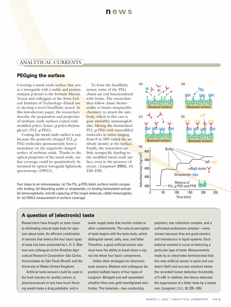
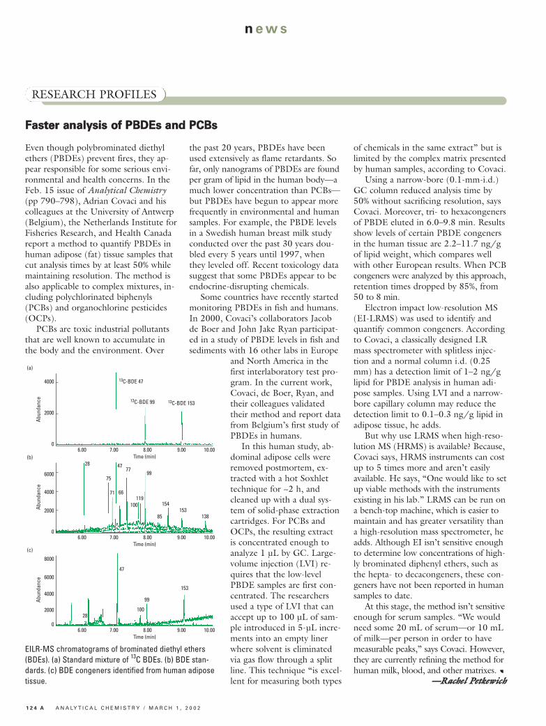
Even though polybrominated diethylethers (PBDEs) prevent fires, they ap-pear responsible for some serious envi-ronmental and health concerns. In theFeb. 15 issue of Analytical Chemistry(pp 790–798), Adrian Covaci and hiscolleagues at the University of Antwerp(Belgium), the Netherlands Institute forFisheries Research, and Health Canadareport a method to quantify PBDEs inhuman adipose (fat) tissue samples thatcut analysis times by at least 50% whilemaintaining resolution. The method isalso applicable to complex mixtures, in-cluding polychlorinated biphenyls(PCBs) and organochlorine pesticides(OCPs).PCBs are toxic industrial pollutants
that are well known to accumulate inthe body and the environment. Over
the past 20 years, PBDEs have beenused extensively as flame retardants. Sofar, only nanograms of PBDEs are foundper gram of lipid in the human body—amuch lower concentration than PCBs—but PBDEs have begun to appear morefrequently in environmental and humansamples. For example, the PBDE levelsin a Swedish human breast milk studyconducted over the past 30 years dou-bled every 5 years until 1997, whenthey leveled off. Recent toxicology datasuggest that some PBDEs appear to beendocrine-disrupting chemicals.Some countries have recently started
monitoring PBDEs in fish and humans.In 2000, Covaci’s collaborators Jacobde Boer and John Jake Ryan participat-ed in a study of PBDE levels in fish andsediments with 16 other labs in Europe
and North America in thefirst interlaboratory test pro-gram. In the current work,Covaci, de Boer, Ryan, andtheir colleagues validatedtheir method and report datafrom Belgium’s first study ofPBDEs in humans.In this human study, ab-
dominal adipose cells wereremoved postmortem, ex-tracted with a hot Soxhlettechnique for ~2 h, andcleaned up with a dual sys-tem of solid-phase extractioncartridges. For PCBs andOCPs, the resulting extractis concentrated enough toanalyze 1 µL by GC. Large-volume injection (LVI) re-quires that the low-levelPBDE samples are first con-centrated. The researchersused a type of LVI that canaccept up to 100 µL of sam-ple introduced in 5-µL incre-ments into an empty linerwhere solvent is eliminatedvia gas flow through a splitline. This technique “is excel-lent for measuring both types
of chemicals in the same extract” but islimited by the complex matrix presentedby human samples, according to Covaci. Using a narrow-bore (0.1-mm-i.d.)
GC column reduced analysis time by50% without sacrificing resolution, saysCovaci. Moreover, tri- to hexacongenersof PBDE eluted in 6.0–9.8 min. Resultsshow levels of certain PBDE congenersin the human tissue are 2.2–11.7 ng/gof lipid weight, which compares wellwith other European results. When PCBcongeners were analyzed by this approach,retention times dropped by 85%, from50 to 8 min.Electron impact low-resolution MS
(EI-LRMS) was used to identify andquantify common congeners. Accordingto Covaci, a classically designed LRmass spectrometer with splitless injec-tion and a normal column i.d. (0.25mm) has a detection limit of 1–2 ng/glipid for PBDE analysis in human adi-pose samples. Using LVI and a narrow-bore capillary column may reduce thedetection limit to 0.1–0.3 ng/g lipid inadipose tissue, he adds.But why use LRMS when high-reso-
lution MS (HRMS) is available? Because,Covaci says, HRMS instruments can costup to 5 times more and aren’t easilyavailable. He says, “One would like to setup viable methods with the instrumentsexisting in his lab.” LRMS can be run ona bench-top machine, which is easier tomaintain and has greater versatility thana high-resolution mass spectrometer, headds. Although EI isn’t sensitive enoughto determine low concentrations of high-ly brominated diphenyl ethers, such asthe hepta- to decacongeners, these con-geners have not been reported in humansamples to date.At this stage, the method isn’t sensitive
enough for serum samples. “We wouldneed some 20 mL of serum—or 10 mLof milk—per person in order to havemeasurable peaks,” says Covaci. However,they are currently refining the method forhuman milk, blood, and other matrixes. a
——RRaacchheell PPeettkkeewwiicchh
6.00 7.00 8.00 10.00Time (min)
4000
Abun
danc
e
2000
0
13C-BDE 47
(a)
13C-BDE 99 13C-BDE 153
9.00
6.00 7.00 8.00 10.00Time (min)
6000
Abun
danc
e
2000
0
28(b)
9.00
4000
75
47
71
77
66
99
119100
85
154153
138
6.00 7.00 8.00 10.00Time (min)
8000
Abun
danc
e
2000
0
28
(c)
9.00
4000
47
99
100
153
6000
EILR-MS chromatograms of brominated diethyl ethers(BDEs). (a) Standard mixture of 13C BDEs. (b) BDE stan-dards. (c) BDE congeners identified from human adiposetissue.
FFaasstteerr aannaallyyssiiss ooff PPBBDDEEss aanndd PPCCBBss

M A R C H 1 , 2 0 0 2 / A N A LY T I C A L C H E M I S T R Y 1 2 5 A
n ews
PEOPLE
Several scientists in the analytical chem-istry community will receive the 2002American Chemical Society awards atthe 223rd National Meeting in Orlan-do, Fla., in April.
Alan G. Mar-shall, professor atFlorida State Uni-versity, will receivethe ACS Award inAnalytical Chem-istry, sponsored byFisher Scientific. The
award is given in recognition of outstand-ing contributions to pure or applied an-alytical chemistry. Marshall is known forhis work in the analytical applications ofFT ion cyclotron resonance ultrahigh-resolution MS.
Ed Yeung, pro-fessor at Iowa StateUniversity, will re-ceive the ACSAward in Chro-matography, spon-sored by Supelco.The award recog-
nizes specific achievements in the fieldof chromatography. Yeung is known forhis many contributions to our fundamen-tal understanding of chromatographyand the development of novel instru-mentation, including single-moleculeadsorption interactions and multiplecapillary systems for DNA sequencingand high-throughput screening.
Brian Chait,Camille and HenryDreyfus Professor atthe Rockefeller Uni-versity, will receivethe Frank H. Fieldand Joe L. FranklinAward for Outstand-
ing Achievement in Mass Spectrometry,sponsored by Bruker Daltonics. Chait isknown for his work in the developmentof mass spectrometric tools for studying
biological processes to aid in, for exam-ple, the elucidation of posttranslationalmodifications.
Takeshi Oka,professor at the Uni-versity of Chicago,will receive the E.Bright WilsonAward in Spec-troscopy, sponsoredby Rohm and Haas.
The award recognizes fundamental con-tributions in all fields of spectroscopy.Oka is best known for his discovery ofH3+, the most abundant molecular ion
in the universe and the initiator of inter-stellar chemistry.
Edward Cussler,professor at the Uni-versity of Minnesota,will receive the ACSAward in Separa-tions Science andTechnology, spon-sored by IBC Ad-
vanced Technologies and Millipore. The award recognizes outstanding accomplishments in the field of fun -damental or applied separation scienceand technology. Cussler is known for his work with membranes for facili -tated transport, temperature-sensitivegels, hollow-fiber extractions, and an artificial gill for keeping a dog alive underwater.
22000022 AACCSS AAwwaarrddss