Embed Size (px)
Citation preview
Sa
CWa
b
c
d
e
a
ARR1AA
KNFCR
1
rcttpcn[gdbpaftn
0d
Analytica Chimica Acta 708 (2011) 141– 148
Contents lists available at SciVerse ScienceDirect
Analytica Chimica Acta
j ourna l ho me page: www.elsev ier .com/ locate /aca
ensitivity evaluation of rhodamine B hydrazide towards nitric oxide and itspplication for macrophage cells imaging
hi-Ming Wub, Yen-Hao Chenc, Kasala Dayanandac, Tsun-Wei Shiuec, Chen-Hsiung Hungd,en-Feng Liawe, Po-Yu Chenb, Yun-Ming Wanga,∗
Department of Biological Science and Technology, Institute of Molecular Medicine and Bioengineering, National Chiao Tung University, 75 Bo-Ai Street, Hsinchu 300, TaiwanDepartment of Medicinal and Applied Chemisrty, Kaohsiung Medical University, 100 Shih-Chuan 1st Road, Kaohsiung 807, TaiwanCollege of Biological Science and Technology, Department of Biological Science and Technology, National Chiao Tung University, 75 Bo-Ai Street, Hsinchu 300, TaiwanInstitute of Chemistry, Academia Sinica, 128 Academia Road Sec. 2, Nankang, Taipei 115, TaiwanDepartment of Chemistry, National Tsing Hua University, 101 Kuang-Fu Road Sec. 2, Hsinchu 300, Taiwan
r t i c l e i n f o
rticle history:eceived 25 April 2011eceived in revised form5 September 2011ccepted 3 October 2011vailable online 8 October 2011
a b s t r a c t
A colorless and non-fluorescent rhodamine derivative, rhodamine B hydrazide (RH), is applied to detectnitric oxide and form fluorescent rhodamine B (RB). The reaction mechanism of RH with NO is proposedin this study. The probe shows good stability over a broad pH range (pH > 4). Furthermore, fluorescenceintensity of RH displays an excellent linearity to the NO concentration and the detection limit is as low as20 nM. A 1000-fold fluorescence turn-on from a dark background was observed. Moreover, the selectivitystudy indicated that the fluorescence intensity increasing in the presence of NO was significantly higher
eywords:itric oxideluorescent probeell imaginghodamine
than those of other reactive oxygen/nitrogen species. In exogenously generated NO detection study,clear intracellular red fluorescence was observed in the presence of S-nitroso-N-acetyl-D,L-penicillamine(SNAP, a kind of NO releasing agent). In endogenously generated NO detection study, increasing incu-bation time of RH with lipopolysaccharied (LPS) pre-treated cells could obtain a highly fluorescent cellimage. These cell imaging results demonstrated that RH can efficiently penetrate into Raw 264.7 cellsand be used for detection of exogenously and endogenously generated nitric oxide.
. Introduction
Nitric oxide (NO) is a gaseous, paramagnetic and highly reactiveadical molecule, which plays various physiological and pathologi-al roles through rapidly reacting with other radicals or metallopro-eins in the organisms. In the animal systems, NO at low concentra-ion acts as a molecular signal transmitter in a variety of biologicalrocesses, such as the blood-pressure regulation of the cardiovas-ular system, the antitumor activity of the immune systems and theeurotransmission of the central and peripheral nervous systems1–9]. On the other hand, high concentration of NO can promote theeneration of reactive nitrogen species (RNS) [3,6,10], which couldegrade lipids, proteins and DNA, with or without the assistancey different kinds of reactive oxygen species (ROS). Since NO isresently regarded as a vital bioregulatory molecule, quantitativessay of the NO concentration either in vitro or in vivo is crucial
or understanding the NO related physiological processes. In ordero comprehend the major functions of NO in biological systems, aumber of methods such as chemiluminescence [11], colorimetry∗ Corresponding author. Tel.: +886 3 5712121x56972; fax: +886 3 5729288.E-mail address: [email protected] (Y.-M. Wang).
003-2670/$ – see front matter © 2011 Elsevier B.V. All rights reserved.oi:10.1016/j.aca.2011.10.005
© 2011 Elsevier B.V. All rights reserved.
[12–15], electron paramagnetic resonance [16–18], electrochem-istry [19–21], and fluorimetry [22–33] have been developed for NOdetection. Compared to other NO detection methods, fluorimetry,which monitor the presence of NO by using a fluorescence probe,has been considered as a favorable way to instantly detect NO withminimal invasiveness, high sensitivity, and high spatial and tempo-ral resolution. This method provides a valuable tool to understandthe versatile roles of NO in the biological system.
Typically, the NO detection mechanism is based on PeT(photoinduced electron transfer) effect. The fluorescence of the NO-sensitive modulator can be quenched by a PeT mechanism in theexcited state. After reacting with NO or the NO oxidized products,the PeT fluorescence quenching property is suppressed and thefluorescence of the probe is restored. The most common PeT sens-ing systems comprise an electron-rich o-diamino aromatic moiety,which can react with NO under aerobic condition to produce flu-orescent triazole derivatives [22,23,30]. However, there are someundesired handicaps, such as complicated and low-yield syntheticprocedures and blank fluorescence, existing in these kinds of NO
probes.Rhodamine dyes are extensively used as fluorescent probesbecause of their high photo-stability, high fluorescence quan-tum yield, high absorption coefficient and broad fluorescence

1 himic
if2tataac2grma
cpfloRdiNowp
2
2
uhfs(oCqhUrTp1puwtno
2
CowsUsuoc
42 C.-M. Wu et al. / Analytica C
n the visible region of electromagnetic spectrum [34]. A spiroorm derivative of rhodamine B (2-amino-3′,6′-bis(diethylamino)-,3-dihydrospiro[iso-indole-1,9′-xanthene]-3-one, RH), which isotally colorless with no absorptions in the visible region, behavess a peroxynitrite (ONOO−) probe when encounters with peroxyni-rite and form the fluorescent rhodamine B-like compound [35]. RHlso reacts with nitrite (NO2
−) at acidic condition (pH < 4) and forms rhodamine derivative with absorption at 561 nm. Importantly, itan also react with NO released by NOC-7 (3-(2-hydroxy-1-methyl--nitrosohydrazino)-N-methyl-1-propanamine) at neutral pH toive a fluorescence at 581 nm [36]. However, the detailed studyelative to the NO detection has not been reported yet. The pri-ary objective of the present study is to investigate the potential
pplication of RH for NO quantification and in vitro detection.In this study, the reaction product of RH with NO under aerobic
onditions, RB, is identified and a possible reaction mechanism isroposed. For realizing the level of fluorescence enhancement, theuorescent properties of RH and RB were studied and compared. Inrder to confirm the stability, the fluorescence intensities of RH andB in various pH conditions were measured. To comprehend the NOetecting ability, time- and concentration-dependent studies were
nvestigated. The correlation between fluorescence intensity andO concentration was also examined. In addition, the selectivityf RH for NO against other reactive oxygen and nitrogen speciesas investigated. Furthermore, in vitro fluorescence images wereerformed.
. Experimental
.1. Materials
All chemicals were obtained from commercial suppliers, andsed as received without further purification. Rhodamine B base,ydrogen peroxide, and lipopolysaccharied (LPS) were purchased
rom Sigma–Aldrich. Hydrazine, sodium nitrite (NaNO2), andodium nitrate (NaNO3) were purchased from Showa. Angeli’s saltNa2N2O3), S-nitroso-N-acetyl-DL-penicillamine (SNAP), and per-xynitrite (as a solution in 0.3 M NaOH) were purchased fromayman Chemical Company. Peroxynitrite was stored at −80 ◦C anduantified by UV–vis spectroscopy immediately prior to use. 4-(2-ydroxyethyl)-1-piperazineethane sulfonic acid (HEPES, free acid,ltrapure Bioreagent) was purchased from MP Biomedicals. Fer-
ous ammonium sulfate (Fe(NH4)2(SO4)2) was purchased from J.. Baker. Dulbecco’s Modified Eagle Medium (DMEM) and sodiumyruvate were purchased from Cellgro. Fetal bovine serum (FBS,0%) was purchased from HyClone. The NO(g) (10% NO/90% N2) wasurchased from SanFu. It was passed through an Ascarite II col-mn to remove higher nitrogen oxides before use [37]. De-ionizedater was produced from Milli-Q reagent water purification sys-
em. Acetonitrile was distilled over calcium hydride (CaH2) underitrogen. Ethanol was distilled over Mg/I2 and subsequently driedver 0.3 nm molecular sieves.
.2. Instruments
Flash column chromatography was performed on reverse phase18 silica gel (25–40 �m). 1H and 13C NMR spectra were obtainedn a Varian VXR-300 spectrometer at 300 MHz and 75 MHz, andere referenced to the internal 1H and 13C solvent peaks. ESI-MS
pectra were recorded on a Micromass Q-Tof mass spectrometer.V–visible absorption spectra were recorded on a Hitachi U-3000
pectrophotometer in 1 cm path length quartz cuvette with a vol-me of 3.5 mL. Fluorescence spectroscopic studies were acquiredn a Hitachi F-7000 spectrophotometer. Bright-field and fluores-ence images were recorded on a fluorescence microscope (IX71,
a Acta 708 (2011) 141– 148
Olympus) equipped with a 100 W mercury lamp, B-2A filters (exci-tation filter: 545–585 nm; emission filter: >610 nm), and a colorCCD camera system.
2.3. Preparation of rhodamine B hydrazide (RH)
A modification of the synthetic procedure of Tong et al. [38]was employed. To 7.2 g of rhodamine B base dissolved in 180 mL ofabsolute ethanol, hydrazine (18 mL) was added dropwisely over 1 hwith vigorous stirring under N2 at room temperature. The stirredmixture was placed in an oil bath and sequentially heated to refluxfor 7 h. The color of the solution changed from dark red to lightorange. The solution was then cooled and the solvent was removedon a rotary evaporator. The residue was added 1 M HCl (300 mL) andadjusted the solution to pH 9.5 by 1 M NaOH. The resulting precipi-tate was filtered and washed 5 times with 10 mL de-ionized water.After removing the solvent in vacuo, the analytically pure product(4.8 g, 67%) was obtained after recrystallization from CH3CN/H2O(1/1, v/v).
ESI-MS, m/z 457.1 ([M+H]+), M+ calculated 456.2.1H NMR (DMSO-d6) ı = 7.76 (m, 1H, ArH), 7.46 (m, 2H, ArH), 6.98
(m, 1H, ArH), 6.37–6.32 (d, 6H, xanthene-H), 4.27 (s, 2H, NH2), 3.32(q, 8H, NCH2CH3), 1.07 (t, 12H, NCH2CH3); 13C NMR (DMSO-d6)ı = 12.46, 43.7, 64.76, 97.42, 105.46, 107.79, 122.16, 123.51, 127.72,128.12, 129.63, 132.39, 148.11, 151.9, 153.04, 165.29.
2.4. Reaction of RH with NO
To a 50 mL of RH (1.0 g, 2.2 mmol) solution in dry acetonitrile,10% NO gas was added under aerobic condition at room tempera-ture. The color of the solution immediately changed from colorlessto deep pink. Then the solvent was removed on a rotary evap-orator. The residue was purified by a column chromatographywith the reverse phase C18 silica gel as stationary phase and theMeOH/H2O (1/1, v/v) solution as eluent. A pink product (0.8 g, 82%)was obtained after drying under vacuo.
ESI-MS, m/z 443.0 ([M+H]+), M+ calculated 442.2.1H NMR (DMSO-d6) ı = 8.23 (d, 1H, ArH), 7.87 (m, 2H, ArH),
7.48 (d, 1H, ArH), 7.10 (d, 2H, ArH), 7.02 (m, 4H, ArH), 3.32 (q, 8H,NCH2CH3), 1.07 (t, 12H, NCH2CH3); 13C NMR (DMSO-d6) ı = 12.4,45.2, 95.8, 112.8, 114.4, 130.1, 130.3, 130.9, 132.6, 155.0, 157.0,166.2.
2.5. Preparation of NO stock solutions
The preparation of NO and its stock solutions were carried outaccording to the reported methods [39,40]. NO can be generatedby slowly dropping 2.0 M H2SO4(aq) into a glass flask containingsaturated NaNO2 water solution. Since O2 will rapidly oxidize NOto form NO2, all apparatus was carefully degassed with argon for30 min to remove O2. The forming gas was passed through a 30%NaOH solution twice and water once to trap NO2 generated fromthe reaction of NO with traces of O2. To produce a saturated NOsolution (1.8 mM, at 20 ◦C) as a stock solution, 10 mL deoxygenatedde-ionized water was bubbled with NO for 30 min and kept underNO atmosphere until being used. Stock solutions were freshly pre-pared for each experiment and stored in a glass flask with a rubberseptum.
2.6. Fluorometric analysis
Fluorescence spectra were recorded by using a Hitachi F-7000spectrophotometer. The slit width was 2.5 nm for both excitationand emission. The photon multiplier voltage was 500 V. Spectrawere routinely acquired at 25.0 ± 0.1 ◦C in quartz cuvettes with a

himic
v5
2
ispi2
2
1s
1N
1N
(as
1w
aHw
iaN
2
1v8tvd(bwri
C.-M. Wu et al. / Analytica C
olume of 3.5 mL. All of the fluorescence spectra were measured at83 nm with excitation wavelength at 510 nm.
.7. Stability studies
Solutions of 50 �M RH (or RB) in 100 mM HEPES with 20% CH3CNn the pH range of 4–12 were prepared by mixing 1 mL of 250 �Molutions of RH (or RB) in CH3CN with 4 mL of 100 mM HEPES whoseH values were adjusted by hydrogen chloride and sodium hydrox-
de. The fluorescence intensities of RH (or RB) were measured after h mixing.
.8. Selectivity studies
Nitric oxide: To a cuvette containing 3 mL RH solution (50 �M, in00 mM HEPES with 20% CH3CN at pH 7.4) was added 416 �L NOtock solution (1.8 mM) under aerobic condition.
Nitrate: To a cuvette containing 3 mL RH solution (50 �M, in00 mM HEPES with 20% CH3CN at pH 7.4) was added 50 �LaNO3(aq) (30 mM) under aerobic condition.
Nitrite: To a cuvette containing 3 mL RH solution (50 �M, in00 mM HEPES with 20% CH3CN at pH 7.4) was added 50 �LaNO2(aq) (30 mM) under aerobic condition.
Hydrogen peroxide: To a cuvette containing 3 mL RH solution50 �M, in 100 mM HEPES with 20% CH3CN at pH 7.4) was added
2.0 �L aliquot of 30% H2O2(aq) under aerobic condition. All of theolutions were prepared at 25.0 ± 0.1 ◦C.
Angeli’s salt (HNO donor): To a gas-tight cuvette containing.8 mg Angeli’s salt was added 3 mL RH (50 �M, in 100 mM HEPESith 20% CH3CN at pH 7.4) under anaerobic condition.
Hydroxyl radical: To a gas-tight cuvette containing ferrousmmonium sulfate (6 mg, 15.3 �mol) and 2.0 �L aliquot of 30%2O2(aq) was added 3 mL RH solution (50 �M, in 100 mM HEPESith 20% CH3CN at pH 7.4) under anaerobic condition.
Peroxynitrite: To a cuvette containing 3 mL RH solution (50 �M,n 100 mM HEPES with 20% CH3CN at pH 7.4) was added a 400 �Lliquot of freshly thawed peroxynitrite solution (3.76 mM in 0.3 MaOH, previously stored at −80 ◦C) under aerobic condition.
.9. Cell viability studies
The cells were grown in 96-well plates at an initial density05 cells per well for 24 h. Subsequently, the 5 �M of RH (2.5%,/v DMSO) was incubated with Raw 264.7 macrophages for 2, 4,, and 12 h. After incubation, the supernatant was removed andhe cells were washed three times with PBS buffer solution. Celliability was evaluated using the 3-(4,5-dimethylthiazol-2-yl)2,5-iphenyl-tetrazoilum bromide (MTT) reduction assay. Briefly, MTT20 �L, 5 mg mL−1) assay was added to each well. After 4 h of incu-
ation, each well was treated with dimethyl sulfoxide (100 �L)ith pipetting. Absorption at 570 nm was measured on a plateeader. Each result was the average of three wells and the viabil-ty of untreated cells was assumed to be 100%. Cell viability (%) of
Scheme 1. The synthet
a Acta 708 (2011) 141– 148 143
Raw 264.7 macrophages at different intervals (2, 4, 8, and 12 h) wascalculated.
2.10. Cell culture and in vitro cell imaging
Raw 264.7 murine macrophage is organized from tumor ascitesinduced by intraperitoneal injection of Abselon Leukemia Virus(A-MuLV) in male mouse. Raw 264.7 murine macrophages wereobtained from the American Type Culture Collection (Manassas,VA). The cells were cultured in Dulbecco’s Modified Eagle Medium(DMEM) and supplemented with 10% fetal bovine serum (FBS), 1%sodium pyruvate, and 1% MEM nonessential amino acids at 37 ◦C ina humidified 5% CO2 atmosphere. For imaging studies, Raw 264.7murine macrophages were passed and plated into poly-d-lysinecoated plates containing 2 mL of DMEM, and incubated at 37 ◦C with5% CO2. For exogenously generated nitric oxide detection studies,the cells were incubated with 5 �M RH (2.5%, v/v DMSO) for 4 h,and further co-incubated with DMEM containing 375 �M SNAP for2 h before being subjected to fluorescence microscope. For endoge-nously generated nitric oxide detection studies, iNOS (induciblenitric oxide synthase) was induced in Raw 264.7 macrophages with1 �g mL−1 of lipopolysaccharide (LPS) for 4 h, and the cells werethen co-incubated with 5 �M RH (2.5%, v/v DMSO) for 2, 4, 6, and8 h. Prior to imaging, the cells were washed three times with 1 mL ofphosphate buffered saline (PBS) and then bathed in 2 mL of PBS. Thefluorescence alterations were monitored by fluorescence micro-scope. For iNOS inhibition study, Raw 264.7 macrophages weresequentially treated with L-NNA (1 mM) for 1 h, LPS for 4 h, andRH for 8 h.
3. Results and discussion
RH was synthesized by the reaction of rhodamine B base withhydrazine followed by a reported procedure [38] with slight mod-ifications (Scheme 1). In order to comprehend the compoundsgenerated after reacting with NO, we dissolved the RH in ace-tonitrile and bubbled 10% NO gas under aerobic condition. Afterpurification and identification, the final fluorescent product (RB)was obtained. A proposed mechanism for RH reacting with NO isshown in Scheme 2. Firstly, NO reacted with its oxidized prod-uct, NO2, to form N2O3 under aerobic condition according to thefollowing equations [41]:
2NO + O2 → 2NO2 (1)
NO2 + NO → N2O3 (2)
Subsequently, N2O3 nitrosylated the hydrazide amino group of RH,and then the amine was converted to diazonium group. In the nextstep, the 5-membered ring was opened and an azide intermedi-ate was formed. Finally, the azide intermediate was converted into
rhodamine B. The absorption and fluorescence spectra of RH andRB were shown in Fig. 1. RH showed a weak absorption at 320 nmand no absorptions between 450 nm and 650 nm, whereas anintense absorption of RB at 560 nm existed. In addition, an obviousic scheme of RH.

144 C.-M. Wu et al. / Analytica Chimica Acta 708 (2011) 141– 148
acting
flR
3
c
Scheme 2. Proposed mechanism of re
uorescence was observed in the fluorescence spectrum of RB, butH had no fluorescence.
.1. Stability study of RH and RB
Since the stability of probes in various pH conditions is con-erned for the biological applicability, the effect of pH values on
Fig. 1. (a) The absorption spectra of RH (dash line) and RB (solid line) an
RH with NO under aerobic condition.
the fluorescence enhancement was measured in the pH range of4−12, and the results were shown in Fig. 2. There was no fluo-rescence observed for RH at pH > 4 indicating that the spirocyclic
moiety can be retained at physiological pH. When the pH valueswere below 4, the RH solutions only showed slight fluorescenceand change to light pink. The increase of fluorescence was observedowing to a ring-opening reaction of the 5-membered ring of RH atd (b) the fluorescence spectra of RH (dash line) and RB (solid line).

C.-M. Wu et al. / Analytica Chimica Acta 708 (2011) 141– 148 145
F(i
psoiu
3
cr(025a5s(citi
L
wtcLp
3
ntoNs1fl(oioeeiet
Fig. 3. (a) Time-dependent fluorescence spectra obtained when various equivalents
ig. 2. The pH dependence of the fluorescence intensity of 50 �M RH (�) and RB�) in 100 mM HEPES buffer with 20% CH3CN at 25.0 ± 0.1 ◦C. The fluorescencentensities were detected at 583 nm with excitation at 510 nm.
H < 4 [42,43]. In contrast, there were almost no fluorescence inten-ities changes in RB in the pH range of 4−12 as shown in Fig. 2. Inther words, the fluorescence intensities of RH and RB were pH-ndependent at pH > 4, which demonstrates a broad pH range forsing RH as a NO probe.
.2. Time- and concentration-dependence of RH reacting with NO
To investigate the NO detection ability, the time- andoncentration-dependent reactions between RH and NO were car-ied out and the results were shown in Fig. 3. To 50 �M RH solutionsin 100 mM HEPES with 20% CH3CN at pH 7.4) were added to.2, 0.4, 0.6, 0.8, and 1.0 equiv. of NO stock solution (1.8 mM) at5.0 ± 0.1 ◦C under aerobic condition. The fluorescence intensity at83 nm increased with time and reached a plateau after 25 mins shown in Fig. 3(a). Furthermore, the fluorescence intensity at83 nm was enhanced with the increasing equivalent of NO(aq), ashown in Fig. 3(b). From Fig. 3(c), an excellent linear correlationR2 = 0.998) between the fluorescence intensity and the NO con-entration could be observed. For RH, the limit of detection (LOD)s the concentration of NO at which the fluorescence signal equalso the blank value plus three-fold of standard deviation, as shownn the following equation [44]:
OD = Xbi + 3Sbi
here Xbi is the mean fluorescence signal of the blank and Sbi ishe standard deviation of the blank. This fluorescence signal wasalculated and compared with the linear correlation to obtain theOD with a value of 20 nM. These results indicated that RH had aotential ability for quantitative NO measurement.
.3. Selectivity
The selectivity of RH for NO against other reactive oxygen anditrogen species requires investigations. The fluorescence intensi-ies of RH reacted with a series of possible competitive reactivexygen or nitrogen species up to 100-fold excess for H2O2, NO3
−,O2
−, HNO, and OH•, and 10-fold excess for ONOO− were mea-ured and the results were shown in Fig. 4. After reacting with00 equiv. of H2O2, NO3
−, NO2−, and HNO, the enhancements of
uorescence intensities were significantly lower than that of NO994-fold). For the comparison of OH• and NO, the concentrationf OH• was 20-fold higher than that of NO, but the fluorescencentensity enhancement of OH• was only 32% of the enhancementf NO. Compared to the result of NO, the fluorescence intensitynhancement of 10 equiv. peroxynitrite was around 81% of the
nhancement from reacting with 5 equiv. of NO. The detections affected when nitric oxide and peroxynitrite simultaneouslyxist. However, it is well-known that peroxynitrite is formed fromhe reaction of nitric oxide and superoxide. The formation ofof NO(aq) were added to a 50 �M RH solution (in 100 mM HEPES with 20% CH3CN atpH 7.4) at 25.0 ± 0.1 ◦C (�ex = 510 nm, �em = 583 nm); (b) concentration-dependentfluorescence spectra based on the intensities at 25 min as shown in (a); and (c)fluorescence intensities upon various equivalents of NO(aq) based on (b).
peroxynitrite is inhibited by an effective superoxide scavengingenzyme, superoxide dismutase (SOD), in biological environment[45].
3.4. Fluorescence imaging of NO in living cells
In order to estimate the exogenously and endogenously gen-erated NO detection ability of RH in living cells, a suitableconcentration of RH is required. There was no significant changein cell viability after the Raw 264.7 macrophages incubated with
5 �M RH (2.5%, v/v DMSO) for 12 h, as shown in Fig. 5. According tothese results, the following cell imaging studies were performed inthe presence of 5 �M RH. For exogenously generated NO detec-tion study, after the cells were incubated with RH for 4 h, the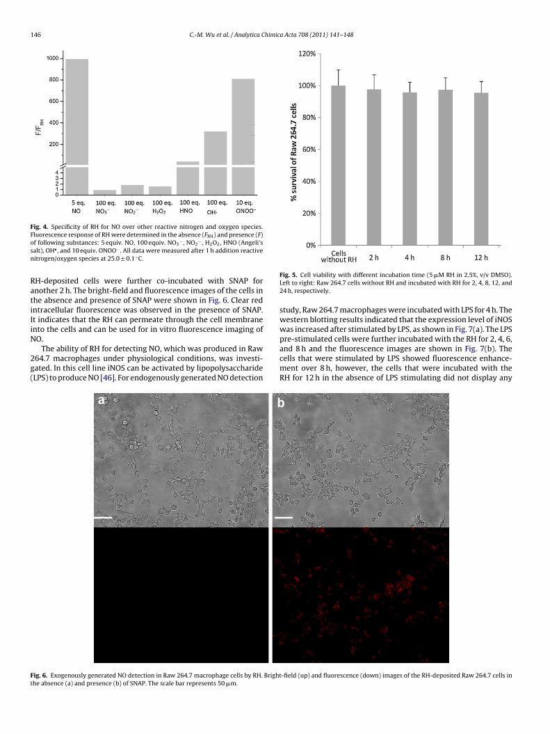
146 C.-M. Wu et al. / Analytica Chimica Acta 708 (2011) 141– 148
Fig. 4. Specificity of RH for NO over other reactive nitrogen and oxygen species.Fluorescence response of RH were determined in the absence (FRH) and presence (F)osn
RatiIiN
2g(
Ft
f following substances: 5 equiv. NO, 100 equiv. NO3− , NO2
− , H2O2, HNO (Angeli’salt), OH• , and 10 equiv. ONOO− . All data were measured after 1 h addition reactiveitrogen/oxygen species at 25.0 ± 0.1 ◦C.
H-deposited cells were further co-incubated with SNAP fornother 2 h. The bright-field and fluorescence images of the cells inhe absence and presence of SNAP were shown in Fig. 6. Clear redntracellular fluorescence was observed in the presence of SNAP.t indicates that the RH can permeate through the cell membranento the cells and can be used for in vitro fluorescence imaging ofO.
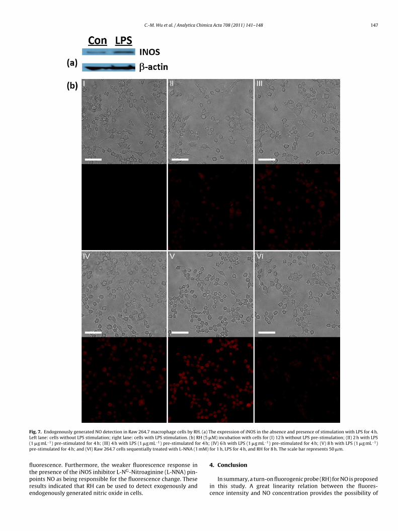
The ability of RH for detecting NO, which was produced in Raw64.7 macrophages under physiological conditions, was investi-ated. In this cell line iNOS can be activated by lipopolysaccharideLPS) to produce NO [46]. For endogenously generated NO detection
ig. 6. Exogenously generated NO detection in Raw 264.7 macrophage cells by RH. Brighthe absence (a) and presence (b) of SNAP. The scale bar represents 50 �m.
Fig. 5. Cell viability with different incubation time (5 �M RH in 2.5%, v/v DMSO).Left to right: Raw 264.7 cells without RH and incubated with RH for 2, 4, 8, 12, and24 h, respectively.
study, Raw 264.7 macrophages were incubated with LPS for 4 h. Thewestern blotting results indicated that the expression level of iNOSwas increased after stimulated by LPS, as shown in Fig. 7(a). The LPSpre-stimulated cells were further incubated with the RH for 2, 4, 6,
and 8 h and the fluorescence images are shown in Fig. 7(b). Thecells that were stimulated by LPS showed fluorescence enhance-ment over 8 h, however, the cells that were incubated with theRH for 12 h in the absence of LPS stimulating did not display any-field (up) and fluorescence (down) images of the RH-deposited Raw 264.7 cells in
C.-M. Wu et al. / Analytica Chimica Acta 708 (2011) 141– 148 147
Fig. 7. Endogenously generated NO detection in Raw 264.7 macrophage cells by RH. (a) The expression of iNOS in the absence and presence of stimulation with LPS for 4 h.L H (5 �( r 4 h;p mM)
fltpre
eft lane: cells without LPS stimulation; right lane: cells with LPS stimulation. (b) R1 �g mL−1) pre-stimulated for 4 h; (III) 4 h with LPS (1 �g mL−1) pre-stimulated fore-stimulated for 4 h; and (VI) Raw 264.7 cells sequentially treated with L-NNA (1
uorescence. Furthermore, the weaker fluorescence response in
he presence of the iNOS inhibitor L-NG-Nitroaginine (L-NNA) pin-oints NO as being responsible for the fluorescence change. Theseesults indicated that RH can be used to detect exogenously andndogenously generated nitric oxide in cells.M) incubation with cells for (I) 12 h without LPS pre-stimulation; (II) 2 h with LPS (IV) 6 h with LPS (1 �g mL−1) pre-stimulated for 4 h; (V) 8 h with LPS (1 �g mL−1)for 1 h, LPS for 4 h, and RH for 8 h. The scale bar represents 50 �m.
4. Conclusion
In summary, a turn-on fluorogenic probe (RH) for NO is proposedin this study. A great linearity relation between the fluores-cence intensity and NO concentration provides the possibility of
1 himic
qrieb
A
Ng
R
[
[
[
[
[
[
[
[[[[
[[
[
[
[[[
[[[
[
[[
[[[[
[[
[[
[
48 C.-M. Wu et al. / Analytica C
uantitative measurement through fluorometric method. A wideange of pH-independent stability allows RH having high stabil-ty at pH > 4. The results of fluorescence imaging of monitoring thexogenously and endogenously generated NO suggest that it wille widely useful for the detection of NO in biological system.
cknowledgements
Funding from National Science Council of Taiwan (Grant Nos.SC 98-2627-M-009- 009 and NSC 97-2113-M-009-016-MY3) isratefully acknowledged.
eferences
[1] R.M.J. Palmer, A.G. Ferrige, S. Moncada, Nature 327 (1987) 524–526.[2] A.R. Butler, D.L.H. Williams, Chem. Soc. Rev. 22 (1993) 233–241.[3] F. Murad, Angew. Chem. Int. Ed. 38 (1999) 1856–1868.[4] R.F. Furchgott, Angew. Chem. Int. Ed. 38 (1999) 1870–1880.[5] L.J. Ignarro, Angew. Chem. Int. Ed. 38 (1999) 1882–1892.[6] J.M. Zimmet, J.M. Hare, Circulation 114 (2006) 1531–1544.[7] D.A. Wink, Y. Vodovotz, J. Laval, F.D. Laval, M.W.J.B. Mitchell, Carcinogenesis 19
(1998) 711–721.[8] B. Mayer, Nitric Oxide, Springer, Berlin, 2000.[9] L.J. Ignarro, Nitric Oxide Biology and Pathobiology, Academic Press, San Diego,
CA, 2000.10] F.L.M. Ricciardolo, P.J. Sterk, B. Gaston, G. Folkerts, Physiol. Rev. 84 (2004)
731–765.11] J.F. Brien, B.E. McLaughlin, K. Nakatsu, G.S. Marks, Methods Enzymol. 268 (1996)
83–92.12] L.C. Green, D.A. Wagner, J.S. Glogowski, P.L.J.S. Wishnok, S.R. Tannenbaum, Anal.
Biochem. 126 (1982) 131–138.13] R.W. Nims, J.F. Darbyshire, J.E. Saavedra, D. Christodoulou, I. Hanbauer, G.W.
Cox, M.B. Grisham, F. Laval, J.A. Cook, M.C. Krishna, D.A. Wink, Methods 7 (1995)48–54.
14] L.A. Ridnour, J.E. Sim, M.A. Hayward, D.A. Wink, S.M. Martin, G.R. Buettner, D.R.
Spitz, Anal. Biochem. 281 (2000) 223–229.15] F. Brown, N.J. Finnerty, F.B. Bolger, J. Millar, J.P. Lowry, Anal. Bioanal. Chem. 381(2005) 964–971.
16] H. Kosaka, M. Watanabe, H. Yoshihara, N. Harada, T. Shiga, Biochem. Biophys.Res. Commun. 184 (1992) 1119–1124.
[[
[[
a Acta 708 (2011) 141– 148
17] Y. Katayama, N. Soh, M. Maeda, ChemPhysChem 2 (2001) 655–661.18] T. Nagano, T. Yoshimura, Chem. Rev. 102 (2002) 1235–1270.19] T. Malinski, Z. Taha, Nature 358 (1992) 676–678.20] K. Ichimori, I.H. Shida, M. Fukahori, H. Nakazawa, E. Murakami, Rev. Sci. Instrum.
65 (1994) 2714–2717.21] F. Bedioui, N. Villeneuve, Electroanalysis 15 (2003) 5–18.22] H. Kojima, N. Nakatsubo, K. Kikuchi, S. Kawahara, Y. Kirino, H. Nagoshi, Y. Hirata,
T. Nagano, Anal. Chem. 70 (1998) 2446–2453.23] H. Kojima, M. Hirotani, N. Nakatsubo, K. Kikuchi, Y. Urano, T. Higuchi, Y. Hirata,
T. Nagano, Anal. Chem. 73 (2001) 1967–1973.24] H. Kojima, Y. Urano, K. Kikuchi, T. Higuchi, Y. Hirata, T. Nagano, Angew. Chem.
Int. Ed. 38 (1999) 3209–3212.25] K.J. Franz, N. Singh, S.J. Lippard, Angew. Chem. Int. Ed. 39 (2000) 2120–2122.26] N. Soh, Y. Katayama, M. Meada, Analyst 126 (2001) 564–566.27] Y. Gabe, Y. Urano, K. Kikuchi, H. Kojima, T. Nagano, J. Am. Chem. Soc. 126 (2004)
3357–3367.28] M.H. Lim, D. Xu, S.J. Lippard, Nat. Chem. Biol. 2 (2006) 375–380.29] E.W. Miller, C.J. Chang, Curr. Opin. Chem. Biol. 11 (2007) 620–625.30] E. Sasaki, H. Kojima, H. Nishimatsu, Y. Urano, K. Kikuchi, Y. Hirata, T. Nagano, J.
Am. Chem. Soc. 127 (2005) 3684–3685.31] H. Zheng, G.Q. Shang, S.Y. Yang, X. Gao, J.G. Xu, Org. Lett. 10 (2008) 2357–
2360.32] Z.J. Tonzetich, L.E. McQuade, S.J. Lippard, Inorg. Chem. 49 (2010) 6338–6348.33] Y. Yang, S.K. Seidlits, M.M. Adams, V.M. Lynch, C.E. Schmidt, E.V. Anslyn, J.B.
Shear, J. Am. Chem. Soc. 132 (2010) 13114–13116.34] M. Beija, C.A.M. Afonso, J.M.G. Martinho, Chem. Soc. Rev. 38 (2009) 2410–2433.35] X.F. Yang, X.Q. Guo, Y.B. Zhao, Talanta 57 (2002) 883–890.36] T. Rieth, K. Sasamoto, Anal. Commun. 35 (1998) 195–197.37] C.F. Works, C.J. Jocher, G.D. Bart, X. Bu, P.C. Ford, Inorg. Chem. 41 (2002)
3728–3739.38] Y. Xiang, A. Tong, P. Jin, Y. Ju, Org. Lett. 8 (2006) 2863–2866.39] J. Ouyang, H. Hong, C. Shen, Y. Zhao, C.G. Ouyang, L. Dong, J.U. Zhu, Z.J. Guo,
K. Zeng, J.G. Chen, C.Y. Zhang, J.F. Zhang, Free Radic. Biol. Med. 45 (2008)1426–1436.
40] K.J. Huang, H. Wang, M. Ma, X. Zhang, H.S. Zhang, Nitric Oxide 16 (2007) 36–43.41] L.J. Ignarro, J.M. Fukuto, J.M. Griscavage, N.E. Rogers, R.E. Byrns, Proc. Natl. Acad.
Sci. U.S.A. 90 (1993) 8103–8107.42] Y. Zhao, X.B. Zhang, Z.X. Han, L. Qiao, C.Y. Li, L.X. Jian, G.L. Shen, R.Q. Yu, Anal.
Chem. 81 (2009) 7022–7030.
43] S. Kang, S. Kim, Y.-K. Yang, S. Bae, J. Tae, Tetrahedron Lett. 50 (2009) 2010–2012.44] EURACHEM, The Fitness for Purpose of Analytical Methods: A Laboratory Guideto Method Validation and Related Topics, LGC, United Kingdom, 1998.45] P. Pacher, J.S. Beckman, L. Liaudet, Physiol. Rev. 87 (2007) 315–424.46] E.M. Conner, M.B. Grisham, Methods 7 (1995) 3–13.


![Analytica Chimica Acta - UT Arlington – UTA · 104 K.A. Schug et al. / Analytica Chimica Acta 713 (2012) 103–110 large pharmaceuticalcompanies[8,9].Infact,onlytwonewclasses of](https://img.dokumen.tips/doc/110x75/5c67dd2009d3f226588c984a/analytica-chimica-acta-ut-arlington-104-ka-schug-et-al-analytica.jpg)


![Analytica Chimica Acta - download.xuebalib.comdownload.xuebalib.com/1dc8WMowcDlH.pdf · vices [10], promising organic thermoelectric materials [20], dye- sensitized solar cells [21],](https://img.dokumen.tips/doc/110x75/5b90021d09d3f28c298d53ca/analytica-chimica-acta-vices-10-promising-organic-thermoelectric-materials.jpg)


![Analytica Chimica Acta - University of California, Santa CruzAnalytica Chimica Acta 1105 (2020) 82e86 about cancer status and progression [5,6]. miR-1290 is a highly sensitive and](https://img.dokumen.tips/doc/110x75/60af0ef23cf3d07be8654c9c/analytica-chimica-acta-university-of-california-santa-cruz-analytica-chimica.jpg)