Embed Size (px)
Citation preview

A N A L Y S I S O F T C E L L H Y B R I D O M A S
II. C o m p a r i s o n s a m o n g T h r e e D i s t i nc t T y p e s o f
M o n o c l o n a l S u p p r e s s o r Fac to r s*
By KENJI OKUDA, MUTSUHIKO MINAMI, SHUICHI FURUSAWA, AND MARTIN E. DORF
From the Harvard Medical School, Department of Pathology, Boston, Massachusetts 02115
Recent advances in cell hybridizat ion techniques have permi t ted the fusion of functional T cell subpopula t ions with tumor cells to yield stable T cell hybr idomas (1-9). Such hybrids immorta l ize the biological activity of an individual cell and permit analysis of monoclonal T cell-derived helper (5) and suppression factors (2, 4, 6, 9, 10). T h e prepara t ion of monoclonal T cell hybr idomas and isolation of biologi- cally active factors derived from monoclonal hybr idoma lines can provide a means of dissecting interact ing T cell subsets that exist within heterogeneous T cell populations. Such an approach permits a more precise unders tanding of the mechanism of T cell interactions.
I m m u n e suppression is one such T cel l-mediated activity in which mult iple T cell populat ions and soluble factors are required (11). Although initial character izat ion of the various suppressor T cell populat ions (Ts) 1 has been possible with heterogeneous Ts populat ions, it remains difficult to analyze the contr ibutions of individual T cell subsets and to insure that a single subset instead of a mixture of subsets is being studied. Fur thermore , the quanti t ies of cells or factors obta ined from conventional cell prepara t ions are often insufficient for detailed analysis. Thus, the establishment of monoclonal T cell hybrids with specific suppressor function provides an ideal tool for the character izat ion of the distinct suppressor cell subsets, their products, and their interactions.
Regulat ion of the i m m u n e response to the hapten 4-hydroxy-3-ni t rophenyl acetyl (NP) has been extensively character ized (12-16). Previous studies using conventional analysis and monoclonal T cell hybr idomas have revealed that in the NP suppressor cell pa thway at least three distinct subsets of T cells are required for suppression (13, 14). T h e first set of Ts, te rmed Tsx, are induced by antigen, bear idiotypic (Id +) receptors, and can function early in the i m m u n e response (10, 12). These Tsl cells, or a factor produced by these cells, can induce a second set of Ts, te rmed Ts2 (10, 13,
* Supported by grants CA 14732 and AI 16677 from the National Institutes of Health and grant PCM 80-04573 from the National Science Foundation.
L Abbreviations used in this paper: C, complement; CGAT, common idiotype on anti-GAT antibodies; CS, cutaneous sensitivity; DMSO, dimethyl sulfoxide; DNFB, 2,4 dinitro-l-fluorohenzene; HBSS, Hanks' balanced salt solution; KLH, keyhole limpet hemocyanin; NP, 4-hydroxy-3-nitrophenyl acetyl hapten; NP b, common idiotype on C57BL anti-NP antibodies; NP-O-Su, NP-O-succinimide ester; PBS, phosphate- buffered saline; RAMG, rabbit anti-mouse immunoglobulin; Ts~, Ts2, Tss, first-, second-, or third-order suppressor T cells, respectively; TsFI, TsF2, TsFa, suppressor factor derived from Tsl, Ts2, or Tss cells, respectively.
1838 J. Exp. MED. © The Rockefeller University Press • 0022-1007/81/12/1838/04 $1.00 Volume 154 December 1981 1838-1851

K. OKUDA, M. MINAMI, S. FURUSAWA, AND M. E. DORF 1839
14). T h e Ts2 cells bear an t i - id io typ ic receptors and function late in the i m m u n e response (13-15). This Ts2 cell popu la t ion or a soluble factor der ived from it act ivates an an t igen -p r imed Ts3 popu la t ion tha t is thought to provide still ano the r suppressor signal (i 4).
T h e Tsn popu la t ion was first descr ibed by Sy et al. (17) in an t igen -p r imed animals . Fu r the r analysis involving the N P system as well as the p-azobenezenearsona te suppressor system has shown tha t the Ts3 popu la t ion possessed idiotypic , Lyt-2, and I-J de te rminan t s (14, 18). 2 In addi t ion , the induct ion of funct ional Ts3 cells is sensitive
to cyc lophosphamide (14, 17). Despi te these results, the exact mechanism by which a Ts3 popu la t i on manifests i m m u n e suppression as well as the na tu re of the Ts3-derived factor r ema ined unknown.
To ob ta in a be t te r unde r s t and ing of the precise na tu re and funct ion of Ts~ cells, we p repa red T cell hybr idomas tha t cor responded to the Ts3 popula t ion . In this com- mun ica t ion we charac ter ize the biological ly active suppressor factors (TsF~) p roduced by these h y b r i d o m a cells and compare these factors wi th previously charac te r ized NP-specific TsF1 and TsFe factors.
M a t e r i a l s a n d M e t h o d s
Mice. All mice were either purchased from The Jackson Laboratory, Bar Harbor, Maine, or were bred in the animal facilities at Harvard Medical School, Boston, Mass. Mice were used at 3-12 mo of age and were maintained on laboratory chow and acidified, clorinated water ad lib.
Antigens. NP-O-Succinimide (NP-O-Su) was purchased from Bioseareh Co., San Rafael, Calif. Dimethylsulfoxide (DMSO) was purchased from Fisher Scientific Co., Pittsburgh, Pa. 2,4-dinitro-l-fluorobenzene (DNFB) and keyhole limpet hemocyanin (KLH) were obtained from Sigma Chemical Co., St. Louis, Mo.
Antisera. Both B10.A(3R) anti-B10.A(5R) (anti-I-J k) and B10.A(5R) anti-B10.A(3R) (anti- I-J b) were produced by immunization with spleen and lymph node cells, as described elsewhere (4). Both monoclonal anti-Thy 1.1 and Thy-1.2 antibodies were ~urchased from New England Nuclear, Boston, Mass. Guinea pig anti-CGAT and anti-NP anti-idiotype antisera were prepared as detailed elsewhere (12, 19). The characterization of these reagents that detect a common idiotype on anti-GAT and anti-NP antibodies, respectively, was described previously (4).
Preparation of Tsa CeUsfrom Antigen Plates. The methods for the preparation and enrichment of NP-binding T cells were described in detail elsewhere (12). 2 In brief, 5 × 10⁷ regional lymph node or spleen cells from CKB(H-2 k, Igh-1 b) and C57BL/6 (H-2 ~, Igh-I ~) mice, which were immunized subcutaneously with 2 mg NP-O-Su or with 100 /~g NP-KLH i.p. in Maalox Perlussis (15), were added to purified anti-mouse immunoglobulin (RAMG)-coated petri dishes to remove B cells. The nonadherent T cells were incubated on NP-bovine serum albumin- coated petri dishes for 45 min at room temperature. Nonadherent cells were removed by gentle swirling, and the plates were placed on ice for 20 min. The antigen-binding cells were collected from the plate by gentle pipetting and used as the source of Ts3 cells.
Hybridization and Screening of Ts3 Hybridoma Lines. Tsa-enriched CKB or C57BL/6 lymphocytes were hybridized with BW5147 T lymphoma cells. The polyethylene glycol-mediated hybrid- izing method was exactly the same as previously reported (4). The hybridized Ts~ candidates were screened using a cytotoxicity test (4) with allele-specific anli-I-J and anti-NP ~' antisera. Three fusions were performed; the CKB-Ts3-3 and CKB-Ts:~-9 lines were derived from one fusion using CKB NP-O-Su-primed lymph node cells. The B6-Ts3-2 line was similarly derived from a fusion between NP-O-Su-primed lymph node cells from C57BL/6 mice and BW5147
2 Sherr, D. It., and M. E. Dorf. Hapten-specific T cell responses to 4-hydroxy-3-nitrophenyl acetyl. XIII. Characterization of a third-order T cell (Tss) involved in suppression of in vitro PFC responses. Manuscript in preparation.

1840 CHARACTERIZATION OF TsF~ SUPPRESSOR FACTOR
tumor cells. The B6-Ts3-19 line was obtained from a third hlsion using antigen-adherent splenic T cells from an NP-KLH-primed C57BL/6 mouse. In addition, the B6-Tsa-8 hybridoma line was derived from still another fusion in which we were screening for Tsz hybrids (Minami and Okuda, unpublished observations). All the Tsa hybrids were periodically passaged over the antigen-coated plates, and the idiotype-adherent cells were used to maintain the cell lines. The B6-Tsa-2, CKB-Ts3-3, and CKB-Ts3-9 hybridoma lines were cloned by limiting dilution; l0 of the 11 clones tested had the same phenotype and suppressive activity as the parental line, but one CKB-Tsa-3 clone lacked functional activity.
All of the hybridomas were cultured in RPMI 1640 containing 8% fetal calf serum and 0.01 M Hepes buffer. The suppressor factors used in the present experiment were collected from the cultured supernatants of cells at an approximate density of 7 × 10⁵ cells/ml in the above medium.
Adsorption and Elution of TsF. The methods of absorption and elution of TsF using protein- conjugated Sepharose-4B columns were described in detail previously (4).
Assay for Suppressive Activity of TsF on NP-mediated Cutaneous Sensitivity (CS) Responses. The assay for NP-specific CS responses was described elsewhere (10, 14). Briefly, each animal was primed subcutaneously with 2 mg of NP-O-Su in DMSO. Unless indicated otherwise, the hybridoma factors were tested in the effector phase, 5 or 6 d after priming. 0.4 ml of each hybridoma supernatant or BW5147 control supernatant was injected i.v. on the day before and the day of antigen challenge. 6 d after immunization, mice were challenged in the left footpad with 0.025 ml phosphate-buffered saline (PBS) solution containing 30 p,g of NP-O-Su (prepared by mixing 25 ~1 of a 2% NP-O-Su/DMSO solution in 0.4 ml PBS). Footpad swelling 3 was measured 24 h after challenge. Swelling was determined as the difference, in units of 10-' cm, between the left and right footpad thickness.
Functional Analysis of TsF~ and TsFa Factors in Cyctophosphamide-treated Antigen-primed Mice. Mice were primed subcutaneously with NP-O-Su on day 0, as described above. 24 h later, they were treated with an i.p. injection of either saline or 50 mg/kg cyclophosphamide in saline. On days 5 and 6, each mouse received 0.4 ml i.v. of control BW5147 tumor supernatant or Ts2- or Tsa- derived factors. On day 6, mice were challenge with NP-O-Su, and CS responses were measured 24 h later.
DNFB Contact Sensitivity Responses. Contact sensitivity was induced by two daily paintings on the shaved abdomen with 25 pl of 0.5% DNFB solution (Sigma Chemical Co., St. Louis, Mo.) in acetone:olive oil (4: i) (4). 6 d after the last painting, 20 p,1 of 0,2% DNFB in the same vehicle was applied to the left ear, and the ear swelling was measured as the difference between the left and right ear thicknesses.
Double Antigen Ear Challenge. Individual mice were immunized with either DNFB alone or with DNFB plus NP-O-Su, as described above. Mice were challenged in the left ear by painting with 0.2% DNFB, injecting 0.015 ml containing 6.0 ~g NP-O-Su (prepared by mixing 0.025 ml of 0.7% NP-O-Su in DMSO with 0.4 ml PBS, pH 7.7), or with both antigens. The incremental ear swelling was measured 24 and 48 h thereafter. The concentration and volume of NP-O-Su used to challenge was predetermined to elicit high specific ear swelling and low nonspecific backgrounds.
Percent Suppression. The percent suppression in the present study was calculated by the following formula: percent suppression = 100 × [(swelling of BW tumor supernatant-injected group - swelling of TsF-injected group)/(swelling of BW tumor supernatant-injected group - swelling of unprimed group)].
Data Analysis. Statistical analysis of the experimental data with respect to controls was calculated using the two-tailed Student's t test.
Resu l t s
Screening of Hybridoma Suppressor Cells. After fusion of ant igen-adherent T cells from NP-pr imed mice with AKR-der ived BW5147 tumor cells, 320 of 2,300 wells developed hybr idoma colonies. 260 of these 320 lines were screened by the microcytotoxicity test using allele-specific ant i-I-J alloantisera. 26% were specifically lysed with anti-I-J alloantisera. 54 of these lines were then screened with guinea pig an t i -NP b or control

K. OKUDA, M. MINAMI, S. FURUSAWA, AND M. E. DORF 1841
anti-CGAT-anti-idiotypic antisera; 39% (21 of 54) of these lines were specifically lysed with anti-NP b idiotype antisera. Supernatants from those lines that were specifically lysed with both anti-I-J and anti-NP b were tested for suppressive activity by admin- istrating 0.4 ml of supernatant the day before and the day of antigen challenge. At least seven lines demonstrated significant levels of supppessive activity. Five of those that demonstrated >50% suppressive activity of the NP-O-Su CS response were selected for further phenotyping and characterization. The five hybridoma lines that constitutively produced suppressor factor were B6-Tsa-2, B6-Ts3-8, B6-Tsa-19, CKB- Tsa-3, and CKB-Tsa-9. Upon retesting, two of the lines (B6-Tsa-8 and B6-Ts3-19) demonstrated variable or no reaction with the anti-I-J b or the anti-NP b idiotypic antisera, although these hybridomas retained their ability to secrete a biologically active suppressor factor (see below). It is not clear why these lines were screened as initially positive, but after continual culture, they appeared to lose their ability to express I-J and idiotypic determinants on their cell surface. Clones of each of these lines were also tested for the presence of I-J and idiotypic determinants, and again the clones appeared to have little or no expression of these antigenic determinants. In contrast, the other three lines have maintained the ability to express cell surface I-J and idiotype determinants.
Specificity of Suppressor Factors. To test the biological activity of these presumptive suppressor hybridomas, we screened for the ability of culture supernatants obtained from cells growing at an approximate density of 7 X 10⁵ cells/ml to suppress in vivo CS responses. As shown in Table I, injection of 0.4 ml of culture supernatant from the B6-Tsa-2, B6-Tss-19, and CKB-Tsa-9 cell lines specifically suppressed NP-O-Su-in-
TABLE I
Specificity of Hybridoma Suppressor Factors *
Source of TsF Strain tested TsF dilution
Swelling response (X 10 -3 cm) ± SE
NP-O-Su DNFB (ear)
(footpad)
BW5147 C57BL/6 Undiluted 27.3 ± 0.9 21.5 ± 0.6 B6-Ts3-2 C57BL/6 Undiluted 13.0 + 0.7~ 22.8 ± 1.7
1/10 12.8 + 1.3~ 1/100 21.0 + 1.4 l/1,000 26.3 ± 0.9
B6-Tsa-19 C57BL/6 Undiluted 15.5 + 0.61l: 26.4 + 2.7 I/I0 14.8 + I.I* 1/100 20.8 + :.9,
1/1,1000 27.5 ± 1.1 BW5147 CKB Undiluted 25.5 ± 0.5 22.8 ± 2.7 CKB-Tsa-9 CKB Undiluted 13.0 ± 1.4, 22.0 ± 0.7
1/10 14.7 ± 0.9~: 1/1oo 25.8 ± 5.6 l/l,O00 25.8 ± i.:
* Groups of four to five mice were immunized with either NP-O-Su or DNFB, The day before and the day of antigen challenge mice were given an i.v. injection of 0.4 ml of either control BW5147 or suppressor factor. The background responses of nonimmunized C57BL/6 mice were 4.1 ± 0.7 and 11.2 "4- 1.2 for NP-O-Su and DNFB, respectively. The background responses of CKB mice were 3.0 ± 0.4 and 2.6 + 0.7 for NP-O-Su and DNFB, respectively.
* Significant suppression, P < 0.005.

1842 CHARACTERIZATION OF TsF,~ SUPPRESSOR FACTOR
duced CS responses. T h e cu l tu re s u p e r n a t a n t s d e m o n s t r a t e d m a x i m u m p l a t e a u levels
o f ac t iv i ty w h e n g iven e i the r u n d i l u t e d or d i lu t ed 1:10. In these expe r imen t s the
pe rcen t o f suppress ion r a n g e d f rom 50-75%. Af te r fu r the r d i lu t ion these ma te r i a l s no
longer d e m o n s t r a t e d s igni f icant levels o f suppress ive act ivi ty .
T h e a n t i g e n specif ic i ty o f these suppressor factors was in i t ia l ly d e m o n s t r a t e d by
the i r inab i l i ty to suppress the D N F B - i n d u c e d con t ac t sens i t iv i ty responses (Tab le I).
T h e N P specif ic i ty o f the B6-Tsa-8 a n d CKB-Tsa -3 suppressor factors was s imi la r ly
es tab l i shed (da ta not shown) .
A l t h o u g h the specif ic i ty o f these suppressor factors was d e m o n s t r a t e d by the a b o v e
cr i te r ia , it cou ld still be a r g u e d tha t specific an t i gen is on ly r e q u i r e d to t r igger the
suppress ive ac t iv i ty , a n d once t r iggered , the suppress ion w o u l d be nonspecif ic . Severa l
e x a m p l e s o f such a n t i g e n - d e p e n d e n t nonspec i f ic suppress ion h a v e been no t ed in the
l i t e r a tu re (22-24) . T o e v a l u a t e the l a t t e r possibi l i ty, g roups o f four to five C 5 7 B L / 6
mice were i m m u n i z e d wi th e i t he r D N F B a lone or D N F B plus N P - O - S u . Af te r 5 d,
the a n i m a l s were g iven e i the r con t ro l BW5147 s u p e r n a t a n t s or cu l tu re s u p e r n a t a n t s
f rom B6-Tsa-2 or B6-Ta-19 cells. T h e mice were then c h a l l e n g e d in the left ea r by
p a i n t i n g wi th D N F B , in j ec t ing 7.0 #g N P - O - S u in 15/.tl PBS (pH 7.7), or wi th b o t h
ant igens . T h e i n c r e m e n t a l ea r swel l ing was m e a s u r e d at 24 a n d 48 h. T h e d a t a in
T a b l e II ver i fy specif ic i ty o f the B6-Tsa-2 a n d the B6-Tsa-19 factors. Thus , these l a t t e r
factors fail to suppress ea r swel l ing responses i n d u c e d a n d e l ic i ted w i t h D N F B or
responses i nduced wi th N P - O - S u plus D N F B p r i m i n g a n d e l ic i ted wi th D N F B alone.
H o w e v e r , the ac t iv i ty o f the B6-Tsa-2 a n d B6-Ts3-19-der ived factors was c o n f i r m e d
by the i r ab i l i ty to suppress ea r swel l ing responses in mice p r i m e d wi th bo th N P - O - S u
plus D N F B a n d c h a l l e n g e d wi th N P - O - S u alone. T o d e t e r m i n e w h e t h e r these
T A B L E II
Specificity of Suppressor Factor in Doubly-Primed or Challenged Recipients *
Antigen priming TsF source Antigen challenge Ear swelling (× 10 -3 cm) ± SE
24 h 48 h
DNFB BW5147 DNFB 13.0 - 1.7 13.3 ± 1.0 DNFB B6-Ts3-2 DNFB 16.0 ± 3.0 16.0 _ 2.3 DNFB B6-Tsa-19 DNFB I1.0 + 1.8 14.0 ± 1,2 - - BW5147 DNFB 1.0 + 0.4 2.0 + 0.4 NP-O-Su + DNFB BW5147 DNFB 15.0 ± 0.7 13.0 ± 1.8 NP-O-Su + DNFB B6-Ts3-2 DNFB 13.0 + 1.4 12.5 ± 1.7 NP-O-Su + DNFB B6-Tsz-19 DNFB 16.0 _+ 2.7 15.0 ± 1.1 - - BW5147 DNFB 1.0 ± 0.4 2.0 ± 0.4 NP-O-Su + DNFB BW5147 NP-O-Su 18.5 ± 1.2 16.5 ± 1.3 NP-O-Su + DNFB B6-Ts3-2 NP-O-Su 8.3 ± 0.% 7.8 ± 1.4:~ NP-O-Su + DNFB B 6 - T s 3 - 1 9 NP-O-Su 8.3 ± 2.7:~ 8.3 ± 1.1~: - - BW5147 NP-O-Su 8.3 ± 1.9 4.3 ± 0.8 DNFB BW547 NP-O-Su + DNFB 16.5 + 1.2 12.8 ± 1.0 DNFB B6-Ts:r2 NP-O-Su + DNFB 19.3 ± 2.5 12.8 ± 1.4 DNFB B6-Ts3-19 NP-O-Su + DNFB t9.0 ± 2.0 t3.8 ± 1.7 - - BW5147 NP-O-Su + DNFB 10.5 ± 1.2 6.5 ± 1.0
* Groups of four to five mice were primed with DNFB alone or with DNFB and NP-O-Su. 5 and 6 d thereafter, they were given TsF or control factors, followed by ear challenge with DNFB, NP-O-Su, or both antigens. The specific ear swelling responses were measured 24 and 48 h after challenge. The data are expressed as the increment of ear swelling (× 1() -:~ cm) + SE.
:~ Significant suppression, P < 0.005.

K. OKUDA, M. MINAMI, S. FURUSAWA, AND M. E. DORF 1843
suppressor factors would nonspecifically suppress DNFB responses when the animals were challenged with both NP-O-Su plus DNFB, another series of animals were tested. As shown in Table II, the presence of NP-O-Su in the challenge site is not sufficient to suppress DNFB-induced contact sensitivity responses. Thus, the data demonstra te that these hybr idoma-der ived suppressor factors are ant igen specific.
Effector-Phase Suppression. To compare this series of NP-specific suppressor factors with the previously characterized Tsl- and Ts2-derived hybr idoma factors, we deter- mined when in the course of the i m m u n e response the various factors were active. 0.4 ml of factor was injected i.v. either on the day of and the day after ant igen p r iming
( induct ion phase) or the day before and the day of ant igen challenge (effector phase). As previously reported (10), the B6-Tsl-29- and CKB-Tsl-17-derived control TsF1 factors only demonstra ted suppressive activity when adminis tered dur ing the induc- tion phase of the i m m u n e response (Table III). In contrast, the previously character-
ized B6-Ts2-28-derived TsF2 factor (20) and the five new hybr idoma factors were only active when adminis tered in the effector phase of the NP-O-Su CS response (Table
III). Genetic Restrictions of Hybridoma Suppressor Factors. To further compare this new series
of hybr idoma suppressor factors with those previously characterized, the genetic restrictions of this new series of factors was determined in comparison with the
monoclonal control Tsl- and Ts2-derived factors. The Tsl factor was adminis tered dur ing the induct ion phase of the i m m u n e response, whereas the Ts2 and the new series of suppressor factors were adminis tered dur ing the effector phase. As shown in Tab le IV and in conf i rmat ion of previous data (10, 20), the C57BL/6-der ived TSl factor from the B6-Ts1-29 cells suppressed NP-specific CS responses in both syngeneic
TABLE III
Comparison of the Ability of TsF to Suppress CS Responses m the Induction vs. Effector Phase
NP-O-Su TsF source Strain tested primed
NP-O-Su-induced swelling (× 10 -3 cm) ± SE
Induction Effector phase phase
None C57BL/6 + 36.4:1:1.4 36.5 ± 1.4 B6-Tsl-29 C57BL/6 + 18.3:1:1.75 35.3 -t- 2.9 B6-Ts2-28 C57BL/6 + 34.9 zl: 1.3 23.5 ± 1.55 B6-Tsa-2 C57BL/6 + 39.5 ± 4.2 13.8 + 2.2:~ B6-Ts3-8 C57BL/6 + 36.0 + 5.4 21.1 ± 2.1:[: B6-Ts3-19 C57BL/6 + 36.8 ± 3.3 19.5 ± 3.0:[: None C57BL/6 - 10.6 ± 2.6 10.6 ~ 2.6 None B10.A + 34.6 ± 3.6 34.6 ± 3.6 CKB-TsI-17 BI0.A + 12.8 ± 1.85 30.4 ± 2.9 CKB-Ts3-3 B10.A + 31.5:1:2.6 18.3 ± 1.75 CKB-Ts3-9 B10.A + 32.3 ± 1.3 16.8:1:2.05 None B10.A - 8.7 ± 1.2 8.7 -e 1.2
* Groups of four to five mice were immunized with NP-O-Su and were given TsF either at the time of antigen priming (induction phase) or antigen challenge (effector phase). The results with the control TsF1 and TsF2 factors were obtained from separate experiments. The data were normalized for presentation.
$ Significant suppression, P < 0.001.

1844 CHARACTERIZATION OF TsFa SUPPRESSOR FACTOR
TABLr IV Genetic Restrictions of Suppressor Factors *
NP-O-Su TsF source priming
Footpad swelling (× 10 -a cm) in recipient strains ± SE
C57BL/6 B6.Igh e B 10.WB CWB C3H.SW (H-2 b , Igh b ) (H-2 b , Igh ~) (H-2 j , Igh b ) (H-2 b , Igh b ) (H-2 b , IgH j )
BW5147 + 24 .8± 1.9 17.4:t: 1.1 20 .8±0 .7 31.2 ± 3.1 2 9 . 8 ± 2 . 3 B6-Tsr29 + 10.7 ± 1.3~ NT§ 9.7 ± 1.0~ NT NT B6-Ts~-28 + 12.6 ± 1.9z~ 16.5 ± 1.9 19.3 ± 0.9 NT NT B6-Tsa-2 + 12.2 ± 0.7~: 18.8 ± 0.3 21.5 ± 0.4 12.3 ± 1.9:[: 29.5 ± 1.0 B6-Tsa-8 + 14.8+ 1.2~ NT 22 .5±2 .1 19.5 ± 2.9z~ 26 .0± 1.5 B6-Tsa-19 + 9. l ± 3.1~: NT 25.1 ± 3.4 14.8 ± 2.6:]: 32.5 ± 4.2 BW5147 - 5.2 ± 1.0 3.5 ± 1.2 5.5 ± 1.3 6.3 ± 1.3 7.8 ± 1.9
* Groups of four to five mice were immunized with NP-O-Su and were given TsF during the induction (TsFi) or effector phase of the immune response. The data are expressed as the increment of footpad swelling ± SE in units of 10 -a cm. Significant suppression, P < 0.05.
§ Not tested.
C57BL/6 (H-2 b, Igh b) and H-2-incompatible B10.WB (H-2 j, Igh b) mice. In contrast, the C57BL/6-derived B6-Tsz-28 factor would not suppress NP-specific CS responses in H-2-incompatible B 10.WB mice. Furthermore, the Ts2 factor also failed to suppress CS responses in Igh congenic B6.Igh e (H-2 b, Igh e) mice. Similarly, the B6-Tsa-2, B6- Ts3-8, and B6-Tsa-19 factors also failed to suppress NP responses in H-2-incompatible B 10.WB mice or in Igh-incompatible B6.Igh e and C3H.SW (H-2 b, Igh j) mice. Because the C57BL/6 and B6.Igh ~ strains as well as the C3H.SW and CWB (H-2 b, Igh b) strains are both congenic with respect to the Igh complex, the data suggest that the Tsz and the presumed Ts3-derived factors are restricted by genes in both the Igh and H-2 complexes. It should be noted that the B6-Tsa-2 cells that produced this factor were a cloned subline. In another series of experiments, factors derived from cloned sublines of CKB-Tsa-3 and CKB-Tsa-9 cells were also shown to be both H-2 and Igh restricted (data not shown). Variations in the magnitude of the NP-specific CS responses in different inbred strains were noted in the data shown in Table IV. This variation appears to correlate with the age of the recipient mice; older animals generally yield larger footpad swelling responses (unpublished observations).
To determine which subregion of the H-2 complex restricted suppressor factor activity, control B6-Ts2-28-derived TsF2, which was previously shown to be I-J b restricted (20), and the new series of hybridoma-derived factors were tested in B 10.MBR (K b, I k, S k, Dq), B 10.A(3R) ( K b, IA b, IB b, IJ b, IE k, IC d, S a, Da), B 10.A(4R) (K k, IA k, IB b, I J b, I C b, S b, ob), and B 10.A(5R) (K b, IA b, IB b, I J k, IE k, IC d, S d, D d) NP- O-Su-primed recipients. The combined results of two independent experiments were normalized and presented in Table V. The C57BL/6 (H-2b)-derived B6-Ts2-28, B6- Tz-2, and B6-Tsz-19 factors suppress NP responses in H-2K-, I-A-, I-B-, and I-J- compatible B 10.A(3R) and I-B-, I-J-, I-E-, I-C-, S-, and H-2D-compatible B 10.A(4R) mice but failed to suppress H-2K-compatible B I0.MBR or H-2K-, I-A-, and I-B- compatible B10.A(5R) recipients. In contrast, the CKB(H-2k)-derived factors that were derived from cloned CKB-Tsz-3 and CKB-Tsz-9 sublines suppressed I-A-, I-B-, I-J-, I-E-, I-C-, and S-compatible BI0.MBR and I-J- and I-E-compatible BI0.A(5R) recipients but failed to suppress I-E-compatible BI0.A(3R) or H-2K- and I-A-corn-
K. OKUDA, M. MINAMI, S. FURUSAWA, AND M. E. DORF 1845
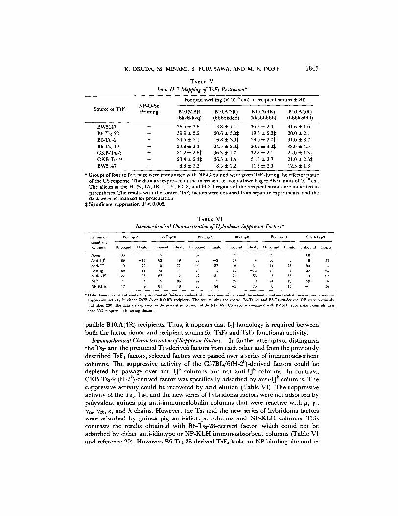
TABLE V
Intra-H-2 Mapping of TsF~ Restriction*
NP-O-Su Source of TsF3 Priming
Footpad swelling (X 10 -3 cm) in recipient strains 2: SE
BI0.MBR B10.A(3R) B10.A(4R) B 10.A(5R) (bkkkkkkc 0 (bbbbkddd) (kkbbbbbb) (bbbkkddd)
BW5147 + 36.5 2:3.6 3.8 2:1.4 36.2 2:2.0 31.6 2:1.6 B6-Ts2-28 + 39.9 2:5.2 20.6 2:3.05 19.3 2: 2.3~: 28.0 2:2.1 B6-Ts3-2 + 34.5 + 2.1 16.8 2:3.35 23.0 :t: 2.0:~ 31.0 2:8.7 B6-Ts3-19 + 39.8 2:2.3 24.5 2:3.05 20.5 2: 3.2:~ 38.0 2:4.5 CKB-Ts3-3 + 21.2 zt: 2.6:~ 36.3 2:1.7 32.8 2:2.1 25.0 2: 1.3:~ CKB-Tss-9 + 23.4 2: 2,3:~ 36.5 2:1.4 31.5 2:2.7 21.0 2: 2.5:~ BW5147 - 8.8 :t: 2.2 8.5 2:2.2 11.3 2:2.3 12.3 :!: 1.3
* Groups of four to five mice were immunized with NP-O-Su and were given TsF during the effector phase of the CS response. The data are expressed as the increment of footpad swelling 2: SE in units of 10 -3 era. The alleles at the H-2K, IA, ]B, I J, IE, IC, S, and H-2D regions of the recipient strains are indicated in parentheses. The results with the control TsF2 factors were obtained from separate experiments, and the data were normalized for presentation.
:~ Significant suppression, P < 0.005.
TABLE VI Immunochemical Characterization of Hybridoma Suppressor Factors*
Immuno- B6-Ts1-29 B6-Ts2-28 B6-Ts3-2 B6-Tsn-8 B6-Ts3-19 CKB-Ts3-9
adsorbent columns Unbound Eluate Unbound Eluate Unbound Eluate Unbound Eluate Unbound Eluate Unbound Eluate
None 83 5 67 65 69 68
Anti-l-J k 89 - 17 63 19 68 - 9 57 4 56 5 0 58
Anti-IJ h 0 72 10 77 - 9 87 6 64 11 73 50 3
Anti-lg 89 I 1 75 17 75 5 65 - 13 45 7 52 - 8
Anti-NP b 22 83 67 12 27 81 2 [ 63 4 83 - 3 62
NP b 71 - I 9 86 92 5 69 9 74 I0 59 6.
NP-KLH 17 69 61 I 0 22 94 - 5 70 0 42 - 1 56
* Hybridoma-derived TsF containing supernatant fluids were adsorbed onto various columns and the unbound and acld-eluted fractions were tested for
suppressive activity in either C57B1/6 or BI0.BR recipients. The results using the control B6-Tsl-'29 and B6-Ts2-28-derived TsF were previously
published (20). The data are expressed as the percent suppression of the NP-CLSu CS response compared with BW5147 supernatant controls. Less
than 3017~, suppression is not significant.
patible B10.A(4R) recipients. Thus, it appears that I-J homology is required between both the factor donor and recipient strains for TsF2 and TsFa funct ional activity.
Immunochemical Characterization of Suppresor Factors. In further a t tempts to dist inguish the Ts2- and the presumed Tsa-derived factors from each other and from the previously described TsF1 factors, selected factors were passed over a series of immunoadsorben t columns. The suppressive activity of the C57BL/6(H-2b)-derived factors could be depleted by passage over ant i - IJ b columns but not ant i - IJ k columns. In contrast, CKB-Ts3-9 (H-2k)-derived factor was specifically adsorbed by ant i - IJ k columns. The suppressive activity could be recovered by acid elut ion (Table VI). The suppressive
activity of the Tsl , Ts2, and the new series of hybr idoma factors were not adsorbed by polyvalent guinea pig an t i - immunog lobu l in columns that were reactive with #, 7z,
72a, yZb, ~, and ~ chains. However, the Tsl and the new series of hybr idoma factors were adsorbed by guinea pig ant i- idiotype columns and N P - K L H columns. This contrasts the results ob ta ined with B6-Ts2-28-derived factor, which could not be adsorbed by either ant i - idiotype or N P - K L H immunoabso rben t columns (Table VI and reference 20). However, B6-Ts2-28-derived TsF2 lacks an NP b i nd i ng site and in
1846 CHARACTERIZATION OF TsF3 SUPPRESSOR FACTOR
cont ras t to the o t h e r factors cou ld be d e p l e t e d on N P b id io type co lumns . T h e
specif ic i ty o f a d s o r p t i o n was con t ro l l ed by d e m o n s t r a t i n g tha t suppress ive ac t iv i ty
cou ld be r ecove red by ac id e lu t ion f rom the a p p r o p r i a t e co lumns . Thus , we c o n c l u d e
tha t this n e w series o f h y b r i d o m a factors b e a r I -J a n d id io typ ic d e t e r m i n a n t s a n d
h a v e a specif ic r ecep to r for N P hap ten . T h e s e fea tures d i s t ingu ish the new series o f
e f fec tor -phase h y b r i d o m a - d e r i v e d suppressor factors f rom the p rev ious ly desc r ibed
TsF2 suppressor factors (20).
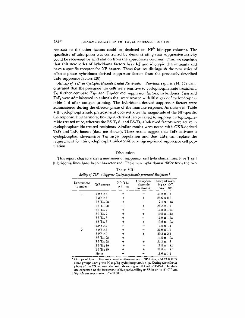
Activity of TsF in Cydophosphamide-treated Recipients. Prev ious repor ts (14, 17) d e m -
o n s t r a t e d tha t the p recu r so r Tsa cells were sensi t ive to c y c l o p h o s p h a m i d e t r e a tmen t .
T o fu r t he r c o m p a r e Tsz- a n d Tsa-der ived suppressor factors, h y b r i d o m a TsF~ a n d
TsFa were a d m i n i s t e r e d to an ima l s tha t were t r ea t ed w i t h 50 m g / k g o f c y c l o p h o s p h a -
m i d e 1 d a f te r a n t i g e n p r iming . T h e h y b r i d o m a - d e r i v e d suppressor factors were
a d m i n i s t e r e d d u r i n g the ef fec tor phase o f the i m m u n e response. As shown in T a b l e
VI I , c y c l o p h o s p h a m i d e p r e t r e a t m e n t does no t a l t e r the m a g n i t u d e o f the NP-spec i f ic
C S response. F u r t h e r m o r e , B6-Tsz-28-der ived fac tor fai led to suppress c y c l o p h o s p h a -
m i d e - t r e a t e d mice , whe reas the B6-Ta-8- a n d B6-Tsa-19-der ived factors were ac t ive in
c y c l o p h o s p h a m i d e - t r e a t e d recipients . S i m i l a r results were n o t e d w i t h C K B - d e r i v e d
TsF2 a n d TsFa factors (da ta not shown). These results suggest t ha t TsF~ ac t iva tes a
c y c l o p h o s p h a m i d e - s e n s i t i v e Tsa t a rge t p o p u l a t i o n a n d tha t TsF3 can rep lace the
r e q u i r e m e n t for this c y c l o p h o s p h a m i d e - s e n s i t i v e a n t i g e n - p r i m e d suppressor cell pop-
u la t ion .
D i s c u s s i o n
T h i s repor t cha rac te r i zes a n e w series o f suppressor cell h y b r i d o m a lines. F ive T cell
h y b r i d o m a lines h a v e been cha rac t e r i zed . T h e s e n e w h y b r i d o m a s dif fer f rom the two
TABLE VII
Ability of TsF to Suppress Cydophosphamide-pretreated Recipients *
Experiment TsF source NP-O-Su Cyclophos- Footpad swell- phamide ing (× 10 -a
number priming treatment cm) ± SE
1 BW5147 + - 24.0 ± 1.6 BW5147 + + 23.6 ± 0.7 B6-Tsz-28 + - 12.3 ± 1.4~: B6-Tsz-28 + + 23.2 ± 1.6 B6-Ts~-2 + - 10.8 ± 1.9~: B6-Tsz-2 + + 10.8 ± 1.1:~ B6-Tsa-8 + - 11.0 ± 1.5:~ B6-Tss-8 + + 13.6 zl: 1.0:~ BW5147 - - 5.8 ± 1.1
2 BW5147 + - 31.0 ± 1.0 BW5147 + + 29.3 ± 2.1 B6-Tse-28 + - 14.8 ± 1.0:~ B6-Ts~-28 + + 31.3 ± 1.8 B6-Ts3-19 .+ - 18.8 ± 1.4:~ B6-Tsa-19 + + 21.8 ± 1.4~: None - - 11.6 -'r 1.2
* Groups of four to five mice were immunized with NP-O-Su, and 24 h later some groups were given 50 mg/kg cyclophosphamide i.p. During the effector phase of the CS response the animals were given 0.4 ml of TsF/d. The data are expressed as the increment of footpad swelling ± SE in units of 10 -3 cm.
~: Significant suppression, P < 0.001.
K. OKUDA, M. MINAMI, S. FURUSAWA, AND M. E. DORF 1847
other types of NP-specific suppressor T cell hybridoma lines previously characterized (10, 20). First, the cells used for fusion are primarily derived from mice that have been immunized with either NP-O-Su or NP-KLH. In the past, we have used spleen cells from animals that were tolerized by intravenously administering NP-coupled syngeneic cells. However, we obtained one Ts3 hybridoma line from spleen cells of mice that were tolerized with NP-coupled syngeneic cells. Although this demonstrates that Tsn cells were also present in tolerized animals, the frequency of such cells must be very low. Thus, only one such hybridoma line has been discovered after the screening of several thousand hybridomas from animals that were tolerized with NP- coupled syngeneic cells.
This new series o f T cell hybridomas bear allele-specific I-J cell surface determinants and generally bear Npb-related idiotypic determinants. The presence of Thy-l .2 marker on most of the hybridoma lines and the Lyt-2.2 marker on the B6-Tss-19 hybridoma (data not shown) is consistent with the previously reported phenotype of the Ts3 population. In a few cases, phenotyping for the Lyt-2.2 marker was not possible because two different monoclonal reagents gave disparate results.
The suppressor factors derived from these hybridoma cell lines were also character- ized. The TsFs suppressor factors differed from the previously characterized TsF1 and TsF2 factors. TsF3 factor acts during the effector phase in contrast to TsF1 factor, which is active only during the induction phase of the CS response. It is not clear why TsF2 or TsF3 factors function only during the effector phase and do not mediate suppression when given during the induction phase of the CS response. One possibility is that these materials have a very short biological half-life. Alternatively, the timing of factor administration with respect to the occurrence of an appropriately differen- tiated target cell population might be critical.
The antigenic properties of TsFs were determined by passing these factors over a series of immunoadsorbent columns. Ysa- and Tss-derived factors had similar char- acteristics. First, both factors bore I-J alad NP b idiotypic determinants and bound to antigen columns; all the suppressor factors lacked constant region immunoglobulin determinants. As reported elsewhere (20), TsF2 factor bound to idiotype columns and apparently possessed an anti-idiotypic receptor (Table VI).
To further distinguish TsF3 factor than TsF1 and TsF2 factors, the genetics of factor activity were analyzed. TsFa factor, like TsF~ factor, only suppressed strains that were matched at both the Igh and H-2 gene complexes (Table IV). In contrast, TsF1 factor lacks at least H-2 i'estrictions, but as described elsewhere (10), is pseudo-restricted by genes in the Igh complex. To further distinguish the TsF3 factors from the TsF2 effector-phase factors, the activity of these factors was tested in animals that had been treated with cyclophosphamide. TsFz factor failed to suppress cyclophosphamide- treated mice, whereas TsFa factor retained its activity in animals pretreated with cyclophosphamide (Table VII). Finally, the target cells of each type of suppressor factor are distinct. Thus, TsF1 functions by inducing a Ts2 population, whereas Ts2 factor appears to trigger a cyclophosphamide-sensitive Ts3 cell population. The target cell of the TsFs factor is unknown. Table VIII summarizes the similarities and differences between Tsl, Ts2, and Tsa cells and the factors derived from these cells.
The literature describes many suppressor cell systems, some of which appear to be antigen specific (24-29), whereas others demonstrate elements of nonspecificity (21- 23, 28). The NP system has been shown to be specific in this and in previous reports

1848 CHARACTERIZATION OF TsF3 SUPPRESSOR FACTOR
TABLE VIII Characteristics of NP-specific Suppressor Factors
Properties of TsF Phenotype of cell producing TsF
Type of suppressor factor
TsF1 TsF2 TsF3
I-J + + + NP b idiotype + -- + Cell type Tsl Ts2 Ts3
MHC determinants I-J I-J I-J Igh-C determinants None None None Igh-V determinants NP b ? NP b Binding specificity NP NP b idiotype NP Phase of action Induction Effector Effector Cellular target Pre-Ts2 Tsa ? Genetic restriction None I-J and Igh I-J and Igh Activity in cyclophosphamide- ? None Yes
treated recipients
(10, 14, 15). The experiments using animals primed or challenged with DNFB together with NP-O-Su strengthen the conclusion that the suppression mediated in the NP system is antigen specific (Table II). The fact that TsF3 factor is Igh restricted differs from the observations in some suppressor cell systems (18, 22, 29). There are several possible explanations for these apparently disparate results. First, the systems and the methods used are quite different in the various suppressor models studied. The different methods for evaluating specificity might account for the results observed. It is noteworthy that Fresno et al. (26) have characterized a specific TsF3-1ike factor, which upon cleavage or partial degradation becomes antigen nonspecific. We have not evaluated this possibility in our system. An alternative possibility is that there are several Ts3-1ike cells, some of which mediate nonspecifie suppression. I f the latter is true, we must conclude that in the NP system the antigen-specific Igh-restricted Ts3 population appears to predominate.
The Ts3 factors show dual genetic restriction, requiring both I-J and Igh region homolog~ for factor activity. Such findings of dual genetic restrictions are not unique to the NP system nor the TsF3-derived factors. Thus, Suzuki et al. (16) and Dietz et al. (30) have recently also reported dual genetic restrictions of suppressor factors. The fact that Igh restrictions are observed suggests that idiotype-anti-idiotype interactions are required between TsF3 and its target cells. Furthermore, these Igh restrictions argue against an antigen-bridging mechanism occurring between the TsF3 and its target cells. The results imply, therefore, that the target of TsF3 might be an anti- idiotypic suppressor or helper cell population. At the present time, we are at tempting to characterize this target population. The I-J restrictions observed with TsF3 factor might reflect a requirement for an acceptor site on the target cell population that is required for triggering or inactivating the target cell. Alternatively, they might reflect a pseudo-genetic restriction similar to that previously characterized for TsF1 factors (10).
The Ts3 cells described in this report constitutively secrete TsF3 factor. This suggests that some of the Ts3 cells are normally present in antigen-primed animals. These Ts3 appear to be fully differentiated and might be involved in the autoregulation of the NP-specific immune response. The question of how these Ts3 cells are activated

K. OKUDA, M. MINAMI, S. FURUSAWA, AND M. E. DORF 1849
remains unanswered. One possibility is that a few Ts2 cells also exist in antigen- primed mice and these Ts2 cells release TsF2 factor that activates a small percentage of the Tsz population. Another possibility is that Tsz ceils might also be activated by other means, e.g., anti-idiotype antibody. Finally, we cannot exclude the possibility that in situ the Tsz cells were not actively secreting TsF8 factor but that after fusion with the BW5147 tumor line the hybrids began to actively secrete TsFz factor. Preliminary experiments suggest that antigen-primed mice possess a population of mature Ts3 cells that do not secrete TsFs unless activated with TsF2 factor. This resting Ts3 population presumably accounts for the bulk of the Tsz cells in antigen- primed mice. Thus, the NP responses of immune animals are not completely sup- pressed until sufficient Ts3 suppressor cells are activated to regulate the immune response.
The characterizaton of three distinct series of suppressor factors specific for the CS response to the NP hapten demonstrates the intricacy of the immunoregulatory pathway. The precise differences between the immunoregulatory molecules responsi- ble for the communications between suppressor T cells is unknown. However, the potential availability of large quantities of monoclonal TsFa, TsFz, and TsF3 factors derived from both the C57BL/6 and CKB series of hybridoma lines should permit molecular comparisons among these functionally distinct T cell-derived products.
SLlmlTlary
Five hybridoma T cell lines were prepared by fusion of Ts3 cells with the BW5147 thymorna. The culture supernatants from these T cell hybrids contained a factor, TsFa, which specifically suppressed 4-hydroxy-3-nitrophenyl acetyl hapten (NP)- hapten cutaneous sensitivity responses. The properties of this new series of hybridoma factors was compared with those of two previously characterized types of NP-specific suppressor factors (TsF1 and TsF2). TsF3 activity was only observed if the factor was administered during the effector phases of the immune response. TsFz bears I-J and C57BL anti-NP antibody idiotypic determinants and has binding specificity for the NP hapten. Furthermore, TsFa does not suppress H-2 (I-J)-incompatible mice. In addition to this H-2 restriction, the monoclonal TsF3 factors also demonstrated an Igh genetic restriction. Finally, the TsF3 factors could be distinguished by their ability to suppress cyclophosphamide-treated recipients.
The authors wish to acknowledge the expert secretarial assistance of Mrs. Nancy Axelrod and Mrs. Teresa Dinse. The authors are especially grateful to Dr. Baruj Benacerraf for his support and advice.
Received for publication 24 August 1981.
References
1. Taniguchi, M., and J. F. A. P. Miller. 1978. Specific suppressive factors produced in hybridomas derived from the fusion of enriched suppressor T cells and a T iymphoma cell line.ft. Exp. Med. 141t:373.
2. Taussig, M., and A. Holliman. 1979. Structure of an antigen-specific suppressor factor produced by a hybrid T cell line. Nature (Lond.). 277:308.
3. Kontiainen, S., E. Simpson, E. Bohrer, P. C. L. Beverly, L. A. Herzenberg, W. C.

1850 CHARACTERIZATION OF TsF3 SUPPRESSOR FACTOR
Fitzpatrick, P. Vogt, A. Torano, I. F. C. McKenzie, and M. Feldmann. 1978. T-cell lines producing antigen-specific suppressor factor. Nature (Lond.) 274:477.
4. Okuda, K., M. Minami, S.-T. Ju, and M. E. Dorf. 1981. Functional association ofidiotypic and I-J determinants on the antigen receptor of suppressor T cells. Proc. Natl. Acad. Sci. U. S. A. In press.
5. Eshhar, Z., R. N. Apte, I. Lowy, Y. Ben-Neriah, D. Givol, and E. Mozes. 1980. T-cell hybridoma bearing heavy chain variable region determinants producing (T,G)-A--L-spe- cific helper factor. Nature (Lond.) 286:270.
6. Kapp, J. A., B. A. Araneo, and B. L. Clevinger. 1980. Suppression of antibody and T cell proliferative responses to L-glutamic acid6°-L-alanine3°-L-tyrosine 1° by a specific monoclonal T cell factor.J. Exp. Med. 152:235.
7. Hewitt, J., and F. Y. Liew. 1979. Antigen-specific suppressor factors produced by T cell hybridomas for delayed-type hypersensitivity. Eur. J. lmmunol. 9:572.
8. Whitaker, R. B . J . T . Nepom, M.-S. Sy, M. Takaoki, C. F. Gramm, I. Fox, R. N. Germain, A. Nisonoff, M. I. Greene, and B. Benacerraf. 1981. Suppressor factor from a T celt hybrid inhibits delayed type hypersensitivity responses to azobenzenearsonate (ABA). Proc. Natl. Acad. Sci. U. S. A. In press.
9. Watanabe, T., M. Kimoto, S. Maruyama, T. Kishimoto, and Y. Yamamura. 1978. Regulation of antibody response in different immunoglobulin classes. V. Establishment of T hybrid cell line secreting IgE class-specific suppressor factor. J. ImmunoL 121:2113.
10. Okuda, K., M. Minami, D. H. Sherr, and M. E. Dorf. 1981. Hapten-specific T cell responses to 4-hydroxy-3-nitrophenyl acetyl. XI. Pseudogenetic restrictions of hybridoma suppressor factors.,]. Exp. Med. 154:468.
11. Germain, R. N., and B. Benaeerraf. 1981. A single major pathway of T-lymphocyte interactions in antigen-specific immune suppression. Scand. J. Immunol, 13:1.
12. Weinberger, Z. J., R. N. Germain, S.-T. Ju, M. I. Greene, B. Benacerraf, and M. E. Dorf. 1979. Hapten-specific T cell responses to 4-hydroxy-3-nitrophenyl acetyl. II. Demonstration of idiotypic determinants on suppressor T cells.J. Exp. Med. 152:161.
14. Sunday, M. E., B. Benacerraf, and M. E. Doff. 1981. Hapten-specific T cell responses to 4-hydroxy-3-nitrophenyl acetyl. VIII. Suppressor cell pathways in cutaneous sensitivity responses.J. Exp. Med. 153:811.
15. Sherr, D. H,, and M. E. Dorf. 1981. Hapten-specific T cell responses to 4-hydroxy-3- nitrophenyl acetyl IX. Characterization of idiotype-specific effector-phase suppressor cells on plaque-forming cell responses in vitro.J. Exp. Med. 153:1445.
16. Suzuki, G., Y. Kumagai, Y. Shiratori, H. Karasuyama, T. Kitahara, R. Abe, K. Hayakawa, K. Okumura, and T. Tada. 1980. Expression of NP b idiotype controlled by genes linked to the Igh-I b allotype on the NP (4-hydroxy-3-nitro-phenyl acetyl) binding T cells. Proc. Jpn. Soc. Imrnunol. 10:12 I.
17. Sy, M.-S., S. D. Miller, J. W. Moorhead, and H. N. Claman. 1979. Active suppression of l-fluoro-2,4-dinitrobenzene-immune T cells. Requirement of an auxiliary T cell induced by antigen.,], Exp. Med. 149:1197.
18. Sy, M.-S., A. Nisonoff, R. N. Germain, B. Benacerraf, and M. I. Greene. 1981. Antigen- and receptor-driven regulatory mechanisms, VIII. Suppression of idiotype-negative, p-azobenzenearsonate-specific T cells results from the interaction of an anti-idiotypic second-order T suppressor cell with a cross-reactive idiot ype-positive, p-azobenzenearsonate- primed T cell target.,/. Exp. Med. 153:1415.
19. Ju, S.-T., T. J. Kipps, J. Theze, B. Benacerraf, and M. E. Doff. 1978. Idiotypic analysis of anti-GAT antibodies. I. Presence of common idiotypic specificities in both responder and nonresponder mice.,/. Immunol. 121:1034.
20. Minami, M., K. Okuda, S. Furusawa, B. Benacerraf, and M. E. Dorf. 1981. Analysis of T

K. OKUDA, M. MINAMI, S. FURUSAWA, AND M. E. DORF 1851
cell hybridomas. I. Characterization of H-2- and Igh-restricted monoclonal suppressor factors. J. Exp. Med. In press.
21. Tada, T. 1977. Regulation of the antibody response by T cell products determined by different I subregions. In The Immune System: Genetics and Regulation. E. Sercarz, L. A. Herzenberg, and C. F. Fox, editors. Academic Press, Inc., New York. 345-361.
22. Sherr, D. H., K. M. Heghinian, B. Benacerraf, and M. E. Dorf. 1979. Immune suppression m vivo with antigen-modified syngeneic cells. III. Distinctions between T-cell tolerance and T-cell-mediated suppression. J. Immunol. 123:2682.
23. Thomas, W. R., F. I. Smith, I. D. Walker, and J. F. A. P. Miller. 1981. Contact sensitivity to azobenzenearsonate and its inhibition after interaction of sensitized cells with antigen- conjugated cells.J. Exp. Med. 153:1124.
24. Alevy, Y. G., and C. J. Bellone. 1980. Anti-phenyltrimethylamino immunity in mice. L-tyrosine-p-azophenyl-trimethyl-ammonium-induced suppressor T cells selectively inhibit the expression of B-cell clones bearing a cross-reactive idiotype. J. Exp. Med. 151-'528.
25. Lynch, R. G., J. W. Rohrer, B. Odermatt, H. W. Gebel, J. R. Autry; and R. G. Hoover. 1979. Immunoregulation of murine myeloma cell growth and differentiation: a monoclonal model of B cell differentiation, lmmunol. Rev. 48:45.
26. Fresno, M., L. McVay-Boudreau, G. Nabel, and H. Cantor. 1981. Antigen-specific T lymphocyte clones. II. Purification and biological characterization of an antigen-specific suppressive protein synthesized by cloned T cells.J. Exp. Med. 153:1260.
27. Benacerraf, B.,J. A. Kapp, P. Debre, C. W. Pierce, and F. DelaCroix. 1975. The stimulation of specific suppressor T cells in genetic nonresponder mice by linear random copolymers of L-amino acids. Transplant. Rev. 26:21.
28. Rich, S. S., and R. R. Rich. 1976. Regulatory mechanisms in cell-mediated immune responses III. I-region control of suppressor cell interaction with responder cells in mixed lymphocyte reactions.J. Exp. Med. 143:672.
29. Chaouat, G., and G. A. Voisin. 1981. Regulatory T cells in pregnancy. V. Allopregnancy- induced T-ceil-suppressor factor is H-2 restricted and bears Ia determinants. Cell. Immunol. 62:186.
30. Dietz, M. H., M.-S. Sy, B. Benacerraf, A. Nisonoff, M. I. Greene, and R. N. Germain. 1981. Antigen- and receptor-driven regulatory mechanisms. VII. H-2-restricted anti-idi- otypic suppressor factor from efferent suppressor T cells..]'. Exp. Med. 153:540.