Embed Size (px)
Citation preview

Trends in Analytical Chemistry, Vol. 31, 2012 Trends
Analysis of microorganismsby capillary electrophoresisJan Petr, Vıtezslav Maier
Microorganisms are well known for their positive, as well as negative, effects on health, which mean that there is a great need for
methods of discovery, identification and determination of microorganisms. In the past decade, capillary electrophoresis (CE)
began to be an interesting tool for analysis of microorganisms, interestingly sometimes with similar dimensions for the separation
capillary and the microorganisms.
This review focuses on the use of CE in the analysis of microorganisms. First, it looks at the origin of microbial surface charge
and then describes key points in the analysis of microbes by capillary zone electrophoresis [first approaches using poly(ethylene
oxide), covalent modification of the inner capillary wall, reversed electroosmotic flow, advances in detection, and on-line
preconcentration] and capillary isoelectric focusing.
ª 2011 Elsevier Ltd. All rights reserved.
Keywords: Capillary coating; Capillary electrophoresis; Capillary isoelectric focusing; Capillary zone electrophoresis; Covalent modification;
Microorganism; Poly(ethylene oxide); Reversed osmotic flow; Surface charge; Zeta potential
Abbreviations: BGE, background electrolyte; CEC, Capillary electrochromatography; CE, Capillary electrophoresis; CFU, Colony-forming unit; CIEF,
Capillary isoelectric focusing; CTAB, Cetyltrimethylammonium bromide; CZE, Capillary zone electrophoresis; DLVO theory, Derjaguin-Landau-
Verwey-Overbeek theory; DESI-IT-MS, Desorption electrospray–ion-trap mass spectrometry; EDTA, Ethylenediaminetetraacetic acid; EOF,
Electroosmotic flow; FISH, Fluorescence in-situ hybridization; InsP6, Myo-inositol hexakisphosphate; MALDI-TOF-MS, Matrix-assisted laser
desorption ionization–time of flight mass spectrometry; IT-MS, ion-trap mass spectrometry; LIF, Laser-induced fluorescence; MES, 2-(N-
morpholino)ethanesulfonic acid; MOPS, 3-(N-morpholino)propanesulfonic acid; MS, Mass spectrometry; PEO, Poly(ethylene oxide); PCR,
Polymerase chain reaction; TBE buffer, Tris-boric acid-EDTA buffer; TMV, Tobacco mosaic virus; Tris, Tris(hydroxymethyl)aminomethane
Jan Petr*, Vıtezslav Maier
Regional Centre of Advanced
Technologies and Materials,
Department of Analytical
Chemistry, Faculty of Science,
Palacky University in Olomouc,
17 listopadu 12, CZ-77146
Olomouc, Czech Republic
*Corresponding author.
Tel.: +420 585 63 4416;
Fax: +420 585 63 4433;
E-mail: [email protected]
0165-9936/$ - see front matter ª 2011
1. Introduction
Microorganisms are well known for theirpositive, as well as negative, effects, thepositive being represented mainly by theiruse in traditional food and beveragepreparation but also in biotechnology andmodern genetic engineering. The negativeeffects are also well known, as pathogenicmicrobes can cause serious diseases thatcan have fatal consequences for livingentities. Both effects mean that there is agreat need to study microorganisms andmethods for their discovery, identificationand determination [1].
The history of the discovery of microbesand microbiology is connected with thediscovery of the microscope in seventeenthcentury [2]. Microbiology was thendeveloped by French chemist Louis Pas-teur, who is probably most famous for hisdevelopment of immunization and pas-teurization [3], and German physicianRobert Koch, who isolated Bacillusanthracis in 1877 and Mycobacterium
Elsevier Ltd. All rights reserved. doi:10.1016/j.trac.2011.07.013
tuberculosis in 1882 [4,5]. The big break-through in identifying microbes came in1884, when Danish scientist Hans Chris-tian Gram developed a method (now wellknown as Gram staining) to discriminatebetween two types of bacteria with similarclinical symptoms – Streptococcus pneumo-niae and Klebsiella pneumoniae [6].
Nowadays, quite a lot of differentmethods for identifying microbes have beenestablished, e.g., different types of staining[7,8], specific antibodies [9], polymerasechain reaction (PCR), and DNA-typingbased identification [10,11]. Moreover,mass spectrometry (MS) and capillaryelectrophoresis (CE) have analyzed intactand lysed microbes [12–15]. These last twotechniques represent interesting innova-tions; MS is a common identification tool forsmall molecules and large molecules (e.g.,proteins) [16,17], and CE is an efficient,high-throughput separation technique[18,19]. In view of increasing interest, thiscritical review covers recent achievementsin CE analysis of microbes.
9

Trends Trends in Analytical Chemistry, Vol. 31, 2012
2. Microbial surface charges and their behavior
2.1. Origin of the surface charge, zeta potentialGenerally, the structure of various cells, includingmicroorganisms, is very complicated. The cell surfacecontains biopolymers (e.g., membrane proteins, recep-tors, phospholipids, polysaccharides) and small mole-cules (e.g., cellular communication molecules orproducts of metabolism) [20–23]. These componentscontain a lot of groups that can dissociate and affect theelectric double layer on cells and their zeta potential. Inaddition, as living systems, bacteria can modify theirsurface composition during the growth phase or as areaction to changes in their environment. These changesalso lead to variations of bacterial surface charge andhydrophobicity [24].
Many authors measured and evaluated zeta (f)potentials of bacteria, supposing that the populationswere fairly monodisperse having relatively uniform col-loidal surface properties [25–29]. Interestingly, cell sur-faces within different cultures of the same bacteria werefound to be heterogeneous [30–32]. Some authors alsostudied the influence of adding some compounds onbacterial f potential. Chatellier et al. [28,29] studied aneffect of adding quaternized polyvinylpyridine on Esche-richia coli cell-surface charge. They observed three fpotential regions with increasing polymer concentration.First, negative f potentials increased up to a criticalconcentration where the cells formed aggregates (f po-tential close to zero), then a constant value of f potentialwas observed. And, after the second critical concentra-tion, the f potential increased to positive values. Theauthors said that probably the adsorption of positively-charged polymer on the cell surface was the reason forsuch behavior. This example, apparently far from CE, iskey for electrophoretic properties. The behavior of bac-teria in CE depends on their f potential and knowledge ofhow to influence the f potential of microorganisms isessential for successful analysis.
2.2. MobilityMathematically, the origin of microbe behavior in anelectric field generally refers to their surface chargeaccording to the well-known function derived by Henry[33,34]:
l ¼ 2ere0f3g
f ðjRÞ ð1Þ
where l is the electrophoretic mobility, er is the relativeelectric permittivity, e0 is the permittivity of the vacuumand f is the zeta potential of an object. The function ofjR is the function of the object radius R and the Debyelength j-1, which represents a thickness of the electricdouble layer formed on the object and depends on theionic strength of the BGE (j �
ffiffi
Ip
). The function f(jR)ranges between 1 and 1.5 (1 for jR << 1; 1.5 for
10 http://www.elsevier.com/locate/trac
jR� 1) where the value 1.5 is called the Smoluchowskilimit, and the Equation (1) is transformed to the well-known Smoluchowski equation [35–37]:
l ¼ ereofg
ð2Þ
The ionic atmosphere of microbes provides the nextimportant feature, which is not explicitly expressed inEquation (1). If electric current is applied, the migrationof such objects results from a balance of three forces –the electric force, the Stokes drag force, and the elec-trophoretic retardation force. The first two forces are wellknown from CE of small molecules and they are coveredin Equation (1). The retardation force is caused by themigration of counter ions in the diffuse part of theelectric double layer of the object in a direction oppositeto the motion of the object (sometimes it is called‘‘relaxation effect’’). Moreover, this relaxation effectmakes electrophoretic mobility more strongly dependenton jR than according to Equation (1). Then, the effect off potential (the surface-charge density) on microbescould be suppressed and migration would depend onlyon the retardation force [33,35–37].
Furthermore, all the previous equations are derived foran ideal structure of bacteria (objects with a sphericalshape). Unfortunately, bacterial morphology is muchmore diverse {e.g., from spherical cocci or rod-shapedbacilli to spiral-shaped spirilla or tightly coiled spiro-chetes (see Fig. 1) [1]}. These irregularly-shaped struc-tures have their own electrophoretic behavior in linewith the increasing influence of relaxation effects.
2.3. AdhesionAdhesion is the next well-known phenomenon withbacteria. Microorganisms have a strong tendency toassociate together or with different microbes or surfaces.They easily form aggregates or co-aggregates (or clus-ters) by means of van der Waals forces, electrostaticforces and acid-base interactions [33,38,39]. Thisaggregation and co-aggregation is more or less ubiqui-tous for all bacteria and was also observed in the earlydays of microbiology.
The physicochemical description of such interactionsis not elementary. Two different approaches used to beaccepted:(1) the thermodynamic approach based on surface free
energies [40,41]; and,(2) the classical Derjaguin, Landau, Verwey, Overbeek
(DLVO) theory based on van der Waals and electro-static interactions and their effect in a certain dis-tance from the cell [36–38,42]. DLVO theory canbe extended by including the acid-base equilibriumand/or steric effects [38,43–46].
DLVO theory describes a balance between repulsiveand attractive electrostatic forces and attractive van derWaals forces. The interaction energy can be expressed as

Figure 1. The diversity of bacterial morphology.
Trends in Analytical Chemistry, Vol. 31, 2012 Trends
a sum of distance-dependent energies of van der Waalsinteraction GW and electrostatic interaction Gel [38]:
GtotðxÞ ¼ GWðxÞ þ GelðxÞ ð3ÞFig. 2 gives an example of calculation of the energies,
where two spherical objects with radius 0.5 lm and zetapotential 15 mV were chosen as a model system. More-over, four Debye lengths j-1 (the electric double layer‘‘thickness’’) of 5.0 nm, 1.0 nm, 0.5 nm and 0.3 nmsimulated the increase of ionic strength of the environ-ment.
It is clearly visible that the electrostatic interactionsbecome extrinsic and aggregates are formed preferably athigh ionic strength. With lower ionic strengths, there isan energy barrier before aggregation with a local energyminimum that increases with increasing object radius.The decrease in the zeta potential causes the decrease inthe energy barrier and both objects tend to form aggre-gates. Indeed, this model does not account for othereffects (e.g., bacterial morphology), but it can providesome basic information about bacterial behavior.
3. Capillary zone electrophoresis
Since microorganisms are charged, they can migrate inthe electric field and therefore they can be analyzed bycapillary zone electrophoresis (CZE). However, CZE ofmicroorganisms brings two main problems: (i) aggrega-tion; and, (ii) interaction with the inner wall of thecapillary. The aggregation tendency of microorganismscan be predicted following DLVO theory. Interactionwith a capillary inner wall can occur since the microbialsurface can contain positive groups that easily interactwith silanol groups of the fused-silica capillary.
3.1. First approachesThe first achievement of CZE analysis of microbes waspublished by Hjerten et al. in 1987 [47]. They describedhow tobacco mosaic virus (TMV) and Lactobacillus caseiwere carried by electroosmotic flow (EOF) through acapillary. TMV was analyzed in 20 mM Tris-HCl pH 7.5and L. casei in 100 mM Tris-acetic acid pH 8.6. Althoughthey did not show any separation, CE started to be aninteresting tool for analysis of such big objects.
In 1993, Ebersole and McCormick [48] successfullyseparated Streptococcus pyogenes, Streptococcus agalactiae,S. pneumoniae and Enterococcus faecalis in TBE buffercontaining 4.45 mM Tris, 4.45 mM boric acid, and 0.10mM EDTA adjusted to pH 9.5 with KOH (see Fig. 3).They also found that the most of the bacteria (�80% forE. faecalis and S. pneumoniae and �60% for S. agalactiae)were viable after CZE separation with the exception ofS. pyogenes (only �10% were viable). Moreover, thepurity of fractions of the two most separated peaks ofS. pneumoniae and Staphylococcus aureus was more than98.5% in the top of the peak and about 20%:80% betweenboth peaks. This paper is one of the most important in thefield of CE analysis of bacteria. Unfortunately, it is notknown as much as would be beneficial for the field, mainlyin the view of the separation and purity of fractions.
Later, Torimura et al. [49] published an interestingpaper dealing with study of the electrophoretic behaviorof nine bacteria (e.g., E. coli, Paracoccus denitrificans,Pseudomonas fluorescens, Acetobacter pasteurianus, andSaccharomyces cerevisiae). They obtained negative elec-trophoretic mobilities (-0.19 – -3.44 x 10⁻⁹ m2/Vs) for allthe microorganisms in phosphate buffer at pH 7.0 withionic strength 19 mM. Moreover, they studied pretreat-ment of E. coli prior to injection into the CE system bycolistin, an antibiotic specific for gram-negative bacteria,
http://www.elsevier.com/locate/trac 11

Figure 2. An example of calculation of distance-dependent energies for two spherical objects with radius of 0.5 nm and zeta potentials of 15 mVusing DLVO theory. More details regarding the theoretical model can be found in [38].
Figure 3. CZE separation of a mixture of Streptococcus agalactiae, Streptococcus pneumoniae and Enterococcus faecalis in TBE buffer contain-ing 4.45 mM Tris, 4.45 mM boric acid, 0.10 mM EDTA adjusted to pH 9.5 with KOH (Reprinted from [48] with permission).
Trends Trends in Analytical Chemistry, Vol. 31, 2012
and by ofloxacin, a synthetic pharmaceutical for bacte-riolysis. Remarkable changes in bacterial bands were
12 http://www.elsevier.com/locate/trac
observed, probably due to the variations in cell-membrane structure.

Trends in Analytical Chemistry, Vol. 31, 2012 Trends
Finally, two groups studied bacterial mobility at theend of the 1990s. Pfetsch and Welsch [50] determinedbacterial mobility in TBE and phosphate buffer, andGlynn et al. [51] in MOPS buffer. Bacterial mobilitiesdecreased with increasing ionic strength. Moreover, theband-width increased with increasing ionic strength,and formation of spikes on the peak profiles was observedwith higher ionic strength. This behavior clearly re-flected changes in the surface-charge density of bacteriaand is similar to CE of nanoparticles [52].
3.2. Use of poly(ethylene oxide)In 1999, Armstrong and co-workers [53] introducedpoly(ethylene oxide) (PEO), known from separation ofproteins [54], for highly efficient separation of microor-ganisms that opened a novel, challenging way for the CEof microorganisms. Fig. 4 shows their separation of S.cerevisiae, P. fluorescens, Enterobacter aerogenes andMicrococcus luteus with apparent efficiencies higher than500,000 plates/m. PEO, in the BGE comprising 4.5 mMTris, 4.5 mM boric acid, and 0.1 mM EDTA, was used forreducing the electroosmotic mobility and possible inter-actions between charged microbes and a capillary wall.They studied the effect of PEO concentration on elec-troosmotic mobility in the range 0–0.035% and con-cluded that use of 0.0125% PEO is most effective foranalysis of microorganisms. They also described dis-crimination by size and shape within the microbes, butthis was probably caused by the choice of sample.Unfortunately, they did not perform a study about thepurity of the peaks, as Ebersole and McCormick did [48].
Figure 4. CZE separation of some microorganisms using PEO
Later, the mechanism of microbial migration in PEO-based systems was proposed by Armstrong et al. [55]and Zheng and Yeung [56]. Armstrong et al. [55] de-signed three models of formation of microbial aggregatesleading to those high apparent efficiencies:(1) the field-induced aggregation model;(2) the hairy-particle model; and,(3) the shape-induced differential mobility model.
The field-induced aggregation model is based on thebehavior of electrorheological fluids. Colloidal particlesforms disk-like aggregates aligned perpendicularly to theelectric field lines and are repelled from a higher electricfield area.
The hairy-particle model (see Fig. 5) is based on aclassical electrokinetic theory of colloids. First, the elec-trical double layer of microbes is reconstituted on thebasis of PEO adsorption on microbial surface togetherwith decreasing conductivity of the microbial zone, andthat also enhances the aggregation tendency. Then, theaggregate with a different global charge and mass isformed and migrates through the capillary.
The shape-induced differential mobility model de-scribes the aggregation of rod-shaped microbes due tothe different effects of the electric field on the randomly-distributed orientations of microbes. In this way, mi-crobes tend to collide and form aggregates. PEO wassuggested to be a mediator in the interaction betweenthe microbes.
Zheng and Yeung [56] used a CCD camera connectedto the capillary to visualize aggregation of Bifidobacte-rium infantis cells. They observed that microbes moved in
(MW 600,000) (Reprinted from [53] with permission).
http://www.elsevier.com/locate/trac 13

Figure 5. The hairy particle model. (A) Initial stage just after the sample is injected. (B) Compression may occur because of the lower conductivityof the sample zone, the countercurrent movement of the negatively-charged “hairy” particles versus the bulk solvent, and the differential chargeat opposite sides of the sample zone. (C) Once compressed, the microbes (which often adhere to one another) move as a single, charged entityrather than as a group of individual particles. (D) The macroparticle moves toward the cathode at a velocity governed by its size-to-charge ratioand the EOF velocity (Reprinted from [54] with permission).
Trends Trends in Analytical Chemistry, Vol. 31, 2012
different directions with different velocities, and that fa-vored collisions and co-aggregation. Moreover, theyshowed the next interesting results:(1) the rate of aggregation was higher if the size of
aggregate increased;(2) the size of aggregate depended on the electric field
used for the analysis; and,(3) the aggregation tendency increased with increasing
microbial concentration and ionic strength of thebuffer.
Both theories [55,56] described the PEO-assistedaggregation tendency of microbes. After these results, weask if it is possible to separate microorganisms using PEObecause they form aggregates and could probably formco-aggregates. This question was considered six yearslater by Haugg et al. [57]. They studied migration ofmicrobial clusters using similar types of bacteria andPEO-based electrolytes. Microbial bands separated by CEwere collected and cultivated. Results showed that bothtypes of microbes were present in one band and only theseparation of different co-aggregates with different globalcharge-to-size ratio was achieved.
Although all these things were quite well known, manyauthors did the separations in PEO electrolytes [58–60].Interestingly, the lack of reproducibility of analyses
14 http://www.elsevier.com/locate/trac
probably caused by formation of aggregates with differentcharge-to-size ratio and memory effects was addressed bymany authors. For example, the positive effect of ultra-sound pre-treatment of microbial sample on the repro-ducibility of aggregate migration using M. luteus, S.cerevisiae, and Alcaligenes faecalis was presented bySchneiderheinze et al. [61]. Generally, many authorshave recommended sonication prior to analyses to pre-vent formation of irreproducible spikes on CE profiles.
Recently, Klodzinska et al. [62] presented a progres-sive study on separation of microorganisms in MESbuffers. They evaluated the zeta potential of S. aureus andE. coli in electrolytes used for CE separation with andwithout PEO in a wide range of pH. The data were thenused for finding the best conditions for CE analysis. Thiscomplex study seems to be the right way for thenext developments in the field of CE analysis of micro-organisms.
3.3. Covalent modification of capillary inner wallIt is well known that the inner wall of a fused-silicacapillary contains silanol groups that can be easily dis-sociated, resulting in interaction with positively-chargedmolecules (e.g., proteins). These silanol groups can bemodified by chemical reactions producing different

Figure 6. Separation of five species of bacteria with trimethylchlorosilane-modified capillary using TBE buffer pH 8.5 and -15 kV (Reprinted from[69] with permission).
Trends in Analytical Chemistry, Vol. 31, 2012 Trends
structures on the surface. Generally, these structures canbe more hydrophobic or carry the possibility for inter-actions (e.g., p-p, van der Waals, or ionic) or providetheir own selectivity [63–66].
In 2003, Buszewski et al. [67] introduced the firstacrylamide-modified capillary for the analysis of E. coli,Proteus vulgaris, Bacillus cereus, and P. fluorescens in Trisbuffer pH 8.0. Similar type of coating was also employed
Figure 7. Staphylococcus aureus analysis by CEC using gigaporous monolitdimethacrylate and trimethylolpropane trimethacrylate as monomers in thglycol and 2-methoxyethanol. Reprinted from [71] with permission).
for the differentiation of S. aureus strains [68]. Later,Szumski et al. [69] studied divinylbenzene andtrimethylchlorosilane covalent modification of silanolgroups on the capillary inner wall. Under these condi-tions, separation of five species, namely E. coli, P. vul-garis, Bacillus meganterium, Micrococcus sp., Arthorobacterglobiformis, was possible using TBE buffer pH 8.5 andnegative-voltage polarity (Fig. 6).
h thermally synthesized with glycidyl methacrylate, triethylene glycole presence of porogen solvent containing 1-decanol, polyethylene
http://www.elsevier.com/locate/trac 15
Trends Trends in Analytical Chemistry, Vol. 31, 2012
Interestingly, in 2008, Buszewski and Klodzinska [70]introduced a monolithic column based on acrylamideand styrene/divinylbenzene for the separation ofS. aureus, E. coli, and P. vulgaris. Also acrylamide-basedmonolithic columns were characterized and theoreticallystudied for the analysis of S. aureus cells in TBE buffer pH8.5 [71]. Here, the analysis from a short part of thecapillary ran over 4 min (Fig. 7).
This type of the capillary modification seems to be verypromising for the future because it can provide differenttypes of interactions (by direct chemical modification ofthe capillary inner wall). These interactions can lead toseparation of microbes or maybe to pre-identification(e.g., by employing reaction agents providing selectiveinteractions with some types of microorganisms).
3.4. Reversed EOFIn 2006, Rodriguez et al. [72] of Armstrong�s group de-scribed another approach for CE analysis of microbesusing the capillary inner wall dynamically coated withcationic surfactant CTAB in Tris/citric acid buffers. It iswell known that CTAB molecules present in the BGE re-verse the EOF [73,74]. However, the next effect of addingCTAB could be in changing the f potential of analyzedmicroorganisms to positive values or values close to zero.Notwithstanding, the purpose of the study [72] was todevelop a highly sensitive method where separation wasnot essential. We describe the basis of the method later,since its main objective is preconcentration.
CTAB coating was also used by some authors to pro-vide more reproducible analyses than using PEO. CTABconcentration was studied in the range 0.5–12 mg/mLwith saturation of the electroosmotic mobility at 4 mg/mL. Increase in CTAB concentration leads to decrease ofthe bacterial signal, so 1 mg/mL was chosen as opti-mum.
Later, Bao et al. [75] studied possible innovations inthe method of preconcentration by replacing CTABsurfactant by monocationic and dicationic ionic liquids.Ionic liquids affect electroosmotic mobility; they can justsuppress the EOF or, similarly to CTAB, reverse the EOFdepending on their structure [76]. Bao et al. [75] con-cluded that ionic liquids could replace or partially re-place CTAB, which may lyse cells.
Generally, the method with reversed EOF is an inter-esting way to analyze microorganisms by CE. Similarlyto the covalent coating, there are many options forinfluencing the mobility of microbes. For example, theuse of didodecyldimethylammonium bromide, whichwas employed in CE of nanoparticles, could be veryinteresting, since it is present only in rinsing solutions,not in a BGE [52].
3.5. The use of non-coated capillaryApart from those articles in the part describing the firstapproaches that studied the use of non-coated capillaries
16 http://www.elsevier.com/locate/trac
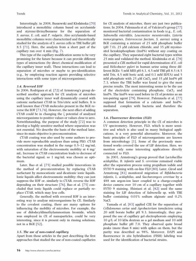
for CE analysis of microbes, there are just two publica-tions. In 2004, Palenzuela et al. of Valcarcel�s group [77]monitored bacterial contamination in foods (e.g., E. coli,Salmonella enteriditis, Leuconostoc mesenteroides, Listeriamonocytogenes, Enterococcus faecium, S. aureus) using aBGE containing a mixture of 25 mM phosphate buffer(pH 7.0), 25 lM calcium chloride, and 35 lM myoino-sitol hexakisphosphate (InsP6) without any coating onthe capillary. They separated eight bacterial types within25 min and validated the method. Klodzinska et al. [78]presented a CZE method for rapid determination of E. coliand Helicobacter pylori in human urine using three dif-ferent BGEs: 5 mM MES pH 6.1, 4.5 mM TBE pH 8.5 (4.5mM Tris, 4.5 mM boric acid, and 0.1 mM EDTA) and 5mM phosphate with 25 lM CaCl2 and 35 lM InsP6 pH7.3, where the TBE buffer was found to give the mostprecise results. The most interesting seems to be the useof the electrolyte containing phosphate, CaCl2 andInsP6. The InsP6 was used before in CE of proteins as anion-pairing agent [79]. However, Palenzuela et al. [77]supposed that formation of a calcium- and InsP6-mediated complex with bacteria and therefore theseparation.
3.6. Fluorescence detection (FLD)A common detection principle in the CE of microbes isUV spectrometry. However, FLD, which is more sensi-tive and which is also used in many biological appli-cations, is a very powerful alternative. Moreover, thebasic principles of FLD of microorganisms are knownfrom microbiology [1]. Some of the previously men-tioned works covered the use of LIF detection. Here, wemention only some interesting applications directlyconnected to FLD.
In 2001, Armstrong�s group proved that Lactobacillusacidophilus, B. infantis and S. cerevisiae remained viableafter the separation process using propidium iodide andSYTO 9 staining with on-line FLD [80]. Later, Girod andArmstrong [81] monitored migration of Bifidobacteriainfantis, L. acidophilus, and Saccharomyces cerevisae by a488 nm argon-ion laser coupled to a charge-coupleddevice camera over 10 cm of a capillary together withSYTO 9 staining. Shintani et al. [82] used the samestaining for LIF detection of Salmonella sp. with TBEbuffer containing 0.01% sodium alginate and 0.2%NaCl.
Yamada et al. [83] applied CZE for the separation ofCellulomonas cartae and Agrobacterium tumefaciens using20 mM borate buffer pH 9.3. Interestingly, they pro-posed the use of capillary gel electrophoresis employing10 g/L of 10 kDa dextran as a gel medium and 10 mMphosphate buffer pH 7.0. They observed quite broadpeaks (more than 8 min) with spikes on them, but thepurity was described as 98%. Moreover, DAPI andfluorescence in situ hybridization (FISH) labeling wasused for the identification of bacterial strains.

Trends in Analytical Chemistry, Vol. 31, 2012 Trends
An interesting application was developed by Horkaet al. [84] for CZE analysis of cultures of E. coli, Candidaalbicans, E. faecalis and Staphylococcus epidermidis with2.5 mM Taurine-Tris buffer at pH 8.6. They introducedfluorescent amphiphilic probe pyrenebutanoate as abuffer additive for the dynamic modification of a capil-lary wall and indirect FLD.
3.7. Advances in detection and identification ofmicrobesIdentification of microorganisms according to mobilityand UV spectra is insufficient because CE does not havecapacity to separate thousands of species of microor-ganisms. For this, additional set-ups are needed foradequate identification.
The first approach was introduced by Yamada et al. [83]in 2001, when FISH labeling (fluorescently labeled oligo-mer probes targeted to complementary RNA sequenceslocated on cell ribosomes) was used to identify C. cartae andA. tumefaciens. Recently, Lantz et al. [85] used FISH withjust 15-min hybridization for rapid identification of C.albicans in blood. This methodology seems to be verypromising for the future, since it combines powerful DNA-labeling with CE. Hrynkiewicz et al. [86] analyzed differentstrains of S. aureus by CZE using PEO and TBE buffer. Re-sults were compared with PCR and nucleic-acid sequenc-ing and commercial microbiological tests. Hrynkiewiczet al. found good agreement between the results.
The final methodology for identification is to use MS,which is a common identification tool of small and large
Figure 8. Microscopic observations of Saccharomyces cerevisiae cells m(B) a zone of dispersed single cells; (C) a zone of aggregated cells; and, (D
molecules. Here, off-line connection was realized usingtwo approaches with MS. First, Petr et al. [87] separatedS. cerevisiae and E. coli in an acrylamide-coated capillary.The microbial zones were collected and analyzed ondesorption electrospray IT-MS. The MS spectra were usedas ‘‘fingerprints’’ of the microorganisms analyzed.Second, MALDI-TOF MS was used by Horka et al. [88].Here, identification was more precise, since the MALDI-TOF-MS experiments reflected the bacterial surface moreprecisely than the DESI-IT-MS measurements. Moreover,MALDI-TOF-MS was an interesting way to identifymicrobes because it has been used to study intactmicroorganisms for many years [89,90].
Finally, three recent studies were concentrated onlyon the detection of microbes for better understanding oftheir electrophoretic behavior. Szumski et al. [91] pre-sented a fluorescence stereomicroscope as an in-linedetector for the separation of bacteria. They studied CEbehavior of H. pylori in 5 mM TBE buffer and proved bystaining that bacteria are able to survive conditions inthe capillary.
Similarly, Petr et al. [92] showed how to connect anoptical microscope with CE to study S. cerevisiae migra-tion. The zoom magnitude enabled study of the migra-tion of microbial aggregates in detail in three ways: (i)using PEO; (ii) with reversed EOF; and, (iii) with covalentcoating. Six different structures of a plug of S. cerevisiaewere observed in different conditions (see similar struc-tures, Fig. 8). Petr et al. noted that the aggregation wasenhanced using the cationic surfactant CTAB in the
igration through a capillary: (A) a capillary filled only with BGE;) a plug-like profile.
http://www.elsevier.com/locate/trac 17

Figure 9. Capillary electrophoresis-based test for microbial contamination. The entire capillary is initially filled with running buffer containingCTAB surfactant. Three injections are made prior to the run: (1) a large plug of sample containing microorganisms; (2) a spacer plug of runningbuffer and CTAB; and, (3) a short plug of blocking agent. Cells present in the sample are represented by ovals (Reprinted from [94] withpermission).
Trends Trends in Analytical Chemistry, Vol. 31, 2012
BGE. Also, memory effects that could cause retardationof microorganisms in the capillary were observed.
Moreover, Xie et al. [93] used optical microscopyconnected to CE instrumentation to study the interactionof E. coli with antibiotic gentamycin. They measuredbacterial mobility distribution and proved that theactivity of gentamycin is dose dependent.
3.8. On-line preconcentration of microbesThe sensitivity of CE analysis is also an importantquestion. Pathogenic populations of microbes can growonly from a few cells, so two major aspects should beaddressed in CE analysis of microbes in practice: the CEmethod should be sensitive to detect ideally only one cell;and, large volume sample injection should be possible.With this in view, we consider on-line approaches topreconcentration for CE of microbes.
In 2006, Rodriguez et al. [72] introduced a ‘‘spacer’’approach (Fig. 9), where sequential injections of differentsolutions were used with reversed EOF caused by addingCTAB. They supposed that nutrient broth (‘‘spacer’’ or‘‘blocking agent’’) swept bacteria cells and favored for-mation of neutral or slightly positive aggregates. LIFdetection with staining of bacteria suppressed the over-lap of nutrient-broth zone during detection. Later, Lantzet al. [94] studied the possibility of changing the nutrientbroth to have more effective aggregation. They testedvarious peptides, zwitterions and zwitterionic surfac-tants, and found caprylyl sulfobetaine to be the best for
18 http://www.elsevier.com/locate/trac
bacterial preconcentration. This ‘‘blocking agent’’ withLIF detection enabled analysis of single bacterial cells.The same group improved this technique using FISHlabeling of S. typhimurium and could detect selectivelyonly a few S. typhimurium cells in a great excess of E. coli[95].
An untraditional concept for analysis of differentmicroorganisms, mainly focused on S. subterranea, wasintroduced by Petr et al. [96]. They compared CEanalysis of S. subterranea cells in capillaries with innerdiameters of 0.10 mm, 0.23 mm and 0.32 mm. Thisapproach together with using large-volume samplestacking and CTAB coating increased injection volume120 times and increased sensitivity 16 times, whichcould be very important in the case of sterility testingwhere CE methods suffer from small injected volumes(one cell in 20 lL sample vial with the injection of 20 nLcauses loss of the probability of a positive match of thiscell to just 0.1%).
Yu and Li [97] used more classical on-line precon-centration technique, large-volume sample stackingwith polarity switching, for analysis of enteropathogenicand enterohemorrhagic E. coli with LIF detection with60-fold preconcentration.
Recently, Oukacine et al. [98] published on the use ofisotachophoresis for the preconcentration of M. luteusand Erwinia carotovora with 4.5 mM Tris, 50 mM boricacid and 3.31 mM HCl pH 7.28 as the leading electrolyteand 13.6 mM Tris, 150 mM boric acid pH 7.94 as the

Trends in Analytical Chemistry, Vol. 31, 2012 Trends
terminating electrolyte. They proved the concept ofisotachophoresis preconcentration by using benzoic acidand antraquinone-1,5-disulfonic acid as model analytes.
Later, the same group published an extension of theirmethod by using simultaneous electrokinetic andhydrodynamic injection of bacteria into a hydroxypro-pylcellulose-coated capillary with an enrichment factoraround 500 [99].
These works are interesting in using isotachophoresis,which is one of the most well-known preconcentrationtechniques in CE. However, as for the previously de-scribed techniques, one can postulate formation ofaggregates here, too. From this viewpoint, the precon-centration is probably due to isotachophoresis-mediatedaggregation.
Figure 10. Separation of tomato pathogens by CIEF with (A) UV and (B) flutively; and, (C) the linearity of the pH gradients (Reprinted from [110] wit
In all the preconcentration approaches published, thepreconcentration seems to be based on faster formationof aggregates and/or retardation of the movement ofaggregates. This clearly leads to preconcentration butside effects appear (e.g., no separation, uncontrolledaggregation, and unknown behavior of aggregates at theelectrolyte boundary). Hence, on-line preconcentrationis a powerful way to analyze microbes, but a lot of workstill needs to be done.
3.9. QuantificationThe last part of the CZE analysis of microbes is devoted toquantification of microbes by CE.
Palenzuela et al. [77] described quantification of bac-terial contamination in foods. Analogically, S. cerevisiae
orometric detection in the pH gradient 2.0–4.9 and 1.8–5.5, respec-h permission).
http://www.elsevier.com/locate/trac 19
Trends Trends in Analytical Chemistry, Vol. 31, 2012
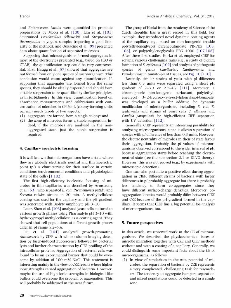
and Enterococcus bacalis were quantified in probioticpreparations by Moon et al. [100], Lim et al. [101]determined Lactobacillus delbrueckii and Streptococcusthermophilus in yogurt samples (reporting a good line-arity of the method), and Oukacine et al. [99] presenteddata about quantification of separated microbes.
Supposing that microorganisms formed aggregates inmost of the electrolytes presented (e.g., based on PEO orCTAB), the quantification step could be very controver-sial. First, Haugg et al. [57] showed that aggregates arenot formed from only one species of microorganism. Thisconclusion would count against any quantification. If,supposing that aggregates are formed from the samespecies, they should be ideally dispersed and should forma stable suspension to be quantified by similar principles,as in turbidimetry. In this view, quantification based onabsorbance measurements and calibrations with con-centration of microbes in CFU/mL (colony-forming unitsper mL) needs proofs of two aspects:(1) aggregates are formed from a single colony; and,(2) the zone of microbes forms a stable suspension; in-
deed, if the microbes are analyzed in the non-aggregated state, just the stable suspension isrequired.
4. Capillary isoelectric focusing
It is well known that microorganisms have a state wherethey are globally electrically neutral and this isoelectricpoint (pI) is characteristic for their surface in certainconditions (environmental conditions and physiologicalstate of the cells) [1,102].
The first high-efficiency isoelectric focusing of mi-crobes in thin capillaries was described by Armstronget al. [53], who separated E. coli, Pseudomonas putida, andSerratia rubidae strains in 20 min. A methylcellulosecoating was used for the capillary and the pH gradientwas generated with Biolyte ampholyte pH 3–10.
Later, Shen et al. [103] analyzed yeast cells cultured tovarious growth phases using Pharmalyte pH 3–10 withhydroxypropyl methylcellulose as a coating agent. Theyshowed that cell populations at different growth phasesdiffer in pI range 5.2–6.4.
Liu et al. [104] analyzed growth-promotingrhizobacteria by CIEF with whole-column imaging detec-tion by laser-induced fluorescence followed by bacteriallysis and further characterization by CIEF profiling of theintracellular proteins. Aggregation of bacterial cells wasfound to be an experimental barrier that could be over-come by addition of 100 mM NaCl. This statement isinteresting mainly in the view of CZE results where higherionic strengths caused aggregation of bacteria. However,maybe the use of high ionic strengths in biological-likebuffers could overcome the problem of aggregation. Thiswill probably be addressed in the near future.
20 http://www.elsevier.com/locate/trac
The group of Horka from the Academy of Science of theCzech Republic has a great record in this field. Forexample, they introduced novel dynamic coating agentsof the capillary e.g., based on non-ionogenic tensidepoly(ethyleneglycol) pyrenebutanoate PB-PEG [105,106], or poly(ethyleneglycole) PEG 4000 [107,108].After these first studies, Horka et al. employed CIEF forsolving various challenging tasks e.g., a study of biofilmformation of S. epidermis [109] and analysis of pathogenicspecies of genus Clavibacter, Xanthomonas andPseudomonas in tomato-plant tissues, see Fig. 10 [110].
Recently, similar strains of yeast with pI differenceless than 0.3 units were separated using a short pHgradient of 2–3.3 or 2.7–4.7 [111]. Moreover, achromophoric non-ionogenic surfactant, poly(ethyl-eneglycol) 3-(2-hydroxy-5-n-octylphenylazo)-benzoate,was developed as a buffer additive for dynamicmodification of microorganisms, including E. coli, S.epidermidis and strains of yeast cells C. albicans andCandida parapsilosis for high-efficient CIEF separationwith UV detection [112].
Generally, CIEF represents an interesting possibility foranalyzing microorganisms, since it allows separation ofspecies with pI difference of less than 0.3 units. However,the electric neutrality of microbes in their pI state favorstheir aggregation. Probably the pI values of microor-ganisms observed correspond to the wider interval of pHbecause aggregation starts before reaching the electro-neutral state (see the sub-section 2.1 or DLVO theory).However, this was not proved (e.g., by experiments withmicroscopic detection).
One can also postulate a positive effect during aggre-gation in CIEF. Different strains of bacteria with largerdifferences in pI probably aggregate but there is probablyless tendency to form co-aggregates since theyhave different surface-charge densities. Moreover, co-aggregation kinetics would probably differ between CIEFand CZE because of the pH gradient formed in the cap-illary. It seems that CIEF has a big potential for analysisof microorganisms, too.
5. Future perspectives
In this article, we reviewed work in the CE of microor-ganisms. We described the physicochemical bases ofmicrobe migration together with CZE and CIEF methodswithout and with a coating of a capillary. Generally, wecould distinguish some important facts about the CE ofmicroorganisms, as follows.(1) In view of similarities in the zeta potential of mi-
crobes, the separation of bacteria by CZE representsa very complicated, challenging task for research-ers. The tendency to aggregate hampers separationand mixed populations could be detected in a singlezone.

Trends in Analytical Chemistry, Vol. 31, 2012 Trends
(2) In view of the pI, it seems that microorganismscould be separated by CIEF. Indeed, electric neutral-ity favors aggregation of microbes and probably co-aggregation could be suppressed.
(3) Identification of microorganisms according tomobility is insufficient. Only CE connection withother set-ups (PCR, FISH or MS) could provide ade-quate information. A sidetrack could be found ingeneral identification of microbial contamination(sterility), in which separation within bacterial spe-cies is unnecessary.
(4) The sensitivity of the CE test is also an importantquestion. Pathogenic populations of microbes cangrow from only a few cells. On this point, two majoraspects should be addressed: the CE method shouldideally be sensitive enough to detect only one cell;and, large-volume sample injection should bepossible. The last problem deals with the typicalinjection volume in CE of tens of nL while the sam-ple volume is in tens of lL. In this configuration(e.g., with one cell in a 20-lL sample vial with injec-tion of 20 nL), the probability of a positive match isjust 0.1% because of the small volume injected.
With many experimental and fundamental problems,the theme of CE analysis of microorganisms is one of themost challenging fields in CE with a lot of questions thatneed to be answered. We noted above some of theimportant tasks (e.g., single-cell identification, problemswith injection volume), but these are not all the aspects.
A study of zeta potential and aggregation tendencycould bring a lot of important results that would lead,together with a suitable capillary coating, to successfulCE separation of microorganisms without co-aggrega-tion. The problem with identifying specific bacterialstrains from thousands of bacterial species remains, but,here, on-line combination of CE with MS and/or otherdetection techniques could be profitable. In such terms,CE could supply or maybe replace microbial tests com-monly used at present.
AcknowledgementsThe financial support by the Research project MSMTMSM 6198959216 and the Operational Program Re-search and Development for Innovations – EuropeanRegional Development Fund (Project CZ.1.05/2.1.00/03.0058) is gratefully acknowledged.
References[1] J.G. Black, Microbiology: Principles and Explorations, Wiley, New
York, USA, 2008.
[2] F.N. Egerton, J. Histor. Biology 1 (1962) 1.
[3] G. Bordenave, Microb. Infect. 5 (2003) 553.
[4] R. Munch, Microb. Infect. 5 (2003) 69.
[5] R. Koch, Berliner Klin. Wochenschr. 19 (1882) 221.
[6] C. Gram, Fortschr. Med. 2 (1884) 185.
[7] F. Agerer, S. Waeckerle, C.R. Hauck, J. Microbiol. Methods 59
(2004) 23.
[8] A. Ghosh, A. Shrivastav, D.A. Jose, S.K. Mishra, C.K. Chandrak-
anth, S. Mishra, A. Das, Anal. Chem. 80 (2008) 5312.
[9] A. Matsuhisa, Y. Saito, H. Ueyama, M. Yamamoto, T. Ohono,
Microbiol. Immunol. 37 (1993) 765.
[10] I.V. Kourkine, M. Ristic-Petrovic, E. Davis, C.G. Ruffolo, A.
Kapsalis, A.E. Barron, Electrophoresis 24 (2003) 655.
[11] C. Zhao, G. Xu, P. Gao, J. Yang, X. Shi, J. Tian, J. Sep. Sci. 28
(2005) 513.
[12] C. Fenselau, P.A. Demirev, Mass Spectrom. Rev. 20 (2001) 157.
[13] R.L. Graham, C. Graham, G. McMullan, Microbial Cell Factor 6
(2007) 26.
[14] M.J. Desai, D.W. Armstrong, Microbiol. Mol. Biol. Rev. 67 (2003)
38.
[15] E. Klodzinska, B. Buszewski, Anal. Chem. 81 (2009) 8.
[16] K.F. Nielsen, J.M. Mogensen, M. Johansen, T.O. Larsen, J.C.
Frisvad, Anal. Bioanal. Chem. 395 (2009) 1225.
[17] G. Mamone, G. Picariello, S. Caira, F. Addeo, P. Ferranti, J.
Chromatogr., A 1216 (2009) 7130.
[18] M.G. Khaledi, High Performance Capillary Electrophoresis: The-
ory, and Applications, Techniques, Wiley, New York, USA,
1998.
[19] S.L. Simpson, J.P. Quirino, S. Terabe, J. Chromatogr., A 1184
(2008) 504.
[20] E. Lewin, L. Cassimeris, V.R. Lingappa, G. Plopper (Editors),
Cells, Jones and Bartlett Publishers, Sudbury, MA, USA, 2006.
[21] J. Ubbink, P. Schar-Zammaretti, Curr. Opin. Colloid Interface Sci.
12 (2007) 263.
[22] T.J. Beveridge, L.L. Graham, Microbiol. Rev. 55 (1991) 684.
[23] T.J. Beveridge, J. Bacteriol. 181 (1999) 4725.
[24] K.A. Soni, A.K. Balasubramanian, A. Beskok, S.D. Pillai, Curr.
Microbiol. 56 (2008) 93.
[25] A. van de Wal, M. Minor, W. Norde, A.J.B. Zehnder, J. Lyklema,
Langmuir 13 (1997) 165.
[26] K.E. Eboigbodin, J.J. Ojeda, C.A. Biggs, Langmuir 23 (2007)
6691.
[27] P. Schar-Zammaretti, M.-L. Dillmann, N. D�Amico, M. Affolter, J.
Ubbink, Appl. Environ. Microbiol. 71 (2005) 8165.
[28] X. Chatellier, J.-Y. Bottero, J. Le Petit, Langmuir 17 (2001) 2782.
[29] X. Chatellier, J.-Y. Bottero, J. Le Petit, Langmuir 17 (2001) 2791.
[30] J.C. Baygents, J.R. Glynn, O. Albinger, B.K. Biesemeyer, K.L.
Ogden, R.G. Arnold, Environ. Sci. Technol. 32 (1998) 1596.
[31] A.E.J. van Merode, H.C. van der Mei, H.J. Busscher, B.P. Krom, J.
Bacteriol. 188 (2006) 2421.
[32] A.E.J. van Merode, H.C. van der Mei, H.J. Busscher, K. Waar, B.P.
Krom, Microbiology 152 (2006) 807.
[33] S.P. Radko, A. Chrambach, Electrophoresis 23 (2002) 1957.
[34] D.C. Henry, Proc. R. Soc. London, Ser. A 133 (1931) 106.
[35] R.W. O�Brien, L.R. White, J. Chem. Soc., Faraday Trans. 77
(1978) 1607.
[36] J.T.G. Overbeek, P.H. Wiersema, in: M. Bier (Editor), Electropho-
resis: Theory, Methods, and Applications, Academic Press, New
York, USA, 1967.
[37] S.S. Dukhin, B.V. Derjaguin, in: E. Matijevic (Editor), Surface and
Colloid Science, Wiley, New York, USA, 1974.
[38] R. Bos, H.C. van der Mei, H.J. Busscher, FEMS Microbiol. Rev. 23
(1999) 179.
[39] K.E. Eboigbodin, J.R.A. Newton, A.F. Routh, C.A. Biggs, Lang-
muir 21 (2005) 12315.
[40] D.R. Absolom, F.V. Lamberti, Z. Policova, W. Zingg, C.J. van Oss,
A.W. Neumann, Appl. Environ. Microbiol. 46 (1983) 90.
[41] H.J. Busscher, A.H. Weerkamp, H.C. van der Mei, A.W.J. van
Pelt, H.P. de Jong, J. Arends, Appl. Environ. Microbiol. 48 (1984)
980.
[42] M.C.M. van Loosdrecht, J. Lynkema, W. Norde, A.J.B. Zehnder,
Microb. Ecol. 17 (1989) 1.
http://www.elsevier.com/locate/trac 21

Trends Trends in Analytical Chemistry, Vol. 31, 2012
[43] C.J. van Oss, R.J. Good, M.K. Chaudhury, J. Colloid Interface Sci.
111 (1986) 378.
[44] J. Wood, R. Sharma, Langmuir 11 (1995) 4797.
[45] M. Elimelech, J. Chem. Soc., Faraday Trans. 86 (1990) 1623.
[46] W.A. Ducker, Z. Xu, J.N. Israelachvili, Langmuir 10 (1994)
3279.
[47] S. Hjerten, K. Elenbring, F. Kilar, J.-L. Liao, A.J.C. Chen, C.J.
Seibert, M.-D. Zhu, J. Chromatogr. 403 (1987) 47.
[48] R.C. Ebersole, R.M. McCormick, Bio/Technology 11 (1993)
1278.
[49] M. Torimura, S. Ito, K. Kano, T. Ikeda, Y. Esaka, T. Ueda, J.
Chromatogr., B 721 (1999) 31.
[50] A. Pfetsch, T. Welsch, Fresenius� J. Anal. Chem. 359 (1997) 198.
[51] J.R. Glynn, B.M. Belongia, R.G. Arnold, K.L. Ogden, J.C.
Baygents, Appl. Environ. Microbiol. 64 (1998) 2572.
[52] J. Petr, B. Teste, S. Descroix, J.-M. Siaugue, P. Gareil, A. Varenne,
Electrophoresis 31 (2010) 2754.
[53] D.W. Armstrong, G. Schulte, J.M. Schneiderheinze, D.J. Westen-
berg, Anal. Chem. 71 (1999) 5465.
[54] N. Iki, E.S. Yeung, J. Chromatogr., A 731 (1996) 273.
[55] D.W. Armstrong, M. Girod, L. He, M.A. Rodriguez, W. Wei, J.
Zheng, E.S. Yeung, Anal. Chem. 74 (2002) 5523.
[56] J. Zheng, E.S. Yeung, Anal. Chem. 75 (2003) 818.
[57] M. Haugg, V. Kaiser, C. Schmidtkunz, T. Welsch, Electrophoresis
30 (2009) 396.
[58] V. Hoerr, A. Stich, U. Holzgrabe, Electrophoresis 25 (2004)
3132.
[59] D. Dai, Y. Chen, L. Qi, X. Yu, Electrophoresis 24 (2003) 3219.
[60] O. Ryparova, J. Petr, M. Kowalska, J. Znaleziona, R. Knob, V.
Maier, I. Frebort, J. Sevcık, Chem. Listy 102 (2008) 1121.
[61] J.M. Schneiderheinze, D.W. Armstrong, G. Schulte, D.J. Westen-
berg, FEMS Microbiol. Lett. 189 (2000) 39.
[62] E. Klodzinska, M. Szumski, E. Dziubakiewicz, K. Hrynkiewicz, E.
Skwarek, W. Janusz, B. Buszewski, Electrophoresis (2010) 1590.
[63] J. Znaleziona, J. Petr, R. Knob, V. Maier, J. Sevcık, Chromatog-
raphia 67 (2008) S5.
[64] P.G. Righetti, C. Gelfi, B. Verzola, L. Castelletti, Electrophoresis 22
(2001) 603.
[65] J. Horvath, V. Dolnik, Electrophoresis 22 (2001) 644.
[66] I. Miksık, Z. Deyl, J. Chromatogr., A 852 (1999) 325.
[67] B. Buszewski, M. Szumski, E. Klodzinska, H. Dahm, J. Sep. Sci. 26
(2003) 1045.
[68] E. Klodzinska, M. Szumski, K. Hrynkiewicz, E. Dziubakiewicz, M.
Jackowski, B. Buszewski, Electrophoresis 30 (2009) 3086.
[69] M. Szumski, E. Klodzinska, B. Buszewski, J. Chromatogr., A 1084
(2005) 186.
[70] B. Buszewski, E. Klodzinska, Electrophoresis 29 (2008) 4177.
[71] B. Buszewski, M. Szumski, E. Klodzinska, R. Jarmalaviciene, A.
Maruska, J. Chromatogr., A 1216 (2009) 6146.
[72] M.A. Rodriguez, A.W. Lantz, D.W. Armstrong, Anal. Chem. 78
(2006) 4759.
[73] T. Tsuda, J. High. Resolut. Chromatogr. 10 (1987) 622.
[74] C.A. Lucy, R.S. Underhill, Anal. Chem. 68 (1996) 300.
[75] Y. Bao, A.W. Lantz, J.A. Crank, J. Huang, D.W. Armstrong,
Electrophoresis 29 (2008) 2587.
[76] E.G. Yanes, S.R. Gratz, M.J. Baldwin, S.E. Robinson, A.M.
Stalcup, Anal. Chem. 73 (2001) 3838.
[77] B. Palenzuela, B.M. Simonet, R.M. Garcıa, A. Rıos, M. Valcarcel,
Anal. Chem. 76 (2004) 3012.
[78] E. Klodzinska, H. Dahm, H. Rozycki, J. Szeliga, M. Jackowski, B.
Buszewski, J. Sep. Sci. 29 (2006) 1180.
[79] G.N. Okafo, H.C. Birrell, M. Greenaway, M. Haran, P. Camilleri,
Anal. Biochem. 219 (1994) 201.
22 http://www.elsevier.com/locate/trac
[80] D.W. Armstrong, L. He, Anal. Chem. 73 (2001) 4551.
[81] M. Girod, D.W. Armstrong, Electrophoresis 23 (2002) 2048.
[82] T. Shintani, K. Yamada, M. Torimura, FEMS Microbiol. Lett. 210
(2002) 245.
[83] K. Yamada, M. Torimura, S. Kurata, Y. Kamagata, T. Kanagawa,
K. Kano, T. Ikeda, T. Yokomaku, R. Kurane, Electrophoresis 22
(2001) 3413.
[84] M. Horka, F. Ruzicka, V. Hola, K. Slais, Electrophoresis 26
(2005) 548.
[85] A.W. Lantz, B. Bisha, M.-Y. Tong, R.E. Nelson, B.F. Brehm-
Stecher, D.W. Armstrong, Electrophoresis 31 (2010) 2849.
[86] K. Hrynkiewicz, E. Klodzinska, H. Dahm, J. Szeliga, M. Jackowski,
B. Buszewski, FEMS Microbiol. Lett. 286 (2008) 1.
[87] J. Petr, O. Ryparova, V. Ranc, P. Hinnerova, J. Znaleziona, M.
Kowalska, R. Knob, V. Maier, I. Frebort, K. Lemr, J. Sevcık,
Electrophoresis 30 (2009) 444.
[88] M. Horka, J. Horky, A. Kubesova, K. Mazanec, H. Matouskova, K.
Slais, Analyst (Cambridge, UK) 135 (2010) 1636.
[89] E. Carbonnelle, C. Mesquita, E. Bille, N. Day, B. Dauphin, J.-L.
Beretti, A. Ferroni, L. Gutmann, X. Nassif, Clin. Biochem. 44
(2011) 104.
[90] P.A. Demirev, C. Fenselau, Annu. Rev. Anal. Chem. 1 (2008) 71.
[91] M. Szumski, E. Klodzinska, B. Buszewski, Microchim. Acta 164
(2009) 287.
[92] J. Petr, O. Ryparova, J. Znaleziona, V. Maier, J. Sevcık, Electro-
phoresis 30 (2009) 3863.
[93] M. Xie, H. Wang, Y. Chen, Electrophoresis 30 (2009) 3884.
[94] A.W. Lantz, Y. Bao, D.W. Armstrong, Anal. Chem. 79 (2007)
1720.
[95] A.W. Lantz, B.F. Brehm-Stecher, D.W. Armstrong, Electropho-
resis 29 (2008) 2477.
[96] J. Petr, C. Jiang, J. Sevcık, E. Tesarova, D.W. Armstrong,
Electrophoresis 30 (2009) 3870.
[97] L. Yu, S.F.Y. Li, J. Chromatogr., A 1161 (2007) 308.
[98] F. Oukacine, L. Garrelly, B. Romestand, D.M. Goodall, T. Zou, H.
Cottet, Anal. Chem. 83 (2011) 1571.
[99] F. Oukacine, J.P. Quirino, L. Garrelly, B. Romestand, T. Zou, H.
Cottet, Anal. Chem. 83 (2011) 4949.
[100] B.-G. Moon, Y. Kim, Bull. Korean Chem. Soc. 24 (2003) 1203.
[101] O. Lim, W. Suntornsuk, L. Suntornsuk, J. Chromatogr., B. 877
(2009) 710.
[102] D. Voet, J.G. Voet, Biochemistry, Wiley, New York, USA, 1995.
[103] Y. Shen, S.J. Berger, R.D. Smith, Anal. Chem. 72 (2000) 4603.
[104] Z. Liu, S.S. Wu, J. Pawliszyn, J. Chromatogr., A 1140 (2007)
213.
[105] M. Horka, F. Ruzicka, J. Horky, V. Hola, K. Slais, Anal. Chem. 78
(2006) 8438.
[106] M. Horka, F. Ruzicka, V. Hola, K. Slais, Electrophoresis 28
(2007) 2300.
[107] M. Horka, F. Ruzicka, J. Horky, V. Hola, K. Slais, J. Chromatogr.,
B 841 (2006) 152.
[108] M. Horka, F. Ruzicka, V. Hola, K. Slais, Anal. Bioanal. Chem.
385 (2006) 840.
[109] F. Ruzicka, M. Horka, V. Hola, M. Votava, J. Microbial Methods
68 (2007) 530.
[110] M. Horka, J. Horky, H. Matouskova, K. Slais, J. Chromatogr., A
1216 (2009) 1019.
[111] M. Horka, F. Ruzicka, V. Hola, K. Slais, Electrophoresis 30
(2009) 2134.
[112] M. Horka, F. Ruzicka, V. Hola, V. Kahle, D. Moravcova, K. Slais,
Anal. Chem. 81 (2009) 6897.
![Capillary thermostatting in capillary electrophoresis · Capillary thermostatting in capillary electrophoresis ... 75 µm BF 3 Injection: ... 25-µm id BF 5 capillary. Voltage [kV]](https://img.dokumen.tips/doc/110x75/5c176ff509d3f27a578bf33a/capillary-thermostatting-in-capillary-electrophoresis-capillary-thermostatting.jpg)