Embed Size (px)
Citation preview

ANALYSIS OF BLOOD FLOW IN ABDOMINAL AORTIC ANEURYSM USING COMPUTATIONAL FLUID DYNAMIC
MOHD YUSUF BIN MOHD JAAFAR
Thesis submitted in fulfilment of the requirementsfor the award of the degree of
Bachelor of Mechanical Engineering
Faculty of Mechanical EngineeringUNIVERSITI MALAYSIA PAHANG
NOVEMBER 2009

ii
SUPERVISOR’S DECLARATION
I hereby declare that I have checked this project and in my opinion, this project is
adequate in terms of scope and quality for the award of the degree of Bachelor of
Mechanical Engineering.
Signature :
Name of Supervisor : MR. MOHAMAD MAZWAN BIN MAHAT
Position : SUPERVISOR
Date : 24 NOVEMBER 2009
iii
STUDENT’S DECLARATION
I hereby declare that the work in this project is my own except for quotations and
summaries which have been duly acknowledged. The project has not been accepted for
any degree and is not concurently submitted for award of other degree.
Signature :
Name : MOHD YUSUF BIN MOHD JAAFAR
ID Number : MA06052
Date : 24 NOVEMBER 2009

iv
Dedicated to my parents

v
ACKNOWLEDGEMENTS
First of all, thanks to Allah S.W.T for the blessing and opportunity for me to finish my final year project. It is a great sense of pleasure that I acknowledge the help and guidance I have receive from numerous people during my time at Universiti Malaysia Pahang especially from my supervisor, Mr. Mohamad Mazwan Bin Mahat. He provided me with energy, enthusiasm and insight to work on this interesting final year project. I am very much thankful to them for all their support in conducting and writing my work. Moreover, I would like to express my heartfelt and sincere for their priceless guidance and support during my final year.
Not forgetting my fellow friends who gave me a lot of ideas and support in completing this project. Without them, I would have been nowhere near completing my project which I also learn the importance of team working. Once again, thanks to Almighty God for giving me the life and strength to finish this project without any major problem.
Lastly, special thanks goes to my beloved parents who always supported me from the very beginning to achieve my goals and sacrifice much in their life for my well-being. I am indebted to their painstaking attitude, which always kept me on the right track.

vi
ABSTRACT
Aneurysm is an abnormalities occurs in blood vessel that involve the size increment and dilations where the cause of the disease not yet been founded. Abdominal aortic aneurysm was occurring at abdomen aorta. The abnormal diameter size of abdominal aorta was cause by ballooning or bulge that lead to changes in blood flow behavior. Investigation on the behavior of blood flow in abdominal aortic aneurysm was taken place to determine the pressure distribution and velocity profile in the aneurysm region. The diameter of normal aorta was 20 mm and the diameter of aneurysm aorta was considered as 60 mm from the literature. The flow of blood in aorta will be disturbedwhen the diameter of aorta was changed. Aneurysm is acting like diffuser that increasesthe pressure and decreases the velocity when blood entering the aneurysm region. Oppositely when it leaving the aneurysm the pressure wills decreases back and the velocity will be increase like nozzle. It was proved that the flow behavior inside aneurysm region would be different compared to the normal aorta from the previous study. The size of aorta influences the pressure and velocity of blood. The flow inside aneurysm region was disturbed due to pressure and velocity changes and resulting the formation of vortex. Reynolds number was establishing by using different initial velocity of blood flow and different diameter of aorta. The simulation of the model was studied under incompressible and non-Newtonian condition which investigated computationally by fluid dynamic software. The studies was done without experimentset-up but based on numerical approach only.

vii
ABSTRAK
Aneurysm adalah suatu penyakit yang berlaku kepada salur darah dimana terjadi ketidaknormalan pada saiz diameter salur darah dan puncanya masih belum diketahui. Abdomen aneurysm berlaku kepada salur darah yang terletak di bahagian perut. Saiz diameter bagi saluran darah abdomen yang tidak normal telah menyebabkan pembesaran atau pengembangan yang membawa kepada perubahan reaksi pengaliran darah. Penyelidikan terhadap keadaan pengaliran darah di dalam aneurysm abdomen dijalankan untuk menentukan taburan tekanan dan profil halaju di dalam kawasan aneurysm. Saiz diameter bagi saluran darah abdomen yang normal adalah 20 mm dan saluran darah aneurysm abdomen ialah 60 mm berdasarkan kepada rujukan dari penyelidikan sebelum ini. Pengaliran di dalam salur darah akan terganggu apabila saiz diameter salur darah tersebut berubah. Halaju dan tekanan darah berubah apabila iasampai kepada kawasan aneurysm di dalam salur darah tersebut. Telah terbukti bahawa pengaliran salur darah di dalam salur darah abdomen aneurysm akan berbeza berbanding dengan pengaliran di dalam salur darah yang normal. Pengaliran di dalam salur darah abdomen telah terganggu kerana terdapat perubahan tekanan dan halaju dan ini telah menyebabkan pembentukan pusaran darah. Nombor Reynold ditentukan dengan menggunakan menggunakan halaju awal darah yang berbeza dan saiz diameter salur darah yang berbeza. Simulasi bagi model ini dikaji berdasarkan parameter aliran mampat dan bukan bendalir Newtonian dimana ianya dikaji secara perkomputeran dengan menggunakan program bendalir dinamik. Kajian ini hanya dijalankan dengan pendekatan berdasarkan pengiraan matematik tanpa melibatkan sebarang eksperimen.

viii
TABLE OF CONTENTS
Page
SUPERVISOR’S DECLARATION ii
STUDENT’S DECLARATION iii
ACKNOWLEDGEMENTS v
ABSTRACT vi
ABSTRAK vii
TABLE OF CONTENTS viii
LIST OF TABLES x
LIST OF FIGURES xi
LIST OF SYMBOLS xiii
LIST OF ABBREVIATIONS xiv
CHAPTER 1 INTRODUCTION
1.1 Introduction 1
1.2 Types of Aneurysm 3
1.3 AAA Formation 7
1.4 Symptom and diagnosis 8
1.5 Treatments 8
1.6 Objectives and Scopes 10
CHAPTER 2 LITERATURE REVIEW
2.1 Introduction 11
2.2 Flow Behaviour in Abdominal Aortic Aneurysm 12
2.3 Flow Disturbance Within AAA Model 19
ix
CHAPTER 3 METHODOLOGY
3.1 Introduction 21
3.2 Simulation Assumption and Parameter 22
3.3 Geometry of Model 22
3.4 Boundary Condition 24
3.5 Governing Equation of Blood Flow 26
3.6 Finite Volume Method 27
3.7 Visualization of Simulation 28
CHAPTER 4 RESULTS AND DISCUSSION
4.1 Introduction 29
4.2 Behaviour of Flow With Different Initial Velocity and Aorta
Diameter
31
4.3 Pressure Distribution 32
4.4 Velocity Profile 37
4.5 Vortex Formation 42
REFERENCES 45
APPENDICES 47

x
LIST OF TABLES
Table No. Title Page
3.1 Parameters in the simulation 22
4.1 Varies initial velocity of blood and aorta diameter that produce certain Reynold Number
31
4.2 Peak Pressure for different inlet velocity 35
4.3 Peak Pressure for different diameter of aorta 36
4.4 Percentage of velocity drop with different value of inlet velocity 38
4.5 Minimum Velocity for different diameter of aorta 39
4.6 Percentage of velocity drop for different diameter of aorta 39
4.7 Correlation obtained in the pressure distribution 41
4.8 Correlation obtained in the velocity profile 41

xi
LIST OF FIGURES
Figure No. Title Page
1.1 Shapes of aneurysm 2
1.2 Abdominal Aortic Aneurysm 3
1.3 Cerebral Aneurysm 4
1.4 Thoracic Aortic Aneurysm 5
1.5 Dissecting Aneurysm 6
1.6 Stented AAA 9
2.1 Axisymmetric AAA 13
2.2 Inlet velocity waveform 13
2.3 Exit pressure waveform 14
3.1 The process in Computational Fluid Dynamic 21
3.2 The geometry model of aneurysm 23
3.3 The model geometry taken from literature 23
3.4 The boundary condition of AAA model 25
3.5 Visualization of blood flow streamlines in AAA model 28
4.1 Pressure distribution in the aneurysm 30
4.2 Velocity profile in the aneurysm 30
4.3 Pressure distribution and velocity profile in the AAA using inlet velocity 0.4 m/s
32
4.4 Pressure distribution and velocity profile in the AAA using inletvelocity 0.5 and 0.6 m/s
33
4.5 Pressure distribution and velocity profile in the AAA using inletvelocity 0.7 and 0.8 m/s
34
4.6 Pressure distribution in AAA with different inlet velocity 35

xii
4.7 Pressure distribution in AAA with different diameter of aorta 36
4.8 Velocity profile in AAA with different inlet velocity 37
4.9 Velocity profile in AAA with different diameter of aorta 38
4.10 Velocity Band Width for different diameter of aorta 39
4.11 Percentage of velocity drop for different diameter of aorta 40
4.12 The vortex formation within aneurysm region 42

xiii
LIST OF SYMBOLS
velocity in the ith direction
Pressure
Body force
Density
Viscosity
Kronecker delta

xiv
LIST OF ABBREVIATIONS
AAA Abdominal Aortic Aneurysm
CFD Computational Fluid Dynamics
CAD Computer Aided Design

CHAPTER 1
INTRODUCTION
1.1 INTRODUCTION
Aneurysm is ballooning or bulge that occurs in a blood vessel cause by disease
or vessel wall that is weaken. The diameter of the blood vessel will increase greater than
its original size and can lead to rupture, bleeding inside the body and often fatal.
Aneurysm consists of four types which is abdominal aortic aneurysm, dissection
aneurysm, thoracic aortic aneurysm and cerebral aneurysm. Type of aneurysm is shown
in figure 1.2 until figure 1.5.
The word aneurysm comes from the Greek aneurysma meaning widening.
Aneurysm consist of different shape that is maybe saccular (balloon like expansions of
only a portion of the wall) or fusiform (gradual dilation of the complete circumference
of the artery) as presented in figure 1.1. The different shapes have not been related to
any specific cardiovascular disease or clinical manifestation (Contran et, 1999).
Abdominal aortic aneurysm can be found in abdominal aorta which is located below
renal arteries above iliac bifurcation). This is a common vascular problem and the rate
of incidence has increased greatly with the increase in life expectancy of the population.
AAA is often not detected at early stages and, in most cases, remains latent until
symptoms occur as their size greatly increases or until they are diagnosed during an
incidental exam (Szilagyi, 1982).

2
Figure1.1: Shapes of aneurysm
Source: http://www.daviddarling.info
Figure 1.1 shows that aneurysm shape consists of saccular, fusiform and
ruptured aneurysm. Every shape consists of different geometry and size. Ruptured
aneurysm is very dangerous to the human. Ruptured aneurysm is cause by weakening of
the arterial wall. Beside the blood pressure inside the artery is very high and can exert
tremendous dynamic force toward arterial wall. Between times, the wall structure
cannot stand any further force and finally this lead to the rupture.
Saccular Aneurysm Fusiform Aneurysm
Ruptured Aneurysm

3
1.2 TYPES OF ANEURYSM
Figure 1.2: Abdominal Aortic Aneurysm
Source: http://aaadoctor.org/
Figure 1.2 shows that abdominal aortic aneurysm is located along the portion of
the aorta that passes through the abdomen . It is a large blood vessel that supplies blood
from the heart to the abdomen, pelvis and leg. The tendency of the aneurysm to break is
base on the size. The larger the aneurysm, the more possiblities for it to break. This type
of aneurysm can occur and develop in anybody, but record often said that males that
over 60 is more likely to be involved.
Lung
Heart
Kidney
Abdominal Aortic Aneurysm

4
Figure 1.3: Cerebral Aneurysm
Source: http://www.nhlbi.nih.gov
Figure 1.3 shows the location of the cerebral aneurysm. It occurs at the blood
vessel (arteries) in the brain. The size of cerebral aneurysm much smaller compare to
abdominal aortic aneurysm, but the threat is same. Typically aneurysms occur at
branching points of arteries. Cerebral aneurysm often under ¼ inches in diameter
especially that is located in front of the brain.
5
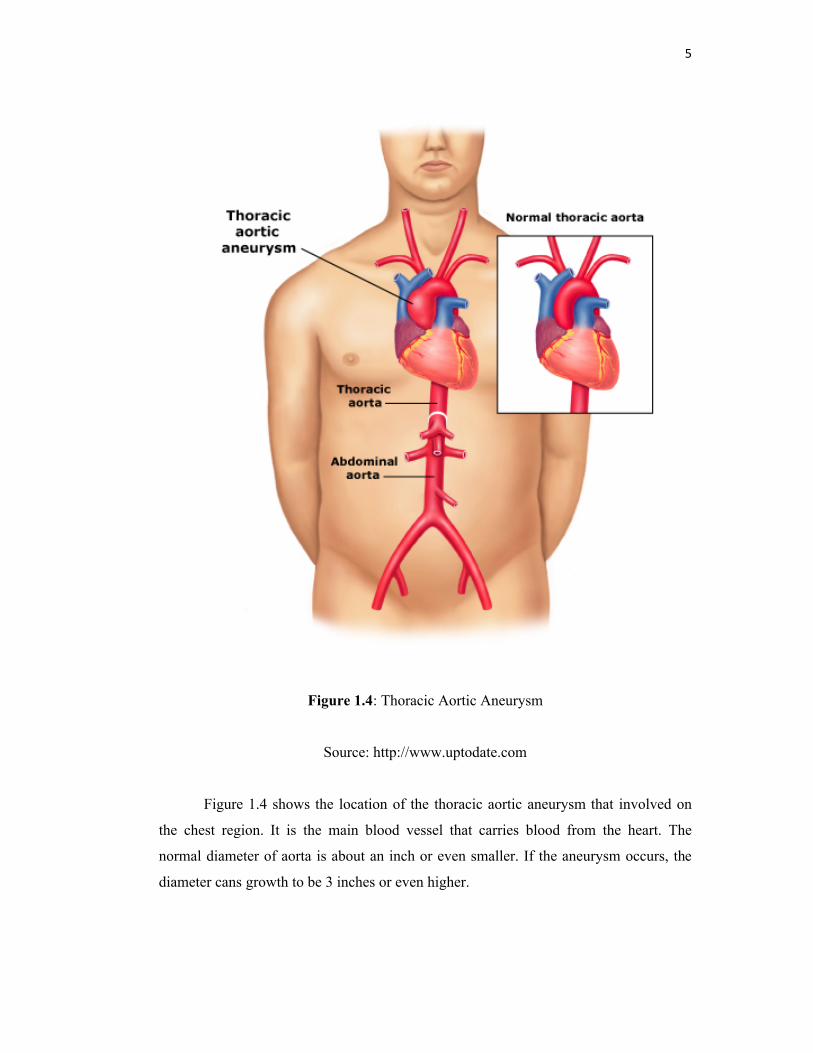
Figure 1.4: Thoracic Aortic Aneurysm
Source: http://www.uptodate.com
Figure 1.4 shows the location of the thoracic aortic aneurysm that involved on
the chest region. It is the main blood vessel that carries blood from the heart. The
normal diameter of aorta is about an inch or even smaller. If the aneurysm occurs, the
diameter cans growth to be 3 inches or even higher.

6
Figure 1.5: Dissecting Aneurysm
Source: http://www.vascularweb.org
Figure 1.5 shows that the location of dissecting aneurysm. The wall of the aorta
rips (dissects) longitudinally. The bleeding that occur on the weaken wall can splits the
wall in this resulting the dissecting aneurysm. It is also because when tear begins in the
wall of the aorta and its three layers is separated. This type of aneurysm can occur
anywhere along the aorta.

7
1.3 AAA FORMATION
The real and exact cause of abdominal aortic aneurysm formation are not been
found yet. From previous studies, the atherosclerosis (buildup of fatty deposits in the
arteries) is being said as the reason, but according to Ernst, 1993, it is clear that the
cause of AAAs is not simply a compilation due to atherosclerosis, but the latter maybe a
secondary response to an injured aortic wall.
Many theories have been suggested to explain the formation of AAAs wall.
Several theories state that structural defect of the aortic wall can lead to the loss of
biomechanical function and it cannot perform under good level of functionality and
finally it leads to the uncontrolled expansion. The theory proposed by Dobrin (1989)
was the aneurysm formation is due to local weakening of the intima and media layers of
the abdominal aorta. Excessive hemodynamic load applied to the aortic walls that have
inadequate strength to withstand it can cause the expansion of AAA.
The strength of the aortic wall is being decreased due to initial depletion of
elastin (the tissue that gives elasticity to blood vessel). Beside that the depletion of
collagen (the tissue that provides stiffness) also was the reason of weakening of the
wall. Where is this action is take place was not specify by Dobrin. Maybe this can be the
future research to find specific place or which wall layers this process would occur.

8
1.4 SYMPTOM AND DIAGNOSIS
Symptoms of aneurysm will depend upon the location of the aneurysm (Li
Zhonghua, 2005). Aneurysm usually cannot be detected during early stages in most
cases, but the symptom only majorly occurs as the size greatly increased. It can be
detect during daily or routine examination. During the occurrence of the symptom, it
can cause severely pain. People who suffer it will face pulsing sensation, difficulty
swallowing, pain, coughing or hoarseness.
The detection of the aneurysm can be done by simple physical examination such
as X-Ray or ultrasonography. Besides that, there are also Angiography, Computed
Tomography (CT) and Magnetic Resonance Imaging (MRI) that being the common
ways to detect Abdominal Aortic Aneurysm.
1.5 TREATMENTS
The treatments for aneurysm have two methods. First is open surgical repair and
second is minimally invasive endovascular repair. For open surgical repair, the patient
chest or abdomen must be operated to allow the installation of graft at the weaken
region or bulge region. The installation of graft will provide new path and allow blood
to move freely compare from previous condition without graft. Blood can flow through
the graph and the collision with the bulge wall can be minimized.
In cerebral aneurysm case, some part of the head skull must remove temporarily
if open surgery method is taken place. We can use tiny metal clips to place at the neck
of the aneurysm. The purpose of this action is to block the blood flow from entering the
aneurysm bulge region. This open surgery method have disadvantages such as the
patient that being treat will face large incision, higher cost of hospitalization, rate of
recovery is low and can lead to longer pain.
The second method is Minimally Invasive Endovascular Repair. This treatment
performed inside patient’s body using long catheters. This method is guided by X-rays.
To perform this method in aortic aneurysm, the small incision must take place at

9
femoral artery. Then the stent graphs being released when it passes through leg artery
until it reach the aneurysm region.
After the graft completely been place at the aneurysm site as plan, blood likely
flow through the new synthetic vessel and it protect the weaken aneurysm wall from the
blood speed and movement that can cause the collision. From these phenomena, the
ruptured aneurysm probability can be decrease and somehow it will be prevented.
Figure 1.6: Stented AAA
Source: http://medicineworld.org

10
1.6 OBJECTIVES AND SCOPES
The first objective of this project is to determine the flow behavior inside
abdominal aortic aneurysms and the second objective is to investigate the flow
disturbances inside the abdominal aortic aneurysm using numerical approach. This
project also includes the aim to obtain pressure distribution and velocity profile along
the aneurysm. Beside that the objective also to find the correlation between inlet
velocity and diameter of aorta to the peak pressure and velocity drop.
In order to achieve these objectives, some limitations were decided to range the
whole study. There are four main types of aneurysms. In this project, only abdominal
aortic aneurysm type will be considered by referring to model geometry from journal.
The analysis of flow behavior takes place on non-stent abdominal aortic aneurysm and
non pulsatile blood flow. All the solutions of the problem presented in this project will
be based on numerical approach only. The result obtains from these analyses hopefully
will explain the flow behavior inside the aneurismal region and flow behavior with
varies inlet velocity and diameter of aorta as the parameter of study.
![Shunting of the Microcirculation After Mesenteric Ischemia and ... · terial blood pressure [SAP, MAP, DAP], heart rate [HR], abdominal blood flow) were recorded using System 6 (Triton](https://img.dokumen.tips/doc/110x75/5e80b881a297cd7cd3039627/shunting-of-the-microcirculation-after-mesenteric-ischemia-and-terial-blood.jpg)