Embed Size (px)
Citation preview
Analysis of Biological Images with EBImage
BioC-2007 Practicals
Oleg [email protected]
August 6, 2007
In this lab the EBImage package is used to analyse a set of images acquired in a large-scale RNAi microscopyscreen and to extract numerical descriptors of the cells in these images. The descriptors define biologicalphenotypes. The descriptors can be further analysed statistically e. g. to classify genes by their phenotypiceffect.
The images used in this lab can be downloaded from http://www.ebi.ac.uk/˜osklyar/BioC2007/dataThese images are raw data coming directly from the automated screening microscope1. The images are 16-
bit grayscale TIFF’s and were measured at two different wavelengths corresponding to two different stainings:DNA content measured in the green channel (suffix G) and one of a cytoplasm protein in red (suffix R). Theexamples in this lab assume that the image files are in the ’data’ sub-directory of the working directory:
> files = dir(path = "data", pattern = "tif")
> print(files)
[1] "Gene1_G.tif" "Gene1_R.tif" "Gene2_G.tif" "Gene2_R.tif" "Gene3_G.tif"[6] "Gene3_R.tif" "Gene4_G.tif" "Gene4_R.tif" "Gene5_G.tif" "Gene5_R.tif"[11] "Gene6_G.tif" "Gene6_R.tif"
> library("EBImage")
1 Handling images
EBImage provides two functions to read images, read.image and choose.image. They both allow users tospecify whether the result should be in grayscale or RGB mode.
An interactive window for loading images is provided by the choose.image function, which is available ifthe package was compiled with GTK+ support:
> x = choose.image(TrueColor)
The read.image function can read local or network files via HTTP or anonymous FTP protocols. Multiplefiles can be loaded into a single object, an image stack. If the source image has multiple frames they all will beread into a stack:
> fG = paste("data", dir(path = "data", pattern = "_G.tif"), sep = "/")
> iG = read.image(fG[1], Grayscale)
> print(dim(iG))
[1] 508 508 4
> fR = paste("data", dir(path = "data", pattern = "_R.tif"), sep = "/")
> iR = read.image(fR[1])
Images can be read from remote URL’s as in the following example:
> baseurl = "http://www.ebi.ac.uk/~osklyar/BioC2007/data/"
> a = read.image(paste(baseurl, c("Gene1_R.tif", "Gene2_R.tif"),
+ sep = ""))
1I have slightly preprocessed for this lab.
1

2 Exploring data
Image objects can be displayed using either the display function or the standard image method for the Imageclass:
> image(iG[, , 1])
> display(iR)
> animate(iR)
Beside displaying one can use hist and standard print methods to explore the data contained in images.For example, for the image stack iR consisting of 4 images, individual histograms look like this:
> split.screen(c(2, 2))
> for (i in 1:4) {
+ screen(i)
+ hist(iR[, , i], xlim = c(0, 1))
+ }
> close.screen(all = TRUE)
Image histogram. Total 258064 pxs
Shades of gray, 0: black, 1: white
Fre
quen
cy
0.0 0.2 0.4 0.6 0.8 1.0
020
0040
0060
0080
0010
000
Image histogram. Total 258064 pxs
Shades of gray, 0: black, 1: white
Fre
quen
cy
0.0 0.2 0.4 0.6 0.8 1.0
050
0010
000
1500
0
Image histogram. Total 258064 pxs
Shades of gray, 0: black, 1: white
Fre
quen
cy
0.0 0.2 0.4 0.6 0.8 1.0
020
0040
0060
0080
00
Image histogram. Total 258064 pxs
Shades of gray, 0: black, 1: white
Fre
quen
cy
0.0 0.2 0.4 0.6 0.8 1.0
020
0040
0060
0080
00
Figure 1: Histograms of individual images in ’Gene1 R.tif’. The frames have significantly different dynamicranges and need to be normalized before intensity-based descriptors can be used.
Try additionally the following commands to investigate the structure of the data.
> str(iR)
> print(iR)
> dim(iR)
> range(iR)
2

Try also to do the same for iG, the images of the DNA content.
3 Image processing
The normalize function allows you to perform normalization of image data to a given range (the default is[0, 1]). Images in a stack can be normalized either separately from each other or simultaneously as parts of a largedataset. In our case, we want to normalize the images each separately. Try the following two normalizationsand compare the outputs:
> iRn.wrong = normalize(iR, separate = FALSE)
> apply(iRn.wrong, 3, range)
[,1] [,2] [,3] [,4][1,] 0.0000000 0.02656858 0.04845574 0.0703125[2,] 0.2718872 0.53049003 1.00000000 0.9562561
> iRn = normalize(iR, separate = TRUE)
> apply(iRn, 3, range)
[,1] [,2] [,3] [,4][1,] 0 0 0 0[2,] 1 1 1 1
> iGn = normalize(iG, separate = TRUE)
> display(iRn)
EBImage provides a set of functions to manipulate images like enhance, contrast, despeckle, denoise.Try them out on the images in iRn. Note that such manipulations can be useful for visualisation, but theyare not appropriate for quantitative analyses, such as when intensity-derived features are used as phenotypicdescriptors.
We can also apply non-linear transformations. Let us define some functions.
> modif1 = function(x) sin((x - 1.2)^3) + 1
> modif2 = function(x, s) (exp(-s * x) - 1)/(exp(-s) - 1)
> modif3 = function(x) x^1.5
The graphs of these functions, for x ∈ [0, 1] and different values of s, are shown on page 4.Try applying the above functions to iRn and iGn and observe the differences. We will proceed with the
following.
> iGn = modif1(iGn)
> iRn = modif1(iRn)
Non-linear transformations of the range [0, 1] into itself are often used in image processing to prepare animage for subsequent operations. I remark as a sidenote that this is also one of the most useful filters inPhotoshop or GIMP when it comes to processing holiday photos.
After the normalization and the above transformation, our histograms look like the following:
> split.screen(c(2, 2))
> for (i in 1:4) {
+ screen(i)
+ hist(iRn[, , i], xlim = c(0, 1))
+ }
> close.screen(all = TRUE)
3

0.0 0.2 0.4 0.6 0.8 1.0
0.0
0.2
0.4
0.6
0.8
1.0
source intensity
targ
et in
tens
ity
modif2(s=4)
modif2(s=−4)
sqrt
modif3
modif1
Figure 2: Non-linear transformations.
Image histogram. Total 258064 pxs
Shades of gray, 0: black, 1: white
Fre
quen
cy
0.0 0.2 0.4 0.6 0.8 1.0
050
0010
000
1500
020
000
Image histogram. Total 258064 pxs
Shades of gray, 0: black, 1: white
Fre
quen
cy
0.0 0.2 0.4 0.6 0.8 1.0
010
000
3000
050
000
7000
0
Image histogram. Total 258064 pxs
Shades of gray, 0: black, 1: white
Fre
quen
cy
0.0 0.2 0.4 0.6 0.8 1.0
010
000
2000
030
000
4000
0
Image histogram. Total 258064 pxs
Shades of gray, 0: black, 1: white
Fre
quen
cy
0.0 0.2 0.4 0.6 0.8 1.0
010
000
2000
030
000
Figure 3: Histograms of individual images in ’Gene1 R.tif’ after normalization.
4

4 Colour modes
For visual representations, images can be converted between grayscale and true colour. A grayscale image canbe converted into one of the RGB channels:
> iRG = channel(iRn, "asred") + channel(iGn, "asgreen")
> display(iRG)
> display(channel(iRG, "gray"))
> display(channel(iRG, "asred"))
Figure 4: False colour representation of the image data.
The channel function can also be used to convert vectors containing colour data from one format to another.Grayscale to RGB integers:
> ch = channel(c(0.2, 0.5, 0.7), "rgb")
> ch
[1] 3355443 8355711 11711154
> sprintf("%X", ch)
[1] "333333" "7F7F7F" "B2B2B2"
Grayscale to X11 hexadecimal color strings:
> channel(c(0.2, 0.5, 0.7), "x11")
[1] "#333333" "#7F7F7F" "#B2B2B2"
Color strings to RGB:
> channel(c("red", "green", "#0000FF"), "rgb")
[1] 255 32768 16711680
RGB to grayscale:
5
> channel(c(3355443L, 8355711L, 11711154L), "gray")
[1] 0.2000000 0.4980392 0.6980392
Images can be stored in files as in the following example for iRG:
> write.image(iRG, "composite.tif")
> compression(iRG) = "JPEG"
> write.image(iRG, "composite.jpg", quality = 98)
5 Creating images and further data manipulation
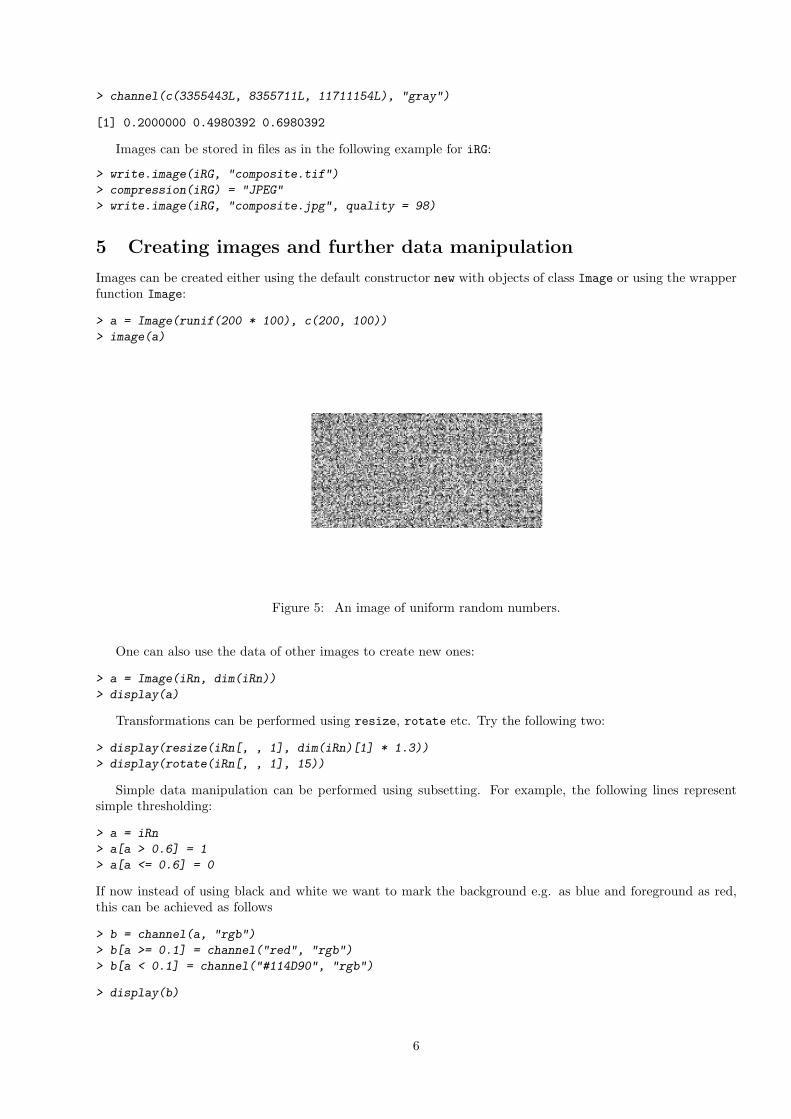
Images can be created either using the default constructor new with objects of class Image or using the wrapperfunction Image:
> a = Image(runif(200 * 100), c(200, 100))
> image(a)
Figure 5: An image of uniform random numbers.
One can also use the data of other images to create new ones:
> a = Image(iRn, dim(iRn))
> display(a)
Transformations can be performed using resize, rotate etc. Try the following two:
> display(resize(iRn[, , 1], dim(iRn)[1] * 1.3))
> display(rotate(iRn[, , 1], 15))
Simple data manipulation can be performed using subsetting. For example, the following lines representsimple thresholding:
> a = iRn
> a[a > 0.6] = 1
> a[a <= 0.6] = 0
If now instead of using black and white we want to mark the background e.g. as blue and foreground as red,this can be achieved as follows
> b = channel(a, "rgb")
> b[a >= 0.1] = channel("red", "rgb")
> b[a < 0.1] = channel("#114D90", "rgb")
> display(b)
6

Figure 6: Marked thresholded images.
6 Image segmentation and image analysis
The purpose of segmentation is to identify the objects of interest in an image. The quality of the segmentationwill generally define the quality of the subsequent feature extraction. We need something better than 6.
We will make use of the fact that we have two images corresponding to the same location of the microscope– one of the nuclei staining and of the cytoplasm protein. Because of the nature of these images it is reasonableto start with segmenting nuclei because they are more clearly separated.
The function thresh provides an implementation of adaptive thresholding that takes into account inequalitiesin background intensity across the image. For the nuclei (the green channel, G), the segmented image can beobtained as follows:
> mask = thresh(iGn, 15, 15, 0.002)
The parameters w, h and offset of the function correspond to the size of objects we expect to find in theimage: objects of different size would require adjustment of these parameters. You can try the results fromusing different parameters.
Some further smoothing of the mask is necessary. A useful set of instruments for this is provided bymathematical morphology, and they are implemented by the morphological operators dilate, erode, openingand closing.
> mk3 = morphKern(3)
> mk5 = morphKern(5)
> mask = dilate(erode(closing(mask, mk5), mk3), mk5)
Here, several operators were used sequentially. You can observe the results of each of these operatorsseparately by looking at the intermediate images. You can also try different kernels, i. e. different parametersfor the function morphKern.
As the next step, one needs to index regions in the segmented image that correspond to the differentobjects. A classic algorithm for segmentation is the computation of the distance map followed by the watershedtransform.
> sG = watershed(distmap(mask), 1.5, 1)
The result is shown in Figure 9.Finally, when we are happy with the results of the watershed transform, we can remove nuclei that are either
too small or too dark or fall on the edge of the images etc.:
7

Figure 7: Preliminary nuclei segmentation.
Figure 8: Nuclei segmentation after noise removal.
8

Figure 9: Nuclei segmentation by watershed (before artefact removal).
> ft = hull.features(sG)
> mf = moments(sG, iGn)
> for (i in seq_along(ft)) ft[[i]] = cbind(ft[[i]], mf[[i]])
> sG = rmObjects(sG, lapply(ft, function(x) which(x[, 3] < 150 |
+ x[, 3] > 10000 | x[, 18] < 30 | 0.4 * x[, 4] < x[, 10])))
The result is shown in Figure 10.Investigate the structure of ft and mf and explain what kind of objects were removed.What we have finally obtained is an IndexedImage for the nuclei, where each nucleus is given an index
from 1 to max(sG). One can now directly use functions like getFeatures or moments etc. to obtain numericaldescriptors of each nucleus.
In principle, the same algorithm as above (distance map – watershed transform) could be used to segmentthe cells, however we often find that neighbouring cells are touching and lead to segmentation errors. But wecan use the already found nuclei as seed points to detect their cells – assuming that each cell has exactly onenucleus. This method falls short of detecting multinuclear cells, but it improves the quality of detection for allother cells tremendously. We start similarly to the nuclei segmentation, however instead of using watershed,we use the function propagate, supplying it with the indexed image of seed points (nuclei). The functionimplements an elegant algorithm that produces a Voronoi segmentation using a metric that combines Euclideandistance and intensity differences between different pixels in the image [?].
> mask = thresh(blur(iRn, 4, 1.5), 25, 25, 0.005)
> mask = erode(erode(dilate(mask, mk5), mk5), mk3)
> sR = propagate(iRn, sG, mask, 1e-05, 1.6)
The result is shown in Figure 11.Again, some artefacts need to be removed. If we want to keep a 1-to-1 match between the cells and the
nuclei we also need to take into account the situation when for some nuclei no cell was detected (the cell canbe dead, or nucleus was a false positive). This is important for those nuclei at the end of the list (for those inthe middle of the list, the corresponding cell will have all descriptors at 0):
> for (i in 1:dim(sR)[3]) {
+ x = sG[, , i]
+ x[x > max(sR[, , i])] = 0
+ sG[, , i] = x
+ }
9

Figure 10: Nuclei segmentation by watershed (after artefact removal).
Figure 11: Cell segmentation by the propagate function (before artefact removal).
10

Now as we have made that there is a 1-to-1 match, we can remove cells that are too small or too large to beplausible, are on the edge of the image, are too dark, etc. We also remove their corresponding nuclei.
> ft = hull.features(sR)
> mf = moments(sR, iRn)
> for (i in seq_along(ft)) ft[[i]] = cbind(ft[[i]], mf[[i]])
> index = lapply(ft, function(x) which(x[, 3] < 150 | x[, 3] >
+ 15000 | x[, 18]/x[, 3] < 0.1 | 0.3 * x[, 4] < x[, 10]))
> sR = rmObjects(sR, index)
> sG = rmObjects(sG, index)
Figure 12: Cell segmentation by propagate (after artefact removal).
Finally, having the indexed images for cells and nuclei, the full set of descriptors can be extracted using thegetFeatures function:
> sG = getFeatures(sG, iGn)
> sR = getFeatures(sR, iRn)
> nucl = do.call("rbind", features(sG))
> dim(nucl)
[1] 213 96
> cells = do.call("rbind", features(sR))
> dim(cells)
[1] 213 96
The resulting matrices have 213 rows (one for each of cell/nucleus) and 96 columns (one for each object descrip-tor).
You can now try to apply the above workflow to the other genes’ data in the example dataset, or you cantry out the following visualisation.
> rgb = paintObjects(sR, iRG)
> rgb = paintObjects(sG, rgb)
> ct = tile(combine(stackObjects(sR, iRn)))
> nt = tile(combine(stackObjects(sG, iGn)))
The result is shown in Figure 13.
11

Figure 13: Results of detection.
12