Embed Size (px)
Citation preview

JOURNAL OF CLINICAL MICROBIOLOGY, OCt. 1991, p. 2273-2279 Vol. 29, No. 100095-1137/91/102273-07$02.00/0Copyright © 1991, American Society for Microbiology
Analysis of Human Rotavirus Strains Prevailing in Bangladeshin Relation to Nationwide Floods Brought
by the 1988 MonsoonMUZAHED UDDIN AHMED,1t SHOZO URASAWA,1* KOKI TANIGUCHI,' TOMOKO URASAWA,1NOBUMICHI KOBAYASHI,1 FUMIYO WAKASUGI,1 A. I. M. M. ISLAM,2 AND HAMID A. SAHIKH3
Department of Hygiene and Epidemiology, Sapporo Medical College, Sapporo 060, Japan,1 and Departments ofCommunity Medicine2 and Pediatrics,3 Mymensingh Medical College, Mymensingh 2202, Bangladesh
Received 5 November 1990/Accepted 18 July 1991
The virologic character of human rotavirus strains prevailing in Bangladesh was investigated in relation tothe devastating nationwide floods brought by the 1988 monsoon. Human rotaviruses contained in stoolspecimens that were collected from inpatients with infantile and adult diarrhea in two hospitals in Mymensinghover a 13-month period (January 1988 to January 1989) and in one hospital in Dhaka over a 3-month period(February to April 1988) were examined for their subgroup, VP7 serotype, and RNA electropherotype. Inconcurrence with the spread of the flood (from the middle of August 1988), the number of infantile and adultdiarrhea patients increased greatly. At the same time, the proportion of rotavirus-positive specimens in alldiarrhea cases also increased remarkably, reaching 54 and 45% in September and October, respectively. Anelectrophoretic analysis of viral RNA revealed 17 distinct patterns of viral RNA (14 long and 3 short electro-pherotypes) and a considerable number of mixed electropherotypes, suggesting the simultaneous infection ofsome patients with more than two rotavirus strains. It was noteworthy that electropherotypes of rotavirusstrains prevailing in the community changed considerably after the spreading of the flood and that thefrequency of virus specimens showing mixed electropherotypes increased significantly during the flood period.These results suggest that sudden environmental change caused by the devastating floods seriously affected theepidemiology of rotavirus infections by increasing the opportunity of transmission of the virus and by reducingthe resistance of the host to infection. In both pediatric and adult patient groups, serotypes 1 and 2 were themost frequent ones detected, followed by serotype 4. Serotype 3 was detected in a single specimen of the 99specimens whose serotypes were determined.
Group A rotavirus is known to be the most common causeof severe diarrhea that occurs every winter among infantsand young children in temperate regions (19). Group Arotavirus is characterized by three major antigens, the sub-group antigen, the VP7 serotype antigen, and the VP4serotype antigen, and its genome consists of 11 double-stranded RNA segments (19). At least two distinct subgroupantigens (subgroups I and II) and seven different VP7serotype antigens (serotypes 1, 2, 3, 4, 8, 9, and 12) have sofar been distinguished in human rotavirus (2, 7, 15, 23, 31,36). Human rotaviruses with subgroup I specificity possessserotype 2, 8, or 12 antigen, while viruses with subgroup IIspecificity have serotype 1, 3, 4, or 9 antigen (2, 7, 15, 23,36). Although the epidemiologic importance of the fourmajor serotypes (1 through 4) has been indicated by theirworldwide distribution, the importance of strains with thenewly described serotypes 8, 9, and 12 remains unknown.Recently, the presence of VP4 serotypes in human rotavirushas been clearly shown by Gorziglia et al. (12) by usingantisera prepared against the VP4 proteins that had beenexpressed by insertion of the VP4 gene into a recombinantbaculovirus. However, since the immunogenicity of the VP4serotype-specific antigen in natural infection or in immuni-zation with human rotavirus is usually low, the serotype
* Corresponding author.t Present address: Department of Medicine, Faculty of Veteri-
nary Science, Bangladesh Agricultural University, Mymensingh2202, Bangladesh.
specificity of human rotavirus is considered to be mostlydetermined by the antigenic specificity of VP7.The understanding of the epidemiologic features of indi-
vidual rotavirus serotypes prevailing worldwide, especiallyin developing countries, is most necessary before the intro-duction of a human rotavirus vaccine, which is now beingdeveloped. For this purpose, a simple method for rotavirusserotyping that does not require virus neutralization in cellculture has been badly needed. Recently, we developed anenzyme-linked immunosorbent assay (ELISA) using sero-type 1-, 2-, 3-, and 4-specific neutralizing monoclonal anti-bodies for directly serotyping rotavirus in stool specimens(32, 34). This method and a similar method developed byother investigators have been employed successfully toserotype rotavirus strains in clinical specimens in a numberof epidemiologic studies (1, 4, 13, 22, 35).
Examination of electrophoretic patterns (electrophero-types) of segmented viral RNA by polyacrylamide gel elec-trophoresis (PAGE) is another indispensable tool in theepidemiology of rotavirus infections (8). Molecular epidemi-ologic studies of rotavirus based on the identification ofRNA electropherotypes of virus strains circulating in acommunity have also been reported by many investigators(3, 6, 9, 25, 26, 28). Our preliminary report (1) described thesubgroup and serotype antigen determination of rotavirusesin stool specimens collected from diarrheic patients in Bang-ladesh between January and June 1988. Collection of speci-mens was continued until January 1989. In this study, thesubgroups, serotypes, and electropherotypes of rotavirusstrains prevailing in Bangladesh were examined in speci-
2273
on June 1, 2018 by guesthttp://jcm
.asm.org/
Dow
nloaded from
2274 AHMED ET AL.
mens obtained from children and adults over a 13-monthperiod. Special attention was given in this study to the effectof the devastating nationwide flood in 1988 on the electro-pherotypes of genomic RNA of prevalent rotavirus strains inBangladesh.
MATERIALS AND METHODS
Virus specimens. Stool specimens were obtained in threehospitals in Bangladesh (1). Specimens of pediatric and adultpatients with diarrhea were collected from the MymensinghMedical College (MMC) and SK hospitals, respectively, inMymensingh between January 1988 and January 1989. Spec-imens from pediatric patients were also obtained from theDhaka Medical College (DMC) hospital in Dhaka betweenJanuary and June 1988. Stool suspensions of about 10% wereprepared in phosphate-buffered saline, clarified, and trans-ported to the Department of Hygiene and Epidemiology,Sapporo Medical College, Sapporo, Japan. All specimenswere screened for the presence of group A rotavirus by anELISA with the monoclonal antibodies YO-156 (reactingwith the group A common antigenic epitope in the innercapsid protein VP6) and YO-2C2 (reacting with the cross-reactive neutralization epitope in the outer capsid proteinVP4) (35). Specimens that reacted with either of the twoantibodies were designated as containing group A rotavirus.Subgrouping and serotyping of human rotavirus. As previ-
ously described (32, 35), subgrouping and serotyping werecarried out by using an ELISA with subgroup I- and II-specific monoclonal antibodies (S2-37 and YO-5, respec-tively) directed at VP6 and serotype 1-, 2-, 3-, and 4-specificmonoclonal antibodies (KU-4, S2-2G10, YO-1E2, and ST-2G7, respectively) directed at the major outer capsid glyco-protein VP7. The color reaction of peroxidase with itssubstrate (o-phenylenediamine dihydrochloride) was mea-sured as A492. The criteria for determining serotype were asfollows: a virus was assigned to a specific serotype when theoptical density value for the reaction with the monoclonalantibody corresponding to that serotype exceeded 0.2 perwell and in addition, when the optical density value for thereaction corresponding to that serotype was greater thantwice the value corresponding to any other serotype (35).
Polyacrylamide gel electrophoresis. Extraction of viralRNA from stool specimens was described previously (21).Electropherotyping of viral RNA was carried out in 10%polyacrylamide slab gels, and silver staining was performedas previously described (21).
Virus isolation and serotype determination of isolates. Virusisolation from selected stool specimens was carried out byusing primary green monkey kidney cell cultures followingthe procedure described previously (37). Virus was passagedat least four times in cell culture for isolation. The serotypesof the isolates were determined by a fluorescence focusreduction neutralization test with hyperimmune serum (30)as well as by the ELISA mentioned above.
Meteorological data. The weather records for Mymensinghfor the year 1988 were obtained from the Department ofIrrigation and Water Management, Bangladesh AgriculturalUniversity, Mymensingh, Bangladesh.
RESULTS
Epidemiological setting relevant to the collection of virusspecimens used in this study. Monthly temperature andrainfall in Mymensingh and the number of pediatric diarrheapatients admitted to the MMC hospital and of adult diarrhea
I 302826
° 24i 22E 20F 18zI. 16
> 500
t 400
a. 3000
200E
z 100l
130 \ 40120 30 S11 20 '.3100 10 .
IL a
'0 70
20 6062 50Z 40
302010
MST MST MST MST MST MST MST MST MST MST MST MST MST
Jan. Feb. Mar. Apr. May Jun. Jul. Aug. Sep. Oct. Nov. Dec. Jan.1988 1989
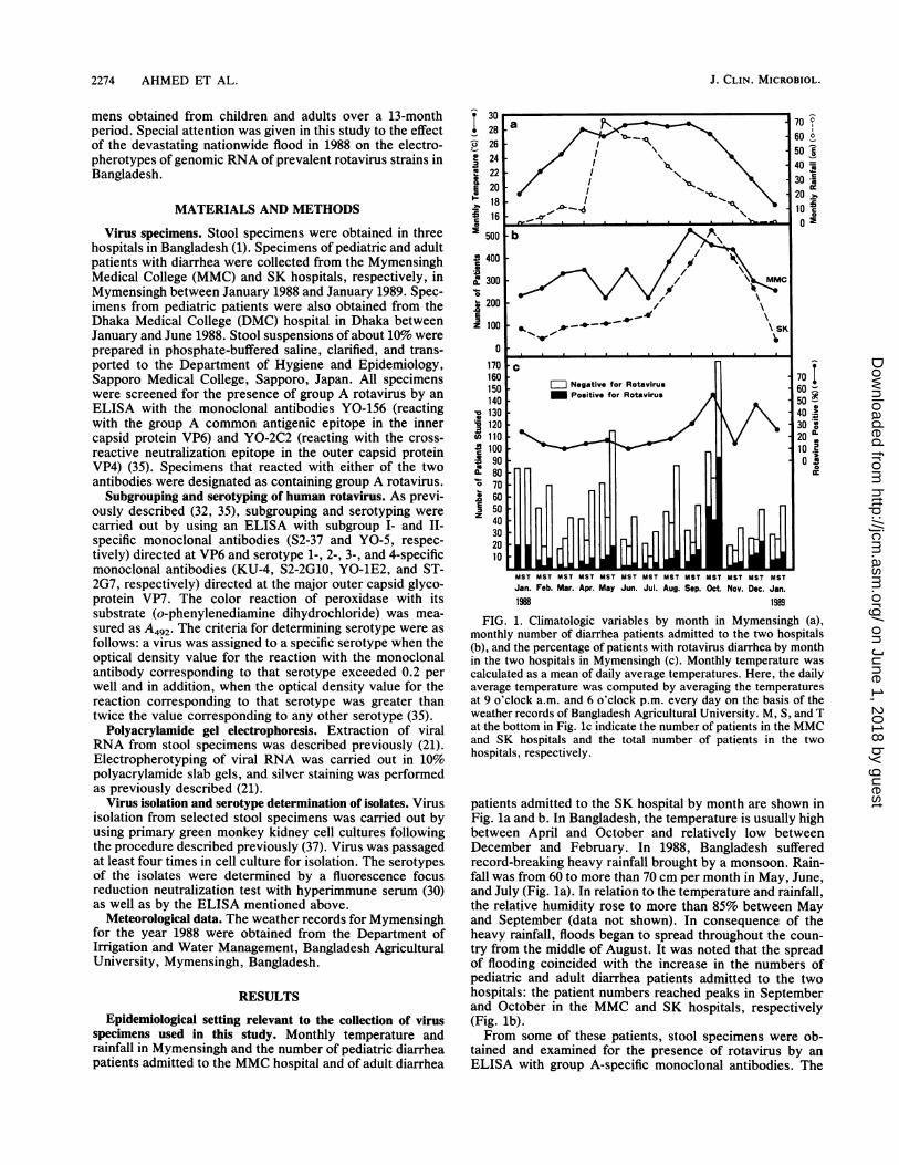
FIG. 1. Climatologic variables by month in Mymensingh (a),monthly number of diarrhea patients admitted to the two hospitals(b), and the percentage of patients with rotavirus diarrhea by monthin the two hospitals in Mymensingh (c). Monthly temperature wascalculated as a mean of daily average temperatures. Here, the dailyaverage temperature was computed by averaging the temperaturesat 9 o'clock a.m. and 6 o'clock p.m. every day on the basis of theweather records of Bangladesh Agricultural University. M, S, and Tat the bottom in Fig. lc indicate the number of patients in the MMCand SK hospitals and the total number of patients in the twohospitals, respectively.
patients admitted to the SK hospital by month are shown inFig. la and b. In Bangladesh, the temperature is usually highbetween April and October and relatively low betweenDecember and February. In 1988, Bangladesh sufferedrecord-breaking heavy rainfall brought by a monsoon. Rain-fall was from 60 to more than 70 cm per month in May, June,and July (Fig. la). In relation to the temperature and rainfall,the relative humidity rose to more than 85% between Mayand September (data not shown). In consequence of theheavy rainfall, floods began to spread throughout the coun-try from the middle of August. It was noted that the spreadof flooding coincided with the increase in the numbers ofpediatric and adult diarrhea patients admitted to the twohospitals: the patient numbers reached peaks in Septemberand October in the MMC and SK hospitals, respectively(Fig. lb).From some of these patients, stool specimens were ob-
tained and examined for the presence of rotavirus by anELISA with group A-specific monoclonal antibodies. The
J. CLIN. MICROBIOL.
on June 1, 2018 by guesthttp://jcm
.asm.org/
Dow
nloaded from

HUMAN ROTAVIRUS INFECTIONS AND FLOOD 2275
TABLE 1. Age distribution of patients with rotavirus-positive stool specimens
No. of specimens in each age groupHospital
0-5 mo 6-11 mo 1 yr 2 yr 3-5 yr 6-12 yr 13-19 yr 29-29 yr 30-39 yr 40-49 yr 50+ yr
DMC 0 10 17 9 6MMC 3 19 23 12 23 23
Jan.-July, 1988 2 6 9 3 2 1Aug. 1988-Jan. 1989 1 13 14 9 21 22
SK 15 35 31 9 6
number of rotavirus-positive and -negative specimens ob-tained in the MMC and SK hospitals and the percentage ofrotavirus-positive specimens of the combined specimensfrom the two hospitals are shown by month in Fig. lc.Overall, there seemed to be no appreciable difference inrotavirus-positive rates by month between the MMC and SKhospitals. The proportion of rotavirus-positive specimens inall diarrhea specimens obtained in the two hospitals bymonth was 10 to 20%, except in January, September, Octo-ber, and December 1988 and in January 1989. An increase inthe proportion of rotavirus diarrhea also seemed to corre-
spond to the spread of the flood after August: the rotavirus-positive rates were 30, 54, 45, and 25% in September,October, and December 1988 and January 1989, respec-
tively.Table 1 shows the number of patients who were positive
for rotavirus in their stool specimens in the two hospitals.The ages of patients with rotavirus diarrhea in the DMChospital were mostly under 2 years. In the MMC hospital,the same age preference was observed for patients betweenJanuary and July 1988. However, after August, rotavirusdiarrhea in patients over 2 years of age increased greatly. Inthe SK hospital, in which only adult patients were treated,rotavirus diarrhea was most frequent among those patientswho were in their twenties and thirties during the studyperiod.
Antigenic characterization of rotaviruses. Subgrouping andserotyping of group A rotaviruses in stools collected frompatients with diarrhea were carried out with an ELISA withsubgroup- and serotype-specific monoclonal antibodies. Asshown in Table 2, of a total of 141 rotavirus-positive speci-mens obtained from patients in the MMC hospital, 101(71.6%) could be subgrouped. Of these, 36 (35.6%) belongedto subgroup I, 62 (61.4%) belonged to subgroup II, and 3(3.0%) showed a dual subgroup specificity (I plus II). Thesubgroup specificity of the other 40 specimens were notdetermined. As to the serotype specificity, 51 (36.2%) of the141 virus-positive specimens were serotyped and 90 (63.8%)remained undetermined. Among the 51 specimens whose
serotypes were determined, 20 (39.2%) were assigned toserotype 2, 15 (29.4%) were assigned to serotype 1, 9 (17.6%)were assigned to serotype 4, and 1 (2.0%) were assigned toserotype 3. Six specimens (11.8%) were doubly reactive toeither serotypes 2 and 4 or serotypes 1 and 2.The results of subgrouping and serotyping of the 104
rotavirus-positive specimens obtained from adult patients inthe SK hospital are shown in Table 3. The patterns ofsubgroup and serotype distribution of rotaviruses from adultpatients were somewhat similar to those of viruses from thepediatric patients mentioned above. The subgroup specific-ities of 78 (75%) of 104 rotaviruses were determined. Ofthese, 33 (42.3%) belonged to subgroup I, 41 (52.6%) be-longed to subgroup II, and 4 (5.1%) showed a dual subgroupspecificity (I plus II). The subgroup specificity of the remain-ing 26 was left undetermined. Of the 104 virus-positivespecimens, 48 (46.2%) were assigned to any one serotype orthe combination of two serotypes, while 56 (53.8%) re-mained undetermined. Of the 48 specimens whose serotypeswere determined, serotypes 1 and 2 were the most frequentones (31.3 and 27.1%, respectively), followed by serotype 4(25%). Eight specimens (16.7%) showed dual serotype spec-ificity of serotype 2 plus 4, serotype 1 plus 2, or serotype 1plus 4. No specimens of serotype 3 were detected.
Sixteen stool specimens containing strains that were sub-grouped but not serotyped with the ELISA and that had a
single RNA electropherotype (see RNA electropherotypes)were inoculated onto primary monkey kidney cell cultures.Five strains were successfully isolated. On the basis of theresults of the ELISA and neutralization tests, one each wasdetermined to be subgroup I-serotype 2 and subgroup II-
serotype 3, two were determined to be subgroup II-serotype4, and one (strain B221) was determined to be subgroup1I-serotype 9.The results of the antigenic characterization of rotaviruses
obtained from pediatric patients in the DMC hospital inDhaka were reported elsewhere (1).RNA electropherotypes of rotavirus. Of the 298 stool spec-
imens that were positive for rotavirus from the three hospi-
TABLE 2. Distribution of subgroups and serotypes of human rotavirus from diarrheic children admittedto the MMC hospital, Mymensingh, Bangladesh
No. of No. of specimens assigned to serotype No. of specimensSubgroup .- of undeterminedspecimens 1 2 3 4 2+4a 1+2a Total serotype
I 36 0 20 0 2 2 0 24 12II 62 13 0 1 6 1 0 21 411+11 3 0 0 0 0 1 2 3 0Undetermined 40 2 0 0 1 0 0 3 37Total 141 15 20 1 9 4 2 51 90
a These specimens showed high optical density values (-0.6) to both serotypes and could not be assigned to a single serotype according to the criteria forserotype determination.
VOL. 29, 1991
on June 1, 2018 by guesthttp://jcm
.asm.org/
Dow
nloaded from

2276 AHMED ET AL.
TABLE 3. Distribution of subgroups and serotypes of human rotavirus from adult diarrheic patients admittedto the SK hospital, Mymensingh, Bangladesh
No. of No. of specimens assigned to serotype No. of specimensSubgroup Ns of undeterminedspecimens 1 2 3 4 2+4a 1+2a 1+4a Total serotype
I 33 0 13 0 2 2 1 0 18 15II 41 14 0 0 9 1 0 1 25 161+11 4 0 0 0 0 1 2 0 3 1Undetermined 26 1 0 0 1 0 0 0 2 24Total 104 15 13 0 12 4 3 1 48 56
a These specimens showed high optical density values (.0.6) to both serotypes and could not be assigned to a single serotype according to the criteria forserotype determination.
tals in Mymensingh and Dhaka, 199 specimens whose quan-tities were large enough to allow the extraction of viral RNAwere subjected to RNA electropherotype determination byPAGE. RNA bands were visible in 159 specimens (79.9%),while no bands were detectable in the other 40 specimens(20.1%). Of these 159 specimens, 82 showed clearly stainedelectrophoretic patterns of viral RNA, which enabled us tofurther classify them into distinct electropherotypes (Fig. 2).In 50 specimens, electropherotypes could not be deter-mined, since in these specimens the staining of RNA seg-ments, especially segments 10 and 11, was not clear enoughto permit their assignment to specific electropherotypes. Theremaining 27 specimens showed mixed RNA patterns (mixedRNA electropherotypes), indicating the presence of extraelectrophoretic bands of viral RNA (Table 4).
Usually electrophoretic patterns of human rotavirus RNAare grouped into two major categories: i.e., a long electro-pherotype, in which RNA segment 11 migrates rapidly, anda short electropherotype, in which the same segment mi-grates slowly. In Fig. 2, the 14 distinct migration patterns ofviral RNA belonging to the long electropherotype (A throughN) and three distinct RNA patterns belonging to the shortelectropherotype (X through Z) that were found in this studyare arranged by the order of detection. Differentiation amongsimilar electropherotypes was made by coelectrophoresis ofviral RNAs (data not shown). All of the virus specimens thatexhibited RNA electropherotypes A, C, and J were serotype1, whereas all of those with electropherotypes E, G, H, M,and N were serotype 4. A single specimen with electrophero-type B was serotype 3. As expected, all those having shortelectropherotypes (X, Y, and Z) were exclusively serotype2. In contrast, serotypes of virus specimens having electro-pherotypes D, F, I, K, and L remained undetermined.
Figure 3 shows several examples from virus specimens ofmixed RNA electropherotypes found in this study. Although
1B C D F F (6 I v K L 1 .X, Yt;.;~~~~~~~~~~1'ilNq;'11^11' M:
FIG. 2. RNA electropherotypes of rotaviruses obtained in Bang-
ladesh.
all of these are examples of mixed patterns of long and shortelectropherotypes, they contained 12 to 17 genomic seg-ments of rotavirus, in contrast to the 11 RNA segmentscharacteristic of rotavirus.The monthly occurrence of rotavirus specimens from the
three hospitals having each RNA electropherotype or amixed electropherotype is shown in Fig. 4. In this figure, thestudy period is divided provisionally into two parts: theperiod between January and July 1988 prior to the spread offlooding in Bangladesh (the preflood period) and the periodbetween August 1988 and January 1989 following the spreadof flooding (the flood period). A rotavirus with electrophero-type A was the most common in the present study. It wasdetected not only in the DMC hospital in Dhaka but also inthe MMC and SK hospitals in Mymensingh (located about120 km north of Dhaka). Furthermore, it was detected in theMMC and SK hospitals in Mymensingh during the entireperiod of the present study. Viruses having long electro-pherotypes B through K and short electropherotype X weredetected only in the preflood period. Of these, viruses withelectropherotypes C, E, and X prevailed in both cities.The strain with electropherotype L was first detected in
July before the flood and again in September after the spreadof the flooding. In contrast, viruses with long electrophero-types M and N and short electropherotypes Y and Z consti-tuted the major epidemic strains during the flood period in
TABLE 4. Electropherotyping of rotavirus RNA and theoccurrence of mixed RNA electropherotype by month
No. of specimens with RNANo. of specimens electropherotypeMO examined
Identified Unidentified Mp xed
Jan. 12 4 7 1 (8.3)Feb. 4 4 0 0Mar. 27 19 4 4 (14.8)Apr. 1 1 0 0May 13 12 1 0June 2 2 0 0July 3 2 1 0Aug. 19 3 11 5 (26.3)Sept. 13 7 4 2 (15.4)Oct. 43 20 9 14 (32.6)Nov. 0 0 0 0Dec. 18 6 12 0Jan. 4 2 1 1(25)Total 159 82 50 27
a The percentage of mixed-pattern specimens from January 1988 throughJuly 1988 was (5/62) 8.1%. The percentage for August 1988 through January1989 was (22/97) 22.7%.
J. CLIN. MICROBIOL.
on June 1, 2018 by guesthttp://jcm
.asm.org/
Dow
nloaded from

HUMAN ROTAVIRUS INFECTIONS AND FLOOD 2277
FIG. 3. Various patterns of mixedrotaviruses obtained in Bangladesh.
RNA electropherotypes of
Mymensingh. Thus, the results shown in this figure indicatedcocirculation of diverse human rotavirus strains in Bang-ladesh as well as an abrupt change of epidemic strains afterthe devastating nationwide floods.
It was noteworthy that a number of virus specimensshowed a mixed pattern of viral RNA, as seen in Fig. 4.Table 4 shows the number of specimens having a mixedRNA pattern obtained in each month. As for the specimensobtained before the flooding, 5 (8.1%) of 62 showed a mixedRNA pattern, whereas of the 97 specimens obtained after theflooding, 22 (22.7%) showed a mixed pattern. The differencein the detection rates of mixed electropherotypes betweenthose before and after the flood was statistically significant(P < 0.05 by the x2 test).
DISCUSSION
Two outer capsid proteins, VP4 and VP7, are known to beinvolved in the neutralization of rotavirus. Of these, theglycoprotein VP7 (encoded by the 8th or 9th genomicsegment of rotavirus) usually determines the serotype spec-ificity (the VP7 serotype) of the virus as defined by theneutralization reaction (18, 27), whereas VP4 (encoded bythe 4th RNA segment) appears to be responsible for cross-neutralization among serotypes and to play a usually minorrole in serotype specificity (29). The inner capsid proteinVP6 (encoded by the 6th RNA segment) is known to governthe other major antigen, i.e., subgroup, of rotavirus (14). Inthe present study, therefore, the serotype and subgroupspecificities of rotaviruses contained in stool specimens weredetermined by an ELISA with serotype-specific monoclonalantibodies (directed at the VP7 of each rotavirus serotype)and subgroup-specific monoclonal antibodies (directed at theVP6 of each rotavirus subgroup).The present study revealed that three rotavirus serotypes,
1, 2 and 4, were prevalent in the pediatric and adult popu-lations in Mymensingh, Bangladesh. A serotype 3 strain wasdetected in only a single case of infantile gastroenteritis.However, as suggested by a previous report (24), the possi-bility remains that the serotype 3 monoclonal antibody usedin this study might have missed some strains because of itslimited reactivity. This limited reactivity was shown to be
MxMxMxMx
DMCHG
EECccE AA
MxMxMxMsMxMxMxMx
MMC (Mx t)
A AA^ A LL ()A M tYXY Mx
MxMxMx
MxxMxMx
SK ~~~~~JJ MxlMxx(1)AA K MM N N
I A AA A MM MM M AA
.||| | | . . s ..M
Jan. Feb. Mar. Apr. May Jun. Jul.1- Pro-Flood Period -1988
Aug. Sep. Oct. Nov. Dec. Jan.Flood Period --
1989
FIG. 4. Monthly change of RNA electropherotic patterns ofhuman rotaviruses collected in three hospitals in Bangladesh. Let-ters enclosed within circles indicate short electropherotypes of viralRNA. Mx indicates mixed RNA electropherotypes.
present by the fact that the only serotype 3 strain isolated inthis study was identified by a neutralization test with specifichyperimmune serum but not by the ELISA with the mono-clonal antibody. Thus, in this study, the prevalence ofserotype 3 strains is considered to be underestimated. Asimilar survey carried out in Dhaka in the same year (1)showed that serotype 2 and 4 rotaviruses were also predom-inant among pediatric patients in Dhaka.Many studies have reported the strong association be-
tween subgroup I and serotype 2 antigens and betweensubgroup II and serotype 1, 3, or 4 antigens of humanrotavirus, although rare exceptions have been reported (19,20). The present study substantially confirmed this associa-tion between subgroup and serotype antigens. However, inthis study, several specimens whose antigenic specificityapparently does not conform with this association (Tables 1and 2) are worthy of mention. Fourteen specimens havingdual serotype specificity (six in the MMC hospital and eightin the SK hospital) were examined by PAGE. Of thesespecimens, 12 were found to be mixtures of virus strainshaving long and short RNA electropherotypes. One speci-men showed an electrophoretic pattern too weak to beanalyzed in detail, and one specimen (having subgroup IIand serotype 1+4 antigens) showed a long electropherotype.When four specimens showing subgroup I and serotype 4specificity (two each in the MMC and the SK hospitals) wereanalyzed by PAGE, they all showed mixed electrophero-types. Thus, the unusual antigenic character of selectedvirus specimens was thought to be mostly due to thepresence of two different viruses in stool specimens. Sincemonoclonal antibodies specific to serotypes 8, 9, and 12 werenot available, the serotyping ELISA used in this study couldnot detect these serotypes in stool specimens. However, invirus isolation experiments performed with selected stoolspecimens, one of the five isolates was found to be serotype
VOL. 29, 1991
.om
on June 1, 2018 by guesthttp://jcm
.asm.org/
Dow
nloaded from

2278 AHMED ET AL.
9, indicating the circulation of this newly described serotypein our study population.
In the present study, 146 (59.6%) of 245 specimens exam-ined could not be serotyped. The failure of serotyping inmore than half of the specimens is considered to be due tothe absence of sufficient numbers of complete double-shelledvirus particles in stool specimens (1), since the monoclonalantibodies used in the ELISA could react only with theindividual serotype-specific neutralization epitopes on VP7in the outer capsid layer of double-shelled particles. In thisconnection, the application of VP7 serotyping by using thepolymerase chain reaction with serotype-specific primers isexpected to greatly improve the efficiency of serotyping.The use of serotype-specific monoclonal antibodies in an
ELISA has made it possible to study more exactly theepidemiology of rotavirus serotypes. These studies, includ-ing the one presented here, have indicated the overallpredominance of serotype 1 rotavirus in most of the coun-tries in which investigations have been made (4, 10, 22, 35).At the same time, these studies suggest that differences existin the prevalence of each serotype by locale and that yearlychanges occur in the frequency of individual serotypes in thesame locales (4, 22, 35).
In the present study, at least 14 different strains havinglong RNA electropherotypes and 3 different strains havingshort RNA electropherotypes were found. The strong asso-ciation between the subgroup and serotype specificities ofhuman rotavirus and the RNA electropherotype of the virus,which has been reported by many investigators (15, 17),were confirmed again in this study: strains with long RNApatterns had subgroup II and serotype 1, 3, or 4 antigens,whereas strains with short RNA patterns had subgroup I andserotype 2 antigens.Robert Black et al. (5) described in their longitudinal
studies of diarrheal diseases in Bangladesh children that theincidence of rotavirus diarrhea did not show marked sea-sonal variation, except for a small peak in December. In1988, however, the record-breaking heavy rainfall broughtby a monsoon seems to have seriously affected the occur-rence of acute gastroenteritis. Since August, when theflooding began to spread throughout the country, the numberof patients with either infantile or adult diarrhea increasedgreatly (Fig. lb). It is said that government surveillancereported 1.64 million cases of diarrhea by the middle ofNovember 1988 (16). It was noteworthy that the nationwidefloods increased the percentage of rotavirus diarrhea of thediarrhea cases reported in addition to the actual number ofrotavirus diarrhea cases. In October, more than half of thediarrhea cases seen in both hospitals were rotavirus-positive(Fig. lc).While cocirculation of diverse human rotaviruses having
individual RNA electropherotypes was demonstrated ineach city, several rotavirus strains (those having electro-pherotypes A, C, E, and X) were found in both citiesstudied. Furthermore, epidemic strains before the floodingcontrasted strongly with those after the flooding: strains withlong RNA patterns B through K and short RNA pattern Xwere prevalent exclusively before the flooding, while thosewith long patterns M and N and short patterns Y and Zformed the majority of epidemic strains during the spreadingof flooding, although two strains (those with patterns A andL) prevailed during both preflood and flood periods.
It was quite unexpected that 27 (17%) of 159 virus speci-mens examined showed mixed RNA patterns and further-more that the majority of them (81.5%) were detected duringthe flood period. A mixed viral pattern which indicates the
presence of strains having different electropherotypes in asingle specimen seems to suggest a simultaneous infection ofindividuals with two or more different strains.
Several significant changes were observed with the spreadof flooding, e.g., an increase in diarrhea patients in the twohospitals, an increased percentage of rotavirus diarrheaamong all diarrhea cases, an increase in rotavirus diarrheaamong children of older ages, an abrupt change in theepidemic strains of rotavirus, and an increased percentage ofmixed rotavirus infection cases. All of these changes reflectthe serious influence of the devastating floods on the epi-demiology of diarrheal diseases, especially of rotavirusdiseases. Reportedly, flood waters inundated two-thirds ofBangladesh and one-third of the total population was ren-dered homeless, at least transiently. Exposure of people toseverely contaminated environments, e.g., food and drink-ing water contaminated by numerous pathogens coupledwith the poor nutritional status of people during the floodperiod, seems to have increased the risk of infection withvarious pathogens, including rotavirus, as well as the risk ofmixed infection with two different rotaviruses.Because of the segmented nature of the rotavirus genome,
reassortment of viral genome segments is known to occureasily both in vitro and in vivo through the process ofsimultaneous infection with two different rotaviruses (11,33). Conditions such as those described above, therefore,may have increased the opportunity for genetic reassortmentto occur between different rotavirus strains and facilitatedthe appearance in nature of new reassortant viruses withunique electropherotypes.The main clinical symptoms of patients as revealed by
examination of patient records were diarrhea, vomiting, andfever. The frequency of each symptom seemed not to differappreciably between the pediatric and adult patient groupsstudied. The duration and severity of each clinical symptomalso did not differ significantly between the two age groups.Thus, although it has been reported that adult rotavirusinfections are usually clinically mild (19), the symptomswere equally severe in both children and adults in this study,as far as hospitalized patients were concerned.
Examinations for bacterial pathogens were not carried outin the present study. However, it is most probable thatdiarrhea due to bacteria was also augmented during the floodperiod, considering the increase in overall diarrhea cases andthe increased percentage of mixed infection cases withrotavirus following the spread of flooding. The generalimpression of clinicians that the diarrhea of patients admit-ted to the hospitals during the flood period was oftenclinically severe might also support the notion that some ofthe patients were infected with not only viral but alsobacterial or protozoal agents.
ACKNOWLEDGMENTS
This work was supported by a grant from the Bangladesh Univer-sity Grants Commission, Dhaka, Bangladesh, and by the JapanSociety for the Promotion of Science, Tokyo, Japan, in the form ofa grant and a fellowship, respectively.
REFERENCES1. Ahmed, M. U., K. Taniguchi, N. Kobayashi, T. Urasawa, F.
Wakasugi, M. Islam, H. Shaikh, and S. Urasawa. 1989. Charac-terization by enzyme-linked immunosorbent assay using sub-group- and serotype-specific monoclonal antibodies of humanrotavirus obtained from diarrheic patients in Bangladesh. J.Clin. Microbiol. 27:1678-1681.
2. Albert, M. J., L. E. Unicomb, and R. F. Bishop. 1987. Cultiva-
J. CLIN. MICROBIOL.
on June 1, 2018 by guesthttp://jcm
.asm.org/
Dow
nloaded from

HUMAN ROTAVIRUS INFECTIONS AND FLOOD 2279
tion and characterization of human rotaviruses with "supershort" RNA patterns. J. Clin. Microbiol. 25:183-185.
3. Beards, G. M. 1982. Polymorphism of genomic RNAs withinrotavirus serotypes and subgroups. Arch. Virol. 74:65-70.
4. Birch, C. J., R. L. Heath, and I. D. Gust. 1988. Use ofserotype-specific monoclonal antibodies to study the epidemi-ology of rotavirus infection. J. Med. Virol. 24:45-53.
5. Black, R. E., K. H. Brown, S. Becker, A. R. M. A. Alim, and I.Huq. 1982. Longitudinal studies of infectious diseases andphysical growth of children in rural Bangladesh. II. Incidence ofdiarrhea and association with known pathogens. Am. J. Epide-miol. 115:315-324.
6. Chiba, Y., C. Miyazaki, Y. Makino, L. N. Mutanda, A. Kibue,E. 0. Lichenga, and P. M. Tukei. 1984. Rotavirus infection ofyoung children in two districts of Kenya from 1982 to 1983 asanalyzed by electrophoresis of genomic RNA. J. Clin. Micro-biol. 19:579-582.
7. Clark, H. F., Y. Hoshino, L. M. Bell, J. Groff, G. Hess, P.Bachman, and P. A. Offit. 1987. Rotavirus isolate W161 repre-senting a presumptive new human serotype. J. Clin. Microbiol.25:1757-1762.
8. Estes, M. K., D. Y. Graham, and D. H. Dimitrov. 1984. Themolecular epidemiology of rotavirus gastroenteritis. Prog. Med.Virol. 29:1-22.
9. Follett, E. A. C., R. C. Sanders, G. M. Beards, F. Hundley, andU. Desselberger. 1984. Molecular epidemiology of human rota-viruses: analysis of outbreaks of acute gastroenteritis in Glas-gow and the west of Scotland 1981/82 and 1982/3. J. Hyg.92:209-222.
10. Georges-Courbot, M. C., A. M. Beraud, G. M. Beards, A. D.Campbell, J. P. Gonzalez, A. J. Georges, and T. H. Flewett.1988. Subgroups, serotypes, and electrophoretypes of rotavirusisolated from children in Bangui, Central African Republic. J.Clin. Microbiol. 26:668-671.
11. Gombold, J. L., and R. F. Ramig. 1986. Analysis of reassort-ment of genome segments in mice mixedly infected with rota-viruses SAl and RRV. J. Virol. 57:110-116.
12. Gorziglia, M., G. Larralde, A. Z. Kapikian, and R. M. Chanock.1990. Antigenic relationships among human rotaviruses as de-termined by outer capsid protein VP4. Proc. Natl. Acad. Sci.USA 87:7155-7159.
13. Gouvea, V., M.-S. Ho, R. Glass, P. Woods, B. Forrester, C.Robinson, R. Ashley, M. Riepenhoff-Talty, H. F. Clark, K.Taniguchi, E. Meddix, B. McKellar, and L. Pickering. 1990.Serotypes and electropherotypes of human rotavirus in theUSA: 1987-1989. J. Infect. Dis. 162:362-367.
14. Greenberg, H. B., A. R. Kalica, R. G. Wyatt, R. W. Jones, A. Z.Kapikian, and R. M. Chanock. 1981. Rescue of noncultivablehuman rotaviruses by gene reassortment during mixed infectionwith ts mutants of cultivatable bovine rotavirus. Proc. Natl.Acad. Sci. USA 78:420-424.
15. Hoshino, Y., R. G. Wyatt, H. B. Greenberg, J. Flores, and A. Z.Kapikian. 1984. Serotypic similarity and diversity of rotavirusesof mammalian and avian origin as studied by plaque-reductionneutralization. J. Infect. Dis. 149:694-702.
16. International Centre for Diarrheal Disease Research, Bangladesh.1988. Glimpse (Newsletter) 10:no. 6.
17. Kalica, A. R., H. B. Greenberg, R. T. Espejo, J. Flores, R. G.Wyatt, A. Z. Kapikian, and R. M. Chanock. 1981. Distinctiveribonucleic acid patterns of human rotavirus subgroups 1 and 2.Infect. Immun. 33:958-961.
18. Kalica, A. R., H. B. Greenberg, R. G. Wyatt, J. Flores, M. M.Sereno, A. Z. Kapikian, and R. M. Chanock. 1981. Genes ofhuman (strain Wa) and bovine (strain UK) rotaviruses that codefor neutralization and subgroup antigens. Virology 112:385-390.
19. Kapikian, A. Z., and R. M. Chanock. 1990. Rotaviruses, p.1353-1404. In B. N. Fields, D. M. Knipe, R. M. Chanock, M. S.Hirsch, J. L. Melnick, T. P. Monath, and B. Roizman (ed.),Virology, 2nd ed. Raven Press, New York.
20. Kitaoka, S., T. Nakagomi, N. Fukuhara, Y. Hoshino, H. Suzuki,0. Nakagomi, A. Z. Kapikian, T. Ebina, T. Konno, and N.Ishida. 1987. Serologic characteristics of a human rotavirusisolate, AU-1, which has a "long" RNA pattern and subgroup I
specificity. J. Med. Virol. 23:351-357.21. Kobayashi, N., I. C. Lintag, T. Urasawa, K. Taniguchi, M. C.
Saniel, and S. Urasawa. 1989. Unusual human rotavirus strainshaving subgroup I specificity and "long" RNA electrophero-type. Arch. Virol. 109:11-23.
22. Matson, D. O., M. K. Estes, J. W. Burns, H. B. Greenberg, K.Taniguchi, and S. Urasawa. 1990. Serotype variation of humangroup A rotaviruses in two regions of the USA. J. Infect. Dis.162:605-614.
23. Matsuno, S., A. Hasegawa, A. Mukoyama, and S. Inouye. 1985.A candidate for a new serotype of human rotavirus. J. Virol.54:623-624.
24. Nishikawa, K., Y. Hoshino, K. Taniguchi, K. Y. Green, H. B.Greenberg, A. Z. Kapikian, R. M. Chanock, and M. Gorziglia.1989. Rotavirus VP7 neutralization epitopes of serotype 3strains. Virology 171:503-515.
25. Rodger, S. M., R. F. Bishop, C. Birch, B. McLean, and I. H.Holmes. 1981. Molecular epidemiology of human rotaviruses inMelbourne, Australia, from 1973 to 1979, as determined byelectrophoresis of genome ribonucleic acid. J. Clin. Microbiol.13:272-278.
26. Schnagl, R. D., S. M. Rodger, and I. H. Holmes. 1981. Variationin human rotavirus electropherotypes occurring between rota-virus gastroenteritis epidemics in central Australia. Infect. Im-mun. 33:17-21.
27. Smith, M. L., I. Lazdins, and I. H. Holmes. 1980. Codingassignments of double-stranded RNA segments of SA 11 rota-virus established by in vitro translation. J. Virol. 33:976-982.
28. Suzuki, H., T. Sato, S. Kitaoka, F. Tazawa, T. Konno, Y.Amano, A. A. Alprecht, E. G. Vera, J. L. Villalta, Y. Numazaki,and N. Ishida. 1986. Epidemiology of rotavirus in Guayaquil,Ecuador. Am. J. Trop. Med. Hyg. 35:372-375.
29. Taniguchi, K., Y. Morita, T. Urasawa, and S. Urasawa. 1987.Cross-reactive neutralization epitopes on VP3 of human rotavi-rus: analysis with monoclonal antibodies and antigenic variants.J. Virol. 61:1726-1730.
30. Taniguchi, K., S. Urasawa, and T. Urasawa. 1985. Preparationand characterization of neutralizing monoclonal antibodies withdifferent reactivity patterns to human rotaviruses. J. Gen. Virol.66:1045-1053.
31. Taniguchi, K., T. Urasawa, N. Kobayashi, M. Gorziglia, and S.Urasawa. 1990. Nucleotide sequence of VP4 and VP7 genes ofhuman rotaviruses with subgroup I specificity and long RNApattern: implication for new G serotype specificity. J. Virol.64:5640-5644.
32. Taniguchi, K., T. Urasawa, Y. Morita, H. B. Greenberg, and S.Urasawa. 1987. Direct serotyping of human rotavirus in stoolsby an enzyme-linked immunosorbent assay using serotype 1-,2-, 3-, and 4-specific monoclonal antibodies to VP7. J. Infect.Dis. 155:1159-1166.
33. Urasawa, S., T. Urasawa, and K. Taniguchi. 1986. Geneticreassortment between two human rotaviruses having differentserotype and subgroup specificities. J. Gen. Virol. 67:1551-1559.
34. Urasawa, S., T. Urasawa, K. Taniguchi, Y. Morita, N. Sakurada,Y. Saeki, 0. Morita, and S. Hasegawa. 1988. Validity of an
enzyme-linked immunosorbent assay with serotype-specificmonoclonal antibodies for serotyping human rotavirus in stoolspecimens. Microbiol. Immunol. 32:699-708.
35. Urasawa, S., T. Urasawa, K. Taniguchi, F. Wakasugi, N. Koba-yashi, S. Chiba, N. Sakurada, M. Morita, 0. Morita, M.Tokieda, H. Kawamoto, Y. Minekawa, and M. Ohseto. 1989.Survey of human rotavirus serotypes in different locales inJapan by enzyme-linked immunosorbent assay with monoclonalantibodies. J. Infect. Dis. 160:44-51.
36. Urasawa, S., T. Urasawa, F. Wakasugi, N. Kobayashi, K.Taniguchi, I. C. Lintag, M. C. Saniel, and H. Goto. 1990.Presumptive seventh serotype of human rotavirus. Arch. Virol.113:279-282.
37. Urasawa, T., S. Urasawa, and K. Taniguchi. 1981. Sequentialpassages of human rotavirus in MA-104 cells. Microbiol. Immu-nol. 25:1025-1035.
VOL. 29, 1991
on June 1, 2018 by guesthttp://jcm
.asm.org/
Dow
nloaded from