Embed Size (px)
Citation preview

1
General Design of Nervous System:
Processing = Integrative System
• 99% of sensory information
discarded
• Synapses determine pathway
of signals
Memory:
Highly facilitated synaptic pathways (sensory input not required to excite pathway)
• Information stored for future use
Skeletal muscle
Smooth muscle
Glandular secretion
Effectors:
Tactile
Visual
Auditor
Olfactory
Receptors:
Output = Motor System
Analogy = Computer
Input = Sensory System
Guyton & Hall – Figure 45.2 Fundamentals of Nervous System

2
Nervous system
Central nervous system (CNS)
Peripheral nervous system (PNS)
Brain Spinal cord Sensory division (afferent)
Motor division (efferent)
Somatic nervous system (voluntary; skeletal muscle)
Autonomic nervous system (involuntary; smooth & cardiac muscle)
Organization of Nervous System:
Integration
Sensory
input
Motor
output
Sympathetic division Parasympathetic division
Fundamentals of Nervous System
• anchor neurons to capillaries
• repair damaged neural tissue
• maintain “blood / brain barrier”
Astrocytes:
• macrophages; engulf invaders
Microglia:
• Insulate neurons (myelin sheath)
Oligodendrocytes:
• line canals / ventricles of brain
• produce cerebrospinal fluid (CSF)
Ependymal cells:
A. Neuroglia (supporting cells – “nerve glue”)
Schwann cells:
• Insulate neurons (myelin sheath)
Histology of Nervous System:
Fundamentals of Nervous System
Central Nervous System (CNS)
(most common) ciliated
Peripheral Nervous System (PNS)
Satellite cells:
• Function similar to astrocytes

3
• Specialized “excitable” cells
• Allow for communication throughout body (via electrical impulses)
1) Dendrites: Receive information (environment / other neurons)
Dendrites
2) Cell body (soma): Integrates information / initiate response
Cell body
3) Axon: Conducts action potential (AP – electrical impulse)
Axon
4) Synaptic terminals: Transmit signal (other neurons / effector organs)
Synaptic
terminals
Axon hillock (AP generation)
Centrioles
(Can not divide)
Neuron Anatomy:
Fundamentals of Nervous System
B. Neurons
• Long-lived (~ 100 years)
• High metabolic rate
Schwann Cells (PNS)
Histology of Nervous System:
• Carry information from sensory receptors to CNS
1) Sensory (Afferent) neurons:
• Carry information from CNS to effector organs
2) Motor (Efferent) neurons:
3) Association neurons (Interneurons):
• Interconnects neurons in brain / spinal cord
Functional Classification of Neurons:
• Specialized “excitable” cells
• Allow for communication throughout body (via electrical impulses)
Fundamentals of Nervous System
B. Neurons
• Long-lived (~ 100 years)
• High metabolic rate
Histology of Nervous System:

4
Structural Classification of Neurons (# of processes) :
• Sensory neurons (e.g., special sense organs)
• Sensory neurons
(PNS)
• Motor neurons
• Interneurons
Multipolar
( 3 processes)
Axon
Trigger zone
Dendrites
Bipolar
(2 processes)
Trigger zone
Axon
Dendrites
Unipolar
(1 process)
Trigger zone Dendrites
Axon
• Specialized “excitable” cells
• Allow for communication throughout body (via electrical impulses)
Fundamentals of Nervous System
B. Neurons
• Long-lived (~ 100 years)
• High metabolic rate
Histology of Nervous System:
Central Nervous System

5
Nervous system
Central nervous system (CNS)
Peripheral nervous system (PNS)
Brain Spinal cord Sensory division (afferent)
Motor division (efferent)
Somatic nervous system (voluntary; skeletal muscle)
Autonomic nervous system (involuntary; smooth & cardiac muscle)
Organization of Nervous System:
Integration
Sensory
input
Motor
output
Sympathetic division Parasympathetic division
Central Nervous System
• ~ 3.5 lbs (35 billion neurons)
• ♂ brain ~ 10% larger than ♀ brain
No correlation exists between brain
size and intelligence…
Gross Anatomy: Cerebrum (forebrain)
Cerebellum
Diencephalon (midbrain)
Brainstem (hindbrain)
Brain:
Central Nervous System

6
Central Nervous System
Embryonic Development of Brain:
Step 1:
Neural plate forms from surface ectoderm
Neural plate
3 week old embryo
Step 2:
Neural plate invaginates; forms neural groove
Neural groove
Neural fold
Step 3:
Neural fold cells migrate; form neural crest
• Neural folds flank neural groove
Neural crest
• Neural crest gives rise to PNS
Step 4:
Neural groove becomes neural tube; sinks deep
• Neural tube gives rise to CNS
Neural tube
4 week old embryo
Marieb & Hoehn – Figure 12.1
Central Nervous System
Embryonic Development of Brain:
Neural
tube
Anterior
Posterior
Primary brain vesicles
Proencephalon
(forebrain)
Mesencephalon
(midbrain)
Rhombencephalon
(hindbrain)
Secondary brain vesicles
5 week old embryo
Telencephalon
(endbrain)
Diencephalon
(interbrain)
Mesencephalon
(midbrain)
Metencephalon
(afterbrain)
Myelencephalon
(spinalbrain)
Adult brain structures
Cerebrum
Diencephalon
Brain stem (midbrain)
Brain stem (pons)
Brain stem (medulla)
Spinal cord
Cerebellum
Marieb & Hoehn – Figure 12.2
How we will consider
brain anatomy

7
Embryonic Development of Brain:
Central Nervous System
Space restriction greatly affects
brain development
Cerebrum
Diencephalon
Brain stem (midbrain) Brain stem (pons)
Brain stem (medulla)
Cerebellum
5 week old embryo
• Flexures develop to fit rapidly growing
brain into membranous skull
Cervical
flexure
Midbrain
flexure
13 week old embryo
• Cerebrum forced to
grow posterior and
lateral (‘horseshoe’)
26 week old embryo
• Convolutions develop to
increase surface area
of brain
Newborn
Marieb & Hoehn – Figure 12.3
Central Nervous System
Basic Layout of Neurons:
White matter: Regions of myelinated axons in CNS
Gray matter: Regions of unmyelinated axons / cell bodies in CNS
Spinal cord
Gray
matter
White
matter
Cerebrum
Gray
matter
Cortex White
matter
Nucleus
Nuclei:
Groups of cell bodies located in
the central nervous system
(analogous to ganglia in PNS)
• Cortex formed by migration of neurons
• Cerebellum similar to cerebrum in its external cortex
• Basic pattern observed in CNS
Marieb & Hoehn – Figure 12.4

8
A. Ventricles: Hollow chambers enclosed within brain (continuous with each other…)
Ventricles lined with ependymal cells
(circulate CSF)
Lateral ventricle
Third ventricle
Cerebral aqueduct
Fourth ventricle
Central canal
Marieb & Hoehn – Figure 12.5 Central Nervous System
Brain Anatomy:
Cerebrospinal fluid (CSF)
• Provide constant, controlled environment
for brain cells
• Protect brain from toxins
• Prevent escape of local neurotransmitters
Choroid plexus:
Vascular network; produces CSF
• Similar ion composition
to blood plasma
• protein content
• 0.5 L / day produced
• Gasses cross freely
Costanzo – Figure 3.36
1) CSF produced by choroid plexus in
ventricles
CSF Circulation:
Central Nervous System
2) CSF flows through ventricles and into
subarachnoid space via lateral and
median apertures
Median aperture
Lateral aperture
3) CSF exits subarachnoid space via
arachnoid villi
Arachnoid villus
Hydrocephalus (‘water on the brain”)
Presence of CSF in subarachnoid space
gives buoyancy to brain (97% weight reduction)
Marieb & Hoehn – Figure 12.26
Lumbar puncture (spinal tap)

9
Cerebrum (forebrain)
Cerebellum
Diencephalon (midbrain)
Brainstem (hindbrain)
Central Nervous System
Occipital
lobe
Parietal
lobe Frontal
lobe
Gyrus (ridge)
Central
sulcus
Temporal
lobe
Parieto-occipital
sulcus
Fissure (deep groove)
Sulcus (groove)
Lateral
sulcus
~ 85% of brain mass
Insula
Marieb & Hoehn – Figure 12.6
Central Nervous System
B. Cerebrum (cerebral hemispheres):
Brain Anatomy:

10
Corpus callosum:
White tract connecting
cerebral hemispheres
1) Cerebral cortex (gray matter)
Cerebral cortex
2) Cerebral white matter
Cerebral
white matter
3) Basal nuclei
Basic regions: (superficial to deep)
Basal nuclei
B. Cerebrum (cerebral hemispheres):
Brain Anatomy:
Central Nervous System
1) Cerebral cortex:
The cerebral
cortex is the
seat of conscious
behavior
Only 2 - 4 mm thick
but comprises 40%
of the brain’s mass
2.5 ft2 of surface area
• Contains 3 types of functional areas:
1) Motor areas (send output)
2) Sensory areas (receive input)
3) Association areas (interpret data)
Functional MRI scan (measure blood flow)
Neural cartography (electrostimulation)
Central Nervous System
B. Cerebrum (cerebral hemispheres):
Brain Anatomy:

11
Primary motor
cortex
Conscious control of skeletal
muscle movements
Marieb & Hoehn – Figure 12.8
Pyramidal cells extend long axons
to the spinal cord, forming pyramidal
tracts, or corticospinal tracts
Somatotopic mapping:
The entire body is spatially
represented in the cerebral cortex
1) Cerebral cortex:
Central Nervous System
B. Cerebrum (cerebral hemispheres):
Brain Anatomy:
• Motor areas:
Homunculus (“little man”):
Marieb & Hoehn – Figure 12.9
Central Nervous System
Note:
One-to-one correspondence between
cortical neurons and muscles misleading;
map really “fuzzy”

12
Primary motor
cortex
Conscious control of skeletal
muscle movements
Premotor cortex
Controls learned motor
skills of repetitious
or patterned nature (e.g., typing)
Broca’s area
Controls muscles involved
in speech production
Frontal eye field
Controls voluntary
movement of eyes
Marieb & Hoehn – Figure 12.8
1) Cerebral cortex:
B. Cerebrum (cerebral hemispheres):
Brain Anatomy:
• Motor areas:
communicates
directly with
primary motor cortex
(often more pronounced in
one hemisphere of brain)
Central Nervous System
Marieb & Hoehn – Figure 12.8
Primary somatorsensory
cortex
Receives information from
sensory receptors in skin &
proprioreceptors in joints
Somatosensory
association
cortex
Integrates / interprets
somatosensory inputs (e.g., temp. / pressure)
Olfactory cortex
Receives olfactory
information
Gustatory cortex
Receives / interprets
sensations of taste
Spatial
discrimination
Primary visual
cortex
Receives visual
information
Visual
association
area
Integrates / interprets
visual inputs (e.g., color / form)
Primary auditory
cortex
Receives auditory
information
Auditory
Association area
Integrates / interprets
auditory inputs (e.g., music / thunder)
Medial
1) Cerebral cortex:
B. Cerebrum (cerebral hemispheres):
Brain Anatomy:
• Sensory areas:
Central Nervous System
(retinotopic mapping) (tonotopic mapping)
• Part of rhinencephalon (‘nose brain’)

13
Marieb & Hoehn – Figure 12.8
Locations where sensations, thoughts,
and emotions become conscious (makes us who we are…)
Anterior association
area
(Prefrontal cortex)
• Intelligence
• Complex learning
• Recall
• Personality
Central Nervous System
1) Cerebral cortex:
B. Cerebrum (cerebral hemispheres):
Brain Anatomy:
• Association areas (multimodal):
(matures slowly)
The American Crowbar Case
Introduction
Central Nervous System
Phineus Gage (1823 – 1860)
Franz Gall (1758 – 1828)
Phrenology:
The brain is the organ of the mind;
contains localized, specific modules

14
Marieb & Hoehn – Figure 12.8
Locations where sensations, thoughts,
and emotions become conscious (makes us who we are…)
Anterior association
area Posterior association
area
Limbic association
area
Processes emotions related
to personal / social interactions
• Pattern recognition
• Spatial recognition
• Sensory grouping
• Language centers
(Wernicke’s area)
Central Nervous System
1) Cerebral cortex:
B. Cerebrum (cerebral hemispheres):
Brain Anatomy:
• Association areas (multimodal):
1) Cerebral cortex:
The cerebral
cortex is the
seat of conscious
behavior
Only 2 - 4 mm thick
but comprises 40%
of the brain’s mass
2.5 ft2 of surface area
• Contains 3 types of functional areas
Central Nervous System
B. Cerebrum (cerebral hemispheres):
Brain Anatomy:
• Contralateral control (e.g., left hemisphere controls right body)
Decussation:
Location where neural pathways cross
• Occur at different locations in CNS
• Lateralization (i.e., hemisphere specialization)

15
Categorical Representational
Central Nervous System
1) Cerebral cortex (gray matter)
Cerebral cortex
2) Cerebral white matter
Cerebral
white matter
3) Basal nuclei
Basic regions: (superficial to deep)
Basal nuclei
B. Cerebrum (cerebral hemispheres):
Brain Anatomy:
Central Nervous System

16
B) Association Fibers:
• Interconnect areas of neural cortex
within a single hemisphere
A) Commissural Fibers (form commissures) :
• Interconnect cerebral hemispheres
C) Projection Fibers:
• Interconnect cerebral hemispheres
with other regions of the brain
Marieb & Hoehn – Figure 12.10
Fiber tracts responsible for communication
between cerebral areas and lower CNS
Commissural
fibers Association
fibers
Projection
fibers
2) Cerebral white matter:
Central Nervous System
B. Cerebrum (cerebral hemispheres):
Brain Anatomy:
Corona radiata:
Point where projection fibers radiate fan-like
through cerebral white matter
Internal capsule:
Compact band of projection
fibers near diencephalon
1) Cerebral cortex (gray matter)
Cerebral cortex
2) Cerebral white matter
Cerebral
white matter
3) Basal nuclei
Basic regions: (superficial to deep)
Basal nuclei
B. Cerebrum (cerebral hemispheres):
Brain Anatomy:
Central Nervous System

17
• Composed of gray matter (neuron cell bodies)
3) Basal nuclei:
Central Nervous System
B. Cerebrum (cerebral hemispheres):
Brain Anatomy:
• Function: 1) Subconscious control of skeletal muscle tone
2) Control stereotypical motor movements (e.g., arm swing)
Caudate
nucleus
Putamen
Globus
pallidus
Lentiform
nucleus
Corpus
striatum
Striped appearance
due to passage of
internal capsule fibers
• Regulate intensity / inhibit unnecessary movements
Cerebrum (forebrain)
Cerebellum
Diencephalon (midbrain)
Brainstem (hindbrain)
Central Nervous System

18
Marieb & Hoehn – Figure 12.12 / 12.13
Thalamus:
Thalamus
• Composes 80% of diencephalon
• Relay station for all information
entering / exiting the cerebral cortex
“Gateway to the
cerebral cortex”
Hypothalamus:
• Autonomic control center
• Center for emotional response
• Body temperature regulation
• Regulation of food / water intake
• Regulation of sleep-wake cycles
• Control of endocrine system
“Control center
of body”
Hypothalamus
Epithalamus:
• Houses pineal gland (melatonin)
and choroid plexus (forms CSF)
Epithalamus
Central Nervous System
C. Diencephalon:
Brain Anatomy:
Limbic system (functional brain system):
• Control emotional states (e.g., fear) / behavioral drives (e.g., sex drive)
• Link conscious (cerebral cortex) with unconscious function (brain stem)
• Psychosomatic illnesses = emotion-induced illness
• Long-term memory storage / retrieval
Alzheimer’s Disease:
“Emotional brain”
Progressive degenerative
disease of the brain
• Memory loss
• Disorientation
• Moodiness / confusion
Central Nervous System
Brain Anatomy: Diencephalon structures:
• Thalamus (anterior thalamic nuclei)
• Hypothalamus
Cerebral cortex structures:
• Cingulate gyrus
• Parahippocampal gyrus
• Hippocampus
• Amygdala Fornix:
Fiber tract linking
regions together

19
Cerebrum (forebrain)
Cerebellum
Diencephalon (midbrain)
Brainstem (hindbrain)
Central Nervous System
Thalamus
Marieb & Hoehn – Figure 12.15
Midbrain:
• Conduction pathways between higher
and lower brain centers
• Visual / auditory reflex centers
Pons:
• Regulate respiration rate / depth
Medulla oblongata:
• Location where fiber tracts from
spinal cord cross over (decussation)
• Produce rigidly programmed, autonomic
behaviors necessary for survival
• Deep gray matter; superficial white matter
Midbrain
• Autonomic reflex center
• Heart rate / blood pressure
• Respiratory rhythm
• Vomiting / hiccupping / etc.
Pons
Medulla
oblongata
Central Nervous System
D. Brain stem:
Brain Anatomy:

20
• Maintains cerebral cortical alertness (e.g., on / off switch)
visual
stimuli
auditory
stimuli
general
stimuli
LSD
Twisting of brain stem can
lead to irreversible coma
Marieb & Hoehn – Figure 12.19
• Filters out repetitive stimuli (~ 99% of stimuli filtered…)
Reticular Activating System (RAS - functional brain system):
Central Nervous System
Brain Anatomy: Aggregation of loosely
clustered neurons:
• Raphe nuclei (midline)
Medial group
Raphe nucleus
Lateral group
• Lateral (small cell) group
• Medial (large cell) group
Cerebrum (forebrain)
Cerebellum
Diencephalon (midbrain)
Brainstem (hindbrain)
Central Nervous System

21
• Gray matter superficial; white matter deep
• Precise timing of muscle coordination (balance, posture, repeated movements)
• All activity subconscious Arbor vitae Folia
Cerebral cortex
sends signal to move
Cerebellar Processing:
Sensory information
from body
Commands to motor
neurons of spinal cord
Maintain
body
coordination
Marieb & Hoehn – Figure 12.17
“Small brain”
(11% TBM)
Central Nervous System
E. Cerebellum:
Brain Anatomy:
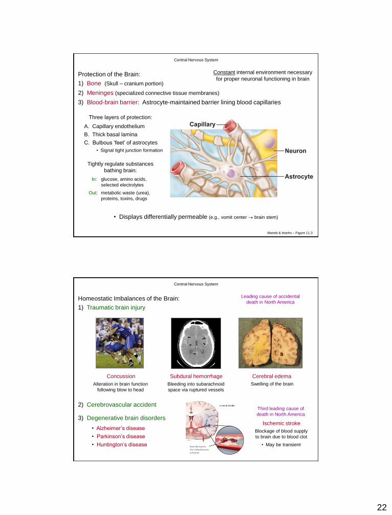
Protection of the Brain:
1) Bone (Skull – cranium portion)
Meningitis:
Inflammation of the meninges
A) Dura mater (“tough mother”)
• Fibrous outer coating (2 layers)
• Protects CNS
B) Arachnoid mater (“spider mother”)
• Delicate middle layer
• Nourishes CNS
C) Pia mater (“gentle mother”)
• Thin inner membrane
• Contains blood vessels
Subarachnoid
space
Bone Dura mater
(periosteal layer)
Arachnoid mater
Pia mater
(filled with CSF)
Marieb & Hoehn – Figure 12.24
2) Meninges (specialized connective tissue membranes)
Central Nervous System
Dura mater (meningeal layer)
Two layers enclose
dural venous sinuses
22
3) Blood-brain barrier: Astrocyte-maintained barrier lining blood capillaries
Marieb & Hoehn – Figure 11.3
In: glucose, amino acids,
selected electrolytes
Tightly regulate substances
bathing brain:
Out: metabolic waste (urea),
proteins, toxins, drugs
• Displays differentially permeable (e.g., vomit center brain stem)
Protection of the Brain:
1) Bone (Skull – cranium portion)
2) Meninges (specialized connective tissue membranes)
Central Nervous System
Constant internal environment necessary
for proper neuronal functioning in brain
Three layers of protection:
A. Capillary endothelium
B. Thick basal lamina
C. Bulbous ‘feet’ of astrocytes
• Signal tight junction formation
Homeostatic Imbalances of the Brain:
1) Traumatic brain injury
Alteration in brain function
following blow to head
Concussion
Leading cause of accidental
death in North America
Bleeding into subarachnoid
space via ruptured vessels
Subdural hemorrhage
Swelling of the brain
Cerebral edema
2) Cerebrovascular accident
Ischemic stroke
Blockage of blood supply
to brain due to blood clot
• May be transient
3) Degenerative brain disorders
Third leading cause of
death in North America
• Alzheimer’s disease
• Parkinson’s disease
• Huntington’s disease
Central Nervous System
23
Nervous system
Central nervous system (CNS)
Peripheral nervous system (PNS)
Brain Spinal cord Sensory division (afferent)
Motor division (efferent)
Somatic nervous system (voluntary; skeletal muscle)
Autonomic nervous system (involuntary; smooth & cardiac muscle)
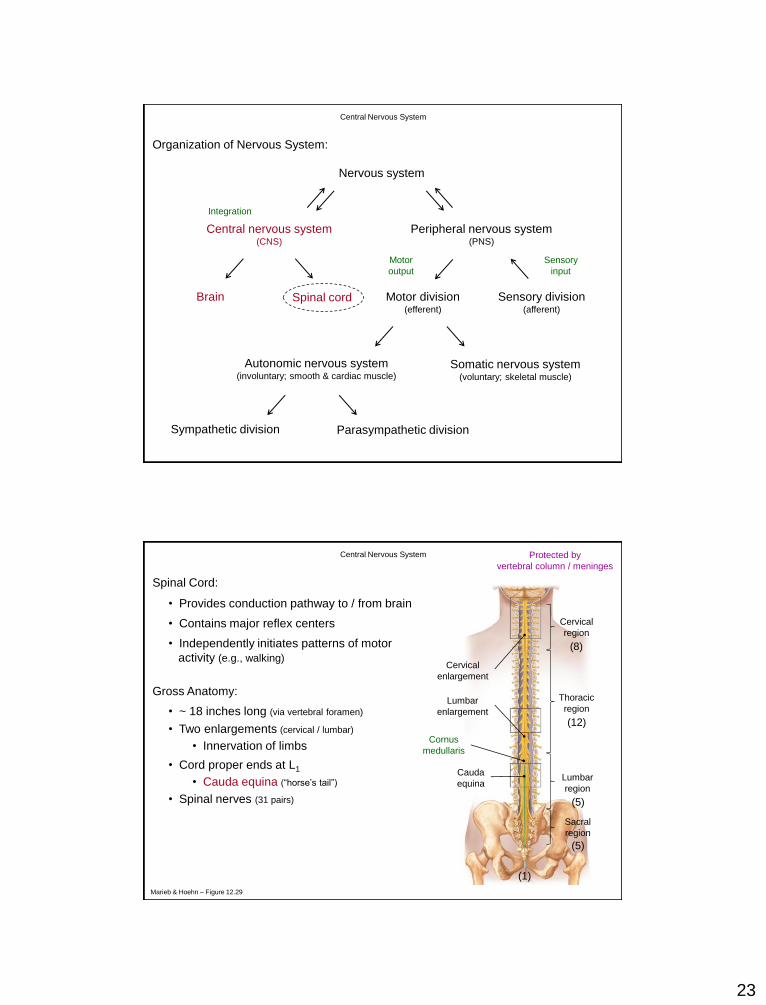
Organization of Nervous System:
Integration
Sensory
input
Motor
output
Sympathetic division Parasympathetic division
Central Nervous System
Spinal Cord:
• Provides conduction pathway to / from brain
• Contains major reflex centers
• Independently initiates patterns of motor
activity (e.g., walking)
Gross Anatomy:
• ~ 18 inches long (via vertebral foramen)
• Spinal nerves (31 pairs)
(8)
(12)
(5)
(5)
(1)
• Two enlargements (cervical / lumbar)
• Cord proper ends at L1
Protected by
vertebral column / meninges
• Cauda equina (“horse’s tail”)
Cervical
region
Thoracic
region
Lumbar
region
Sacral
region
Cervical
enlargement
Lumbar
enlargement
Cauda
equina
(8)
(12)
(5)
(5)
(1)
Marieb & Hoehn – Figure 12.29
• Innervation of limbs
Central Nervous System
Cornus
medullaris

24
Cross-sectional Anatomy:
Central canal
Posterior median sulcus
Anterior median fissure
Posterior
funiculus
Lateral
funiculus
Anterior
funiculus
Ascending tracts:
Carry information to brain
Descending tracts:
Carry information from brain
Transverse tracts:
Carry information across cord
Characteristics:
1) Decussation present
2) Multi-neuron pathways
3) Somatotopy exhibited
4) Symmetrical arrangement
White
matter
Gray
matter
Spinal Cord:
Central Nervous System
Marieb & Hoehn – Figure 12.33
Cross-sectional Anatomy:
Spinal Cord:
Central Nervous System

25
Central canal
Posterior median sulcus
Anterior median fissure
Posterior
horn
(interneurons)
Lateral
horn
(visceral motor neurons)
Anterior
horn
(somatic motor neurons)
Posterior
funiculus
Lateral
funiculus
Anterior
funiculus
White
matter
Gray
matter
Gray
commissure
Cross-sectional Anatomy:
Spinal Cord:
Central Nervous System
Sensory
neuron
Interneuron
Motor
neuron
Dorsal Root
Ventral Root
Dorsal root
ganglion
Spinal
nerve
Cross-sectional Anatomy:
Spinal Cord:
Central Nervous System

26
Organization of Gray Matter:
Spinal Cord:
Central Nervous System
Homeostatic Imbalances of the Spinal Cord:
1) Spinal cord trauma
Damage to spinal cord leading
leading to functional / sensory loss
Paralysis / Paresthesias
Transection of spinal cord
between T1 and L1
Paraplegia Quadriplegia
2) Poliomyelitis 3) Amyotrophic lateral sclerosis (ALS)
Transection of spinal cord
between C4 and C7
Destruction of ventral horn
motor neurons by poliovirus
Lou Gehrig’s
disease
Progressive destruction of ventral
horn motor neurons (autoimmune?)
Central Nervous System


![Lecture Two: The Analogy Theory [‘AT’] · Lecture Two: The Analogy Theory ... 2. [AT] claims: OM-judgments justified by an argument from analogy ... iPaul Bartha, “Analogy and](https://img.dokumen.tips/doc/110x75/5b1ae5387f8b9a28258e143b/lecture-two-the-analogy-theory-at-lecture-two-the-analogy-theory-.jpg)