Embed Size (px)
Citation preview
1
2

Anal cancer is rare in the general population but high and growing in at‐risk populations which are men who have sex with men regardless of HIV status. Women are considered at risk when they are HIV+, have a history of cervical dysplasia, vulvar cancer, or anal condyloma. Incidence rates as stated on the slide.
3

There are morphologic and biological similarities between AIN and CIN. There is a causal relationship link between high risk HPV and the development of anal carcinoma. HPV types 16/18 account for 85% of the cases. Gardasil vaccine has been approved to prevent anal cancer. There are no FDA approved test for anal HPV testing; however, some labs offer this type of testing off‐label. The 2001 Bethesda guidelines includes an appendix for anal cytology.
4
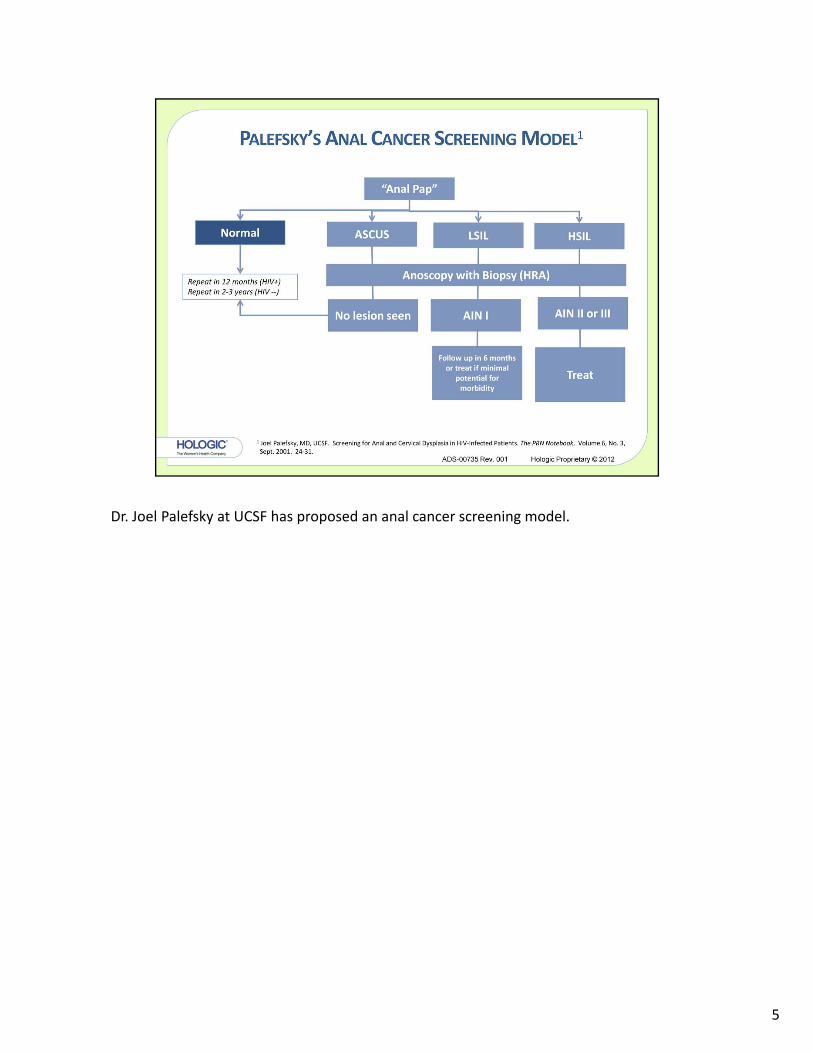
Dr. Joel Palefsky at UCSF has proposed an anal cancer screening model.
5

The anal canal is a 3‐4 cm long tubular structure which is surrounded by smooth muscle. The canal extends from the anal verge to the rectal mucosa and is delineated by the anal‐rectal transformation zone. Samples should be taken from the entire canal to ensure proper sampling.
6

The patient may be positioned either dorsal lithotomy position (The dorsal (or supine) position means to lie on one's back. The lithotomy position is where the patient has his/her feet elevated above the hips and sometimes above the head depending on the procedure, in stirrups. This is the most common position for childbirth and pelvic exams. ) or lateral recumbent. (the posture assumed by the patient lying on the left side with the right thigh and knee drawn up)
Specimens may be collected “blind” without the aid of proctoscope or anoscope. Specimens are often collected under high resolution anoscopy ( this type of collection requires that the collector is certified in the use of this instrument).
7

8

The lab should prepare the specimen on the TP Processor using a Blue filter, using sequence 2 on the TP 2000 or the Non Gyn sequence on the TP 5000.
9

According to Bethesda 2001 for an anal pap to be considered satisfactory it should contain 2,000‐3,000 nucleated squamous cells. For the Thin Prep specimen this would equate to 1 ‐2 nucleated squamous cells per high power field.
10

This field of view would demonstrate an adequate specimen showing numerous nucleated squamous cells along with expected background mucous and anucleated squames.
11
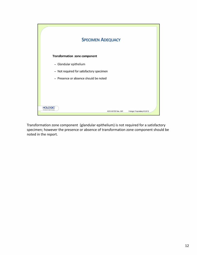
Transformation zone component (glandular epithelium) is not required for a satisfactory specimen; however the presence or absence of transformation zone component should be noted in the report.
12

Glandular epithelium from the transformation zone note the similarity to endocervical cells. The same criteria for normal glandular epithelium would apply.
13

Frequently obscuring material may be observed in anal paps. This obscuring material would include fecal material, bacteria, inflammation, mucus, and blood. All of these materials may hinder microscopic evaluation just as in other cytologic samples.
14
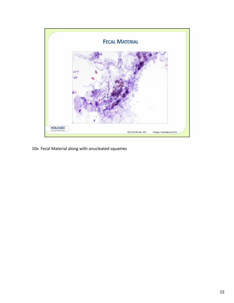
10x Fecal Material along with anucleated squames
15

20x Vegetable or food material along with anucleated squames
16

Mucus present obscuring nucleated squamous cells.
17
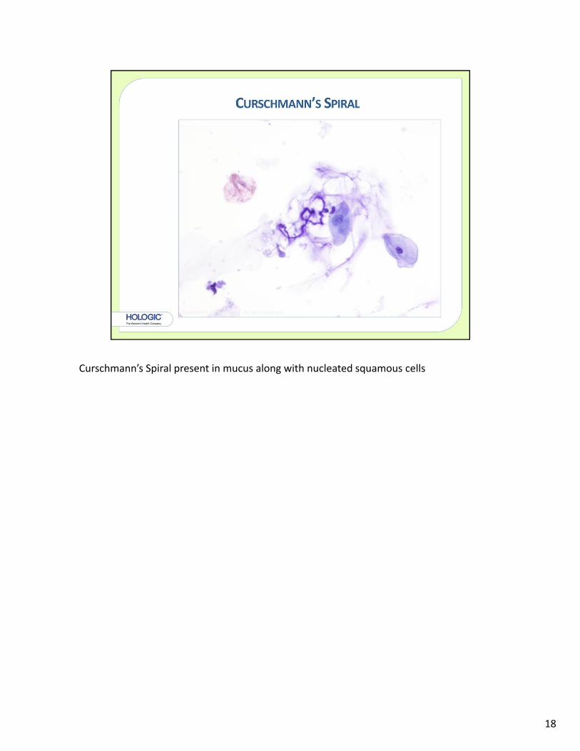
Curschmann’s Spiral present in mucus along with nucleated squamous cells
18

Bacteria present obscuring cellular material
19

20
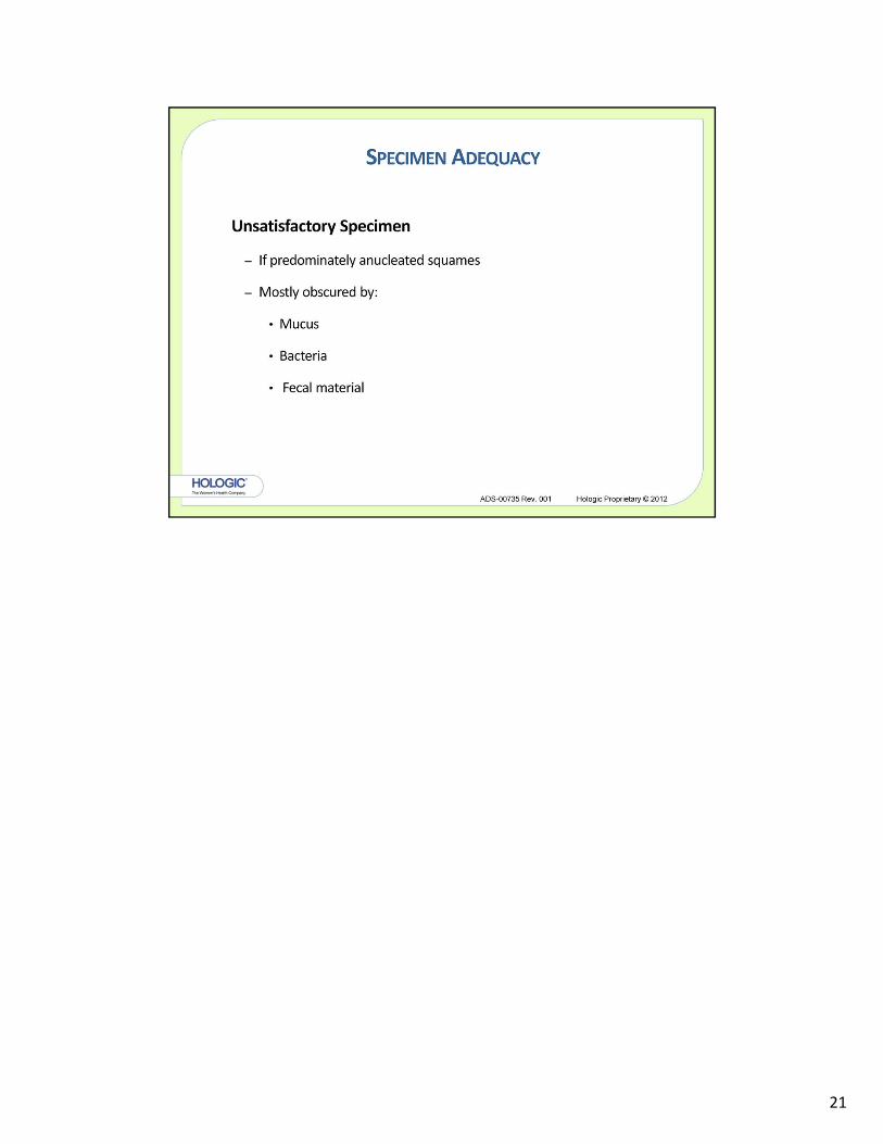
21

22

23

This field of view show anucleated and nucleated squamous cells, mucus, and glandular cells
24
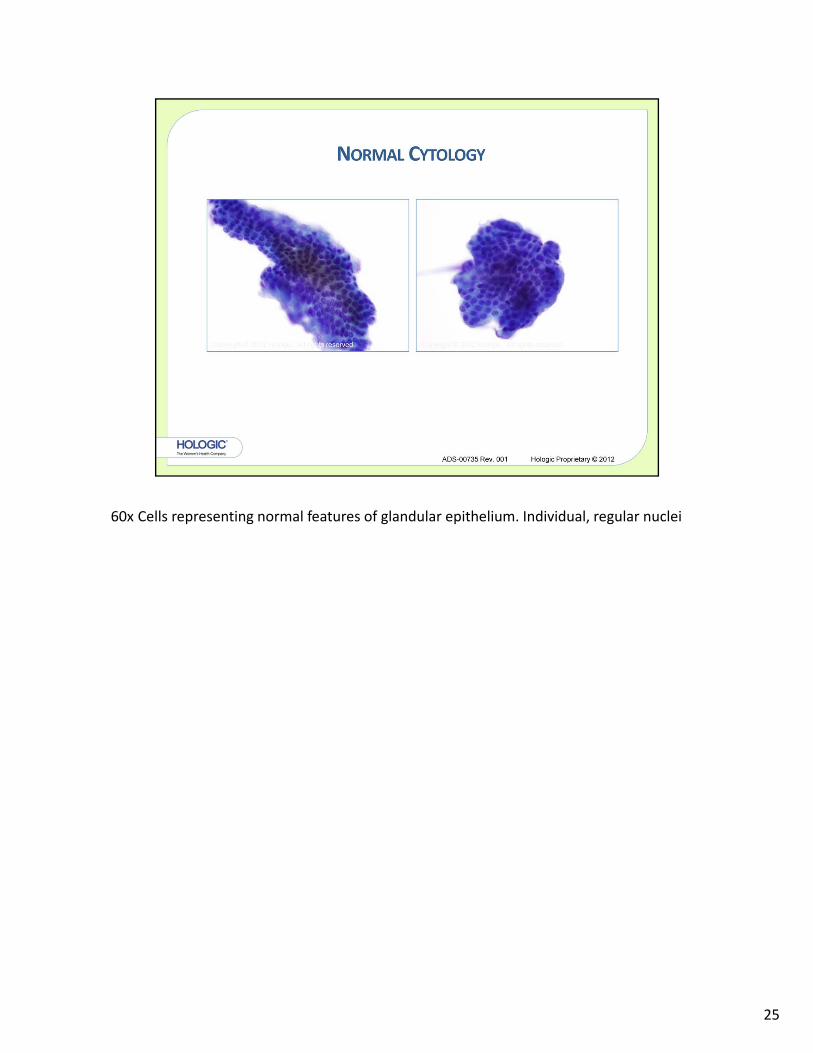
60x Cells representing normal features of glandular epithelium. Individual, regular nuclei
25

40x Glandular epithelium in columnar picketed fence appearance
26

10x Fungal spores present along with normal squamous epithelium
27
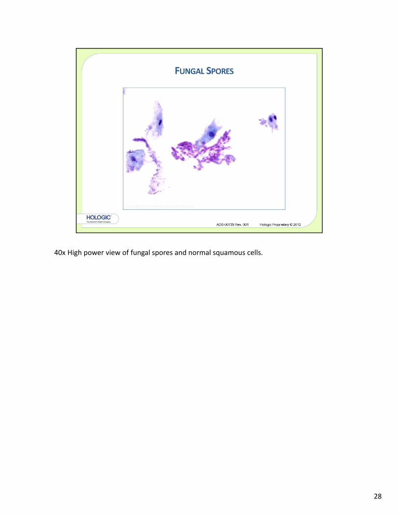
40x High power view of fungal spores and normal squamous cells.
28

40x
29
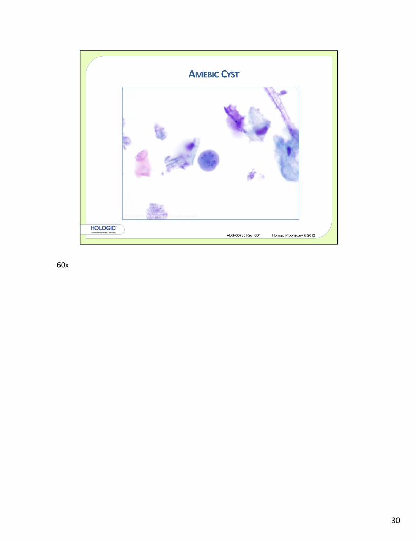
60x
30

40x
31

40x
32

33

40X: Atypical cells of undetermined significance. Atypical mature squamous cells that do not meet the criteria required for an interpretation of low‐grade intraepithelial lesion. (LSIL) Specific criteria similar to cervical cytology:
•Cells are found in sheets or singly
• Nuclei 21/2 – 3 times the size of an intermediate nucleus
• Uniform chromatin distribution
34

40X: Atypical cells of undetermined significance. Atypical mature squamous cells that do not meet the criteria required for an interpretation of low‐grade intraepithelial lesion. (LSIL)
35

36

40X: Criteria for low‐grade intraepithelial lesions (LSIL) are the same as they are for cervical specimens:
•Cells occur singly and in sheets•Cytologic changes are usually confined to cells with “mature” or superficial‐type cytoplasm•Increased Nuclear Detail
•Irregular Nuclear Membrane
•Nuclei 3‐4X Intermediate Nucleus•Sharp, Irregular Cytoplasmic Cavitation (HPV Effect)
•Cytoplasmic Keratinization more prominent than in cervical squamous lesions
37

40X: Criteria for low‐grade intraepithelial lesions (LSIL) are the same as they are for cervical specimens:
•Cytologic changes are usually confined to cells with “mature” or superficial‐type cytoplasm•Cells occur singly and in sheets•Cytologic changes are usually confined to cells with “mature” or superficial‐type cytoplasm•Increased Nuclear Detail
•Irregular Nuclear Membrane
•Nuclei 3‐4X Intermediate Nucleus•Sharp, Irregular Cytoplasmic Cavitation (HPV Effect)
•Cytoplasmic Keratinization more prominent than in cervical squamous lesions
38

40X: Criteria for low‐grade intraepithelial lesions (LSIL) are the same as they are for cervical specimens:
•Cells occur singly and in sheets•Cytologic changes are usually confined to cells with “mature” or superficial‐type cytoplasm•Cells occur singly and in sheets•Cytologic changes are usually confined to cells with “mature” or superficial‐type cytoplasm•Increased Nuclear Detail
•Irregular Nuclear Membrane
•Nuclei 3‐4X Intermediate Nucleus•Sharp, Irregular Cytoplasmic Cavitation (HPV Effect)
•Cytoplasmic Keratinization more prominent than in cervical squamous lesions
39

40

40X: Criteria for high‐grade intraepithelial lesions (HSIL) are the same as they are for cervical specimens:
41

40X: Criteria for high‐grade intraepithelial lesions (HSIL) are the same as they are for cervical specimens:
42

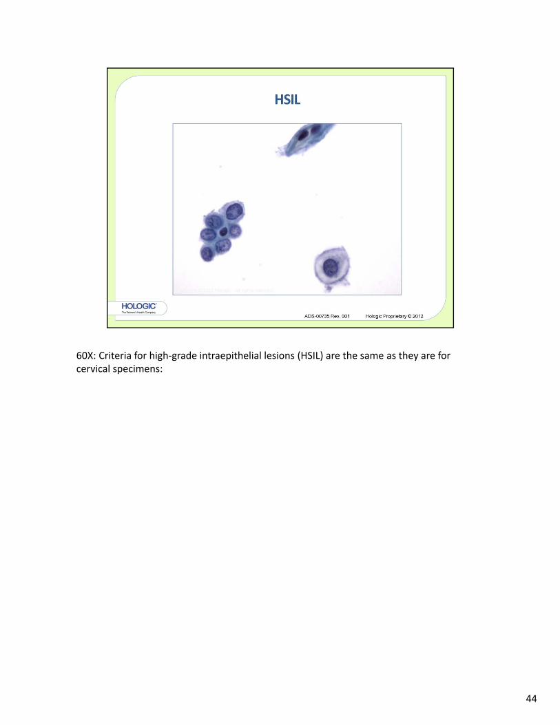
60X: Criteria for high‐grade intraepithelial lesions (HSIL) are the same as they are for cervical specimens:
43
60X: Criteria for high‐grade intraepithelial lesions (HSIL) are the same as they are for cervical specimens:
44
45

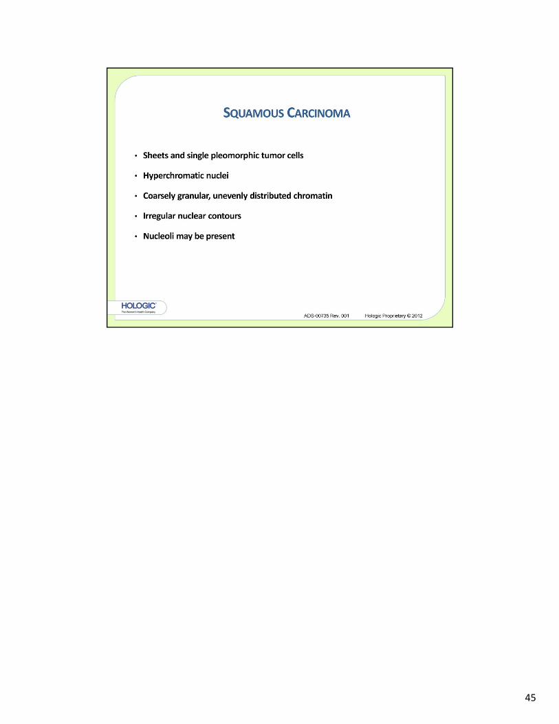
40X: squamous carcinoma is difficult to diagnose with cytology alone due to the lack of tumor diathesis which may be due to the fact that the rectum is a closed system which would allow exfoliated material and cellular debris to be excreted with feces.
46
47

This 40X image shows a group of benign rectal columnar cells. Notice how closely they resemble the normal endocervical component of the Pap Test.
48

The cells in these images taken at 40x are readily identified as those arising in a high grade lesion. They show high nuclear to cytoplasmic ratios, abnormal chromatin patterns and irregular nuclear membranes.
49
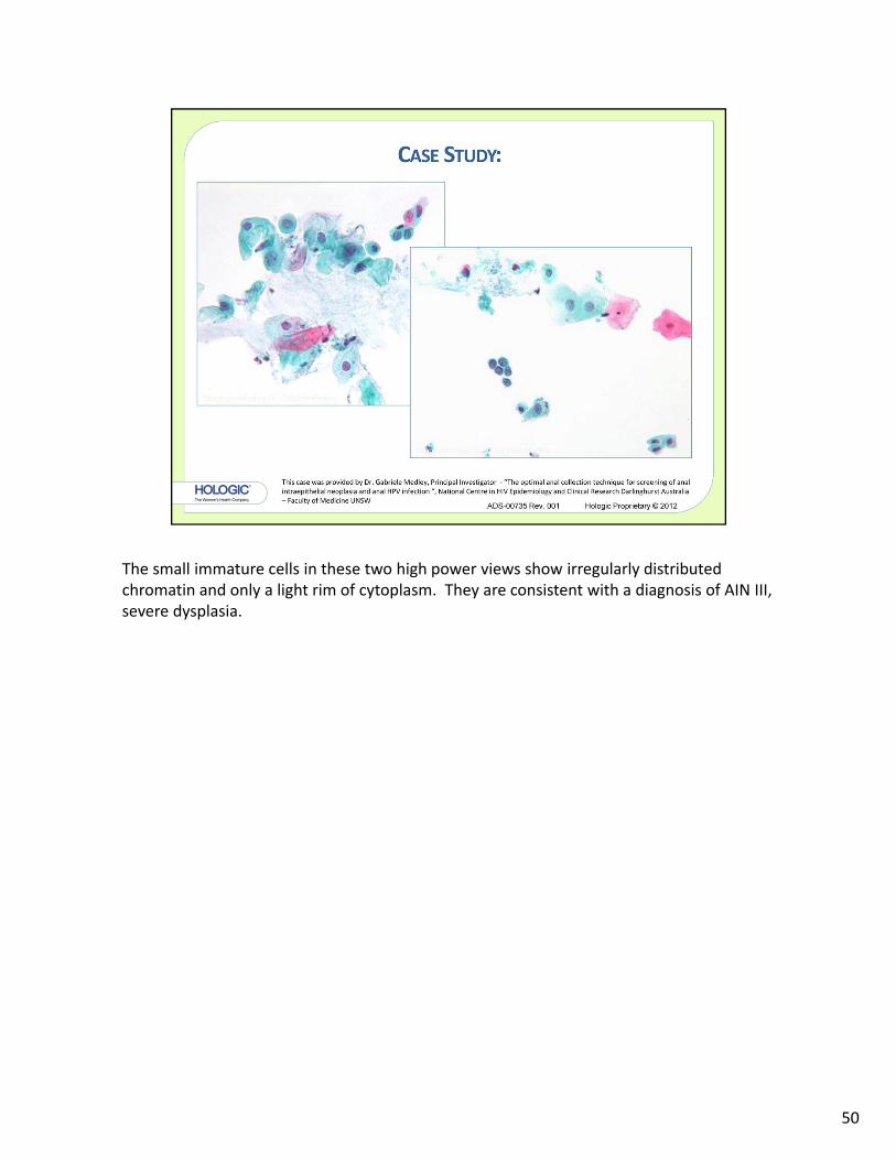
The small immature cells in these two high power views show irregularly distributed chromatin and only a light rim of cytoplasm. They are consistent with a diagnosis of AIN III, severe dysplasia.
50

Histologic section of the lesion at 20x
51

52
53

Morphology I
Slide: 54