Embed Size (px)
Citation preview

JOURNAL OF BACTERIOLOGY, Nov. 1984, p. 764-7700021-9193/84/110764-07$02.00/0Copyright C 1984, American Society for Microbiology
Vol. 160, No. 2
An Acidic or Basic Amino Acid at Position 26 of the b Subunit ofEscherichia coli F1F0-ATPase Impairs Membrane Proton
Permeability: Suppression of the uncF469 Nonsense MutationDAVID A. JANS, LYNDALL HATCH, ANTHONY L. FIMMEL, FRANK GIBSON, AND GRAEME B. COX*
Department of Biochemistry, John Curtin School of Medical Research, Australian National University, Canberra ACT2601 Australia
Received 8 March 1984/Accepted 22 May 1984
The uncF469 allele differed from normal in that a G -( A base change occurred at nucleotide 77 of the uncFgene, resulting in a TAG stop codon rather than the tryptophan codon TGG. Two partial revertant strains wereisolated which retained the uncF469 allele but formped a partially functional b-subunit, d,ue to suppression of theuncF469 nonsense mutation. From the altered isoelectric points of the b-subunits from these' strains, it wasconcluded that the suppressor gene of partial revertant strain AN1956 inserts an acidic amino acid for the TAGcodon, and that the suppressor gene of partial revertant strain AN1958 inserts a basic amino aci4. Themembranes of both partial revertant strains showed impaired permeability to protons on removal of Fl-ATPase. The membranes of both strains, however, were able to carry out oxidative phosphorylation, and theATPase activities of both were resistant to the inhibitor dicyclohexylcarbodiimide.
The subunits of the proton-translocating adenosine tri-phosphatase complex of Escherichia coli are encoded by thegenes of the unc operon, mapping at 84 min on the E. colichromosome (1). As established by genetic complementationstudies (7, 8, 13) and the determination of the complete DNAsequence (11, 31), the unc operon comprises nine genestranscribed in the order uncIBEFHAGDC, with the unclgene promoter proximal. The uncI gene appears not toencode a structural component of the F1F0-ATPase (11). Thea-, c-, and b-subunits of the membrane Fo portion are codedfor by the uncB, uncE, and uncF genes, respectively (8),whereas the b-, a-, Sy-, -, and e-subunits of the soluble Fl-ATPase are encoded by the uncH, uncA, uncG, uncD, anduncC genes, respectively (7, 15).The b-subunit of the F1F0-ATPase has a strongly hydro-
phobic N-terminal region, extending from residues 1 to 33,whereas the remainder of the molecule is highly polar (11). Aphospholipid analogue with a photoreactive group at thephospholipid head group almost exclusively labels aminoacid residues asparagine 2, cysteine 21, and tryptophan 26 ofthe b-subunit (17), suggesting that the hydrophobic N-terminus of the b-subunit traverses the membrane. Thehydrophilic remainder of the b-subunit appears to projectinto the cytoplasm in an a-helical hairpin structure (34, 35),interacting with the subunits of the F1-ATPase. The conclu-sions of Perlin et al. (29) and Hoppe et al. (16) based onprotease treatment of F1-stripped membranes, lend supportto this view.
In the present paper, the nature of the uncF469 mutation(8) is examined, and the properties of two partial revertantstrains are described.
MATERIALS AND METHODSEnzymes and chemicals. Restriction endonucleases and T4
virus DNA ligase were obtained from Amersham AustraliaPty Ltd. All chemicals used were of the highest qualityavailable.
* Corresponding author.
Bacterial strains and plasmids. All of the bacterial strainsused were derived from E. coli K-12 and are described,together with the plasmids used, in Table 1.
Genetic techniques. Techniques used for genetic experi-ments were as outlined previously (7, 13, 14).
Preparation of plasmids. Plasmid DNA was prepared asdescribed by Selker et al. (32). Chromosomal DNA wasprepared by the method described by Saito and Miura (30).DNA sequencing. Nucleotide sequences were determined
by the method of Maxam and Gilbert (22); end-labeling ofDNA was performed with DNA polymerase and [a-32P]deoxyadenosine triphosphate (22).Media and growth of organisms. The mineral-salts minimal
medium used and additions were as described previously(13). Cells for the preparation of membranes were grown in14-liter fermentors essentially as described previously (4).The medium in the fermentors was supplemented with 5%(vol/vol) Luria broth (21). Turbidities of cultures were mea-sured with a Klett-Summerson colorimeter.
Preparation of cell membranes. Preparation and treatmentof membranes were as previously described (5).
Two-dimensional gel electrophoresis. Membranes washed in5 mM TES [N-tris(hydroxymethyl)methyl-2-aminoethane-sulfonic acid] buffer containing p-aminobenzamidine wereexamined by two-dimensional gel electrophoresis as de-scribed previously (33). This technique is essentially that ofO'Farrell (28), involving isoelectric focusing in the firstdimension and sodium dodecyl sulphate-polyacrylamide gra-dient gel electrophoresis in the second dimension.Other methods. ATPase and atebrin fluorescence-quench-
ing activities were assayed as described previously (12).Oxidative phosphorylation and dicyclohexylcarbodiimide(DCCD) sensitivity of ATPase activity were measured asdescribed by Cox et al. (5). Protein concentrations weredetermined with Folin phenol reagent (20).
RESULTSTwo mutations in the uncF gene that result in the nonin-
corporation ipto membranes of the b-subunit of the F1F0-ATPase have been described previously (8). Strains carrying
764
on March 10, 2020 by guest
http://jb.asm.org/
Dow
nloaded from

SUPPRESSION OF THE uncF469 ALLELE 765
TABLE 1. Strains of E. coli and plasmids used
Bacterial strain Relevant genotype' Notes and referencesor plasmid
AN1529 pAN54IuncF469 argH pyrE entA recA Partial diploid strain carrying uncF469 allele on both F-plas-mid and chromosome (13)
uncB402 thr leu hsdR hsdM recA
pAN192IuncB402 thr leu hsdR hsdM recA
uncB402 argH pyrE entA recAuncE429 argH pyrE entA recAuncF469 argH pyrE entA recAuncA401 argH pyrE entA recAuncG428 argH pyrE entA recAuncD409 argH pyrE entA recAuncC424 argH pyrE entA recA
uncH241 argH pyrE entA recA
argH ilvC pyrE entA
uncF476 argH pyrE entA recA
uncF469 argH pyrE entA
argH pyrE entA
AN1956 uncF469 argH pyrE entA sup
AN1958 uncF469 argH pyrE entA sup
AN1456 uncE429 thr leu hsdR hsdM recA
pACYC184 Cmr Tcr
pAN54 pyrE+ uncB+E+F469H+A+G+D+C+ ilvC+ argH+
Cmr Tcs undB+E+F469H+A+
Cmr Tcs uncB+E+F+H+A+
pyrE+ uncB+E+F469H+A+G+D+C+ ilvC+ argH+ sup
pyrE+ uncB+E+F469H+A+G+D+C+ ilvC+ argH+
Cmr Tcs uncB+E+F469H+A+
Cmr Tcs uncB+E+F+H+A+
Cmr Tcs uncB+E+F469H+A+
Prepared by transduction of uncB402 allele into an ilvC de-rivative of C600 (7)
Described in this paper
An isogenic set of strains carrying reference unc alleles(available from Coli Genetic Stock Center) in AN346 back-ground (6, 7, 8, 12)
Obtained by transduction of the uncH241 (18) allele fromstrain RH141 (obtained from R. D. Simoni) into AN346
(2)
uncF476 allele in AN346 background (8)
uncF469 allele in AN346 background (8)
Revertant of uncF469 allele described in this paper
Partial revertant of uncF469 allele described in this paper
Partial revertant of uncF469 allele described in this paper
Prepared by transduction of uncE429 allele into an ilvC de-rivative of C600 (8)
(3)
F-plasmid carrying uncF469 allele isolated as described pre-viously (8)
Described in this paper
(7)
F-plasmid carrying unc operon from partial revertant strainAN1956. Prepared as described previously (13)
F-plasmid carrying unc operon from partial revertant strainAN1958. Prepared as described previously (13)
Multicopy plasmid carrying unc genes derived from partialrevertant strain AN1956. Described in this paper
Multicopy plasmid carrying unc genes derived from revertantstrain AN2033; described in this paper
Multicopy plasmid carrying unc genes derived from partialrevertant strain AN1958; described in this paper
a Chromosomal gene nomenclature is that of Bachmann et al. (1). Plasmid nomenclature is that of Novick et al. (28).
these mutations lack membrane-bound ATPase activity, andmembrane preparations are unable to bind Fl-ATPase. Thenature of one of these mutations, the uncF469 allele, was
determined by sequencing the mutant unc gene.Cloning and sequencing of the uncF469 allele. DNA (both
chromosomal and plasmid) was prepared from the partialdiploid strain AN1529 (uncF4691uncF469) and digested withthe restriction endonuclease HindIII. This DNA was mixed
with HindIll-digested pACYC184 plasmid DNA and treatedwith T4 virus DNA ligase. Strain AN1453 (uncB402, recA)was transformed with the ligated DNA mixture, and selec-tion was made for growth on succinate-chloramphenicolminimal medium. One colony was selected at random andpurified, and this strain (AN2000) was shown to carry a
plasmid (pAN192) with an insert corresponding in size tothat of plasmid pAN51 (uncB+E+F+H+A+) (7).
AN1453
AN2000
AN727AN943AN1440AN730AN1273AN818AN802
AN2015
AN346
AN1521
AN1419
AN2033
pAN192
pAN51
pAN185
pAN187
pAN194
pAN209
pAN211
VOL. 160, i984
on March 10, 2020 by guest
http://jb.asm.org/
Dow
nloaded from

766 JANS ET AL.
Plasmid pAN192 was used to transform a series of strains,each carrying one of the unc alleles uncB402, uncE429,uncF469, uncF476, uncH241, or uncA401. Selection was
made on rich medium containing chloramphenicol, andchloramphenicol-resistant transformants were tested for theability to grow on succinate. Complementation, as judged bygrowth on succinate minimal medium, was obtained betweenplasmid pAN192 and the uncB402, uncE429, uncH241, andancA401 alleles, but not with the uncF469 or uncF476 allele.The complete DNA sequence of the uncF469 allele was
determined from plasmid pAN192 (uncB+E+F469 H+A+) byusing the seqUencing strategy described previously (D. A.Jans, A. L. Fimmel, L. Hatch, F. Gibson, and G. B. Cox,Biochem. J., in press) and compared with the correspond-ing normal DNA sequence derived from plasmid pAN51(uncB+E+F+H+A+). The nucleotide sequence of theuncF469 allele differed from normal in that a G -+ A base
change had occurred at nucleotide 77 of the uncF gene,resUilting in the replacement of the tryptophan codon (atposition 26 of the amino acid sequence) by the TAG stopcodon. Other than this, the sequence agreed with the se-
quence of the uncF gene published previously (11, 19, 25;Jans et al., in press).
Isolation and genetic complementation of revertants ofstrain AN1419 (uneF469). Cells of strain AN1419 (uncF469)were spread on solid succinate minimal medium supplement-ed with 0.05% Casamino Acids and incubated at 37°C. Afterseveral days, revertant colonies were subcultured, andgrowth oil succinate minimal medium with and without0.05% Casamino Acids was tested. Three strains selected forfUrther study were strain AN2033, which grew on succinateminimal medium with or without 0.05% Casamino Acids,and strains AN1956 and AN1958, which were unable to growon succinate without 0.05% Casamino Acids. Growth ratesin liquid succinate minimal medium supplemented with0.05% Casamino Acids were determined (Fig. 1). StrainAN1419 (uncF469) grew very slowly whereas strain AN2033appeared to be a full revertant, growing at a normal rate.Strains AN1956 and AN1958 grew at rates intermediatebetween the two (Fig. 1), confirming that they were partialrevettants.
500
300
200
150
0
Time(hrs)FIG. 1. Growth curves in succinate minimal medium. Minimal
medium was supplemented with 0.05% Casamino Acids. Opticaldensities of liquid cultures were measured in Klett units. Symbols:0, strain AN346 (unc+); V, strain AN2033 (unc+); V, partialrevertant strain AN1956; 0, partial revertant strain AN1958; and O,strain AN1419 (uncF469).
TABLE 2. ATPase and oxidative phosphorylation activities ofmembranes
ActivityNADH ATP for-
ATPase oxidaseStrain (,umol/min (ng-atoms mation
per mg of of 0 per (nmollmin PlO ratioprotein)a min per mg per mg of
of protein)b Protein)'AN1419 (uncF469) 0 187 <0.1 0AN346 (unc+) 1.5 70 17 0.24AN1956 (uncF469 0.6 81 5.3 0.07
partial revertant)AN1958 (uncF469 0.6 69 8.2 0.12
partial revertant)AN2033 (uncF469 1.3 72 15 0.21
full revertant)a ATPase activities were determined on membrane preparations as de-
scribed previously (12).b Oxygen uptake by the membrane preparations was measured by using an
oxygen electrode (5).c ATP formed from ADP in the presence of 32p, was trapped as glucose-6-
phosphate. The reaction was stopped by the addition of trichloroacetic acid,and Pi was extracted after the addition of molybdate (5).
The unc genes of the partial revertant straitis AN1956 andAN1958 were incorporated into F-plasmids by the methoddescribed previously (13). The derived partial diploid strainsAN1986 (carrying the unc genes of partial revertant strainAN1956 on the F-plasmid pAN185) and AN1988 (carryingthe unc genes of partial revertant strain AN1958 on the F-plasmid pAN187) were used as donors in complementationtests with female strains carrying mutations in each of thegenes of the unc operon. F-plasmid pAN185 (derived fromstrain AN1956) complemented all mutations, but F-plasmidpAN187 (derived from strain AN1958), although it comple-mented the uncB402, uncE429, uncH241, uncA401,uncG428, uncD409, and uncC424 mutant alleles, did notcomplement either the uncF469 or uncF476 alleles, identicalto the complementation pattern of F-plasmid pAN54(uncF469). This result suggested that the second mutationresponsible for partial reversion of strain AN1958 is notpresent on F-plasmid pAN187.Membrane properties of strain AN1419 (uncF469) and its
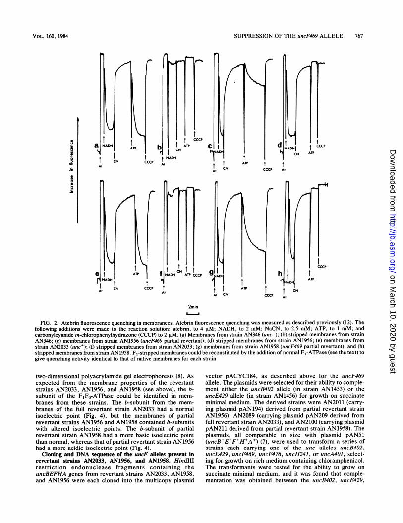
revertants. Membranes were prepared from strain AN1419(uncF469) and the revertant strains AN2033, AN1956, andAN1958. ATPase activities (with and without DCCD),atebrin fluorescence-quenching activities, and oxidativephosphorylation activities were assayed. The membranes ofstrain AN2033 were normal (see Table 2 and Fig. 2 and 3),confirming that this strain was a full revertant. Membrahesfrom partial revertant strains AN1956 and AN1958 support-ed significant levels of oxidative phosphorylation (Table 2),but retained NADH-dependent atebrin fluorescence-quenching activity on removal of the F1-ATPase (Fig. 2).ATP-dependent atebrin fluorescence-quenching activity inthese stripped membranes could be reconstituted to the levelof native membranes by the addition of purified normal Fl-ATPase. It was concluded that retention of NADH-depen-dent atebrin fluorescence-quenching activity of Fl-ATPaseremoval from membranes of strains AN1956 and AN1958was not due to attached F1-subunits. The ATPase activitiesof the membranes from both strains were resistant to theinhibitor DCCD (Fig. 3).
Two-dimensional gel electrophoresis of membranes of strainAN1419 (uncF469) and its revertants. The b-subunit is absentfrom membranes of strain AN1419 (uncF469), as judged by
J. BACTERIOL.
on March 10, 2020 by guest
http://jb.asm.org/
Dow
nloaded from
SUPPRESSION OF THE uncF469 ALLELE 767
0 ^ 1 ti ' h:
AD' A TAD]CCPI ATP~~~~~~N
Att NADH
0t C Ct tACN t t C
CCCP AV~~~N CC A
At~~~~~~~~~~~~~~~~~~~~
Fi e quenc g f NAD tCeAP ws u shr pr
folloingdditons ere Nade to th ecinsltin tbi,t , ;ND,tAM;NC,t . MATP,t1mM an
carbonylcyanidemcopnT(CCCPa ATPpCNAt~~~~~~~~AAt~~~~~~~~~~~~~~~~A
2mmn
FIG. 2. Atebrin fluorescence quenching in membrances. Atebrin fluorescence quenching was measured as described previously (12). Thefollowing additions were made to the reaction solution: atebrin, to 4 ~LM; NADH, to 2 mM; NaCN, to 2.5 mM; ATP, to 1 mM; andcarbonylcyanide m-chlorophenylhydrazone (CCCP) to 2 p.M. (a) Membranes from strain AN346 (unc'); (b) stripped membranes from strainAN346; (c) membranes from strain AN1956 (uncF469 partial revertant); (d) stripped membranes from strain AN1956; (e) membranes fromstrain AN2033 (unc+); (f) stripped membranes from strain AN2033; (g) membranes from strain AN1958 (uncF469 partial revertant); and (h)stripped membranes from strain AN1958. F1-stripped membranes could be reconstituted by the addition of normal F1-ATPase (see the text) togive quenching activity identical to that of native membranes for each strain.
two-dimensional polyacrylamide gel electrophoresis (8). Asexpected from the membrane properties of the revertantstrains AN2033, AN1956, and AN1958 (see above), the b-subunit of the F1FO-ATPase could be identified in mem-branes from these strains. The b-subunit from the mem-branes of the full revertant strain AN2033 had a normalisoelectric point (Fig. 4), but the membranes of partialrevertant strains AN1956 and AN1958 contained b-subunitswith altered isoelectric points. The b-subunit of partialrevertant strain AN1958 had a more basic isoelectric pointthan normal, whereas that of partial revertant strain AN1956had a more acidic isoelectric point (Fig. 4).
Cloning and DNA sequence of the uncF alleles present inrevertant strains AN2033, AN1956, and AN1958. HindIIIrestriction endonuclease fragments containing theuncBEFHA genes from revertant strains AN2033, AN1958,and AN1956 were each cloned into the multicopy plasmid
vector pACYC184, as described above for the uncF469allele. The plasmids were selected for their ability to comple-ment either the uncB402 allele (in strain AN1453) or theuncE429 allele (in strain AN1456) for growth on succinateminimal medium. The derived strains were AN2011 (carry-ing plasmid pAN194) derived from partial revertant strainAN1956), AN2089 (carrying plasmid pAN209 derived fromfull revertant strain AN2033), and AN2100 (carrying plasmidpAN211 derived from partial revertant strain AN1958). Theplasmids, all comparable in size with plasmid pAN51(uncB+E+F+H+A+) (7), were used to transform a series ofstrains each carrying one of the unc alleles uncB402,uncE429, uncF469, uncF476, uncH241, or uncA401, select-ing for growth on rich medium containing chloramphenicol.The transformants were tested for the ability to grow onsuccinate minimal medium, and it was found that comple-mentation was obtained between the uncB402, uncE429,
VOL. 160, 1984
on March 10, 2020 by guest
http://jb.asm.org/
Dow
nloaded from

768 JANS ET AL.
~60
40a40
I--
20
00 2 4 6 8 10 12
DCCD concentration (lg/ml.)
FIG. 3. Inhibition of ATPase activity by DCCD. Membranes (0.3mg of protein) were incubated at 30°C in 5 ml of the ATPase assaymixture together with the indicated amount of DCCD. The mixturewas sampled at intervals, the rate was determined for each DCCDconcentration, and the percent inhibition was calculated. Symbols:V, membranes from strain AN346 (unc+); *, membranes fromstrain AN2033 (unc+); V, membranes from partial revertant strainAN1956; and 0, membranes from partial revertant strain AN1958.
uncH241, and uncA401 alleles and all three plasmids. Theseresults suggested that the second mutations of the revertantstrains AN1956 and AN1958 had not occurred in the uncB,uncE, uncH, or uncA genes. Complementation occurred
between plasmid pAN209 (derived from full revertant strainAN2033) and both uncF alleles, as expected, but plasmidspAN194 (derived from partial revertant strain AN1956) andpAN211 (derived from partial revertant strain AN1958) wereunable to complement either the uncF469 or uncF476 allele.The complete DNA sequences of the uncF alleles present
in the three revertant strains were determined as for theuncF469 allele (see above). That of the uncF allele present inthe full revertant strain AN2033 was normal, being identicalto that of plasmid pAN51 (uncB+E+F+H+A+). StrainAN2033 was thus confirmed as a full revertant of theuncF469 nonsense mutation. The nucleotide sequences ofthe uncF alleles present in partial revertant strains AN1956and AN1958 were the same, being identical to that of theuncF469 allele in that TAG replaced the normal TGGtryptophan codon at position 26 of the amino acid sequence.Partial revertant strains AN1956 and AN1958 thus containedthe uncF469 allele, but as membranes prepared from thesestrains possessed a b-subunit of normal molecular weight(see above), it was concluded that both strains probablycontained tRNA suppressor genes capable of inserting anamino acid at the TAG stop codon.
DISCUSSIONThe uncF469 allele is a nonsense mutation, resulting in the
termination of translation of the b-subunit of the F1F0-ATPase of E. coli at amino acid 26. This confirmed thefindings of Noumi and Kanazawa (26) who localized theuncF469 mutation to the first 198 bases of the uncF gene byrecombination. The fact that the TGG -* TAG (Trp 26 --
stop) mutation constitutes the sole mutation of importance instrain AN1419 (uncF469) and its derivatives is indicated bythe characterization of strain AN2033. This strain is a fullphenotypic revertant in which the stop codon has reverted to
.......~~~~~0 ,E,) .,,... ..A C
s __
> - _-- a;;-
_$ *_ R.
_ _
_.._
._^ .. 4. X
.4'-sb .. ,w -i_=f*_.
.*=*-..i,_*__ ... ,,, F.. __ _g W. .Ab.fi_
_ _ -.. w _._-. .
_r _
B -9 - _(|
0 S _, . " .... ..... ..... ....... .. _ . ;.. _s _ vs _ __ _
__ _.. ... - . _. .. __ _ tc.. PE _ S_ 3> 1 _ it
;>b_ ...._ _ . ,. CD
qui.
.8;,440- Iy
:,4A it"A.S.,
FIG. 4. Two-dimensional gel electrophoresis of membrane preparations. (A) Membranes from strain AN1956 (uncF469 partial revertant);(B) membranes from strain AN1958 (uncF469 partial revertant); (C) membranes from strain AN1419 (uncF469); and (D) membranes fromstrain AN2033 (unc+). Samples (1 ml containing ca. 20 mg of protein) of membranes washed in low-ionic-strength buffer in the presence of p-aminobenzamidine were extracted twice with 5 ml of cold acetone. The dried residue was solubilized in ca. 0.8 ml of lysis buffer (29), and 50 ,ulused for electrophoresis. In the first dimension, ampholines with pH's ranging from 5 to 7 and from 3.5 to 10 were present at 1.2 and 0.8% (wt/vol), respectively. In the second dimension, an acrylamide gradient of 10.5 to 24.5% (wt/vol) was used. The arrows labeled a, ,B, y, 8 and bidentify the corresponding subunits of the F1FO-ATPase. Unlabeled arrows indicate the normal positions of the absent subunits.
J. BACTERIOL.
a -11.
,...O
ILPWIW-46 .001- - -
-
. -400
-IW.W
on March 10, 2020 by guest
http://jb.asm.org/
Dow
nloaded from

SUPPRESSION OF THE uncF469 ALLELE 769
the original tryptophan codon, viz., TAG -) TGG (stopTrp 26). It is noteworthy that all genes distal to the uncFgene are apparently unaffected in expression (as shown bygenetic complementation by plasmids carrying the uncF469allele) despite the early occurrence of the TAG codon in theuncF469 gene coding sequence. A similar nonpolar effect onexpression of genes distal to a stop codon is observed forstrains carrying the uncB402 allele, reported by Fillingame etal. (9) to be a nonsense mutation suppressible by phageencoding the Su3 suppressor (which inserts the amino acidtyrosine for TAG).
In this study, two partial revertants of the uncF469mutation were found to retain the uncF469 nonsense muta-tion, yet membrane preparations from both contained fullF1FO-ATPase assemblies with the b-subunit present. It isdeduced that these strains contain suppressors enablingsynthesis of the b-subunit. The amber suppressors in thepartial revertant strains AN1956 and AN1958 insert differentamino acids. The suppressor of strain AN1956 is clearlypresent on the repaired F-plasmid Flll derived from strainAN1956 (F-plasmid pAN185), as this plasmid complementsthe uncF469 allele and the nonsuppressible uncF476 allele.This suppressor therefore maps in the vicinity of the uncgenes on the E. coli chromosome (81 through 90 min). Itinserts an acidic amino acid for the TAG stop codon, asdemonstrated by the isoelectric point of the b-subunit fromthis strain. The only tRNA anticodon specifying an acidicamino acid that can mutate via a single base substitution tobe complementary to TAG is that for glutamate (GAGTAG). It thus seems reasonable to propose that the acidicamino acid inserted for TAG in partial revertant strainAN1956 is most probably glutamate.
In contrast, the suppressor of strain AN1958 does not mapin the unc region of the E. coli chromosome, as the repairedF-plasmid pAN187 derived from this strain does not comple-ment uncF mutants. Furthermore, this suppressor inserts abasic amino acid in place of the TAG stop codon, on thebasis of the isoelectric point of the b-subunit of strainAN1958. The only tRNA anticodon specifying a basic aminoacid that can mutate via a single base change to recognizeTAG is that for lysine (AAG -* TAG). It can therefore bereasonably proposed that the basic amino acid inserted bythe suppressor of partial revertant strain AN1958 is mostprobably lysine.Amino acid substitutions in place of tryptophan 26, situat-
ed in the region of the polar head groups of the lipid bilayernear the cytoplasmic side of the membrane (17) clearly havea significant effect on proton transfer through the Fo-ATPase. Insertion of either an acidic (strain AN1956) or abasic (strain AN1958) amino acid at position 26 impairsmembrane proton permeability. Nagle and Morowitz (23)and Nagle and Tristram-Nagle (24) have proposed thatproton transfer occurs via the progress of protons along thehydrogen bonds of polar amino acid side chains such asaspartate, glutamate, serine, threonine, tyrosine, lysine, andarginine. The fact that mutations introducing additionalcharged or polar amino acid side chains into the Fo-sector,such as the uncF476 allele (Gly 9 -- Asp) in the b-subunit(Jans et al., in press) or the uncE513 allele (Ala 25 -- Thr) inthe c-subunit (A. L. Fimmel, unpublished data), as well asthe basic or acidic amino acids instead of tryptophan in thesuppressed uncF469 mutation reported here, all inhibittransmembrane H+-translocation when they might perhapsbe expected to facilitate it is hard to reconcile with hydro-gen-bonded chain theory. That either lysine or glutamate atposition 26 of the b-subunit located in the phospholipid head
groups of the lipid bilayer near the cytoplasmic side of themembrane (17) has an identical inhibitory effect on protontransfer does not support such models of membrane protonmovement.The membrane properties of haploid strains AN1956 and
AN1958 are of considerable interest. The oxidative phos-phorylation levels attained by the membranes of both,relative to ATPase activities, were similar to that of the fullrevertant strain AN2033. However, the membranes of par-tial revertant strains AN1956 and AN1958 remained protonimpermeable on removal of the Fl-ATPase. In addition, themembrane-bound ATPase activities of both were relativelyinsensitive to the inhibitor DCCD. Such properties have nowbeen seen in a number of unc mutant strains, viz., strainscarrying multiple copies of the uncE408 or uncE463 alleles(5), and strains carrying the uncES01 and uncE502 alleles(10).
ACKNOWLEDGMENTSWe thank D. Webb, B. Rowell, and B. Webb for skilled technical
assistance. We thank R. D. Simoni for uncH mutant strains.
LITERATURE CITED1. Bachmann, B. J. 1983. Linkage map of Escherichia coli K-12,
edition 7. Microbiol. Rev. 47:180-230.2. Butlin, J. D., G. B. Cox, and F. Gibson. 1973. Oxidative
phosphorylation in Escherichia coli K12: the genetic and bio-chemical characterization of a strain carrying a mutation in theuncB gene. Biochim. Biophys. Acta 292:366-375.
3. Chang, A. C. Y., and S. N. Cohen. 1978. Construction andcharacterization of amplifiable multicopy DNA cloning vehiclesderived from the P15A multicopy cryptic miniplasmid. J. Bac-teriol. 134:1141-1156.
4. Cox, G. B., N. A. Newton, F. Gibson, A. M. Snosweli, and J. A.Hamilton. 1970. The function of ubiquinone in Escherichia coli.Biochem. J. 117:551-562.
5. Cox, G. B., D. A. Jans, F. Gibson, L. Langman, A. E. Senior,and A. L. Fimmel. 1983. Oxidative phosphorylation by mutantEscherichia coli membranes with impaired proton permeability.Biochem. J. 216:143-150.
6. Downie, J. A., A. E. Senior, F. Gibson, and G. B. Cox. 1979. Afifth gene (uncE) in the operon concerned with oxidative phos-phorylation in Escherichia coli. J. Bacteriol. 137:711-718.
7. Downie, J. A., L. Langman, G. B. Cox, C. Yanofsky, and F.Gibson. 1980. Subunits of the adenosine triphosphatase complextranslated in vitro from the Escherichia coli unc operon. J.Bacteriol. 143:8-17.
8. Downie, J. A., G. B. Cox, L. Langman, G. Ash, M. Becker, andF. Gibson. 1981. Three genes coding for subunits of the mem-brane sector (FO) of the Escherichia coli adenosine triphospha-tase complex. J. Bacteriol. 145:200-210.
9. Fillingame, R. H., M. E. Mosher, R. S. Negrin, and L. K. Peters.1983. H+-ATPase of Escherichia coli: uncB402 mutation leadsto loss of x-subunit of Fo sector. J. Biol. Chem. 258:604-609.
10. Fimmel, A. L., D. A. Jans, L. Langman, L. B. James, G. R. Ash,J. A. Downie, A. E. Senior, F. Gibson, and G. B. Cox. 1983. TheF1F0-ATPase of Escherichia coli. Substitution of proline byleucine at position 64 in the c-subunit causes loss of oxidativephosphorylation. Biochem. J. 213:451-458.
11. Gay, N. J., and J. E. Walker. 1981. The atp operon: nucleotidesequence of the promoter and the genes for the membraneproteins, and the 8 subunit of Escherichia coli ATP synthase.Nucleic Acids Res. 9:39 3919-3926.
12. Gibson, F., G. B. Cox, J. A. Downie, and J. Radik. 1977. Partialdiploids of Escherichia coli carrying normal and mutant uncalleles affecting oxidative phosphorylation. Biochem. J.162:665-670.
13. Gibson, F., G. B. Cox, J. A. Downie, and J. Radik. 1977. Amutation affecting a second component of the Fo portion of themagnesium ion-stimulated adenosine triphosphatase of Esche-
VOL. 160, 1984
on March 10, 2020 by guest
http://jb.asm.org/
Dow
nloaded from

770 JANS ET AL.
richia coli K12. the uncC424 allele. Biochem. J. 164:193-198.14. Gibson, F., J. A. Downie, G. B. Cox, and J. Radik. 1978. Mu-
induced polarity in the unc operon of Escherichia coli K-12. J.Bacteriol. 134:728-736.
15. Gunsalus, R. P., W. S. A. Brusilow, and R. D. Simoni. 1982.Gene order and gene polypeptide relationships of the proton-translocating ATPase operon (unc) of Escherichia coli. Proc.Natl. Acad. Sci. U.S.A. 79:320-324.
16. Hoppe, J., P. Friedi, H. U. Schairer, W. Sebald, K. vonMeyenburg, and B. B. Jorgensen. 1983. The topology of theproton translocating Fo component of the ATP synthase fromEscherichia coli K12: studies with proteases. EMBO J. 2:105-110.
17. Hoppe, J., C. Montecucco, and P. Friedl. 1983. Labelling ofsubunit b of the ATP synthase from Escherichia coli with aphotoreactive phospholipid analogue. J. Biol. Chem. 258:2882-2885.
18. Humbert, R., W. S. A. Brusilow, R. P. Gunsalus, D. J. Klionsky,and R. D. Simoni. 1983. Escherichia coli mutants defective inthe uncH gene. J. Bacteriol. 153:416-422.
19. Kanazawa, H., K. Mabuchi, T. Kayano, T. Noumi, T. Sekiya,and M. Futai. 1981. Nucleotide sequence of the genes for Focomponents of the proton-translocating ATPase from Esche-richia coli: prediction of the primary structure of Fo subunits.Biochem. Biophys. Res. Commun. 103:613-620.
20. Lowry, 0. H., N. J. Rosebrough, A. L. Farr, and R. J. Randall.1951. Protein measurement with the Folin phenol reagent. J.Biol. Chem. 193:265-275.
21. Luria, S. E., and J. W. Burrous. 1957. Hybridization betweenEscherichia coli and shigella. J. Bacteriol. 74:461-476.
22. Maxam, A. M., and W. Gilbert. 1980. Sequencing end-labelledDNA with base-specific chemical cleavages. Methods Enzymol.65:499-560.
23. Nagle, J. F., and H. J. Morowitz. 1978. Molecular mechanismfor proton transport in membranes. Proc. Natl. Acad. Sci.U.S.A. 75:298-302.
24. Nagle, J. F., and S. Tristram-Nagle. 1983. Hydrogen-bondedchain mechanisms for proton conduction and proton pumping.
J. Memb. Biol. 74:1-14.25. Nielsen, J., F. G. Hansen, J. Hoppe, P. Friedl, and K. von
Meyenburg. 1981. The nucleotide sequence of the atp genescoding for the Fo subunits a, b, c and the F1 subunit 8 of themembrane bound ATP synthase of Escherichia coli. Mol. Gen.Genet. 184:33-39.
26. Noumi, T., and H. Kanazawa. 1983. Mutants of Escherichia coliH+-ATPase defective in the b-subunit of F, and the b-subunit ofFo. Biochem. Biophys. Res. Commun. 111:143-149.
27. Novick, R. P., R. C. Clowes, S. N. Cohen, R. Curtiss III, N.Datta, and S. Falkow. 1976. Uniform nomenclature for bacterialplasmids: a proposal. Bacteriol. Rev. 40:168-189.
28. O'Farrell, P. H. 1975. High resolution two-dimensional electro-phoresis of proteins. J. Biol. Chem. 250:4007-4021.
29. Perlin, D. S., D. N. Cox, and A. E. Senior. 1983. Integration ofF, and the membrane-sector of the proton-ATPase of Esche-richia coli: role of subunit "b" (uncF protein). J. Biol. Chem.258:9793-9800.
30. Saito, H., and K. Miura. 1963. Preparation of transformingdeoxyribonucleic acid by phenol treatment. Biochim. Biophys.Acta 72:619-629.
31. Saraste, M., N. J. Gay, A. Eberle, M. J. Runswick, and J. E.Walker. 1981. The atp operon: nucleotide sequence of the genesfor the y, P, and e subunits of Escherichia coli ATP synthase.Nucleic Acids Res. 9:5287-5296.
32. Selker, E., K. Brown, and C. Yanofsky. 1977. Mitomycin C-induced expression of trpA by Salmonella typhimurium insertedinto the plasmid ColEl. J. Bacteriol. 129:388-394.
33. Senior, A. E., D. R. H. Fayle, J. A. Downie, F. Gibson, and G. B.Cox. 1979. Properties of membranes from mutant strains ofEscherichia coli in which the P-subunit of the adenosine triphos-phatase is abnormal. Biochem. J. 180:111-118.
34. Senior, A. E. 1983. On the secondary and tertiary structure ofmembrane proteins involved in proton translocation. Biochim.Biophys. Acta 726:81-95.
35. Walker, J. E., M. Saraste, and N. J. Gay. 1982. Escherichia coliF1-ATPase interacts with a membrane protein component of aproton channel. Nature (London) 298:867-869.
J. BACTERIOL.
on March 10, 2020 by guest
http://jb.asm.org/
Dow
nloaded from