Embed Size (px)
Citation preview
University of RichmondUR Scholarship Repository
Master's Theses Student Research
1969
An ultrastructural investigation on the origin ofmurine Langerhans cellsStanley Powell Tompkins
Follow this and additional works at: https://scholarship.richmond.edu/masters-theses
Part of the Biology Commons
This Thesis is brought to you for free and open access by the Student Research at UR Scholarship Repository. It has been accepted for inclusion inMaster's Theses by an authorized administrator of UR Scholarship Repository. For more information, please [email protected].
Recommended CitationTompkins, Stanley Powell, "An ultrastructural investigation on the origin of murine Langerhans cells" (1969). Master's Theses. 1321.https://scholarship.richmond.edu/masters-theses/1321

AN ULTRASTRUCTURAL INVESTIGATION ON THE
ORIGIN OF MURINE LANGERHANS CELLS
BY
STANLEY POWELL TOMPKINS
EXAMINING COMMITTEE
d'&� COMMITTEE�
APPROVlll>:

AN ULTRA.STRUCTURAL INVESTIGATION ON THE
ORIGIN OF MURINE LANGERHANS CELLS
BY
STANLEY POWELL TOMPKINS
B. S., OLD DOMINION COLLEGE, 1967
A THESIS
SUBMITTED TO THE FACULTY OF THE GRADUATE SCHOOL
OF THE UNIVERSITY OF RICHMOND
IN PARTIAL FULFILLMENT OF THE REQUIREMENTS FOR THE
DEGREE OF MASTER OF SCIENCE IN BIOLOGY
AUGUST, 1969

I
II
III
"I)/
V
VI
VII
VIII
IX
X
TABLE OF CONTENTS
Abstract
Acknowledgements
Introduotion
Methods and Materials
Observations
Disoussion
Conolusion
Literature Cited
Figures
Vita
i
ii
iii
1
4
7
12
22
23
28
44
ii
ABSTRACT
Electron microsoopic investigations have revealed two types
of oells populating normal skin of mammals: keratinocytes and
nonkeratinocytes. The nonkeratinocytes were represented as
melanocytes and epidermal Langerhans cells. This study reports
the description and occurrence of another cell population found
in both normal and neural crest-free skin from mice of the PEI'
strain.
The cells of this population have been designated semikeratino
oytes. characterized as dendritic cells possessing irregular
nuclei, and found in both the basal and suprabasal cell layers.
The cytoplasm of these cells possessed few to no tonofilaments
and tonofibrils, a reduced number of ribosomes, mitochondria,
numerous vesicles, and minidesmosomes along the plasma membrane.
Since the semikeratinocytes found in both normal and neural
crest-free mouse skin possessed characteristics similar to both
epidermal Langerhans cells and keratinocytes, the evidence
presented suggests that the semikeratinocytes are divisional
products of basal keratinooytes and differentiate into epidermal
Langerhans cells.

ACKNOWLEDGEMENTS
I wish to express my sincere appreciation to Dr. Willie
Mathews Reruns, Jr., whose patience, talent, guidance, and
understanding have been instrumental 1n fulfilling this goal.
His zeal for the biological sciences has been a continuous
inspiration to me throughout rrry graduate studies, and will,
I am sure, continue to influence me 1n the years to come.
I wish to thank Drs. Francis B. Leftwich and Wilton R.
Tenney, members of rrry thesis committee, for their prudent
evaluation and valuable criticism which were indispensible in
the final preparations of this manuscript.
iii
I would like to thank Mesdames Shirley Craig and Edith
Sulley, Department of Anatomy, Medical College of Virginia,
for their helpful technical suggestions and assistance which
were essential 1n the preparations of specimens used in this
study. I also want to thank Dr. J. Samuel Gillespie, Jr.,
Director of the Virginia Institute for Scientific Research,
for the use of the laboratory and darkroom facilities, and
especially, for the use of the RCA EMU-JB electron microscope,
all of which were vital to the completion of this study.
Lastly, I want to express my deepest appreciation to my
wife, Pat, whose encouragement and devotion made this goal possible.
This study was supported by NIH grant AM 11864, and the A. H.
Robins Company, Richmond, Virginia.

1
INTRODUCTION
The mammalian epidermis has been described as consisting of
three different cell populations: keratinocytes, pigment cells,
and Langerhans cells. Ultrastructural investigations of mam."llalian
skin within the last decade have resulted in criteria whereby
these oell types can be differentiated upon electron microscopic
examination.,
Ultrastruoturally the keratinocyte is characterized by being
a columnar or cuboidal epidermal cell which possesses a round to
oval nucleus and a plasma membrane having numerous desmosomal
attachments. Tonofilaments and tonofibrils, melanosomes, and
.keratohyalin granules are found in cells of the upper stratum
spinosum and stratum granulosum (Odland and Reed, 1967; Snell,
1967). During their assent from the basal layer, the keratino
cytes experience a nattening so that the long axis of the cells
appears parallel to the surface of the integument •
. Pigment cells are characterized by being dendritic basal
cells which possess a round to oval nucleus which, at times,
may have a slight indentation, and a plasma membrane which lacks
desmosomal attachments (Birbeck, � �., 1961; Zelickson, 1967).
The cytoplasm contains premelanosomes and melanosomes, lacks
tonofilaments and tonofibrils, and appears relatively clear when
compared with ihe cytoplasm of keratinocytes (Birbeok, !:!:, !!_., 1961).
2
Epidermal Langerhans cells are described as dendritic cells
of the basal and suprabasal cell layers which possess a convoluted
nucleus and a plasma membrane which lacks desmosomes. Their
cytoplasm lacks tonofilaments and tonofibrils, and it appears
relatively clear as compared with the cytoplasm of the keratino
cytes (Birbeck, et !1_., 1961). In addition, the cytoplasm contains
a characteristic organelle, known as the Langerhans granule, which
appears rod-shaped or tennis racket-shaped in cross section, and
disc-shaped in top view (Birbeck, et&•, 1961; Breathnach, 1964;
Zelickson, 1966; Wolff, 1967; Sagebiel and Reed, 1968).
The ultrastructural similarity of the epidermal melanocyte
and the epidermal Langerhans cell led many investigators to believe
that the Langerhans cells were derived from melanocytes as either
effete melanocytes (Birbeck, � !1_., 1961), transitional products
(Breathnach, .2:l!. &•, 1963; Zelickson, 1965; Mishima, 1966), or
post-divisional products (Breathnaoh, 1964, 1965; Breathnach and
Goodwin, 1965; Zelickson, 1966). The Langerhans cell-melanooyte
relationship was challenged when organelles identical to Langer
hans granules were found in the tumor histiocytes of histio
cytosis-X, suggesting a mesodermal origin (Tarnowski and Hashimoto,
1967 ).
To add support to the speculation of a Langerhans cell
melanocyte relationship, Reruns,�&•, (1967) found no evidence
for the presence of cells with characteristic Langerhans granules
or melanosomes in murine skin experimentally deprived of its neural
crest derivatives. In a more conclusive study, Breathnach,

� al., (1968) found nonkeratinooytes in the .epidermis of mouse
skin which had been deprived of its neural crest derivatives.
Many ?f the nonkeratinocytes possessed granules within the cytoplasm,
which were identi�al to the Langerhans granule. This evidence was
purported to disprove all existing hypotheses which proposed that
epidermal Langerhans cells were derived from melanocytes, and
· renewed speculation as to their origin and function.
Adding support to a possible mesodermal origin of the epidermal
Langerhans cell was evidence by Kiistala and Mustakallio (1968)
and by Pruniceras {1969). On the other hand, Wolff and Winklemann,
(1967a), and Lessard,�!!_., (1968) suggested a possible relationship
between the keratinocyte. and the epidermal Langerhans cell. Another
possible origin has been introduced by the report of an unidenti
fiable dendritio cell type in the epidermis of the guinea pig
(Lessard, et!!_., 1967), and by the presence of an "unclassi-
fiable nonkeratinocyte11 in mouse skin by Quevedo (1968). Quevedo
(1968) considers these cells to be preousors of Langerhans cells
as well as melanocyteso
The present study was undertaken to investigate the occur
rence of epidermal Langerhans cells in both neural crest-free
and normal PET mouse skin, and to present evidence for a possible
origin of the epidermal Langerhans cells of murine skin.
4
METHODS AND MATERIALS
The tissues used in this investigation were obtained from
.mice of the PET strain (Reams, 1967a). Mice or the PET strain
were used because of availability and the background of information
on the. developmental history of their neural crest cells (Mayer
and Reams, 1962; Reams, 1967b).
Epidermal tissue free of neural crest cells was obtained by
implanting limb buds from 10-day-old mouse embryos into the coeloms
of 72-hour White Leghorn chick embryo hosts. At the same time,
limb buds possessing neural crest cells, from 12½-day-old mouse
embryos were implanted into the coeloms of 72-hour White Leghorn
chick embryos. These served as controls. After 15 days of intra
coelomic cultivation, the grafts were recovered and small strips
of skin were removed and immediately fixed. Under the steroscopic
microscope, it was evident that the grafts from the 10-day-old
embryos were deprived of their neural crest derivatives because
colorless hairs were seen protruding from the grafts. In contrast,
the grafts from the 12½-day-old embryos possessed black hairs,
indicating the presence of neural crest elements.
Normal skin was obtained .from 15 to 18-day-old embryos,
newborn, and J-day-old mice. Strips of skin, measuring 5 x 10 Illlll,
.,., ... ---....__,_�
were removed i'rom the dorsal surfaoes·or the above, immediately
placed into cold fixative, and sliced into smaller strips, 1 x 3 Illlll.

s
Samples of normal skin were removed from 15-day-old and
adult mice which had been anesthetized with an intraperitoneal
injection of Diabutal (sodium pentabarbital) •. As soon as the
animals were immobile, a commerical depilatory, "Nair," was applied
to their dorsal surfaces. The 11Nair 11 was removed after five minutes
leaving the surfaces hairless. While the mice were still alive,
S x 10 mm strips of skin were removed and immediately placed into
cold fixative where they were sliced into strips, 1 x J mm.
All of the strips of skin were fiXed for two hours in 1"p
osmium tetroxide buffered at pH 7.4 with veronal acetate (Palade,
1952). Following two five-minute rinses in veronal acetate buffer,
pH 7.4, the tissue was dehydrated through a series of acetones
maintained at 4°0. The tissue remained for five minutes in each
of 201,, 50%, 801,, and 95% acetone, and for fifteen minutes in each
of two 100% acetone baths. It was then placed into 1001, acetone
at room temperature (25°0) for fifteen minutes.
The hardened strips of skin were carried through an
infiltration and embedding process using an araldite compound
(Durcupan-A0M, Fluka Company, Switzerland). The procedure required
the tissue to be passed through the following series of acetone
Durcupan miXtures: J:1 acetone to Durcupan, 1:1 acetone:to
Durcupan, and 1 :3 acetone to Durpupan. The tissue remained in
each mixture for one hour before being left overnight in 1001,
Durcupan. The tissue was then placed into flat molds (S x 15 .mm)
.f'illed with 1ooi Durcupan and allowed to polymerize in a 60°c
oven for 48 to 72 hours.

6
Both thick and thin sections were obtained on a Reichert
"OmU 2 11 ultramicrotome with either glass or diamond knives. The
thick sections (0.5 to 1.0..,.., thick) were mounted on glass slides
for exa.'lrl.nation with a phase microscope to check fixation and the
orientation of the tissue in the plastic blocks, Thin sections
(60-90 m.l' thick) were mounted on copper grids (100 mesh, 3 mm
O.D.) which had been coated with a film of o.1oi parlodion.
The thin sections were routinely stained with a 1i aqueous
solution of uranyl acetate for two minutes and with lead citrate
(Reynolds, 1963) for three minutes. To emphasize such structures
as basement membranes, desmosomes, and tonofibrils. some thin
sections were stained with lead citrate (Reynolds, 1963) for one
minute, followed by staining with potassium permanganate (Lawn,
1960) for fifteen minutes.
The thin sections were examined on a RCA EMU-3B electron
microscope. Electron micrographs were taken on 3¼ x 4 inch glass
Kodak projector slide plates from which prints were obtained
following photographic enlargement.
7
OBSERVATIONS
The following observations were made while evaluating over
JOO electron micrographs taken during the examination of over 200
sections from normal and neural crest-free PET mouse skin.
Normal�• There appeared to be no significant ultra
structural differences between the various epidermal tissue types ---------��--,,.._---,,."�·.,.._._,."7" ____ ... .,...",,.,...�--• -"""''"""""'
used ,a,s __ e,campl�s of no�al s��•- _ K��:-�<;,.T!!,.,,,,��.,!1?,��p����ooytes
were found in all age gro��s. Nonkeratinocytes were cells which
did not possess the typical characteristics of keratinocytes
and were represented as melanocytes and Langerhans cells.
The keratinocytes of the normal skin types were characterized
as being cuboidal to columnar in shape and possessing nuclei
which were either round, oval, ·or slightly indented. The plasma
membrane possessed numerous desmosomes of normal appearance (Fig. 1).
The cytoplasm contained numerous mitochondria, and tonofilaments
and tonofibrils (Figs. 1 and 2). Ribosomes and vesicles were
seen scattered throughout the cytoplasm. · Cisternae of rough
endoplasmic reticulum and Golgi complexes were also seen.
Melanosomes were <;>bserved in cells of both the basal and suprabasal
levels of the epidermis. • Keratoh_yalin granules were found only in
the suprabasal layers of the upper stratum spinosum and stratum
granulosum.
Pigment cells as melanocyt�s were observed only in the basal

8
cell layers of normal mouse skin. The nuclei appeared slightly
indented (Figs. 1 and 2). The plasma membrane, in contrast to
keratinocytes, lacked desmosomes. The cytoplasm appeared more
electron dense than that of the adjacent keratinocytes and possessed
numerous vesicles, mitochondria, and melanosomes, but lacked tono
filaments and tonofibrils (Fig. 2).
Nonkeratinocytes, which could be classified as Langerhans cells,
were observed in skin from 1 S-day-old PET mice. These cells were
observed in both basal and suprabasal cell layers. The nuclei
were convoluted and the plasma membranes lacked desmosomes (Figs. 3,
4, and 5). The cytoplasm of these cells, like that of melanocytes,
appeared more electron dense than that of the adjacent keratinocytes
and lacked tonofilaments and tonofibrils. The cytoplasm also
possessed numerous vesicles and mitochondria, Golgi complexes,
and cisternae of the endoplasmic reticulum. Occasionally, centrioles
and membrane-bound melanosomes were seen in the cytoplasm of these
cells (Figs. J and 4).
Cytoplasmic Langerhans granules, organelles characteristic of
Langerhans cells were observed (Figs. J, 4, and 5). However, no
more than one granule per section per cell was seen. Most of the
granules appeared to be rod-shaped with rounded ends and a central
dense line. A rod-shaped organelle with what appeared to be a
vesicle attached to one end was seen to be continous with the
plasma membrane (Fig. S). Two Langerhans cells were observed
adjacent to one another and appeared to be the products of a recent
mitotic division (Fig. 4).

Often nonkeratinocytes were observed which could not be . ' ' . . . • , , ,,..
.� · ·�+·· ,,, ., ... ,,-� ... ,� �:-. ". '
9
definitely identified as either melanocytes or Langerhans cells
due to.the absence of characteristic granules (Fig. 6). This was
particularly true for cells located in the basal cell layer where
both types of nonkeratinocytes were found (Fig. 6). These cells
possessed characteristics applicable to both types of nonkeratino
cytes: they lacked desmosomes and fibrillar elements, and had a
cytoplasm that was more electron dense than the cytoplasm of
adjacent keratinocytes.
Another type of cell frequently encountered was characterized
as a dendritic cell with an irregular nucleus (Fig. 7). In this
cell there was a scarcity of desmosomes along the plasma membrane.
Those which were seen appeared as minidesmosomes when compared to
the size-of the average desmosome possessed by keratinooytes (Figs.
4 and 7). The cytoplasm of these semikeratinocytes was relatively
clear compared with adjacent keratinocytes. It possessed numerous
mitochondria and vesicles, and cisternae of the endoplasmic
reticulum. A few tonofilaments were scattered throughout the
cytoplasm (Fig. 7). The number of ribosomes was greatly reduced
as compared to a typical keratinocyte (Fig. 7).
Neural crest-free�- The skin recovered from the limb bud
transplants after 15 days of intracoelomic incubation was equiva
lent in age to skin from the limbs ·or 4-day-old PET mice which
had developed normally. As seen with the light microscope, the
skin from the 10-day-old limb bud grafts appeared to be free of
its neural crest derivatives as colorless hairs were seen protruding

10
from the surface of the grafts. In contrast, the presence of black
hairs protruding from the surface of the 12½-day-old limb bud grafts
used as controls indicated that these grafts were not deprived of
their neural crest derivatives.
Ultrastructurally, the general appearance of the epidermis
from the graft tissue was similar to the epidemis from normal
skin. Typical keratinocytes were seen throughout the entire
epidermis. No cell� were observed which could be identified as
nonkeratinocytes, melanocytes or Langerhans cells. However,
two types of cells were seen which were classified as semi
keratinocytes.
One type of semikeratinocyte, type A, was characterized as
being a dendritic cell with an irregular to convoluted nucleus
(Figs. 8-14). The cytoplasm of these cells was relatively clear
when compared with the adjacent keratinocytes, and possessed
mitochondria, vesicles, cisternae of the endoplasmic reticulum,
a reduced number of ribosomes, and few to no tonofilaments. The
plasma membrane had only a few minidesmosomes (Figs 8-14). Several
cells were seen with dendritic processes (Figs. 11 and 12). These
cells were ovserved in both the basal and suprabasal cell layers.
One cell of this type possessed a vesicle which appeared to contain
the remnant of an autophagocytized desmosome (Figs. 13 and 14).
Other semikeratinocytes of type A appeared to be in the process
of forming convoluted nuclei. One cell was located in the basal
layer (Fig. 9), while another appeared to be migrating out of the
basal layer (Fig. 10).
11
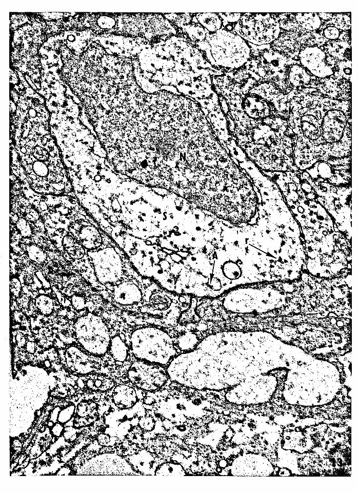
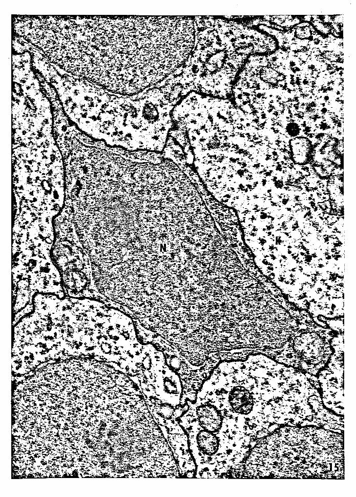
The second type of semikeratinocyte, type B, was also characterized
as being a dendritic cell with an irregular nucleus (Figs. 15 and 16).
The cytoplasm of these cells was more electron dense than that or
the adjacent keratinocytes. It possessed mitochondria, numerous
vesicles, cisternae of the endoplasmic reticulum, and scattered
ribosomes (Figs. 15 and 16). The plasma membranes 0£ these cells
possessed a few minidesmosomes, as did the cells of type A. Several
of these cells had dendritic processes extending from the main
body of the �ell into the superficial cell layers. Cells of type B
were found in close proximity to one another in both basal and
suprabasal levels. In the suprabasal cell layers, two cells were
observed separated by two keratinocytes. In the basal layers, these
cells were found, at times, to be separated by six to ten keratinocytes.
Occasionally, two cells or type B were seen adjacent to one another
which suggested that they were the products or a recent mitotic
division.
12
DISCUSSION
Early investigations into the nature of the epidermal Langerhans
cells were conducted solely by histochemical methods. Fan,��••
(1959) described epidermal Langerhans ·cells in the guinea pig as
gold positive, high-level branched cells of the epidermis. They
considered these as effete or worn out melanocytes because these
cells lost their affinity for gold when melanocyte proliferation
was stimulated. Mishima. and Miller-Milinska (1961) demonstrated
not only high-level branched cells, but also basal melanocytes with
osmium iodide in-human skin, Since both cell types'were osmium
iodide positive, they considered. this as positive proof for a
Langerhans cell-melanocyte relationship. Because of the non
specificity of many histochemical methods, e. g. gold chloride,
and osmium iodide, a more positive method of identifying epidermal
Langerhans cells was needed.
Birbeck, � al., (1961) described. the ultrastructure and the
occurrence of epidermal Langerhans cells in vitiliginous and
normal human skin. This ultrastructural description of Langerhans
cells established. the criteria with which to distinguish these cells
from all other cells populating the epidermis. The Langerhans cells
were characterized. as dendritic cells, �ccurring in both basal
and suprabasal layers and possessing convoluted nuclei. The plasma
membrane of these cells lacked desmosomes, and the cytoplasm was
---

13
clear compared with adjacent keratinooytes due to the lack of
fibrillar elements. The cytoplasm also possessed conspicuous
rod-shaped organelles or granules. The granules were described
as diso-shaped, possessing a 11two-dimensional array of particles."
In cross-section these disc-shaped granules appeared as rods with
a central dense line representing the lattice of particles. Birbeck,
� al., (1961) noted that the ultrastructure of the epidermal Langer
hans cells was similar to that of the epidermal melanocyte, except
for the appearance of the nuclei and the structure of the character
istic granules. The nuclei of epidermal melanocytes did not possess
the degree of indentation or oonvolution, and the premelanosomes
and melanosomes .had a different structure compared with that of the
Langerhans granule. In normal skin, melanocytes were found in the
basal layer, whereas Langerhans cells occurred in the suprabasal
layers. In vitiliginous skin, which lacked melanocytes, Langerhans
cells occurred in both the basal and suprabasal cell layers. Due
to the strong similarities between the melanocytes and the epidermal
Langerhans cells, and the occurrence of basal Langerhans cells in
vitiligo, Birbeck, tl al., (1961) considered the epidermal Langerhans
cells effete melanocytes, agreeing with Fan,� al., (1959).
In human skin treated with thorium-X to increase melanocyte
turnover, Breathnach, et al., (1963) reported the occurrence of basal
melanocytes, suprabasal epidermal Langerhans cells, and a third cell
type, lacking granular structures common to the other two. The
third cell type was considered to be a transitional cell type between
the melanocyte and the Langerhans.cells. Thus it was suggested
14
that the suprabasal Langerhans cells were transitional products of
basal melanocytes.
With the reported occurrence of epidermal Langerhans cells
in both basal and suprabasal layers of normal ginea pig skin
(Breathnach, et al., 1964; Breathnach and Goodwin, 1965), Breathnach, --
(1965) suggested that Langerhans cells were post-divisional products
of basal melanocytes. One of the daughter cells, having the character
istics of a Langerhans cell, migrated into the suprabasal layers
never to become melanogenic. The other daughter cell remained in
the basal cell layer, became rnelanogenic, and eventually divided,
forming the next generation of basal and suprabasal dendritic cells.
Reports by Zelickson (1965), investigating normal skin, and
by l'.d.shima ( 1966) • examining stripped human skin. of transitional
cells containing both melanosomes and Langerhans granules, renewed
speculation of a transitional relationship between melanocytes and
Langerhans cells •. This was challenged, however, by Breathnach and
Wyllie (1965) who showed membrane.-bound melanosomes within the
cytoplasm of Langerhans cells in both normal and vitiliginous human
skin. This suggested that the melanosomes had beeh phagocytized
by the Langerhans cells.
Evidence to support a melanocyte-epidermal Langerhans cell
. relationship was tendered by Reaills, et &• , ( 1967) who found no
nonkeratinocytes which could be identified as either melanocytes
or Langerhans cells in neural crest-free murine skin equivalent in
age to skin from J-day-ol� post natal mice. The neural crest-free
skin was obtained from 10-day-old mouse limb buds which had been
-
1.5
grafted into the coelom of chick embryos. In a more extensive
investigation, Breathnach, � al., (1968) found nonkeratinocytes
possessing Langerhans granules in neural crest-free mouse skin
equivalent in age to skin from 10 and 25-day-old ·post natal mice.
The neural crest-free skin was also from 10-day mouse embryo limb
buds, but had been grafted onto the spleen of ad.ult mice. The
results of this study disproved existing hypotheses concerning a
melanocyte-Langerhans cell relationship.·
· Evidence challenging a neural crest origin for the epidermal
Langerhans cell was presented before the report by Breathnach,
� tl•, (1968). Tarnowski and Hashimoto (1967) show·ed Langerhans
granules in tumor histiocytes, macrophages, of human skin with
histiocytosis-X, suggesting a mesodermal origin for epidermal
Langerhans cells. Hashimoto and Tarnowski (1968), and Kiistala
and Mustakallio (1968) support this hypothesis by showing histio
cytes containing Langerhans granules crossing .the basement membrane.
They suggest that the cells were moving from the dermis into the
epidermis. This was contrary to an earlier report by Zelickson (1965),
which stated that cells containing Langerhans granules were passing
through the basement membrane of normal skin, while moving from
the epidermis into the dermis. The concept of mesodermal origin
for the epidermal Langerhans cells has been presented also by
Prunieras (1969) in reviewing the history and current concepts
regarding the epidermal Langerhans cells.
Reruns and Greco (unpublished) have found evidence against a
mesodermal origin for epidermal Langerhans cells as a result of
---
16
their fluorescent-antibody studies in normal mouse skin. After
they injected tagged anti-lymphocyte serum into mice, they examined
skin samples and found no tagged cells within the eipdermis.
The possibility that epidermal Langerhans cells are derived
from yet another cell population has been presented (Lessard,�
�., 1967, 1968; Quevedo, 1968). An unidentifiable dendritic cell
was demonst�ated with ATPase in regenerating epidermis of the guinea
pig following epidermal stripping (Lessard,� al., 1967). In, a
later report from the ,same study, Lessard, et al., (1968) re�or.ted
that an increase in the number of epidermal Langerhans cells first
appeared on the sixth day following epidermal stripping, and attained
its normal cell population by the 15th day. Quevedo (1968) reported
the.occurrence of 11unclassifiable nonkeratinocytes 11 from birth in
normal mouse skin,' while epidermal L·angerhans cells and melanocytes
were not -identified until the 6th day after birth. Because of this
evidence he felt that the "unclassifiable nonkeratinocyte" gave
rise to both melanocytes and epidermal Langerhans cells.
In the present study, epidermal-Langerhans cells were not
identified in normal skin from PET mice varying in age from late
term embryos to J-day-old post partum. However, cells which could
be identified as epidermal Langerhans cells, by the presence of
characteristic granules, were seen in both basal and suprabasal
cell layers of the epidermis in PET mice 15-days post partum and
older (Figs. 3, 4, and 5), agreeing with Quevedo (1968).
No nonkeratinocytes were observed in murine skin which had
been deprived of its neural crest derivatives, substantiating work

17
by Reruns, et al., (1967). This find was contrary to the report by
Breathnach, � �-, (1968) which indicated the presence of epidermal
Langerhans cells in the epidermis of neural crest-free murine skin.
Two factors, the length of the incubation period, and the nature
of the host tissue, must be considered when comparin� the reports
by Reruns, et al., (1967) and Breathnach, et al., (1968) with the -- --
present study. Breathnach, et al., (1968) grafted neural crest�
free limb buds from 10-day-old embryos onto the spleens of adult
mice. The splenic site, they noted, had been proven to contain
cells which posess Langerhans granules. After J or 5 weeks of
incubation, the graft skin was equivalent in age to skin from 10
and 25-day-old post partum mice, respectively, ages at which Langer
hans granules have been identified within nonkeratinocytes of normal
mouse epidermis (Quevedo, 1968). In contrast, the present study
found no nonkeratinocytes in neural crest-free skin equivalent in
age to skin from a 4-day-old mouse, indicating that nonkeratino
cytes containing Langerhans granules appear in mouse skin between
the 4th and 6th days following birth.
The semikeratinocytes observed in normal skin of all ages and
in neural crest-free skin (Reazns and Tompkins, 1969) are believed
to be similar to, if not identical to, the "unclassifiable
keratinocytes 11 described to constitute a constant population within
mouse skin from birth (Quevedo,1968), and to the unidentifiable
dendritic cells common in regenerating epidermis (Lessard, et�., 1967).
Present evidence suggests that the semikeratinocytes are
divisional products of basal ke�atinocytes which have the potential-
18
to differentiate into epidermal Langerhans oells. Following mitotic
division of a basal keratinocyte, the semikeratinocytes have the
appearance of cells designated as type A. They possess a relatively
clear cytoplasm, an irregular to convoluted nucleus, a reduced
number of ribosomes, and a few minidesmosomes along the plasma
membrane (Fig. 8). These type A cells either remain in the basal
layer and undergo differentiation (Fig. 9), or they migrate out of
the basal cell layer and undergo differentiation in the suprabasal
cell layers (Fig. 10).
During the early phases of diffe�entiation, the semikeratino
cytes begin to acquire dendritic shapes (Figs. 11 and 12), the
nuclei appear irregular in shape (Figs 11 and 12), and auto
phagocytosis of the minidesmosomes is initiated (Figs. 13 and 14).
The autophagocytized desmosome lining one side of the vacuole in
Figures 13 and 14 is similar to the late stages of desmosome au�o
phagocytosis in trypsinized embryological epidermal cells described
by Overton (1968).
In the later phases of differentiation, the semikeratinocytes
have the appearance of cells designated. as type B. These are more
dendritic in shape, the cytoplasm becomes more dense, and few to
no minidesmosomes remain along the plasma membranes (Figs. 15 and
16). By the sixth day following the start of differentiation,
the semikeratinocytes have acquired all of the characteristics of
epidermal Langerhans cells (Quevedo, 1968).
It has been suggested that since epidermal Langerhan� cells
constitute such a constant population within the epidermis, a close
19
relationship exists between an epidermal Langerhans cell and the
surrounding keratinocytes, forming an epidermal Langerhans cell
unit, which may be analogous to the epidermal melanin unit (Wolff
and Winklemann, 1967b). If this is true, and if the epidermal
Langerhans cells are related to the keratinocytes, then once the
correct ratio between epidermal keratinocytes and epidermal Langer
hans cells has been reached, differentiation of semikeratinocytes . .
stops and they remain in the epidermis to be exfoliated with the
other epidermal cells (Fig. 7). The population of semikeratinocytes
is perpetuated by the basal keratinocytes.
The presence of centrioles in epidermal Langerhans cells
(Fig. 3), the observation of two epidermal Langerhans cells juxtaposed.
(Fig. 4), the report of DNA synthesis by Langerhans cells (Giacometti
and Montagna, 1967), and the report of mitotic activity by Langer
hans cells (Hashimoto and Tarnowski, 1968) suggest that epidermal
Langerhans cells constitute a self-sustaining cell population under
normal conditions. However, in the event of epidermal injury,
disrupting the epidermal Langerhans cell unit (Wolff and Winklemann,
1967), semikeratinocytes are stimulated to differentiate into
epidermal Langerhans cells to return the oell population to normal.
Following epidermal stripping of the guinea pig, an increase in
the number of epidermal Langerhans cells was not observed until
the sixth day (Lessard·, et al. , 1968), which agrees with the first
appearance of identifiable epidermal Langerhans cells in mouse
skin (Quevedo, 1968).
Any study into the nature and origin of the epidemal Langerhans
20
qell must also include consideration of its characteristic cyto
plasmic granule first described by Birbeck, � !!•, (1961). The
presence of cytoplasmic organelles having similar descriptions in
various tissues has been reported by a number of investigators.
The first three-dimensional description was made by Wolff (1967),
immediately followed by Sagebiel and Reed (1968). Both have
designated the organelle to be known as the Langerhans granule,
and suggested that the granule is formed by the collapse of vesicles.
Two sites have been suggested for the origin of these granules:
a. they are formed by the collapse of Golgi vesicles which move
to the periphery of the cell (Breathnach, 1964, 1965; Zelickson,
1965, 1966); and b. they are formed by an infolding of the plasma
membrane (Breathnach, 1964). Support for the latter mode of
formation was presented when granules identical with Langerhans
granules were shown being formed by a cytoplasmic villus folding
back onto the plasma membrane forming organelles continuous with
the plasma membrane (Tarnowski and Hashimoto, 1967; Hashimoto
and Tarnowski, 1968). The presence of Langerhans granules in
lysosome-like vacuoles (Breathnach, 1965; Tarnowski and Hashimoto, 1967),
strengthens the hypothesis presented by Hashimoto and Tarnowski (1968)
suggesting that once the granules have been formed by the plasma
membrane, the granules and their contents are digested by the
lysosomes. Added support for the infolding theory was presented
by Cancilla (1968) who found that only the Langerhans granules
continuous with the plasma membrane were stained.when exposed to
lanthanum.
21
From the above, the author is inclined to agree with the theory
which suggests that Langerhans granules are formed by an infolding
of the plasma membrane. However, the real significance of these
organelles is questioned since they have been found in a variety
of mammalian tissues, as well as in invertebrates (Tarnowski and
Hashimoto, 1967; Hashimoto and Tarnowski, 1968).

CONCLUSION
The presence of four different cell populations in both
nomal and neural crest-free mouse epidermis is described.
These cell populations consist of keratinocytes, pigment cells,
Langerhans cells, and semikeratinocytes. The latter possess
characteristics of both keratinocytes and Langerhans cells.
22
· They are characterized by the presence of an irregular nucleus,
few to no tonofilaments and a reduced number of ribosomes in
the cytoplasm, and minidesmosomes along the plasma membrane.
Evidence is presented which suggests that the semikeratinocytes
are derived from the basal keratinocytes and differentiate into
epidermal Langerhans cells.

23
LITERATURE CITED
Birbeck, M. s., A. D. Breathnach, and J. D. Everall (1961)
An eleotron mioroscope study of basal melanocytes and
high-level clear cells (Langerhans cells) in vitiligo.
Journal 2f Investigative Dermatology, 37(1) : 51-64.
Breathnaoh, A. S., M. S. Birbeck, and J. D. Everall (1963)
Observations bearing on the relationship between Langerhans
cells and melanocytes. Annals 2f the New� Academy 2f
Science, 1 OO(Part I) : · 223-238.
Breathnach, A. s. (1964) Observations on cytoplasmic organelles
in Langerhans cells of human epidermis. Journal of
Anatomy, 98(2) : 265-270.
(1965) The cells of Langerhans.· International
Review of CytolotQ', 18: 1-28.
Breathnach, A. s. and D. P. Goodwin (1965) Electron microscopy
of non-keratinocytes in the basal layer of white spotted
guinea-pig. Journal 2£ Anatomy, 99(2) : 377-387.
Breathnach, A. s. and L. M-A. Wyllie (1965) Melanin in Langerhans
cells. Journal 2f. Investigative Dermatology, 45(5) : 401-403.
Breathnach, A. S., W. K. Silvers, J. Smith, and S. Reyner (1968)
Langerhans cells in mouse skin experimentally deprived of
its neural crest component. Journal 2£ Investigative
Dermatolog;y,,50(2) : 147-160.

24
Cancilla, P. A. (1968) Demonstration of the Langerhans granule
by lanthanum. Journal of� Biology, 38: 248-2.52.
Fan, J., R. J. Schonfeld, and R. Hunter (1959) A study of the
epide:rmai clear cells with special reference to their
relationship to the cells of Langerhans.
Investigative Dermatology, 32: 44.5-4.50.
Journal of
Giacometti, L. and w. Montagna (1967) Langerhans cells: Uptake
of tritiated thymidine. Science, 1.57: 439-440.
Hashimoto·, K. and W. M. Tarnowski (1968) Some new aspects of the
Langerhans cell. Archives 2£ Dermatology, 97: 4.50-464.
Kiistala, U. and K. K. Mustakallio (1968) The presence of
Langerhans cells in human dermis with special reference
to their potential mesenchymal origin. Acta�-
venercol, 48 : 115-122.
Lawn, A. M. (1960) The use of potassium permanganate as an
electron-dense stain for sections of tissue embedded in
epoxy resin. Journal 2£ Biophysical� Biochemical
Cytology, 7: 197.
Lessard, R. J., K. Wolff, and R. K. Winklemann (1967) Disappearance
and degeneration of Langerhans cells after epidermal
injury. Clinical Research, 15(2) : 521.
(1968) The disappearance and degeneration of Langerhans
cells following epidermal injury. Journal of Investigative
Dermatology, 50(2) : 171-179.
Mayer, T. c.· and w. M. Reams, Jr. (1962) An experimental analysis
and description of the melanocytes in the leg musculature
25
of the PET strain of mice. Anatomical Record, 142: 431-442.
Mishima, Y. and A. Miller-Milinska (1961) Junctional and high
level dendritic cells revealed with osmium iodide reaction
in human and animal epidermis under conditions of hyper
pigmentation and depigmentation. Journal of Investigative
Dermatology, 32: 445-450.
Mishima, Y. (1966) Melanosomes in phagocytic vacuoles in Langerhans
cells. Journal of Cell Biology, 30(2) : 417-423.
Odland, G. F. and T. H. Reed (1967) Epidermis. In: Ultrastructure
of Normal� Abnormal�. ed. by A. L. Zelickson. Lea
and Febiger, Philadelphia, pp. 54-75.
Overton, J. (1968) The fate of desmosomes in trypsinized tissue.
Journal of Experimental Zoology, 168(2) : 203-214.
Palade, G. E. (1952) A study of fixation for electron micros_copy.
Journal 2f Experimental Medicine, 95 : 285.
Punieras, M. (1969) Interactions between keratinocytes and dendritic
cells. Journal of Investigative Dermatology, 52(1) 1-17.
Quevedo, W. C., Jr. and J. Smith (1968) Electron microscope
observation on postnatal 1 loss 1 of interfollicular epidermal
melanocytes in. mice. Journal of Cell Biology, 39 : :108a.
Reruns, W. M., Jr. (1966) Pigment cell population pressure within
the skin and its role in the pigment cell invasion of extra
epidermal tissues. In: Advances.� Biology£! the �. ed.
by W. Montagna and F. Hu, Pergamon Press, New York, Vol. VIII,
pp._489-494.
(1967) Registration of the Pet mouse strain.·
26
Mouse Strains No. 5, Jackson Laboratories, Bar Harbor, Maine.
Reams, W. M., Jr., R. B. Scoggins, and A. S. Zelickson (1967)
An experimental and EM analysis of the origin of Langerhans
cells. � � Bulletin, 14(2) : '.38.
Reams, w. M., Jr. ,·and s. P. Tompkins (1969) An EM study of the
dendritic keratinocytes of the PEI' mouse. � ASB Bulletin,
16(2) : 64.
· Reams, W. M., Jr. and P. P. Greco (1969) Personal communication.
Reynol�s, E. s. (1963) The use of lead citrate at high pH as an
electron opaque stain in. EM. Journal 2f. � Biology,
17: 208.
Sagebiel, R. w. and T. H. Reed (1968) Serial reconstruction of the
characteristic granule of the Langerhans cell. Journal
of Q@ Biology, 36: 595-602.
Snell, R. (1967) An electron microscopic study of the human
keratinocyte. Zeitschritt fur Zellforschung, 79: 492-506.
Tarnowski, W. M. and K. Hashimoto (1967) Langerhans cell granules
in histiocytosis-X. Archives of Dermatology, 96: 298-304.
Wolff, K. (1967) The fine structure of the Langerhans c·ell granule.
Journal 2f � Biology, 35 : 468-473.
Wolff, K. and R. K. Winklemann (1967a) Ultrastructural localization
of nucl�oside triphophatase in Langerh�s cells. Journal
of Investigative Dematology, 48(1) : 50-,54.
-------
(1967b) Quantitative studies on the Langerhans cell
population of guinea pig epidermis. Journal of
Investigative Dermatology, 48(6) : 504-513.

Zelickson. A. s. (1965) The Langerhans cell. Journal of
Investigative Dermatology. 44(3) : 201-21·2.
27
(1966) Granule formation in the Langerhans cell.
Journal 2£ Investigative Dermatology. 47 (5) : 498-502.
(1967) Melanocyte, melanin granule, and Langerhans ------
cell. In: Ultrastructure £f Normal and Abnormal�'
ed. by A. s. Zelickson. Lea and Febiger, Philadelphia,
pp. 163-181.
28
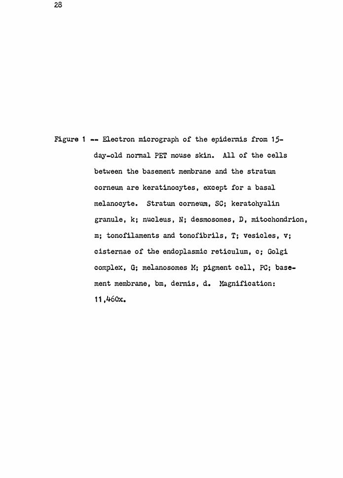
Figure 1 -- Electron micrograph of the epidermis from 1.5-
day-old normal PET mouse skin. All of the cells
between the basement membrane and the stratum
corneum are keratinooytes, except for a basal
melanocyte. Stratum corneum, SC; keratohyalin
granule, k; nucleus, N; desmosomes, D, mitochondrion,
m; tonofilaments and tonofibrils, T; vesicles, v;
cisternae of the endoplasmic reticulum, c; Golgi
complex, G; melanosomes M; pigment cell, PC; base
ment membrane, bm, dermis, d. Magnification:
11,460x.

29
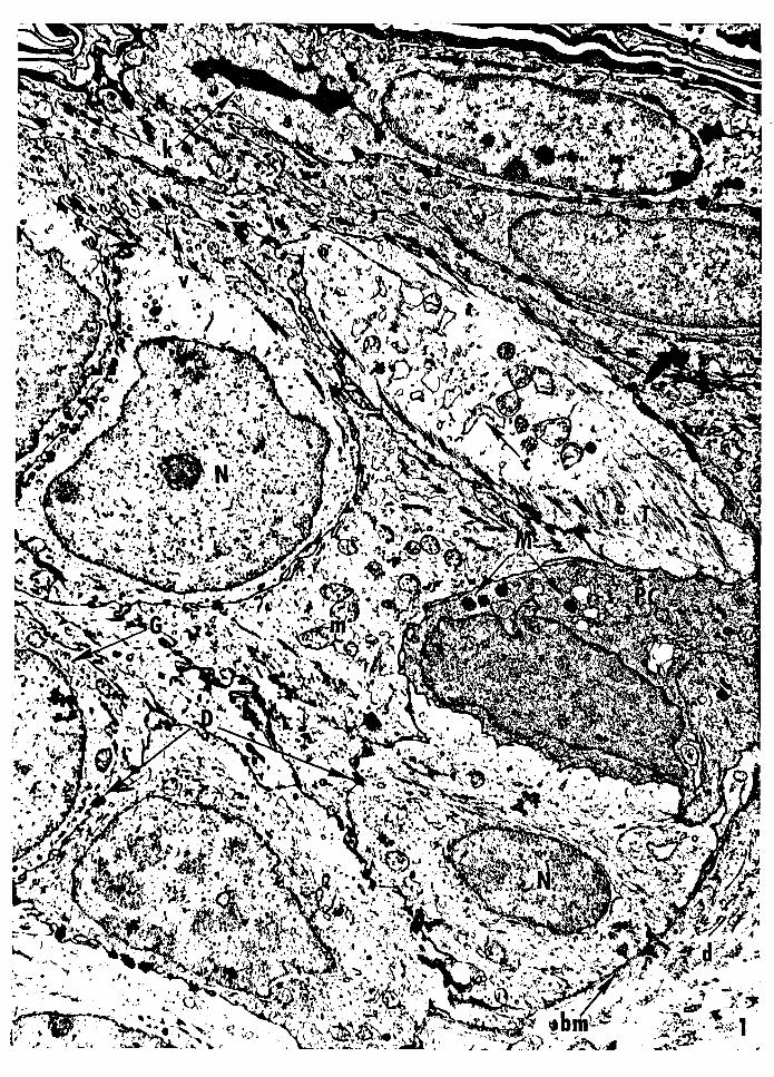
Figure 2 -- Electron micrograph of a dendritic basal melo
cyte of the epidermis from 15-day-old nonnal PET
mouse skin. The nucleus is irregular, and the
cytoplasm is more electron dense than the adjacent
keratinocytes. The cytoplasm lacks tonofilaments
and tonofibrils, but contains melanosomes. The
plasma membrane lacks desmosomes. Nucleus, N;
melanosomes, M; tonofilaments and tonofibrils, T;
keratinocyte, K; dermis, d; desmosome, D.
Magnification: 19,680.
JO
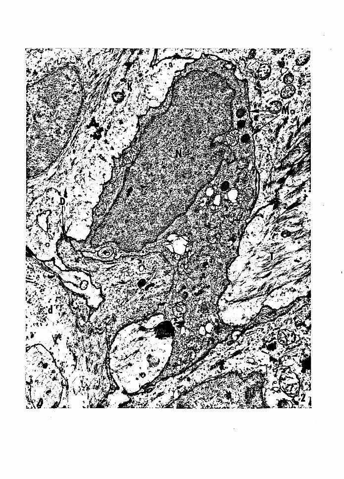
Figure J -- Electron micrograph or a basal epidermal Langerhans
cell from 1 ,5-day-old PET mouse skin. The nucleus is
convoluted, and the cytoplasm is more electron dense
than the adjacent keratinocytes. The cytoplasm lacks
tonofilaments and tonofibrils, and the plasma membrane
lacks desmosomes. A characteristic Langerhans granule
and a centriole are present in the cytoplasm. Nucleus,
N; Langerhans granule, G; centriole, C; keratinocyte, K;
dermis, d. Magnification: 46, 1 OOx.

! ,· ; I.· ·•:•
31
Figure 4 -- Electron micrograph of two juxtaposed suprabasal
epidermal Langerhans cells from 15-day-old PET mouse
skin. The nucleus is convoluted and the cytoplasm is
more electron dense than that of the surrounding kera
tinocytes. The cytoplasm lacks tonofilaments and tono
fibrils, and the plasma membrane lacks desmosomes. A
Langerhans granule is present in the cytoplasm of the
lower cell, and membrane bound melanosomes are present
in the cytoplasm of the upper cell. Nucleus, N;
Langerhans granule, G; membrane bound melanosome, M;
keratinocyta, K; tonofilaments and tonofibrils, T;
desmosomes, D. Magnification: 22,960x.

32
Figure S -- Electron micrograph of two juxtaposed suprabasal
epidermal Langerhans cells from 15-day-old PET mouse
skin. The nucleus is convoluted and the cytoplasm
is more electron dense than that of the surrounding
keratinocytes. The cytoplasm lacks tonofilaments
and tonofibrils, and the plasma membrane lacks
desmosomes. A rod-shaped granule, attached to what
appears to be a vesicle, is continuous with the
plasma membrane. Nucleus, N; tonofilaments and
tonofibrils, T; Langerhans granule, G; keratinocyte,
K. Magnification: 48,405x.

f, �-.,r-' .•. ,.J,:.:.
' ... �
! •
,:"'l ' '

33
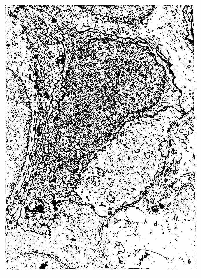
Figure 6 -- Electron micrograph of a basal nonkeratinocyte
from 1.5-day-old PET mouse skin. This cell has
characteristics of both epidermal Langerhans cells
and basal melanocytes, exc:ept it lacks the charac-,
teristic granules. Nucleus, N; electron dense
cytoplasm, c; keratinocyte, K; dermis, d. Magni
fication: 22,128x.
, , .�
-:\� :, . . ."-'
� d . •' ... ,·!J\
. . . ,. •
. ' ,, . ... "
... •
•>
-t�r-· ... --� .. -�. :" .:t .. .. �
6 . . ·. ,.
L
Figure 7 -- Electron micrograph of part of a suprabasal
dendritic semikeratinocyte from J-day-old normal
PET mouse epidermis. The cytoplasm is relatively
clear compared to the surrounding keratinocytes,
and contains a reduced number of ribosomes. ¥J.ni
desmosomes are seen along the plasma membrane.
Ribosomes, r; minidesmosomes, md; semikeratino
cyte, S; keratinocyte, K. Magnification: 16,110x.
35
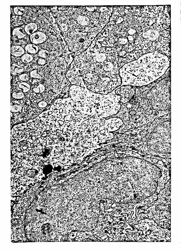
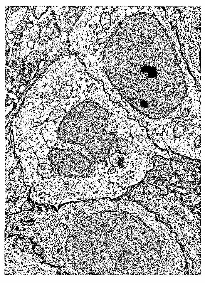
Figure 8 -- Electron micrograph of a basal semikeratinocyte
(type A) from neural crest-free PET mouse epidermis.
The nucleus is irregular and the cytoplasm is relatively
clear compared to that of the adjacent keratinocytes.
The cytoplasm contains a reduced number of ribosomes,
and the plasma membrane possesses several minides
mosomes. Nucleus, N; ribosomes, r; minidesmosomes,
md; semikeratinocyte, S; keratinocyte, K. Magni
fication: 1S,280x.
36
Figure 9 -- Electron micrograph of a basal semikeratinocyte
(type A) from neural crest-free epidermis. The
nucleus appears to be acquiring a convoluted. shape.
Nucleus, N; semikeratinocyte, S; minidesmosome, mi;
dermis, d. Magnification: ;6,900x.


37
Figure 10 -- Electron micrograph of a semikeratinocyte (type A)
migrating out of the basal layer into the suprabasal
layers. The nucleus appears to be acquiring a con
voluted shape. Nucleus, N; semikeratinocyte, S;
dermis, d. Magnification: 19,680x.


JS
Figure 11 Electron micrograph of a dendritic suprabasal
semikeratinocyte (type A). The nucleus is irregular
and the cytoplasm is relatively clear compared to that
of the adjacent keratinocytes. The cytoplasm contains
fewer ribosomes than the adjacent keratinocytes.
Nucleus, N; semikeratinocyte, S; ribosomes, r;
keratinocyte, K. Magnifiaction: 22,960x.
--
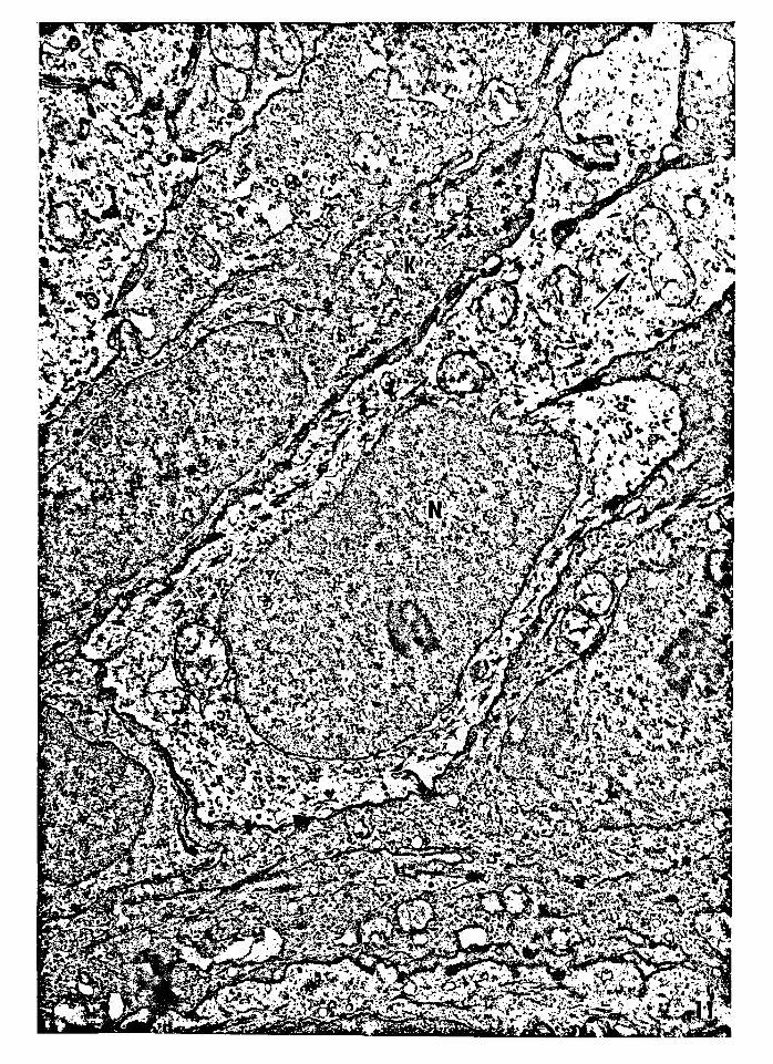
39
Figure 12 -- Electron micrograph of suprabasal dendritic
semikeratinocytes (type A). Semikeratinocyte, s.
Magnification: 12,606x.
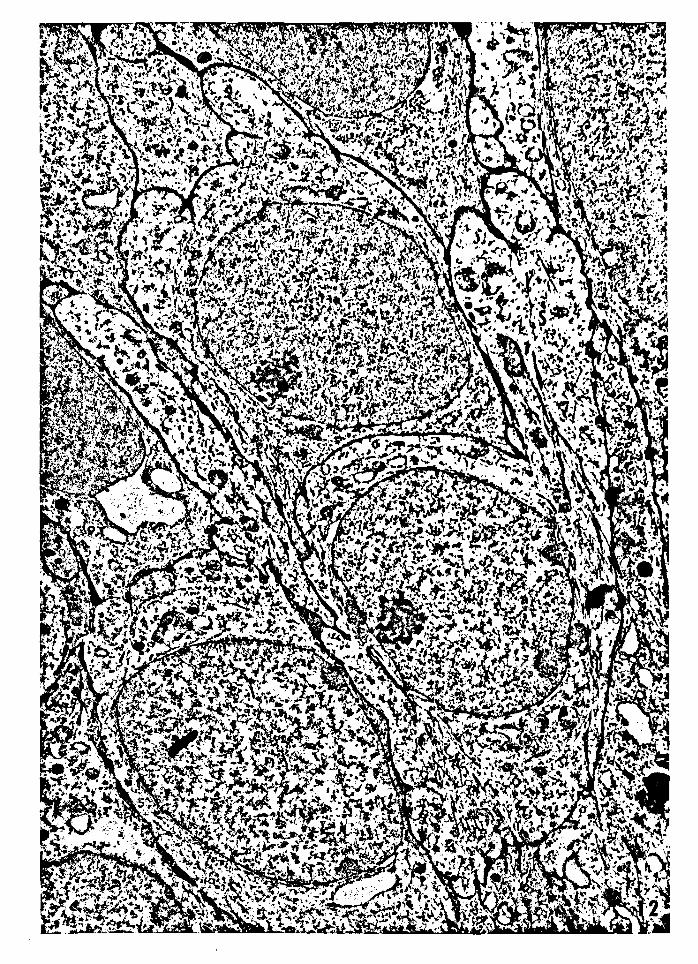
40
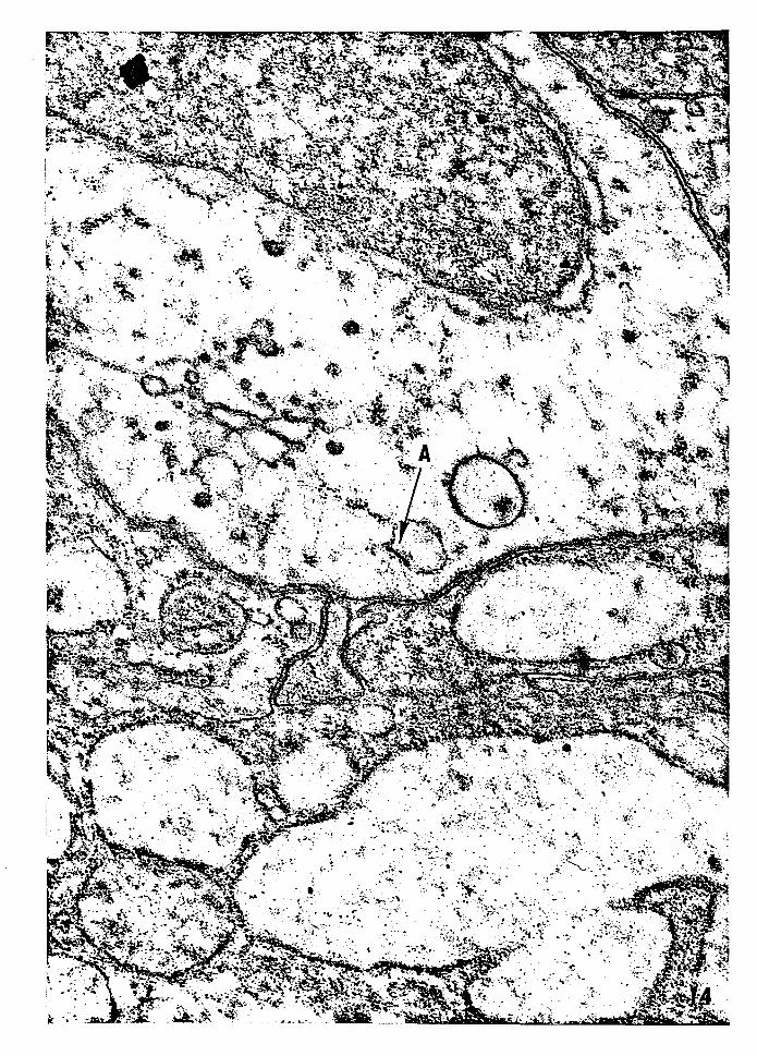
Figure 13 -- Electron micrograph of a suprabasal semikeratino
cyte (type A). The nucleus is irregular and the cyto
plasm is relatively clear compared to that of the sur
rounding keratinocytes. The cytoplasm contains few
ribosomes, and a vacuole is lined with the remnant of
an autophagocytized desmosome. Nucleus, N; ribosomes,
r; remnant of an autophagocytized desmosome, A; semi
keratinocyte, S; keratinocyte, K. Magnification:
29,910x.

41
Figure 14 -- Electron micrograph of a high magnification of
Figure 13 showing the vacuole oontaining an auto
phagocytized desmosome (A). Magnification: 44,850x.
42
Figure 15 -- Electron micrograph of suprabasal dendritic semi
keratinocyte (type B). The nucleus is irregular and
the cytoplasm is more electron dense than that of
the adjacent keratinocytes. The plasma membrane
appears to lack desmosomes. Nucleus of the semi
keratinocyte, N; keratinocyte, K. Magnification:
34,114x.
4J
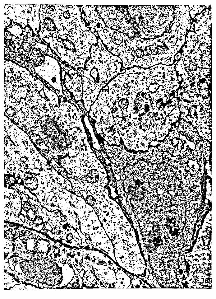
Figure 16 -- Electron micrograph of a basal dendritic semi
keratinocyte (type B). The nucleus is irregular
and the cytoplasm is more electron dense than that
of the adjacent keratinocytes. A dendritic process
is extending into the superficial cell layers. The
plasma membrane possesses a few minidesmosomes.
Nucleus of the semikeratinocyte, N; minidesmosomes,
mi; dendritic process, P; keratinocyte, K; dermis, d.
Magnification: 17,056x.

VITA
Stanley Powell Tompkins was born in Portsmouth, Virginia,
on April 18, 1943. He received his elementary and secondary
education through the Norfolk County public schools and was
graduated from Churchland High School in June, 1961. He began
his 'undergraduate studies at Old Dominion College in September,
1961. After attending Virginia Polytechnic Institute, he
returned to Old Dominion College where he received a Bachelor
of Science degree in biology in June, 1967.
44
In September, 1967, he entered the Graduate School at the
University of Richmond to pursue studies leading to a Master of
Science degree in biology. During his graduate studies, he was
elected to membership in the Beta Beta Beta Biological Honor
Society and the Atlantic Estuarine Research Society. He assisted
in the General Biology laboratories, and was a research assistant
in electron microscopy in the Department of Dermatology at the
Medical College of Virginia, under the supervision of Dr. W. M.
Reruns, Jr. While engaged in this research, he was a coauthor
on the following paper:
Reams, W. M., Jr. and S. P. Tompkins (1969) An
EM study of the dendritic keratinocytes of the PEI' mouse. � � Bulletin, 16(2) : €:A.

Upon receiving a Master of Science degree in biology in
August, 1969, he plans to pursue a professional career through
studies in the School of Dentistry at the Medical College of
Virginia.
He is married to the former Patricia Virginia Tynes.
4S