Embed Size (px)
Citation preview

Case ReportAn Ulcerated Ileal Gastrointestinal Stromal Tumor Disguised asAcute Appendicitis
Ashish Lal Shrestha 1 and Girishma Shrestha2
1Department of General Surgery, United Mission Hospital, Tansen, Palpa, Nepal2Department of Pathology, Patan Academy of Health Sciences, Lagankhel, Kathmandu, Nepal
Correspondence should be addressed to Ashish Lal Shrestha; [email protected]
Received 7 March 2018; Accepted 26 April 2018; Published 5 June 2018
Academic Editor: Boris Kirshtein
Copyright © 2018 Ashish Lal Shrestha and Girishma Shrestha. This is an open access article distributed under the CreativeCommons Attribution License, which permits unrestricted use, distribution, and reproduction in any medium, provided theoriginal work is properly cited.
Background. Gastrointestinal stromal tumor (GIST) of the ileum is not a common differential to consider in the management ofacute right iliac fossa (RIF) pain and tenderness. Finding of a normal-looking appendix intraoperatively should arouse thesurgeon to explore further and look for other unanticipated pathologies. We present a case, clinically diagnosed as acuteappendicitis and intraoperatively found to be an ulcerated ileal GIST. Case Presentation. A 28-year-old female without previouscomorbidities presented to the emergency unit with sudden pain around the umbilicus that later migrated and localized to theRIF for one day. There was associated intermittent fever and anorexia without urinary symptoms. Abdominal examinationrevealed guarding and rebound tenderness at RIF. Examination by 2 senior surgeons at different points of time, the same day,made a clinical diagnosis of acute appendicitis. Ultrasonogram (USG) was inconclusive. At laparotomy through Lanz incision,the appendix was found to be normal and no other pathology was identified on walking bowel up to 3 ft proximal to ileocecaljunction (ICJ). Just when closure was thought of, an ulcerated lesion could be seen through the medial aspect of the incision. Onfurther exploration, a 7× 5 cm ulcerated lesion arising from the antimesenteric border of the ileum was noted with localizedinterloop hemoperitoneum and inflammatory exudates. Ileal segmental resection anastomosis was done with peritonealtoileting. The lesion was subsequently reported to be an ulcerated malignant GIST. Conclusion. The commonest cause of RIFpain with localized peritonitis is an acutely inflamed appendix. Dilemma arises when the appendix is found to look normal.Further exploration is indicted to not miss other findings.
1. Introduction
The term “GIST” was first introduced by Mazur and Clark in1983 to include the nonepithelial tumors of digestive tractthat lack ultrastructure of smooth muscle cells and immuno-histochemical properties of Schwann cells. GISTs are knownto arise from the interstitial cells of Cajal that are regarded asthe pacemaker cells, constituting a part of the autonomicnervous system of the gut and controlling intestinal peri-stalsis [1]. GISTs may vary in presentation and sometimesmimic other commoner conditions. We report an interestingcase of an ulcerated small bowel GIST that behaved clinicallylike acute appendicitis. The clinical presentation, investiga-tive findings, and management are discussed along withrelevant literatures.
2. Case Presentation
A 28-year-old female with insignificant past medico surgicalhistory presented with one day of acute onset pain in theperiumbilical region that later migrated and confined to theRIF. She had associated intermittent fever, nausea, and lossof appetite. She did not have any urinary symptoms, bowelirregularities, or gynecological complaints. Abdominal exam-ination was performed by two senior surgeons at twodifferent occasions; the same day had findings of guardingand rebound tenderness at RIF. Hematological tests showedpolymorphonuclear leukocytosis with left shift. Biochemicaltests and urinalysis were normal. Urinary pregnancy testwas negative. Abdominal radiographs were unremarkable.USG could not visualize appendix and was inconclusive
HindawiCase Reports in SurgeryVolume 2018, Article ID 1320107, 5 pageshttps://doi.org/10.1155/2018/1320107

except for probe tenderness in RIF. CT scan of the abdomencould not be done due to unavailability. A clinical diagnosisof acute appendicitis was made assigning an Alvarado scoreof 9/10. Laparotomy was performed using the Lanz incisionin RIF. Intraoperatively appendix was found to be normalwithout evidence of inflammation or infection in RIF. Inview of symptoms and signs, a possibility of other pathologywas thought. Walking the bowel proximally up to 3 feet(1m) did not show a Meckel’s diverticulum or any othersmall bowel lesions. There were no obvious mesentericlymph nodal enlargement and pelvic organs looked pristine.Approaching closure, just when the medial edge of theincision was retracted superomedially, a hemorrhagic lesionseemed to appear little deeper in the mid abdomen. There-fore, the incision was extended transversely from the medialedge to explore further. Entire bowel was explored and thisrevealed an ulcerated lesion measuring 7× 5 cm arising fromthe antimesenteric border of the ileum 8 feet (2.5m) fromICJ with localized interloop hemoperitoneum and inflam-matory exudates as shown in Figure 1. Resection of ilealsegment containing the lesion was performed followed byrestoration of bowel continuity and peritoneal toileting.The lesion was subsequently reported to be an ulceratedmalignant ileal GIST.
Histopathologically, gross examination confirmed theoperative findings, and the cut section revealed a nodularlesion protruding out of the serosal surface measuring7× 5 cm along with 2 lymph nodes each measuring 2× 1 cm.
Microscopically, the growth from the ileum had villouslining epithelium with focal ulceration. The submucosalregion had a circumscribed nodule with proliferation ofloosely cohesive spindle cells; some of which were arrangedin vague storiform pattern and others in long fascicles. Therewere areas with epitheloid cells forming small anastomosingnests and cords. The areas in between these showed skenoidfibers along with focal areas of hemorrhage, infarction, andcongestion as shown in Figure 2. The mitotic figures wereseen (8/50 high-power field). The lymph nodes were micro-scopically identified to be reactive, and the resected marginsof the ileum were free of tumor.
Based on tumor size and mitotic activity, possibility ofa malignant GIST was suggested along with immunohisto-chemical analysis (CD117 and CD34) for further confir-mation. The patient had an uneventful recovery and wasdischarged on the 8th postoperative day. She was advised toreview a week later at the outpatients but failed to report.All possible contacts were used to trace her, but she remainedinaccessible and lost to follow-up.
3. Discussion
It is a common clinical situation to have a patient present-ing with periumbilical pain subsequently localizing to theRIF associated with vomiting with or without nausea andfever. The classical symptom complex called Murphy’striad is often observed and tends to occur in the samesequential order [2]. The findings of guarding at the RIF withMcBurney’s point tenderness are suspicious of acute appen-dicitis along with various named signs [3]. Leukocytosis
and neutrophilic left shift added to the USG findings ofa noncompressible blind tube> 6mm in RIF with probetenderness strongly impress upon the surgeon to wait nofurther before embarking on an emergency appendectomy.The usual finding is that of an inflamed appendix with orwithout associated complications (gangrene, perforation,or periappendicular collection). Figure 3 shows an uncom-plicated appendicitis in a different patient.
The annual global incidence of appendicitis is reported tobe 11 cases per 10,000 population [4]. In one study, the sen-sitivity and specificity of clinical examination to diagnoseappendicitis were 99% and 76% and the same for USG were99% and 91%, respectively [5]. Various scoring systems havealso been devised to aid accurate preoperative diagnosis, forexample, Alvarado, Ohhmann, Eskelinen, and RIPASA, andreport a wide range of variability in sensitivity, specificity,and predictive validity in different comparative studies [6].Despite our long-term experience in treating this condition,there have been several incidences of finding an unantici-pated pathology intraoperatively and the “On Table Sur-prise” does not stop to amaze us even now. In most series, anegative appendectomy rate of 10–20% is considered accept-able though newer studies quote an even lesser rate [7, 8]. Anormal-looking appendix certainly arouses the surgeon tosuspect something sinister and thence the usual tendency tolook for conditions like an inflamed Meckel’s diverticulum,the incidence of which is said to be 2%. The other pathologiesthat may be encountered are mesenteric lymphadenitis, largeor small bowel diverticulitis, right ureteric pathology, and awide variety of gynecological ailments like ruptured ovarianfollicle with midcycle ovulatory bleeding (Mittelschmerz’s),ovarian torsion, salpingitis, and ruptured ectopic pregnancyespecially in women of child bearing age [9]. But a rupturedsmall bowel GIST is certainly not the prime suspect underusual circumstances. GISTs are known to us since thetime they were first reported by Mazur and Clark in1983. They have constantly made their presence felt invarious case reports globally when they were not recognizedpreoperatively. Refractory peptic ulcer disease, gastrointesti-nal bleeding, pneumomediastinum, acute diffuse peritonitis,abdominal abscesses, and sudden perforation with hemoper-itoneum have all been the various modes of presentation ofGISTs [10–15]. One similar incidence of a GIST mimicking
Figure 1: The intraoperative image of the ulcerated ilealGIST arising from the antimesenteric border with interloophemoperitoneum and inflammatory exudates.
2 Case Reports in Surgery
appendicitis was found reportedly from the jejunum; how-ever, ours was one from the ileum [16]. The usual age ofpresentation of GIST is 40 to 60 years and the common sitesof origin are the stomach and followed by small bowel andcolorectum and rarely esophagus. Although many are diag-nosed incidentally, some with advanced disease present withsymptoms that include nonspecific abdominal pain and largeabdominal masses. Occasionally, luminal erosion of a highlyvascular GIST may present with a life-threatening gastroin-testinal hemorrhage, while on account of luminal narrowing,the other forms of presentation may be obstruction andperforation. Tumor rupture in this regard seems to be moredreadful condition; in that, it carries risks of tumor dissemi-nation that can be difficult to treat apart from hemoperito-neum and acute abdomen. Some may even present with
metastasis to the liver and peritoneum and very rarely tothe lungs. Also of note are local spread to adjacent viscera likethe intestine, omentum, and diaphragm. Cross-sectionalimaging with a CT scan is helpful in identifying the extentof lesion and studying the characters like necrosis, ulceration,calcification, ascites, and local and distant metastasis thatdenote the aggressive nature of primary lesion to plan asubsequent operative therapy. PET scan is considered animportant adjunct to CT in evaluation and in order to assessresponse to chemotherapy [1]. In an acute setting, and in aperipheral set up like ours, both these modalities are onlyof theoretical value. Similarly, endoscopic ultrasound andFNAC are invaluable in preoperative tissue diagnosis incenters where the expertise is available. Definitive diagnosisis possible with histopathological examination of the tissueaided by immunohistochemistry (IHC); the current panelof which includes CD117, smooth muscle actin, CD34,desmin, and S-100. Unfortunately, in our case, it was unavail-able and had to be sent to a tertiary care center on receivingthe histopathological report. Since the patient did not followup in the postoperative period, it could not be done. Anextensive search in PubMed, Medline, and Google in refer-ence to GIST misdiagnosed as appendicitis was done from2000 till now. Only 4 cases were found to have been reportedworldwide of which 1 was in the stomach, 2 were in thejejunum, and 1 was in the ileum [16–19]. This was the secondreport of similar presentation of GIST in the ileum. In all thecases, treatment approach was surgical with laparoscopicresection in 1 and open resection in the rest. Our patientunderwent open resection with an uneventful recovery.
(a) (b)
(c)
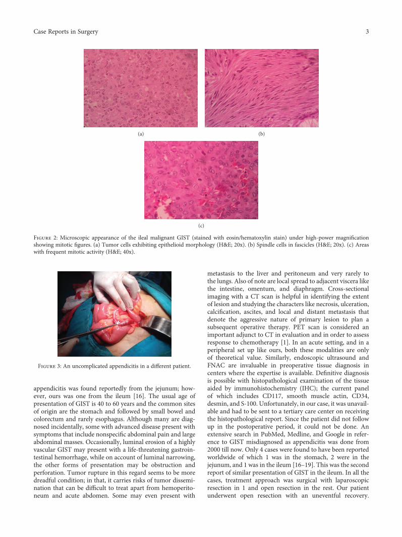
Figure 2: Microscopic appearance of the ileal malignant GIST (stained with eosin/hematoxylin stain) under high-power magnificationshowing mitotic figures. (a) Tumor cells exhibiting epithelioid morphology (H&E; 20x). (b) Spindle cells in fascicles (H&E; 20x). (c) Areaswith frequent mitotic activity (H&E; 40x).
Figure 3: An uncomplicated appendicitis in a different patient.
3Case Reports in Surgery

Had she returned for follow-up, she should have beenevaluated for metastatic disease and further management.But since that was not possible, we could neither plan furthertreatment nor prognosticate her disease. In general, progno-sis of GIST depends upon the size of the tumor and to themitotic rate: tumors> 10 cm or with a mitotic rate of >5 per50 HPF having higher risk of recurrence, metastatic spread,and a poorer prognosis. Other prognostic factors includetumor-free surgical margins, tumor rupture, and c-kit muta-tion [17]. The IHC and other molecular studies could not bedone in our patient.
4. Conclusion
In essence, an ulcerated malignant GIST of the ileummasquerading as acute appendicitis is a common presenta-tion of an uncommon diagnosis. Disproportionate symptomsand signs inconsistent with a normal-looking appendix ontable should alert the surgeon to suspect other possiblecauses no matter how remote. Negative appendectomyshould not be taken lightly and mandates thorough explo-ration of the entire length of bowel. Definitive diagnosis ispossible on histopathological evaluation aided by IHC.Resection with negative margins and further therapy basedon IHC panel forms the backbone of management. Theawareness of the clinical presentation and good pathologicalexpertise are important adjuncts in the diagnosis. Surgery isthe mainstay of treatment in the acute presentation.
Abbreviations
GIST: Gastrointestinal stromal tumorRIF: Right iliac fossaUSG: UltrasonogramICJ: Ileocecal junctionCT: Computed tomographyIHC: Immunohistochemistry.
Consent
Written informed consent was obtained from the patient forpublication of this case report and accompanying images.
Conflicts of Interest
The authors declare no competing interests regarding thepublication of this paper.
Authors’ Contributions
Ashish Lal Shrestha participated in the surgical and peri-operative management of the patient and conception anddesign of the report and wrote the paper. GirishmaShrestha performed the histopathological analysis of thereport. Both have been involved in the diagnosis and patientcare. Both authors read and approved the final paper. Boththe authors were involved in planning, analysis of the case,and writing of the paper.
Acknowledgments
The authors would like to thank the ward staff of thehospital for providing support and helping in managementof the patient.
References
[1] M. Zinner and S. Ashley, Maingot’s Abdominal Opera-tions, McGraw-Hill Professional, New York, NY, USA,11th edition, 2006.
[2] R. S. Lawson, “Murphy’s triad,” British Medical Journal, vol. 1,no. 5745, pp. 401-402, 1971.
[3] A. Sachdeva and A. K. Dutta, Advances in Pediatrics, JPMedical Ltd, 2012.
[4] A. Petroianu, “Diagnosis of acute appendicitis,” InternationalJournal of Surgery, vol. 10, no. 3, pp. 115–119, 2012.
[5] J. S. Park, J. H. Jeong, J. I. Lee, J. H. Lee, J. K. Park, andH. J. Moon, “Accuracies of diagnostic methods for acuteappendicitis,” The American Surgeon, vol. 79, no. 1, pp. 101–106, 2013.
[6] H. Erdem, S. Çetinkünar, K. Daş et al., “Alvarado, Eskelinen,Ohhmann and Raja Isteri Pengiran Anak Saleha appendicitisscores for diagnosis of acute appendicitis,” World Journal ofGastroenterology, vol. 19, no. 47, pp. 9057–9062, 2013.
[7] D. Papeš, S. Sršen Medančić, A. Antabak, I. Sjekavica, andT. Luetić, “What is the acceptable rate of negative appendec-tomy? Comment on prospective evaluation of the added valueof imaging within the Dutch National Diagnostic AppendicitisGuideline - do we forget our clinical eye?,” Digestive Surgery,vol. 32, no. 3, pp. 181-182, 2015.
[8] M. Colson, K. A. Skinner, and G. Dunnington, “High negativeappendectomy rates are no longer acceptable,” The AmericanJournal of Surgery, vol. 174, no. 6, pp. 723–727, 1997.
[9] D. J. Humes and J. Simpson, “Acute appendicitis,” BMJ,vol. 333, no. 7567, pp. 530–534, 2006.
[10] M. Mokhtare, T. Taghvaei, and H. Tirgar Fakheri, “Acutebleeding in duodenal gastrointestinal stromal tumor,” MiddleEast Journal of Digestive Diseases, vol. 5, no. 1, pp. 47–51, 2013.
[11] M. Sugimoto, T. Hikichi, Y. Shioya et al., “A case of gastroin-testinal storomal tumor with pneumomediastinum,” Fukush-ima Journal of Medical Science, vol. 59, no. 2, pp. 97–101, 2013.
[12] P. Rubini and F. Tartamella, “Primary gastrointestinal stromaltumour of the ileum pre-operatively diagnosed as an abdomi-nal abscess,” Molecular and Clinical Oncology, vol. 5, no. 5,pp. 596–598, 2016.
[13] J. D. Jones, S. Oh, C. Clark, and R. Pawa, “A bleeding duodenalGIST masquerading as refractory peptic ulcer disease,” ACGCase Reports Journal, vol. 3, no. 4, article e189, 2016.
[14] S. Ulusan, Z. Koc, and F. Kayaselcuk, “Spontaneously rupturedgastrointestinal stromal tumor with pelvic abscess: a casereport and review,” Gastroenterology Research, vol. 2, no. 6,pp. 361–363, 2009.
[15] W. Attaallah, Ş. Coşkun, G. Özden, H. Mollamemişoğlu, andC. Yeğen, “Spontaneous rupture of extraluminal jejunal gas-trointestinal stromal tumor causing acute abdomen andhemoperitoneum,” Turkish Journal of Surgery, vol. 31, no. 2,pp. 99–101, 2015.
[16] M. Ajduk, D. Mikulić, B. Sebecić et al., “Spontaneously rup-tured gastrointestinal stromal tumor (GIST) of the jejunum
4 Case Reports in Surgery

mimicking acute appendicitis,” Collegium Antropologicum,vol. 28, no. 2, pp. 937–941, 2004.
[17] E. Elangovan, “A rare case of jejunal GIST presenting asacute abdomen,” University Journal of Surgery and SurgicalSpecialities, vol. 3, no. 2, 2017.
[18] S. A. Badger, M. Yousaf, and W. J. Campbell, “A case of agastrointestinal stromal tumour presenting as acute abdo-men,” Irish Journal of Medical Science, vol. 174, no. 3,pp. 84-85, 2005.
[19] C. Agalar, S. Benli, B. Manoglu, T. Egeli, and M. U. Unlu,“Laparoscopic resection of ileal GIST, mimicking acuteappendicitis,” General Surgery Reports, vol. 1, no. 1, 2017.
5Case Reports in Surgery

Stem Cells International
Hindawiwww.hindawi.com Volume 2018
Hindawiwww.hindawi.com Volume 2018
MEDIATORSINFLAMMATION
of
EndocrinologyInternational Journal of
Hindawiwww.hindawi.com Volume 2018
Hindawiwww.hindawi.com Volume 2018
Disease Markers
Hindawiwww.hindawi.com Volume 2018
BioMed Research International
OncologyJournal of
Hindawiwww.hindawi.com Volume 2013
Hindawiwww.hindawi.com Volume 2018
Oxidative Medicine and Cellular Longevity
Hindawiwww.hindawi.com Volume 2018
PPAR Research
Hindawi Publishing Corporation http://www.hindawi.com Volume 2013Hindawiwww.hindawi.com
The Scientific World Journal
Volume 2018
Immunology ResearchHindawiwww.hindawi.com Volume 2018
Journal of
ObesityJournal of
Hindawiwww.hindawi.com Volume 2018
Hindawiwww.hindawi.com Volume 2018
Computational and Mathematical Methods in Medicine
Hindawiwww.hindawi.com Volume 2018
Behavioural Neurology
OphthalmologyJournal of
Hindawiwww.hindawi.com Volume 2018
Diabetes ResearchJournal of
Hindawiwww.hindawi.com Volume 2018
Hindawiwww.hindawi.com Volume 2018
Research and TreatmentAIDS
Hindawiwww.hindawi.com Volume 2018
Gastroenterology Research and Practice
Hindawiwww.hindawi.com Volume 2018
Parkinson’s Disease
Evidence-Based Complementary andAlternative Medicine
Volume 2018Hindawiwww.hindawi.com
Submit your manuscripts atwww.hindawi.com


![Physical exercise for the treatment of non-ulcerated ...arquivos.info.ufrn.br/arquivos/20141612267d... · [Intervention Protocol] Physical exercise for the treatment of non-ulcerated](https://img.dokumen.tips/doc/110x75/5f0a5ba37e708231d42b3fe1/physical-exercise-for-the-treatment-of-non-ulcerated-intervention-protocol.jpg)