Embed Size (px)
Citation preview

ARTICLE
Received 1 Apr 2016 | Accepted 18 Oct 2016 | Published 8 Dec 2016
An oxygen-sensitive toxin–antitoxin systemOriol Marimon1, Joao M.C. Teixeira1, Tiago N. Cordeiro1,w, Valerie W.C. Soo2, Thammajun L. Wood2,
Maxim Mayzel3, Irene Amata1,w, Jesus Garcıa1,4, Ainara Morera1,w, Marina Gay4, Marta Vilaseca4,
Vladislav Yu Orekhov3, Thomas K. Wood2 & Miquel Pons1
The Hha and TomB proteins from Escherichia coli form an oxygen-dependent toxin–antitoxin
(TA) system. Here we show that YmoB, the Yersinia orthologue of TomB, and its single
cysteine variant [C117S]YmoB can replace TomB as antitoxins in E. coli. In contrast to other TA
systems, [C117S]YmoB transiently interacts with Hha (rather than forming a stable complex)
and enhances the spontaneous oxidation of the Hha conserved cysteine residue to a -SOxH-
containing species (sulfenic, sulfinic or sulfonic acid), which destabilizes the toxin. The
nuclear magnetic resonance structure of [C117S]YmoB and the homology model of TomB
show that the two proteins form a four-helix bundle with a conserved buried cysteine
connected to the exterior by a channel with a diameter comparable to that of an oxygen
molecule. The Hha interaction site is located on the opposite side of the helix bundle.
DOI: 10.1038/ncomms13634 OPEN
1 Biomolecular NMR Laboratory, Organic Chemistry Section, Inorganic and Organic Chemistry Department, University of Barcelona, Baldiri Reixac 10-12,Barcelona 08028, Spain. 2 Department of Chemical Engineering and Department of Biochemistry and Molecular Biology, Pennsylvania State University,University Park, Pennsylvania 16802, USA. 3 Swedish NMR Centre, Gothenburg University, PO Box 465, Gothenburg SE-40530, Sweden. 4 Institute forResearch in Biomedicine (IRB-Barcelona), The Barcelona Institute of Science and Technology, Baldiri Reixac 10-12, Barcelona 08028, Spain. w Presentaddresses: X-ray and Neutron Science, Niels Bohr Institute, Copenhagen, 2100, Denmark (T.N.C.); CNRS FRE 3630 (affiliated with Univ. Paris Diderot,Sorbonne Paris Cite), Institut de Biologie Physico-Chimique, Paris 75006, France (I.A.); Departament de Fısica Fonamental, Martı i Franques, Barcelona 1-1108028, Spain (A.M.). Correspondence and requests for materials should be addressed to T.K.W. (email: [email protected]) or to M.P. (email:[email protected]).
NATURE COMMUNICATIONS | 7:13634 | DOI: 10.1038/ncomms13634 | www.nature.com/naturecommunications 1
Antimicrobial resistance is a major threat to global health1.Biofilms are resistant communities of microorganismsattached to surfaces and encapsulated in a matrix2,3.
Biofilms are involved in 80% of human bacterial infections4 andconfer antibiotic resistance5.
Oxygen consumption in the biofilm generates oxygen gradi-ents6,7 and may be linked to biofilm dispersal8. Selective celldeath, for example, in anoxic regions, creates channels thatfacilitate nutrient-waste product exchange9–11 and biofilmdispersion12. Dispersed cells are more susceptible to antibioticsthan those in biofilms, although they are also distinct fromplanktonic cells in terms of gene expression and pathogenicity13.
Classical toxin–antitoxin (TA) systems are based on silencingof a stable toxin by a labile antitoxin that, when inactivated,releases the toxin, resulting in a reduction in metabolism. TAsystems modulate the generation of persister cells14, phageinhibition15 and biofilm regulation16.
The Hha/TomB TA system is part of the first group of TAsystems identified in biofilms17. The overexpression of Hha(haemolysin expression modulating protein) causes cell lysisand reduction of biofilm formation as well as increases biofilmdispersal18. An engineered variant of Hha caused nearlycomplete biofilm dispersal via cell lysis19,20, and deletion ofhha led to no dispersal of Escherichia coli BW25113 (ref. 20)corroborating the role of Hha in biofilm dispersal. TomB (toxinoverexpression modulator in biofilms, previously known asYbaJ) inactivates Hha toxicity18 although the mechanism hasnot been elucidated.
Here we report that the TomB antitoxin activity isoxygen-dependent and that its Yersinia orthologue, YmoB canreplace TomB as the Hha antitoxin. A single cysteine variant[C117S]YmoB is similarly active in vivo and promotes theoxidation of the single conserved cysteine of Hha to -SOxH(sulfenic, sulfinic and sulfonic acids) by air in the absence of anyexternal source of reactive oxygen species. Oxidation of Hha orthe introduction of a negative charge at the position of thecysteine residue, mimicking the presence of -SOx
� , causes itsdestabilization and results in reduced toxicity.
The three-dimensional (3D) structure and also the Hhatransient interaction site of [C117S]YmoB were determined bynuclear magnetic resonance (NMR). Relevant structural featuresinclude a buried cysteine residue located close to the Hha-bindingsite and connected to the opposite side of the four-helix bundle bya narrow channel with a width comparable to that of an oxygenmolecule. The built-in oxygen sensor in this TA system mayprovide a mechanism for environmental regulation of cellactivity.
Taken together, these observations suggest that Hha/TomBrepresent a TA system based on the inactivation of the toxin(Hha) by oxidation with molecular oxygen mediated by theantitoxin (TomB).
ResultsTomB antitoxin activity is oxygen-dependent. We tested thehypothesis that the Hha-TomB pair was an oxygen-dependentTA system by measuring E. coli growth at increasing agitationrates (Fig. 1). Overexpression of Hha has a toxic effect in minimalmedium, resulting in a decrease of the growth rate and the celldensity in the stationary phase. Growth in rich medium showedan even higher toxicity18. Simultaneous overexpression of Hhaand TomB in minimal medium results in biphasic growthgradually evolving from the slow growth rate observed when Hhais overexpressed in the absence of TomB to the faster growthobserved in the controls not expressing Hha. A significantantitoxin effect of TomB was observed after B4 h, and after 6 h,
cultures overexpressing both Hha and TomB reached the samecell densities as the controls.
Experiments at the two extreme agitation rates were repeatedwhile simultaneously measuring oxygen saturation, using anoxygen-sensitive polarographic electrode (Fig. 1b). Oxygen levelsreflect the balance between air uptake, enhanced by fast agitation,and oxygen consumption by the metabolic activity of the bacteria.At an agitation rate of 100 r.p.m., the oxygen saturation levels ofthe control and HhaþTomB expressing cultures reached aminimum of 1% while at an agitation rate of 250 r.p.m., theoxygen levels were always higher than 30%. Oxygen levelsincreased when the cultures approached the stationary phase. Thelonger lag time for the antitoxin activity at slow agitation rates isconsistent with oxygen being required for TomB activity as anantitoxin.
At the agitation rate of 250 r.p.m., the oxygen levels of thecultures overexpressing only Hha were always higher than thecontrols, reflecting a lower oxygen consumption in the presenceof the toxin. At 100 r.p.m., cultures expressing HhaþTomB oronly Hha reached similar oxygen levels (10% saturation) in latestationary phase in spite of the fact that the cell density, asmeasured by the turbidity at 600 nm, was lower in the culturesexpressing only the toxin. However, in these cultures, the numberof colony forming units (CFU) was lower than in those expressingalso the antitoxin (Supplementary Table 1). These observationssuggest that 10% oxygen saturation is enough to enable TomBantitoxin activity. Interestingly, the observation that the numberof CFU is higher in the samples overexpressing the two proteins(HhaþTomB) than in those only expressing Hha rules out thepossibility that the observed toxicity of Hha is only related to themetabolic stress associated with protein overexpression andconfirms that the toxicity of Hha can be compensated by TomB.
These observations suggest an oxygen-dependent TA mechan-ism that we investigated in detail by combining chemical biologyand structural studies.
YmoB protects E. coli cells from Hha toxicity. A non-redundantcollection of 250 TomB homologues was obtained by BLASTsearch using as a query the TomB sequence from E. coli K-12strain (D64776). The sequences (Supplementary Fig. 1), repre-sentative of the YbaJ-superfamily, contain from one to fivecysteines. Cysteine 18 (in 244 sequences) and a second cysteinelocated towards the C-terminus (235/250 sequences) are mostconserved. Attempts to obtain suitable constructs to carry outstructural studies of TomB failed, so we focused on its Yersinia’shomologue YmoB, which has 58.1% pairwise identity, 73.6%pairwise similarity and contains the two most conserved cysteines(C18 and C117, Fig. 2a). Its single cysteine variant [C117S]YmoBproved suitable for structural determination by NMR, as shownbelow.
We first explored if YmoB and [C117S]YmoB could function-ally replace TomB in preventing Hha-associated toxicity in E. coliin planktonic cells and in biofilms. Planktonic cell culturesoverexpressing Hha and either of the two YmoB variants reachedthe same cell density of the controls not expressing Hha oroverexpressing both Hha and TomB (Fig. 2b), although theantitoxin activity of the YmoB variants showed a longer lag timethan TomB. The C117S substitution had no significant effect onYmoB antitoxin activity in planktonic cells.
Overexpression of Hha causes a fourfold decrease of standardbiofilm formation (Fig. 2c). The co-expression of TomB or YmoBtotally restored biofilm formation, although the C117S variantwas slightly less efficient. Regarding biofilm morphology, Hhaoverexpression leads to amorphous biofilms with a higherpercentage of dead cells (Live/Dead cells ratio of 5.6±1.4,
ARTICLE NATURE COMMUNICATIONS | DOI: 10.1038/ncomms13634
2 NATURE COMMUNICATIONS | 7:13634 | DOI: 10.1038/ncomms13634 | www.nature.com/naturecommunications

quantitative analysis by COMSTAT21 software). Instead, whenYmoB was co-expressed, the Live/Dead cell ratio significantlyincreased to 35.4±8.5 and the biofilm was composed bymicrocolonies uniformly spread on the entire surface (Fig. 2d).Thus, YmoB or [C117S]YmoB can functionally replace TomB asthe Hha antitoxin in E. coli.
[C117S]YmoB enhances Hha oxidation in vitro. Hha has asingle cysteine residue (C18) highly conserved in the Hha-familyof proteins. We hypothesized that the oxygen-dependent anti-toxin activity of TomB and [C117S]YmoB towards Hha could berelated to the oxidation of this conserved cysteine residue.
In addition to their capacity to form disulfide-bonds, cysteinethiol groups can be oxidized to sulfenic (-SOH), sulfinic (-SO2H)and sulfonic (-SO3H) acids22. Sulfenic acid formation is reversiblebut sulfinic and sulfonic acid are irreversibly formed and result inthe introduction of a negative charge. Hha has an unusual highproportion (33%) of charged residues and its structure is highlysensitive to electrostatic effects23. The additional chargeintroduced by cysteine oxidation could destabilize Hha andprevent its inherent toxicity.
To investigate the effect of YmoB on the oxidation of C18 ofHha in vitro, we used mass spectrometry. Identical samples ofHha were incubated in the presence and in the absence of[C117S]YmoB for 30 min. Hha was isolated by size exclusionchromatography (SEC), digested with trypsin and analysed by
mass spectrometry to identify cysteine-containing peptides intheir reduced and oxidized (-SOxH) forms. Figure 3a shows adecrease in the reduced form and an increase in the oxidizedforms of Hha in the presence of [C117S]YmoB. The differenceswith the untreated Hha samples are statistically significantaccording to a Student t-test with P¼ 0.03 (for sulfonic acid)and P¼ 0.02 (reduced and other oxidized forms). Variants ofYmoB in which F111 had been mutated showed no effect(Fig. 3b). The choice of the phenylalanine mutants was based onthe structural studies described below. Interestingly, even in theabsence of YmoB, Hha contains a significant proportion ofpeptides with oxidized forms of cysteine suggesting that the effectof YmoB is to enhance its spontaneous oxidation. The Hhahomologue in Yersinia, the protein YmoA, showed similaroxidation effects (Supplementary Fig. 2).
A negative charge at the cysteine position destabilizes Hha. Wemimicked the effect of adding a negative charge at the cysteineposition by mutating it to glutamic acid, and we measured thegrowth of E. coli Dhha expressing the [C18E]Hha variant. Ascontrols, we used wild-type Hha and a previously studied C18Ivariant24. While [C18I]Hha retains its capacity to act as a toxin,the presence of a negative charge at this position substantiallyreduces the toxicity of the C18E variant (Fig. 4a).
The expression levels of wild type, C18I and C18E variants ofHha are comparable after 4 h of induction at 37 �C (Fig. 4b).
% O
2 sa
tura
tion
% O
2 sa
tura
tion
100
80
60
40
20
0
100
80
60
40
20
0
100 r.p.m.
1.6
1.4
1.2
1.0
0.8
0.6
0.4
0.2
0.00 1 2 3 4 5 6 7 8 9 10
0 1 2 3 4 5 6 7 8 9 10
1.6
1.4
1.2
1.0
0.8
0.6
0.4
0.2
0.0
3.0
2.0
1.0
0.03.0
2.0
1.0
0.0
O.D
. (60
0 nm
)
O.D
. (60
0 nm
)O
.D. (
600
nm)
3.0
2.0
1.0
0.03.0
2.0
1.0
0.00 2
Time (h) Time (h)
Time (h)
4 6 8 10
150 r.p.m.
200 r.p.m.
250 r.p.m.
ControlHha+Hha+ TomB+
Control
O.D.(600 nm)
%O2saturation
Hha+Hha+ TomB+
100 r.p.m.
250 r.p.m.
a b
Figure 1 | TomB antitoxin activity depends on the agitation rate. (a) Growth curves of E. coli K-12 MG1655 Dhha cells harbouring pCA24N-hha and
pBAD30-tomB at (from top to bottom) 100, 150, 200 and 250 r.p.m. agitation rates. Control cultures with no expression of Hha or TomB (squares) were
compared with cultures expressing only Hha (circles) and co-expressing both Hha and TomB (diamonds). Growth curves were measured in triplicate at
37 �C in tryptone minimal medium (TMM; 10 g l� 1 tryptone and 2.5 g l� 1 NaCl). Error bars represent the sample s.d. (b) Growth curves and simultaneous
measurements of the oxygen saturation during culture growth at 150 r.p.m. (top) and 250 r.p.m. (bottom) agitation rates.
NATURE COMMUNICATIONS | DOI: 10.1038/ncomms13634 ARTICLE
NATURE COMMUNICATIONS | 7:13634 | DOI: 10.1038/ncomms13634 | www.nature.com/naturecommunications 3

TomB *YmoB
ControlHha+Hha+ TomB+Hha+ YmoB+Hha+ [C117S] YmoB+
Sta
ndar
d bi
ofilm
form
atio
n
O.D
. (60
0 nm
)0.4
0.3
0.2
0.1
0
1.8
1.2
0.6
0.0
Time (h)
Dead Dead+LiveLive
Control Hha Hha+TomB+
Hha+YomB+
Hha+[C117S]YomB+
2 4 6 8 10
Hha
1 un
it ce
ll: 1
0 µm
Hha
+W
TY
moB
+
*a
b c
d
Figure 2 | Yersinia’s YmoB protects E. coli from toxic effect of Hha. All experiments were carried out using E. coli K-12 MG1655 Dhha cells harbouring
pCA24N-hha and pBAD30-tomB, pBAD30-ymoB or pBAD30-ymoB(C117S), at 37 �C and in TMM. (a) Sequence alignment of TomB/YmoB proteins.
Asterisks highlight the position of conserved cysteine residues at positions 18 and 117. (b) Growth curves of control cultures with no expression of a TA pair
of proteins (squares) were compared with cultures overexpressing: Hha (circles), Hha and TomB (diamonds), Hha and YmoB (triangles up) or Hha and
[C117S]YmoB (triangles down). Experiments were performed in duplicate, and the error bars are the sample standard deviation. (c) Microtitre plate biofilm
assay. Total biofilm formation was measured at OD540nm and was standardized as OD540nm/OD620nm. Experiments were performed in duplicate, and the
error bars are the sample standard deviation. (d) Representative IMARIS (BITplane, Zurich, Switzerland) images of flow cell biofilm with Hha
overexpression (up) and Hha and YmoB co-expression (down). Dead cells (left) are shown in red and live cells (centre) in green, while superimpositions of
dead and alive cells are shown on the right. OD, optical density.
100a b
Hha
Hha + [C117S]YmoB
*
**
HhaHha + [F111L, C117S]YmoB
Hha + [F111Y, C117S]YmoB80
60
40
20
% C
ox/C
tota
l
0No Ox Sulfenic Sulfinic Sulfonic
100
80
60
40
20
% C
ox/C
tota
l
0No Ox Sulfenic Sulfinic Sulfonic
Figure 3 | [C117S]YmoB enhances the oxidation of Hha. The bars represent the percentage of peptide spectrum matches (PSM) to the various oxidation
states relative to the total PSM of cysteine-containing peptides in the mass spectra of trypsin-treated samples of Hha. Parallel experiments were performed
by incubating Hha for 30 min without any added YmoB variant, or in the presence of (a) [C117S]YmoB, or (b) [F111L,C117S]YmoB or [F111Y,C117S]YmoB.
The experiments were run in triplicate and each sample was analysed in duplicate. The error bars are the standard deviations. Asterisks mark statistically
significant deviations according to a Student t-test with P¼0.02. [C117S]YmoB enhanced oxidation of YmoA was measured under different conditions and
is shown in Supplementary Fig. 2.
ARTICLE NATURE COMMUNICATIONS | DOI: 10.1038/ncomms13634
4 NATURE COMMUNICATIONS | 7:13634 | DOI: 10.1038/ncomms13634 | www.nature.com/naturecommunications
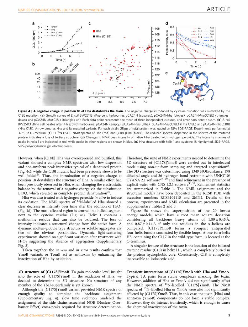
However, when [C18E] Hha was overexpressed and purified, thisvariant showed a complex NMR spectrum with low dispersionand non-uniform peak intensities typical of a denatured protein(Fig. 4c), while the C18I mutant had been previously shown to bewell folded24. Thus, the introduction of a negative charge atposition 18 destabilizes the structure of Hha. A similar effect hadbeen previously observed in Hha, when changing the electrostaticbalance by the removal of a negative charge via the substitutionE53Q, which resulted in complete protein denaturation25.
Hha was also treated with hydrogen peroxide in vitro to induceits oxidation. The NMR spectra of 15N-labelled Hha showed aclear decrease in intensity over time after the addition of H2O2
(Fig. 4d). The most-affected region is centred in a helical segmentnext to the cysteine residue (Fig. 4e). Helix 1 contains amethionine residue that can also be oxidized. The loss ofintensity indicates a structural change from the native form. Adynamic molten-globule type structure or soluble aggregates aretwo of the obvious possibilities. Dynamic light-scatteringexperiments showed no significant variation after treatment withH2O2 suggesting the absence of aggregation (SupplementaryFig. 3).
Taken together, the in vivo and in vitro results confirm thatYmoB variants or TomB act as antitoxins by enhancing theinactivation of Hha by oxidation.
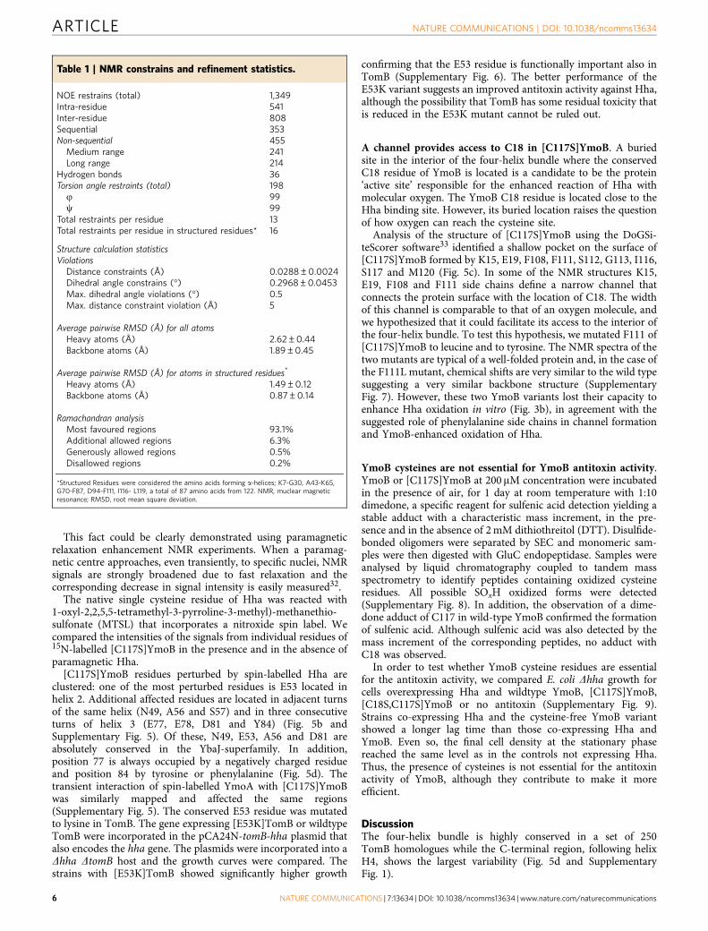
3D structure of [C117S]YmoB. To gain molecular level insightinto the role of [C117S]YmoB in the oxidation of Hha, wedecided to determine its 3D structure. No structure of anymember of the YbaJ-superfamily is yet known.
Although the [C117S]YmoB variant provided NMR spectra ofenough quality to complete the backbone assignment(Supplementary Fig. 4), slow time evolution hindered theassignment of the side chains associated NOE (Nuclear Over-hauser Effect) cross-peaks required for structure determination.
Therefore, the suite of NMR experiments needed to determine the3D structure of [C117S]YmoB were carried out in interleavedmode using non-uniform sampling and targeted acquisition26.The 3D structure was determined using 1349 NOE/distance, 198dihedral angle and 36 hydrogen bond restraints with UNIO’10/CYANA 2.1 software27–29 and final refinement in the presence ofexplicit water with CNS 1.2.1 software30,31. Refinement statisticsare summarized in Table 1. The NMR assignment and thestructural models have been deposited in the RCSB PDB withaccession numbers RCSB104115 and 2MN2. Details of theprocess, experiments and NMR calculation are presented in theSupplementary Tables 2 and 3.
Figure 5a shows the superposition of the 20 lowestenergy models, which have a root mean square deviationconsidering all backbone heavy atoms of 1.89±0.45 Å,and 0.87±0.14 Å if only the residues in the a-helices arecompared. [C117S]YmoB forms a compact antiparallelfour-helix bundle connected by flexible loops. A one-turn helixH5, containing the C117 in the wild-type form, is located at theC-terminus.
A singular feature of the structure is the location of the isolatedcysteine residue (C18) in helix H1, which is completely buried inthe protein hydrophobic core. Consistently, C18 is completelyinaccessible to iodoacetic acid.
Transient interactions of [C117S]YmoB with Hha and YmoA.Typical TA pairs form stable complexes masking the toxin.However, addition of Hha or YmoA did not significantly affectthe NMR spectra of 15N-labelled [C117S]YmoB. The NMRspectra of 15N-labelled Hha or YmoA were also not significantlyaffected by [C117S]YmoB. Thus, in this case, the toxin (Hha) andantitoxin (YmoB) components do not form a stable complex.However, they do interact transiently, which is enough to causethe chemical inactivation of the toxin.
5 EmptyHhaHha (C18E)
Hha (C18E)
Hha
Hha C
18I
Hha C
18E
Hha (C18I)
0 2 4 6 8Time (h)
kDaEm
pty
Hha
4025
15
10
59.0
1H (p.p.m.) 15N
(p.
p.m
.)
8.5 8.0 7.5 7.0
128
4
3O
.D. (
600
nm)
2
1
0
126
122
Cys18
Lys8
118
116
114
112
110
108
1060.8
K8D10L12R14
R26
N31
D48
L51
W68
Pea
k in
tens
ity r
atio
0.6
0.4
0.2
00 1 2 3 4
124
120Time (h)
a
b
c d
e
Figure 4 | A negative charge in position 18 of Hha destabilizes the toxin. The negative charge introduced by cysteine oxidation was mimicked by the
C18E mutation. (a) Growth curves of E. coli BW25113 Dhha cells harbouring: pCA24N (squares), pCA24N-hha (circles), pCA24N-hha(C18E) (triangles
down) and pCA24N-hha(C18I) (triangles up). Each data point represents the mean of three independent cultures, and error bars denote s.e.m. (b) E. coli
BW25113 Dhha cell lysates after 4 h growth harbouring: pCA24N (empty), pCA24N-hha (Hha), pCA24N-hha(C18E) (Hha C18E) and pCA24N-hha(C18I)
(Hha C18I). Arrow denotes Hha and its mutated variants. For each strain, 25 mg of total protein was loaded on 18% SDS-PAGE. Experiments performed at
37 �C in LB medium. (c) 1H–15N HSQC NMR spectra of Hha (red) and [C18E]Hha (black). The reduced spectral dispersion in the spectra of the mutated
protein indicates a loss of tertiary structure. (d) Changes in NMR peak intensity of native Hha treated with hydrogen peroxide. The intensity changes of
peaks in helix 1 are indicated in red, while peaks in other regions are shown in blue. (e) Hha structure with helix 1 and cysteine 18 highlighted. SDS–PAGE,
SDS–polyacrylamide gel electroporesis.
NATURE COMMUNICATIONS | DOI: 10.1038/ncomms13634 ARTICLE
NATURE COMMUNICATIONS | 7:13634 | DOI: 10.1038/ncomms13634 | www.nature.com/naturecommunications 5
This fact could be clearly demonstrated using paramagneticrelaxation enhancement NMR experiments. When a paramag-netic centre approaches, even transiently, to specific nuclei, NMRsignals are strongly broadened due to fast relaxation and thecorresponding decrease in signal intensity is easily measured32.
The native single cysteine residue of Hha was reacted with1-oxyl-2,2,5,5-tetramethyl-3-pyrroline-3-methyl)-methanethio-sulfonate (MTSL) that incorporates a nitroxide spin label. Wecompared the intensities of the signals from individual residues of15N-labelled [C117S]YmoB in the presence and in the absence ofparamagnetic Hha.
[C117S]YmoB residues perturbed by spin-labelled Hha areclustered: one of the most perturbed residues is E53 located inhelix 2. Additional affected residues are located in adjacent turnsof the same helix (N49, A56 and S57) and in three consecutiveturns of helix 3 (E77, E78, D81 and Y84) (Fig. 5b andSupplementary Fig. 5). Of these, N49, E53, A56 and D81 areabsolutely conserved in the YbaJ-superfamily. In addition,position 77 is always occupied by a negatively charged residueand position 84 by tyrosine or phenylalanine (Fig. 5d). Thetransient interaction of spin-labelled YmoA with [C117S]YmoBwas similarly mapped and affected the same regions(Supplementary Fig. 5). The conserved E53 residue was mutatedto lysine in TomB. The gene expressing [E53K]TomB or wildtypeTomB were incorporated in the pCA24N-tomB-hha plasmid thatalso encodes the hha gene. The plasmids were incorporated into aDhha DtomB host and the growth curves were compared. Thestrains with [E53K]TomB showed significantly higher growth
confirming that the E53 residue is functionally important also inTomB (Supplementary Fig. 6). The better performance of theE53K variant suggests an improved antitoxin activity against Hha,although the possibility that TomB has some residual toxicity thatis reduced in the E53K mutant cannot be ruled out.
A channel provides access to C18 in [C117S]YmoB. A buriedsite in the interior of the four-helix bundle where the conservedC18 residue of YmoB is located is a candidate to be the protein‘active site’ responsible for the enhanced reaction of Hha withmolecular oxygen. The YmoB C18 residue is located close to theHha binding site. However, its buried location raises the questionof how oxygen can reach the cysteine site.
Analysis of the structure of [C117S]YmoB using the DoGSi-teScorer software33 identified a shallow pocket on the surface of[C117S]YmoB formed by K15, E19, F108, F111, S112, G113, I116,S117 and M120 (Fig. 5c). In some of the NMR structures K15,E19, F108 and F111 side chains define a narrow channel thatconnects the protein surface with the location of C18. The widthof this channel is comparable to that of an oxygen molecule, andwe hypothesized that it could facilitate its access to the interior ofthe four-helix bundle. To test this hypothesis, we mutated F111 of[C117S]YmoB to leucine and to tyrosine. The NMR spectra of thetwo mutants are typical of a well-folded protein and, in the case ofthe F111L mutant, chemical shifts are very similar to the wild typesuggesting a very similar backbone structure (SupplementaryFig. 7). However, these two YmoB variants lost their capacity toenhance Hha oxidation in vitro (Fig. 3b), in agreement with thesuggested role of phenylalanine side chains in channel formationand YmoB-enhanced oxidation of Hha.
YmoB cysteines are not essential for YmoB antitoxin activity.YmoB or [C117S]YmoB at 200mM concentration were incubatedin the presence of air, for 1 day at room temperature with 1:10dimedone, a specific reagent for sulfenic acid detection yielding astable adduct with a characteristic mass increment, in the pre-sence and in the absence of 2 mM dithiothreitol (DTT). Disulfide-bonded oligomers were separated by SEC and monomeric sam-ples were then digested with GluC endopeptidase. Samples wereanalysed by liquid chromatography coupled to tandem massspectrometry to identify peptides containing oxidized cysteineresidues. All possible SOxH oxidized forms were detected(Supplementary Fig. 8). In addition, the observation of a dime-done adduct of C117 in wild-type YmoB confirmed the formationof sulfenic acid. Although sulfenic acid was also detected by themass increment of the corresponding peptides, no adduct withC18 was observed.
In order to test whether YmoB cysteine residues are essentialfor the antitoxin activity, we compared E. coli Dhha growth forcells overexpressing Hha and wildtype YmoB, [C117S]YmoB,[C18S,C117S]YmoB or no antitoxin (Supplementary Fig. 9).Strains co-expressing Hha and the cysteine-free YmoB variantshowed a longer lag time than those co-expressing Hha andYmoB. Even so, the final cell density at the stationary phasereached the same level as in the controls not expressing Hha.Thus, the presence of cysteines is not essential for the antitoxinactivity of YmoB, although they contribute to make it moreefficient.
DiscussionThe four-helix bundle is highly conserved in a set of 250TomB homologues while the C-terminal region, following helixH4, shows the largest variability (Fig. 5d and SupplementaryFig. 1).
Table 1 | NMR constrains and refinement statistics.
NOE restrains (total) 1,349Intra-residue 541Inter-residue 808Sequential 353Non-sequential 455
Medium range 241Long range 214
Hydrogen bonds 36Torsion angle restraints (total) 198j 99c 99
Total restraints per residue 13Total restraints per residue in structured residues* 16
Structure calculation statisticsViolations
Distance constraints (Å) 0.0288±0.0024Dihedral angle constrains (�) 0.2968±0.0453Max. dihedral angle violations (�) 0.5Max. distance constraint violation (Å) 5
Average pairwise RMSD (Å) for all atomsHeavy atoms (Å) 2.62±0.44Backbone atoms (Å) 1.89±0.45
Average pairwise RMSD (Å) for atoms in structured residues*
Heavy atoms (Å) 1.49±0.12Backbone atoms (Å) 0.87±0.14
Ramachandran analysisMost favoured regions 93.1%Additional allowed regions 6.3%Generously allowed regions 0.5%Disallowed regions 0.2%
*Structured Residues were considered the amino acids forming a-helices; K7-G30, A43-K65,G70-F87, D94-F111, I116- L119, a total of 87 amino acids from 122. NMR, muclear magneticresonance; RMSD, root mean square deviation.
ARTICLE NATURE COMMUNICATIONS | DOI: 10.1038/ncomms13634
6 NATURE COMMUNICATIONS | 7:13634 | DOI: 10.1038/ncomms13634 | www.nature.com/naturecommunications

One of the most conserved regions includes cysteine 18 and itsenvironment: position 15 is exclusively occupied by lysine orarginine, position 19 is preferentially occupied by glutamic andaspartic acid and position 17 by leucine, placing C18 in a peculiarhydrophobic but polarized environment. The hydrophobicenvironment is completed by aromatic residues in position 108(phenylalanine appears in 225/250 sequences) and in position 111(phenylalanine in 240/250 sequences) forming an access channelto C18 in [C117S]YmoB (Supplementary Fig. 10).
The residues transiently interacting with Hha and their nearestneighbours are also highly conserved: E53, N49, A56, E77, E78,D81, D82 and F84.
Thus, the TA mechanism demonstrated in vivo for Hha/TomBand that is operative also for the artificial Hha/YmoB pair is likelyto be a general feature in the entire YbaJ-superfamily. However,in spite of the large similarities, there are distinct subfamilies thatdiffer, for example in the number of cysteine residues. In thisrespect, TomB and YmoB are not the closest members of thefamily but, nevertheless, YmoB could functionally replace TomB
as an antitoxin. We also demonstrated similar oxidation processesin YmoA, suggesting that a similar TA system may also exist inYersinia sp.
A homology model of TomB was built from the structure of[C117S]YmoB using Modeller34. TomB has four cysteines, inpositions 18, 25, 110 and 124. C110 is located at the end of helixH4, not far from the location of cysteine 117 in YmoB, and C124,located at the C-terminus of TomB that extends beyond the endof YmoB.
TomB cysteines 18 and 25 are located in helix H1 and are bothburied in the interior of the four-helix bundle. Interestingly C18and C25 are located symmetrically, but in opposite directions,with respect to the Hha interaction site. The residues flankingC18 (Leu-18Cys-Glu) and C25 (Asp-25Cys-Leu) form a symme-trical pattern. The residues surrounding C18 in the 3D structureof [C117S]YmoB and in the homology model of TomB areidentical.
The conservation of the buried cysteine residues, their locationclose to the Hha interaction site, and the fact that C18 in YmoB
Leu46
Ser88
Glu77
H4
H5
H1
H3
H2
Cys18
N-term
C-term
1.0
0.5
0.0Pro
babi
lity
1.0
5
70 75 80 85 90 95 100 105 110 115 120 125 130
10H3
H1
H4
H2
15 20 25 30 35 40 45 50 55 60 65
*
0.5
0.0Pro
babi
lity
Phe108 Glu19
Lys15
Phe111
Cys18
Ser112
Gly113
Ser117 Met120
lle116
Ser57
180°
*
* * * * * * *0.99 0.99 0.98 0.90 0.97 0.97 0.64 0.64 0.17 0.17 0.03 0.03 0.03 0.000.02
b a c
d
Figure 5 | NMR structure of [C117S]YmoB as a model for TomB structure. (a) Superimposition of 20 lowest energy models. Helical regions (H1: 7–30;
H2: 43–65; H3: 70–87; H4: 94–111; and H5: 116–119) are shown in white and non-structured regions in blue. C18 is coloured yellow. The same colour is used
for H1 in the topological representations. Helices H3 (negatively charged) and H4 (positively charged) are represented in red and blue, respectively.
(b) Surface representation of the residues broadened in the presence of paramagnetically labelled Hha. Those located in helices H2 and H3 are shown
in red. Additional affected residues, (mainly in loops) are marked in orange (see Supplementary Fig. 4). The structures in a,b are rotated 180�. Most of the
residues contacting Hha are located in one side of the structure. (c) Expanded view of the pocket and channel defined by conserved residues F108 and F111
(purple), K15 (blue), E19 (red) and S117 (C117 in wild-type YmoB, yellow) connecting the surface with the location of the sulfur atom of C18. M120, also
susceptible to oxidation and close to C117, is shown in orange. The entry of the channel is located in the opposite face from the Hha-binding site. S117 is
occupied by cysteine in the wild-type form of YmoB. (d) Logo representation of the sequence variability within the YbaJ-superfamily. Secondary structure of
[C117S]YmoB is represented. An asterisk highlights residues forming the pocket surrounding C18. Some of the sequences terminate after position 116. The
proportion of sequences that extended to a given position is shown and colour-coded in circles at the C-terminal end of the logo representation.
NATURE COMMUNICATIONS | DOI: 10.1038/ncomms13634 ARTICLE
NATURE COMMUNICATIONS | 7:13634 | DOI: 10.1038/ncomms13634 | www.nature.com/naturecommunications 7
experiences similar oxidation processes as those observed in Hhasuggest that this residue may be involved in the oxidationenhancement mechanism. However, the fact that a variant ofYmoB without any cysteine retained some capacity to act as anantitoxin indicates that the cysteines of YmoB are not essential.On the other hand, mutations affecting the channel leading to theYmoB C18 site prevented the oxidation of Hha. Thus, theenvironment around YmoB C18, rather that the cysteine residueitself is probably sufficient for YmoB-enhanced oxidation,although the cysteine residue may play a role, for examplethrough its transient and reversible oxidation to sulfenic acid.TomB seems to have this site duplicated while the Hha-bindingsite is not. These structural variations among the YbaJ super-family probably reflect adaptions to the required levels ofantitoxin activity or their oxygen dependency.
The cysteine residues of Hha and YmoA are partially oxidizedspontaneously by air in vitro even in the absence of TomB orYmoB giving a mixture of sulfenic, sulfinic and sulfonic acid.However, the oxidation is enhanced in the presence of[C117S]YmoB. This cysteine residue is conserved in nearly allmembers of the Hha family but its function was unknown. It islocated in a loop between to helical regions, only partially solventexposed, and located in the interface between regions withopposite charge35,36. The unusual sensitivity to oxidation may berelated to this particular environment. Interestingly, C18 inYmoB is also located in a buried region but surrounded bycharged residues.
Reactions of free molecular oxygen have a high-activationbarrier associated with the symmetry of its electron spin tripletground state37. The presence of polar residues, electric dipoles orinteractions breaking the local symmetry enhances the capacity ofmolecular oxygen to oxidize cysteine to SOxH species22,38.
Hha oxidation leads to a loss of structure thus explaining itsinactivation by TomB in the presence of oxygen. The Hhastructure was also destabilized by a mutation introducing anegative charge at the position of C18. The sensitivity of Hha tocharge modifications had been previously observed and isconsistent with its unusual high density of charged residues23,25.
Overall, these results strongly suggest that the TA systemformed by proteins from the Hha-YbaJ superfamily acts througha mechanism that does not fall in any of the currently acceptedTA classes. While known TA systems are based on a stable toxinthat is neutralized by forming a complex with a labile antitoxin,the Hha-YbaJ system is based on the chemical inactivation of thetoxin caused by oxidation, enhanced in the presence of theantitoxin. This process does not require the formation of a stableTA complex but takes place through transient interactions.
The described mechanism links the TA system to theconcentration of oxygen, making proteins from the YbaJ-superfamily effective antitoxins only for highly oxygenated cells.Strong oxygen concentration gradients in biofilms could cause theantitoxin to be less efficient in cells located in anoxic regions,resulting in selective death of these cells and the creation ofcavities in the biofilm matrix allowing the escape of planktoniccells, or detachment of entire microcolonies facilitating biofilmdispersion.
The structure of [C117S]YmoB provides hints on the chemicalorigin of this activity and suggests that the members of the YbaJ-superfamily could be targets for antibacterial treatments to inhibitthe formation of biofilms, one of the strongest contributors to thepressing antibiotic resistance problem.
MethodsAntitoxin activity of TomB, and YmoB variants. Growth curves. One colony ofE. coli K-12 MG1655 Dhha cells harbouring pCA24N-hha (under the control ofT5lac promoter, activated by 1 mM IPTG (isopropyl b-D-1-thiogalactopyranoside),
selected with 100 mg ml� 1 of ampicillin) and pBAD30-tomB or pBAD30-ymoB orpBAD30-ymoB(C117S) or pBAD30-ymoB(C18S)(C117S) (under the controlof araC promoter, activated by 0.1% arabinose, selected with 50 mg ml� 1 ofchloramphenicol) was pre-cultured overnight at 37 �C and 250 r.p.m. in 30 ml oftryptone minimal medium (TMM: 10 g l� 1 tryptone and 2.5 g l� 1 NaCl).
Growth curves were started by adding 4 ml of preculture to 200 ml of TMM(with corresponding antibiotics and chemical inducers) and the experiments werecarried out with the incubators and the room maintained at 37 �C.
Oxygen saturation measurements. Dissolved oxygen was measured as thepercentage of saturation using a polarographic electrode (VWR DO210) using thebuilt-in temperature and ionic-strength compensation. The electrode was cleanedwith 14% ammonium hydroxide for 2 min and MilliQ water for 1 min to preventcross-contamination.
Colony forming unit measurements. The number of CFUs was obtained byplatting 10ml of a 108 dilution of samples taken at selected optical densities.
Microtiter biofilm assays. Single colonies of E. coli K-12 MG1655 Dhha cellsharbouring pCA24N-hha and pBAD30-tomB or pBAD30-ymoB or pBAD30-ymoB(C117S) were inoculated into 25 ml TMM medium (with the correspondingantibiotic) and incubated overnight at 37 �C and 250 r.p.m. shaking speed. Twoindependent cultures were prepared for each strain, diluted with fresh TMM(with the corresponding antibiotic) to obtain an OD600 below 0.05, and 300 ml ofculture were added into separate wells of a 96 well plate. Blanks were prepared withpure TMM medium. The plate was incubated at 37 �C without shaking, for 15 h.To record the total growth, we measured OD620. Supernatant was discarded and theplate was washed by dipping three times in MilliQ water, 300ml of 0.1% crystalviolet were added in each well and the plate was incubated for 20 min at roomtemperature. Staining solution was discarded and the plate was washed by dippingthree times in MilliQ water. An amount of 300ml of 95% ethanol was added to eachwell and was incubated for 5 min. We measured the total biofilm reading OD540
after mixing for 50 s. Standard biofilm formation was calculated as OD540/(OD620�OD620). OD measurements were done using Infinite M1000 PRO plate reader.
Flow cell biofilm assays. Strain E. coli K-12 MG1655 Dhha pCA24N-hhapBAD30-ymoB was streaked on LB agar plates with the corresponding antibiotics.One-day old single colonies were inoculated in 25 ml of TMM overnight at 37 �C at250 r.p.m. Overnight cultures were diluted to reach OD600 below 0.05 in 150 mlTMM with the corresponding antibiotics. Diluted cultures under stirring at 37 �Cwere pumped to the flow cell at 10 ml h� 1 for 3 h to facilitate cell attachment to theglass slide surface. Culture medium was replaced with 200 ml of fresh TMMcontaining the corresponding antibiotics and inducers and then fed to the flow cellat the same rate for 15 h. After incubation, the flow cell unit was disconnected andblocked (never allowing it to dry). It was placed at room temperature and theSYTO9 mix was added using a 5 ml syringe. The flow cell was incubated withSYTO9 in the dark for 20 min at room temperature. Eight images in randompositions were taken with an Olympus FV100 confocal microscope using a longworking distance � 40 Olympus LWDCDPLAN 40LP lens. 3D images of each spotwere obtained using IMARIS software, and quantitative analysis of biofilm wasperformed using COMSTAT software. For the SYTO9 stain preparation, theLIVE/DEAD BacLight Bacterial Viability Kit L7007 was used. Three millilitre ofdye was prepared in a 30 ml glass beaker mixing; 7.5 ml of component A (SYTO9 dye, 1.67 mM / propidium iodide, 1.67 mM), 7.5 ml of component B (SYTO 9 dye,1.67 mM / propidium iodide, 18.3 mM) and 3 ml of TMM.
YmoB mutagenesis. Mutations in YmoB (cysteine to serine, phenylalanine toleucine, phenylalanine to tyrosine) were introduced using the QuickChange sitedirected mutagenesis Kit (Stratagene) with the appropriate complementarymutagenic primers. We confirmed the constructs by DNA sequencing.
[C117S]YmoB 3D NMR structure.. [C117S]YmoB sample preparation for NMRexperiments. 13C/15N-labelled [C117S]YmoB variant was expressed in E. coli BL21(DE3) in minimal medium with 15NH4Cl and/D-[13C] glucose as the sole sourcesof nitrogen and carbon respectively39. Cells were lysed by sonication. Solubleprotein was purified by His-Tag affinity chromatography (Ni-NTA agaroseresin QIAGEN) and samples were eluted using buffer A (20 mM Trizmahydrochloride, 800 mM NaCl, 400 mM imidazole, 5 mM DTT and pH¼ 8.00).Protein was further purified by SEC using HiLoad Superdex 75 prepgradecolumns (GE Healthcare) and buffer B (pH¼ 7.00 buffered with 20 mM[NaH2PO4þNa2HPO4], 150 mM NaCl, 1 mM tris-carboxyethylphosphine(TCEP), 0.1 mM ethylenediaminetetraacetic acid (EDTA) and 0.01% w/v NaN3).Fractions containing the purified protein were concentrated using a centrifugalconcentrator VIVASPIN 20 10,000 MWCO PES to the mentioned concentrationsfor NMR experiments.
NMR acquisition, processing and analysis. NMR spectra listed in SupplementaryTable 2 were acquired at 1.4 mM protein concentration, in buffer A and 25 �C on600 or 800 MHz Bruker spectrometers equipped with a cryo-pulse-field gradienttriple-resonance probe. NMR spectra listed at Supplementary Table 3 wereacquired at 1.0 mM protein concentration in buffer B and 25 �C on 900 or 800 MHzAgilent spectrometers equipped with a cryo- (900 MHz) and room temperature(800 MHz) pulse-field gradient triple- resonance probes. Hydrogen bonds wereidentified from a H/D exchange experiment. A lyophilized sample of 1.4 mM[C117S] YmoB was suspended in D2O reaching the same concentration, and a
ARTICLE NATURE COMMUNICATIONS | DOI: 10.1038/ncomms13634
8 NATURE COMMUNICATIONS | 7:13634 | DOI: 10.1038/ncomms13634 | www.nature.com/naturecommunications

two-dimensional 1H-15N-heteronuclear single quantum correlation (HSQC)experiment was acquired after 4 min of sample preparation. Stereospecificassignments of valine and leucine methyl groups were obtained from a constanttime 1H–13C HSQC on a 10% 13C/100% 15N [C117S]YmoB 1.4 mM sample inbuffer B. One fresh sample of 15N–13C labelled [C117S]YmoB at 1.0 mM in bufferB was used for NOE correlation spectroscopy (NOESY) experiments and anequivalent fresh sample for total correlation spectroscopy (TOCSY) experiments.Protein distance restraints were obtained from 3D 1H–15N edited NOESY HSQC,3D 1H–13C edited NOESY (aromatic optimized in D2O), and 3D 1H–13C editedNOESY HSQC in D2O experiments. NOESY and TOCSY spectra were recordedusing gradient sensitivity-enhanced pulse sequences from the BioPack library(Agilent Inc.). Mixing time in NOESY experiments was set to 100 ms and to 14 msin TOCSY experiments. The non-uniform sampling (NUS) schedules wereprepared by program nussampler from the MDDNMR software package40. Whenappropriate, the sampling probability density was biased for aliphatic 13C-aliphaticevolution according to the single-bond 13C–13C homonuclear coupling constant of35 Hz. When appropriate, sampling was exponentially weighted according to themaximum evolution time. Data processing and analysis were carried out withMDDNMR, NMRPipe41, NMRViewJ42 and CARA43. Proton chemical shifts werereferenced using 4,4-dimethyl-4-silapentane-1-sulfonic acid as an internalstandard, whereas 15N and 13C chemical shifts were indirectly referenced.
Structure determination. NOE distance restraints were obtained iteratively usingthe Unio’10/CYANA 2.1 (refs 27–29) suite program and manually inspected.Structure was determined by simulated annealing using the torsion angle dynamicsimulation program CYANA 2.1 and further water refinement with CNS 1.2.1(refs 30,31). Protein structure calculation was based on Unio’10/CYANA generatedupper distances using only unambiguously assigned restraints derived fromNOESY experiments, hydrogen bond restraints based on H/D 1H-15N-HSQCexchange experiments and dihedral angle restraints calculated with TALOSþ(ref. 44). An ensemble of 100 protein structures was generated and the 20 lowestenergy models were checked using PROCHECK-NMR45. None of the structurescontained distance or dihedral angle violations 40.5 Å or 5�, respectively. Inter-helical angles were measured with Chimera v10.1 (ref. 46). Ramachandran analysisis as follows: 93.1% most favoured regions, 6.3% additional allowed regions, 0.5%generously allowed regions and 2.2% disallowed regions. Molecular imageswere generated with Chimera, Gimp (http://www.gimp.org/) and InkScape(https://inkscape.org/en/). Pocket identification was carried out with DogSiteScorerserver33 using default settings. The TomB homology model was built usingModeller34 starting with YmoB as template structure and only rebuildingthe regions surrounding insertions or deletions in the sequence alignment.Logo representation was generated using web service from http://weblogo.threeplusone.com47 and manually coloured using InkScape.
Paramagnetic relaxation enhancement (PRE-NMR) experiments. 15N-labelled[C117S]YmoB, Hha and YmoA proteins were prepared as previously described for13C/15N-labelled [C117S]YmoB. For MTSL-protein functionalization, reductantwas firstly removed using PD10 desalting column (GE). Commercially availableMTSL tag (1-oxyl-2,2,5,5-tetramethyl-3-pyrroline-3-methyl)-methanethiosulfonatefrom Santa Cruz Biotechnology) was dissolved in acetone and 10 molar equivalentsof MTSL were added to the protein sample and left reacting overnight at 4 �Cunder stirring conditions. Excess of MTSL was removed using P10 desaltingcolumn in buffer containing 20 mM [NaH2PO4þNa2HPO4] pH¼ 7, 150 mMNaCl, 0.1 mM EDTA. Protein samples were prepared fresh and measured imme-diately after preparation. NMR experiments were recorded at 298 K with a600 MHz Bruker Avance III spectrometer equipped with a TCI cryo-probe. NMRspectra were analysed using CCPNMR Analysis48.
Mass spectrometry. Hha oxidation by [C117S]YmoB. A solution containing500mM Hha and 500 mM [C117S]YmoB was incubated for 30 min in 20 mM[NaH2PO4þNa2HPO4], 150 mM NaCl, 0.1 mM EDTA pH¼ 7.00. Parallelexperiments were carried out with the same batch of Hha with [F111L,C117S]Y-moB, [F111Y, C117S]YmoB or without any YmoB variant. Hha and YmoB wereseparated by SEC in 50 mM ammonium acetate buffer. Hha was digested withtrypsin and the resulting peptides were analysed by nanoHPLC-ESI-MS/MS(nano high-performance liquid chromatography coupled to tandem massspectrometry with nanoelectrospray ionization) in a LTQ-FT Ultra (ThermoScientific) mass spectrometer and processed and analysed with Xcalibur software(versus 2.02 SR2). Ion deconvolution to zero charged monoisotopic masses wasperformed using Xtract algorithm in Xcalibur software.
The relative abundance of the oxidized species was estimated from the ratio ofthe peptide spectrum matches (PSM) corresponding to peptides with oxidizedcysteine (sulfenic, sulfinic or sulfonic acids) with respect to the PSM number of allcysteine-containing peptides. The incubation and digestion steps were carried outat least in duplicate and at least two separate samples of each peptide mixture wereanalysed by mass-spectrometry.
In a separate experiment the relative abundance of oxidized peptides wasalso calculated considering the chromatographic peak areas of the correspondingpeptides using Skyline 3.1 software49, obtaining equivalent results (data notshown).
Extended air oxidations of YmoB, [C117S]YmoB and YmoA. YmoB or[C117S]YmoB at 200 mM protein concentration and YmoA alone or in the pre-sence of [C117S]YmoB were incubated in the presence of air for 12 days in abuffered solution of 20 mM [NaH2PO4þNa2HPO4] pH¼ 7.0, 150 mM NaCl,0.1 mM EDTA and 0.01% w/v NaN3) with or without DTT (2 mM) and in thepresence of dimedone in a 1:10 ratio. Peptides were generated with endoproteinaseGluC digestion and analysed as described above.
Toxicity tests of Hha and TomB variants.. Overnight cultures of E. coli BW25113Dhha harbouring pCA24N, pCA24N-hha, pCA24N-hha(C18E) or pCA24N-hha(C18I) were inoculated into 25 ml of LB medium with 30 mg ml� 1 chlor-amphenicol and 1 mM IPTG at an OD600 of 0.04. For TomB and [E53K]TomB,overnight cultures of E. coli ATCC25404 Dhha DtomB harbouring pCA24N-tomB-hha or pCA24N-tomB(E53K)-hha were inoculated into 25 ml of LB medium with30 mg ml� 1 chloramphenicol at an OD600 of 0.05. After an hour of inoculation,0.1 mM IPTG was added. Growth was evaluated periodically by measuring OD600.For each strain, two or three independent cultures were evaluated. To check totalprotein expression, cultures at an OD600 of 0.5 were induced with 1 mM IPTG for4 h. Cells were sonicated, centrifuged, and 25 mg of cell lysates (supernatant) wereresolved via 18% SDS–polyacrylamide gel electroporesis. Total protein of celllysates was quantified using the Pierce BCA Protein Assay Kit (Thermo Scientific,Waltham, MA).
Stability tests of [C18E]Hha and oxidized Hha. The hha(C18E) mutation wasgenerated in a pET15b expression plasmid35 that produces a His6-tagged form ofHha. The 15N-labelled wild type and [C18E] proteins were purified by His-Tagaffinity chromatography (Ni-NTA agarose resin QIAGEN) by eluting with 20 mMTrizma hydrochloride, 800 mM NaCl, 400 mM imidazole, 5 mM DTT andpH¼ 8.00. A final purification step on a Superdex 75 column in 20 mM sodiumphosphate, 150 mM NaCl, 1 mM TCEP, 0.01% w/v NaN3, pH¼ 7.00 yielded pureproteins as monomers.
Before H2O2 treatment, TCEP was removed by passing the protein solutionthrough a PD-10 column. After measuring the reference NMR spectra, thewild-type protein was treated with 5 mM H2O2 and spectra were recorded every130 min.
Data availability. All relevant data are available from the authors. NMR assign-ment and the structural models have been deposited in the RCSB PDB withaccession numbers RCSB104115 and 2MN2.
References1. Laxminarayan, R. et al. Antibiotic resistance-the need for global solutions.
Lancet Infect. Dis. 13, 1057–1098 (2013).2. Hung, C., Zhou, Y., Pinkner, J. S. & Have, B. Escherichia coli biofilms have an
organised and complex extracellular matrix structure. mBio 4, e00645–13(2013).
3. Sauer, K., Rickard, A. H. & Davies, D. G. Biofilms and biocomplexity. Microbe2, 347–353 (2007).
4. Costerton, B. Microbial ecology comes of age and joins the general ecologycommunity. Proc. Natl Acad. Sci. USA 101, 16983–16984 (2004).
5. Ito, A., Taniuchi, A., May, T., Kawata, K. & Okabe, S. An increase in antibioticresistance of Escherichia coli in mature biofilms. Appl. Environ. Microbiol. 75,4093–4100 (2009).
6. Stewart, P. S. & Franklin, M. J. Physiological heterogeneity in biofilms. Nat.Rev. Microbiol. 6, 199–210 (2008).
7. Karatan, E. & Watnick, P. Signals, regulatory networks, and materials that buildand break bacterial biofilms. Microbiol. Mol. Biol. Rev. 73, 310–347 (2009).
8. Kostakioti, M., Hadjifrangiskou, M. & Hultgren, S. J. Bacterial biofilms:development, dispersal, and therapeutic strategies in the dawn of thepostantibiotic era. Cold Spring Harb. Perspect. Med. 3, a010306 (2013).
9. McDougald, D., Rice, S. A., Barraud, N., Steinberg, P. D. & Kjelleberg, S. Shouldwe stay or should we go: mechanisms and ecological consequences for biofilmdispersal. Nat. Rev. Microbiol. 10, 39–50 (2012).
10. Asally, M. et al. Localized cell death focuses mechanical forces during 3Dpatterning in a biofilm. Proc. Natl Acad. Sci. USA 109, 18891–18896 (2012).
11. Wilking, J. N. et al. Liquid transport facilitated by channels in Bacillus subtilisbiofilms. Proc. Natl Acad. Sci. USA 110, 848–852 (2013).
12. Ma, L. et al. Assembly and development of the Pseudomonas aeruginosa biofilmmatrix. PLoS Pathog. 5, e1000354 (2009).
13. Petrova, O. E. & Sauer, K. Escaping the biofilm in more than one way:desorption, detachment and dispersion. Curr. Opin. Microbiol. 30, 67–78(2016).
14. Wang, X. & Wood, T. K. Toxin/antitoxin systems influence biofilm andpersister cell formation and the general stress response. Appl. Environ.Microbiol. 77, 5577–5583 (2011).
15. Pecota, D. & Wood, T. K. Exclusion of T4 phage by the hok/sok killer locusfrom plasmid R1. J. Bacteriol. 178, 2044–2050 (1996).
NATURE COMMUNICATIONS | DOI: 10.1038/ncomms13634 ARTICLE
NATURE COMMUNICATIONS | 7:13634 | DOI: 10.1038/ncomms13634 | www.nature.com/naturecommunications 9
16. Wen, Y., Behiels, E. & Devreese, B. Toxin-antitoxin systems: their role inpersistence, biofilm formation, and pathogenicity. Pathog. Dis. 70, 240–249(2014).
17. Gonzalez Barrios, A. F., Zuo, R., Ren, D. & Wood, T. K. Hha, YbaJ, and OmpAregulate Escherichia coli K12 biofilm formation and conjugation plasmidsabolish motility. Biotechnol. Bioeng. 93, 188–200 (2006).
18. Garcıa Contreras, R., Zhang, X.-S., Kim, Y. & Wood, T. K. Protein translationand cell death: the role of rare tRNAs in biofilm formation and in activatingdormant phage killer genes. PLoS ONE 3, e2394 (2008).
19. Hong, S. H. et al. Synthetic quorum-sensing circuit to control consortialbiofilm formation and dispersal in a microfluidic device. Nat. Commun. 3, 613(2012).
20. Hong, S. H., Lee, J. & Wood, T. K. Engineering global regulator Hha ofEscherichia coli to control biofilm dispersal. Microb. Biotechnol. 3, 717–728(2010).
21. Heydorn, A. et al. Quantification of biofilm structures by the novel computerprogram COMSTAT. Microbiology 146, 2395–2407 (2000).
22. Reddie, K. G. & Carroll, K. S. Expanding the functional diversity ofproteins through cysteine oxidation. Curr. Opin. Chem. Biol. 12, 746–754(2008).
23. Cordeiro, T. N., Garcıa, J., Bernado, P., Millet, O. & Pons, M. A three-proteincharge zipper stabilizes a complex modulating bacterial gene silencing. J. Biol.Chem. 290, 21200–21212 (2015).
24. Cordeiro, T. N. et al. A single residue mutation in Hha preserving structure andbinding to H-NS results in loss of H-NS mediated gene repression properties.FEBS Lett. 582, 3139–3144 (2008).
25. Fernandez de Alba, C. et al. Essential residues in the H-NS binding site of Hha,a co-regulator of horizontally acquired genes in Enterobacteria. FEBS Lett. 585,1765–1770 (2011).
26. Jaravine, V. A. & Orekhov, V. Y. Targeted acquisition for real-time NMRspectroscopy. J. Am. Chem. Soc. 128, 13421–13426 (2006).
27. Herrmann, T., Guntert, P. & Wuthrich, K. Protein NMR structuredetermination with automated NOE assignment using the new softwareCANDID and the torsion angle dynamics algorithm DYANA. J. Mol. Biol. 319,209–227 (2002).
28. Herrmann, T., Guntert, P. & Wuthrich, K. Protein NMR structuredetermination with automated NOE-identification in the NOESY spectra usingthe new software ATNOS. J. Biomol. NMR 24, 171–189 (2002).
29. Guntert, P. Automated NMR structure calculation with CYANA. Methods Mol.Biol. 278, 353–378 (2004).
30. Adams, P. D. et al. Crystallography & NMR system: a new software suite formacromolecular structure determination. Acta Cristallogr. D Biol. Crystallogr.D54, 905–921 (1998).
31. Nederveen, A. J. et al. RECOORD: a recalculated coordinate database of 500þproteins from the PDB using restraints from the BioMagResBank. Proteins 59,662–672 (2005).
32. Iwahara, J. & Clore, G. M. Detecting transient intermediates in macromolecularbinding by paramagnetic NMR. Nature 440, 1227–1230 (2006).
33. Volkamer, A., Kuhn, D., Grombacher, T., Rippmann, F. & Rarey, M.Combining global and local measures for structure-based druggabilitypredictions. J. Chem. Inf. Model. 52, 360–372 (2012).
34. Marti-Renom, M. A. et al. Comparative protein structure modeling of genesand genomes. Annu. Rev. Biophys. Biomol. Struct. 29, 291–325 (2000).
35. Yee, A. et al. An NMR approach to structural proteomics. Proc. Natl Acad. Sci.USA 99, 1825–1830 (2002).
36. McFeeters, R. L. et al. The high-precision solution structure of Yersiniamodulating protein YmoA provides insight into interaction with H-NS.Biochemistry 46, 13975–13982 (2007).
37. Bagiyan, G. A., Koroleva, I. K., Soroka, N. V. & Ufimtsev, A. V. Oxidation ofthiol compounds by molecular oxygen in aqueous solutions. Rus. Chem. Bull.Int. Edit. 52, 1135–1141 (2003).
38. Salsbury, Jr F. R., Knutson, S. T., Poole, L. B. & Fetrow, J. S. Functional siteprofiling and electrostatic analysis of cysteines modifiable to cysteine sulfenicacid. Protein Sci. 17, 299–312 (2008).
39. Marley, J., Lu, M. & Bracken, C. A method for efficient isotopic labeling ofrecombinant proteins. J. Biomol. NMR 20, 71–75 (2001).
40. Orekhov, V. Y. & Jaravine, V. A. Analysis of non-uniformly sampled spectrawith multi-dimensional decomposition. Prog. Nucl. Magn. Reson. Spectrosc. 59,271–292 (2011).
41. Delaglio, F. et al. NMRPipe: a multidimensional spectral processing systembased on UNIX pipes. J. Biomol. NMR. 6, 277–293 (1995).
42. Johnson, B. A. Using NMRView to visualize and analyze the NMR spectra ofmacromolecules. Methods Mol. Biol. 278, 313–352 (2004).
43. Keller, R. L. J. The Computer Aided Resonance Assignment Tutorial (CANTINAVerlag. Goldau, 2004).
44. Shen, Y., Delaglio, F., Cornilescu, G. & Bax, A. TALOSþ : A hybrid method forpredicting protein backbone torsion angles from NMR chemical shifts.J. Biomol. NMR. 44, 213–223 (2009).
45. Laskowski, R. A., Rullmannn, J. A., MacArthur, M. W., Kaptein, R. &Thornton, J. M. AQUA and PROCHECK-NMR: programs for checking thequality of protein structures solved by NMR. J Biomol. NMR 8, 477–486 (1996).
46. Pettersen, E. F. et al. UCSF chimera—a visualization system for exploratoryresearch and analysis. J. Comput. Chem. 25, 1605–1612 (2004).
47. Crooks, G., Hon, G., Chandonia, J. & Brenner, S. NCBI GenBank FTP SiteWebLogo: a sequence logo generator. Genome Res. 14, 1188–1190 (2004).
48. Vranken, W. F. et al. The CCPN data model for NMR spectroscopy:development of a software pipeline. Proteins 59, 687–696 (2005).
49. MacLean, B. et al. Skyline: an open source document editor for creating andanalysing targeted proteomics experiments. Bioinformatics 26, 966–968 (2010).
AcknowledgementsWe thank Cristina Madrid and Antonio Juarez for helpful discussions and for providingplasmids and Laura Villarreal and Mar Vilanova for help in MS experiments and LaarebIrrem Mohamad for help in DLS experiments. This work was partially supported byfunds from the Spanish MINECO (BIO2010-15683 and BIO2013-45793R –cofoundedwith European Structural Funds, FEDER-), the EC FP7 BioNMR project (contract261863) and the ARO (W911NF-14-1-0279). T.K.W. is the Biotechnology EndowedProfessor at the Pennsylvania State University. NMR experiments were performed at theICTS NMR LRB and CCITUB (University of Barcelona) and the Swedish NMR Centre(Goteborg University). MS experiments were performed at IRB Barcelona MS CoreFacility (member of the BMBS COST Action BM 1403 and Proteored, PRB2-ISCIII(IPT13/0001—ISCIII-SGEFI / ERDF). IRB Barcelona is the recipient of a Severo OchoaAward of Excellence from MINECO (Government of Spain). Development of Chimerapackage by UCSF is supported by NIGMS P41-GM103311.
Author contributionsO.M. performed and analysed structural, functional and mass spectrometry experiments.J.M.C.T, V.W.C.S. T.L.W. and J.G. prepared and analysed mutants. A.M. performedoxygen dependency experiments. J.M.C.T., M.G. and M.V. performed and analysed massspectrometry experiments. J.M.C.T. performed experiments leading to the identificationof the Hha binding site. T.N.C. and I.A. contributed to NMR experiments and structuralanalysis. M.M. and V.Y.O. performed interleaved-NUS NMR experiments. J.G. andT.N.C. screened and optimized constructs and NMR conditions. T.K.W. designed andanalysed functional experiments. M.P. conceived and supervised the project and wrotethe paper with contributions from all other co-authors.
Additional informationSupplementary Information accompanies this paper at http://www.nature.com/naturecommunications
Competing financial interests: The authors declare no competing financial interests.
Reprints and permission information is available online at http://npg.nature.com/reprintsandpermissions/
How to cite this article: Marimon, O. et al. An oxygen-sensitive toxin–antitoxin system.Nat. Commun. 7, 13634 doi: 10.1038/ncomms13634 (2016).
Publisher’s note: Springer Nature remains neutral with regard to jurisdictional claims inpublished maps and institutional affiliations.
This work is licensed under a Creative Commons Attribution 4.0International License. The images or other third party material in this
article are included in the article’s Creative Commons license, unless indicated otherwisein the credit line; if the material is not included under the Creative Commons license,users will need to obtain permission from the license holder to reproduce the material.To view a copy of this license, visit http://creativecommons.org/licenses/by/4.0/
r The Author(s) 2016
ARTICLE NATURE COMMUNICATIONS | DOI: 10.1038/ncomms13634
10 NATURE COMMUNICATIONS | 7:13634 | DOI: 10.1038/ncomms13634 | www.nature.com/naturecommunications


![Type II toxin/antitoxin MqsR/MqsA controls type V toxin ......is ghoT [yjdO, ghost cells (Wang et al., 2012)]. GhoT is a membrane toxin that produces the ghost-cell phenotype (lysed](https://img.dokumen.tips/doc/110x75/5fa80fdce5abf545555543f8/type-ii-toxinantitoxin-mqsrmqsa-controls-type-v-toxin-is-ghot-yjdo-ghost.jpg)