Embed Size (px)
Citation preview

Dr. Leow Wei QiangAssociate Consultant Department of Pathology, Histopathology and Cytology Sections Gastrointestinal and Hepatopancreatobiliary Service
An Overview of the Pathology of Gastric
Cancers: pathogenesis, classifications and new
understandings.
2
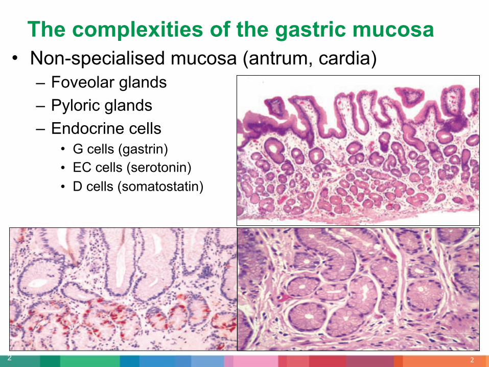
• Non-specialised mucosa (antrum, cardia) – Foveolar glands– Pyloric glands– Endocrine cells
• G cells (gastrin)• EC cells (serotonin)• D cells (somatostatin)
!
The complexities of the gastric mucosa
3
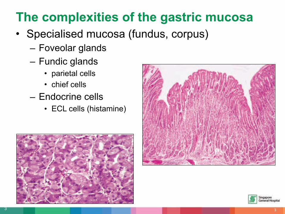
• Specialised mucosa (fundus, corpus) – Foveolar glands– Fundic glands
• parietal cells • chief cells
– Endocrine cells• ECL cells (histamine)
"
The complexities of the gastric mucosa

4
• In 2005, Barry J. Marshall and Robin Warren were awarded the Nobel Prize in Physiology for their discovery of Helicobacter pylori and its role in gastritis and peptic ulcer disease.
• Persistent infection induces a series of phenotypical changes that occur before the development of ‘intestinal type’ adenocarcinoma.
• H.pylori’s potent urease activity releases ammonia, which increases expression of nitrosated compounds, which can induce DNA damage.
• Studies suggest at least 4x increased risk of gastric lesions in patients with H.pylori infection.
#
Helicobacter pylori

5
• Diet rich in salt-preserved or smoked foods, combined with low intakes of fruits and vegetables.
– Increased intraluminal formation of nitrosated compounds ! DNA damage.– Protective effect of antioxidants (controversial)
• Bile reflux, particularly after gastric surgery, increases risk of gastric cancers.
• Cigarette smoking, increases risk by 2-3x.
• Helicobacter pylori.
• Polymorphisms of interleukin 1 gene, which plays a role in hypochlorhydria and atrophy, is associated with risk of gastric carcinoma.
• In contrast to intestinal type adenocarcinomas, diffuse type adenocarcinoma shows equal incidence in all geographic areas and occur in younger individuals, suggesting influence by genetic factors over environmental.
Multifactorial etiologies
$

6
• Chronic gastritis!Atrophy!Intestinal metaplasia!Dysplasia!Cancer.
Correa cascade of multistep gastric carcinogenesis
%

7
• A phenotypic change from normal gastric epithelium to intestinal type.
• Due to chronic inflammation.
• Attempts to subclassify them into complete and incomplete, but have l imited clinical significance.
• Rather than the subtype, the extent and severity of IM is predictive of the cancer risk.
&
Intestinal metaplasia (IM)

8
• Incidence of gastric dysplasia closely parallels incidence of adenocarcinoma.– Represents a direct neoplastic precursor lesion– Most dysplastic lesions have an intestinal phenotype (Type 1)
• Dysplasia is also a marker for risk for cancer elsewhere in the stomach.– Up to 12.5% cited in a Japanese study
• 57% of gastric cancers found during surveillance of gastric dysplasia are considered early gastric cancers.
• There are interpretive variations in the diagnosis of such lesions.– Negative for dysplasia (benign, reactive, metaplastic)– Dysplasia (unequivocal features; further classified into low grade and
high grade)– Indefinite for dysplasia (ambiguous morphological pattern)
Dysplasia / Intraepithelial neoplasia
'

9
Dysplasia
(
• Gland size• Budding, cribriform profiles• Surface maturation• Inflammation• Intraluminal necrotic debris• Mucin depletion• Nuclear size• Nuclear stratification• Nuclear shape• Hyperchromasia• Prominent nucleoli• Increased / atypical mitoses

10
Dysplasia: the lows and the highs
)*
+,-./0123.456781691:.;<=8310.3;810>3?3;@.1;2.38,;>1@9,;A.;<=8310.=0,-29;>.1;2.763<2,6@01@9B9=1@9,;
C9>D./0123.456781691:.9;.1229@9,;A.=,[email protected],66.,B.;<=8310.7,8109@5A.?10E32.;<=8310.783,?,07D96?A.=,?783F.>81;2.6@0<=@<036

11
Intramucosal carcinoma
))

12 )!
13
• WHO definition: Gastric carcinomas are malignant epithelial neoplasms. They represent a biologically and genetically heterogeneous group of tumours with multifactorial etiologies, both environmental and genetic. They are characterised by broad morphological heterogeneity with respect to patterns of architecture and growth, cell differentiation and histogenesis.
)"
Gastric Carcinoma
14
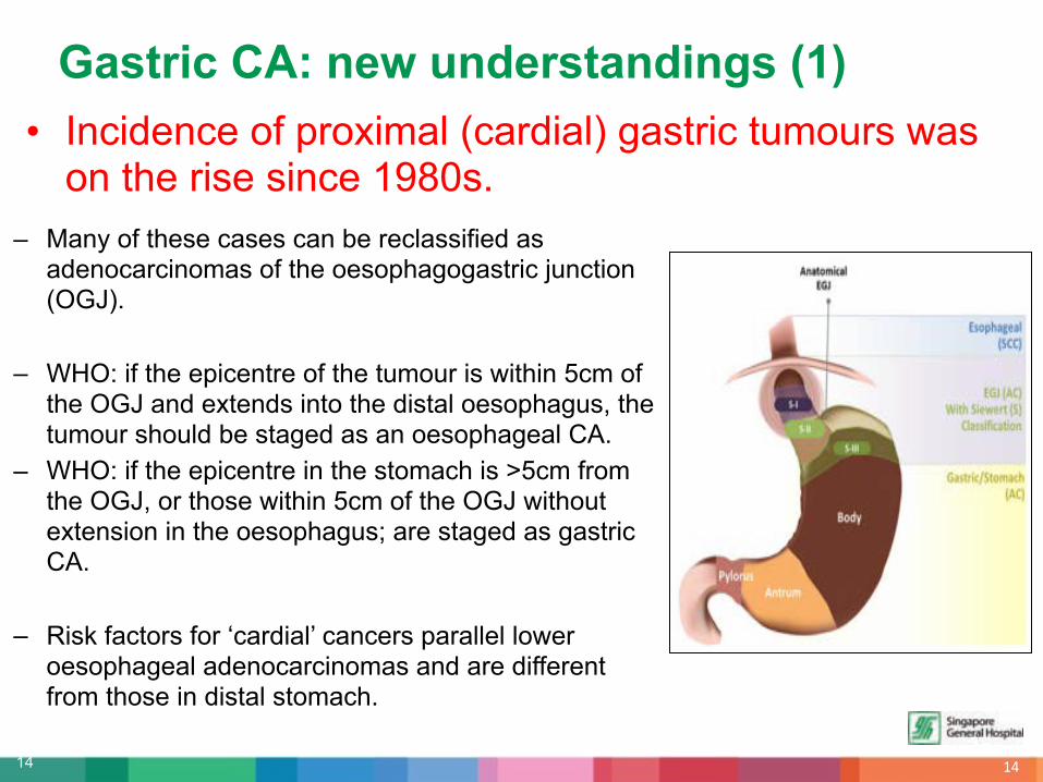
• Incidence of proximal (cardial) gastric tumours was on the rise since 1980s.
)#
Gastric CA: new understandings (1)
– Many of these cases can be reclassified as adenocarcinomas of the oesophagogastric junction (OGJ).
– WHO: if the epicentre of the tumour is within 5cm of the OGJ and extends into the distal oesophagus, the tumour should be staged as an oesophageal CA.
– WHO: if the epicentre in the stomach is >5cm from the OGJ, or those within 5cm of the OGJ without extension in the oesophagus; are staged as gastric CA.
– Risk factors for ‘cardial’ cancers parallel lower oesophageal adenocarcinomas and are different from those in distal stomach.
15
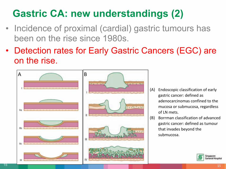
• Incidence of proximal (cardial) gastric tumours has been on the rise since 1980s.
• Detection rates for Early Gastric Cancers (EGC) are on the rise.
)$
Gastric CA: new understandings (2)
GHI J;2,6=,79=.=81669B9=1@9,;.,B.31085.>16@09=.=1;=30:.23B9;32.16.123;,=10=9;,?16.=,;B9;32.@,.@D3.?<=,61.,0.6<K?<=,61A.03>1028366.,B.+L.?3@6M.
GNI N,00?1;.=81669B9=1@9,;.,B.12O1;=32.>16@09=.=1;=30:.23B9;32.16.@<?,<0.@[email protected];O1236.K35,;[email protected]<K?<=,61M
H N

16
• As a result of increased number of upper endoscopies being performed worldwide.– 15 - 21% of newly diagnosed cases in Western studies– More than 50% in Japan
• Most occurs in males, fifties.
• Symptoms are usually mild. – epigastric pain, dyspepsia, asymptomatic
• Tumour size is usually small (2 - 5cm).
• Located on lesser curve.
• Multiple primary sites occur in 3-13%, and is associated with worse prognosis.
Early Gastric Cancers
)%

17
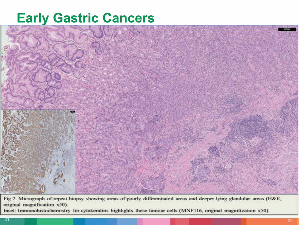
• Majority of EGCs are well differentiated.– Tubular carcinoma (52%)– Papillary carcinoma – Signet ring cell carcinoma– Poorly differentiated carcinoma
• With resection, the prognosis is excellent.– 5 year survival rates >90%
• Size of tumour and depth of invasion are the two most important prognostic indicators.
Early Gastric Cancers
)&

18
Early Gastric Cancers
)'

19
Early Gastric Cancers
)(

20
Early Gastric Cancers
!*
21
Early Gastric Cancers
!)

22
Early Gastric Cancers
!!
23
• Incidence of proximal (cardial) gastric tumours has been on the rise since 1980s.
• Detection rates for Early Gastric Cancers (EGC) are on the rise.
• There is no entirely satisfactory histologic classification of gastric carcinoma.
!"
Gastric CA: new understandings (3)

24
• The perfect classification scheme should be:
– Used prevalently in clinical work and research
– Able to cover all gastric primary lesions
– High reproducibility and validity
– Prognostically relevant
!#
‘Proliferative Classificationitis’
25
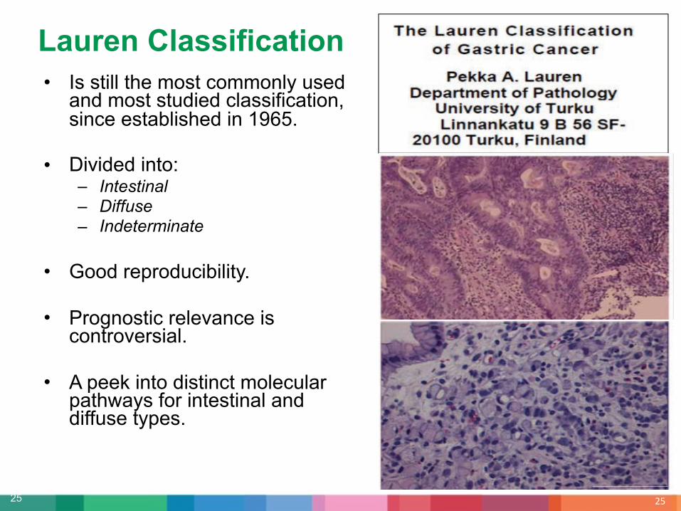
• Is still the most commonly used and most studied classification, since established in 1965.
• Divided into:– Intestinal– Diffuse– Indeterminate
• Good reproducibility.
• Prognostic relevance is controversial.
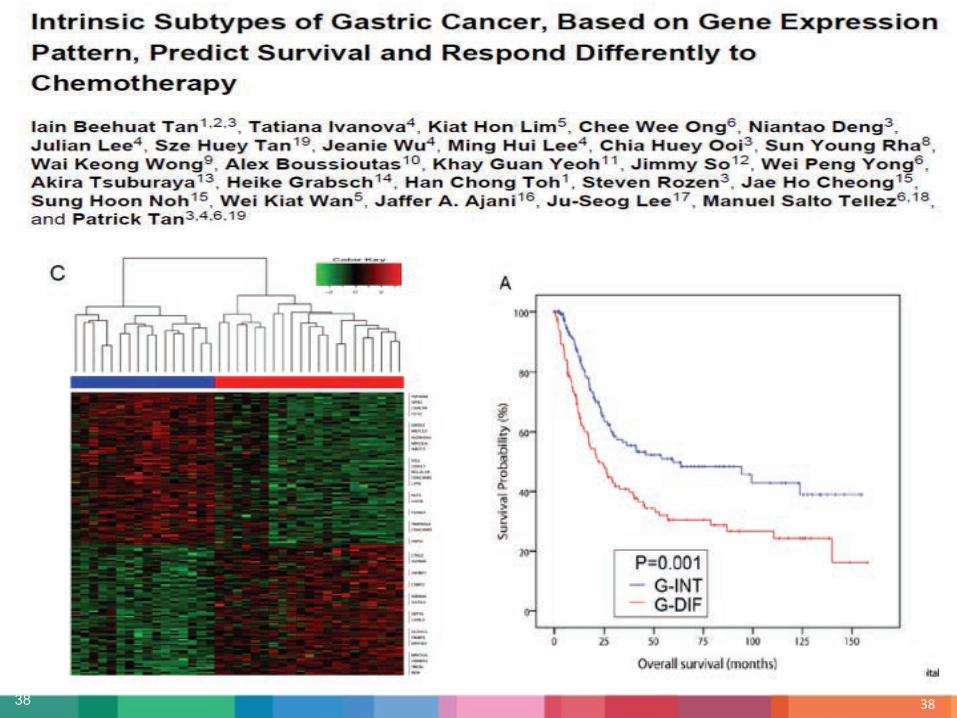
• A peek into distinct molecular pathways for intestinal and diffuse types.
!$
Lauren Classification

26 !%26

27 !&
WHO Classification, 4th Edition, 2010
P9><03.!.Q17988105.123;,=10=9;,[email protected],R3=@9,;6.89;32.K5.;3,7816@9=.=3886

28 !'
WHO Classification, 4th Edition, 2010

29 !(
WHO Classification, 4th Edition, 2010

30 "*
Gastric Carcinoma with Lymphoid Stroma• Also known as medullary
carcinoma or lymphoepithelioma-like carcinoma.
• >80% associated with EBV infection.
• Hispanic males.
• Pushing border with irregular syncytial sheets of tumour cells within a rich lymphocytic stroma.
• Prognosis better than other gastric cancers.

31 ")
Hepatoid / AFP producing Carcinoma• Hepatoid carcinoma resembles
hepatocellular carcinoma with large polygonal cells and prominent eosinophilic cytoplasm.
• AFP producing carcinoma shows well differentiated tubular or papillary architecture with clear cytoplasm.
• Both expresses AFP in immunohistochemistry and patient serum AFP may be raised.
• Aggressive tumour, 5 year survival rate ~ 12%.

32 "!
Ming Classification• Based on growth pattern.
• Divided into:– Expanding– Infiltrative
• Good reproducibility.
• Simple.
• No prognostic significance.

33
• Up to now, no histological classification system could provide additional prognostic information beyond what the TNM system does.
• Initial studies suggested that the amount of intra-cellular mucin could be a prognostic factor, but later studies could not confirm this.
• Not widely used.
""
Goseki Classification
rich

34 "#
Goseki Classification

35 "$
Correlated Classifications
• There has been good correlation between all the classification systems.
• We routinely report the WHO, Lauren and Ming classification in our reports.
36
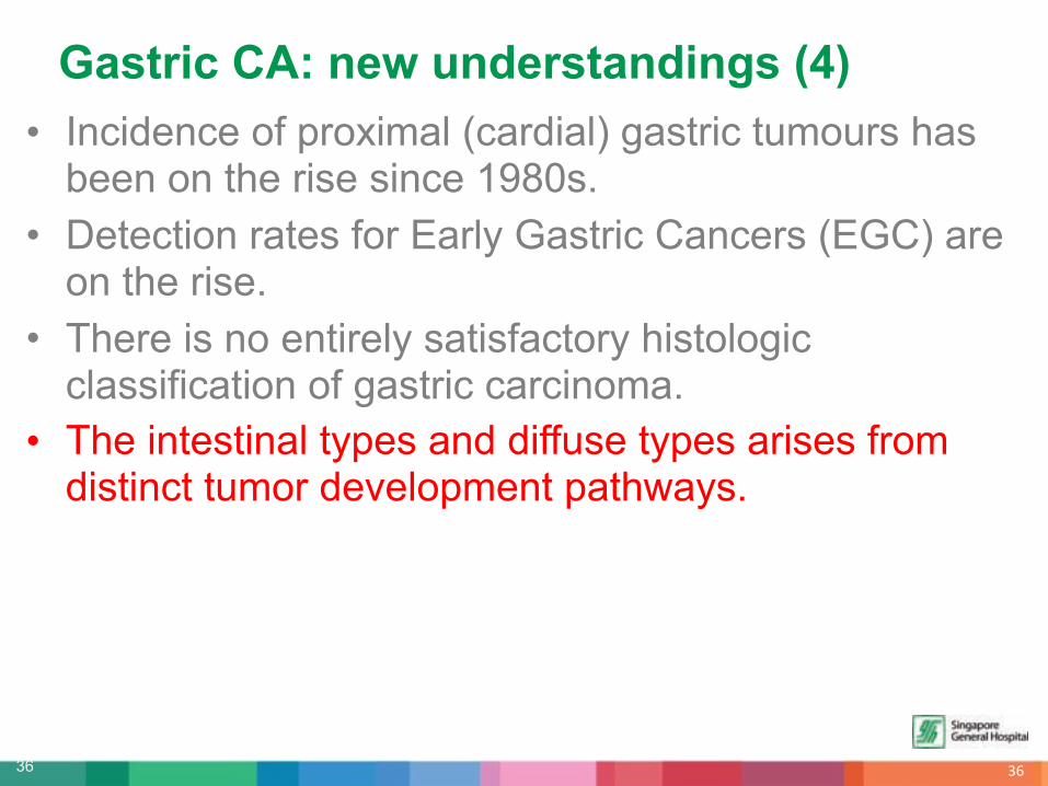
• Incidence of proximal (cardial) gastric tumours has been on the rise since 1980s.
• Detection rates for Early Gastric Cancers (EGC) are on the rise.
• There is no entirely satisfactory histologic classification of gastric carcinoma.
• The intestinal types and diffuse types arises from distinct tumor development pathways.
"%
Gastric CA: new understandings (4)

37
• 2 - 10% of all gastric carcinomas are diagnosed in young patients (<40 years).
• An equal gender distribution is reported.
• Most are of the diffuse type and are not associated with gastric atrophy and intestinal metaplasia.
• 10 - 25% of these young patients have a positive family history, suggesting genetic aetiologic factors.
• Different genomic profile between younger and older patients with gastric carcinoma.
"&
Gastric Carcinoma in Young Patients
38 "'

39 "(
".?1R,0.?,83=<810.?3=D1;96?6:.
)M SD0,?,6,?18.9;6@1K989@5M.• T;@36@9;18.@573:.'UA.)&UA.!*U.>19;6.1;2."7A.$U.8,6636.• 49BB<63.@573:.)!UA.)"U.>19;6.1;2.#UA.)$UA.)%UA.)&7.8,6636.• S,;@09K<@36.@,.B,=18.>3;3.1?789B9=1@9,;6.V.CJW!A.XJYM.
!M X9=0,61@[email protected];6@1K989@5M.• H66,[email protected]@D.9;@36@9;18.6<K@573M.• J/PWZXHQ[A.QT"[[email protected],[email protected];.0381@9,;M.
"M J79>3;3@9=.18@301@9,;6M.• 4LH.D5730?3@D581@9,;6• STXQ.GS7/.9681;2.?3@D581@,0.7D3;,@573I.V.JN\.166,=91@32
]^D3;.9;@30703@9;>[email protected]@[email protected].<63B<8.@,.=,;69230.>16@09=.=1;=30.;,@.16.,;3.2963163A.K<@[email protected]@.8316@.@-,.?1R,0.6<[email protected]@D.<;9U<3.B31@<036M_

40 #*
H691;.1;2.L,;ZH691;.>16@09=.=1;[email protected]@9;=@.>3;3.69>;1@<036.0381@32.@,.9;B81??1@9,;.1;2.9??<;[email protected]@[email protected];@91885.166,[email protected]@D.;,;ZH691;.>16@09=.=1;=306M

41 #)
42 #!
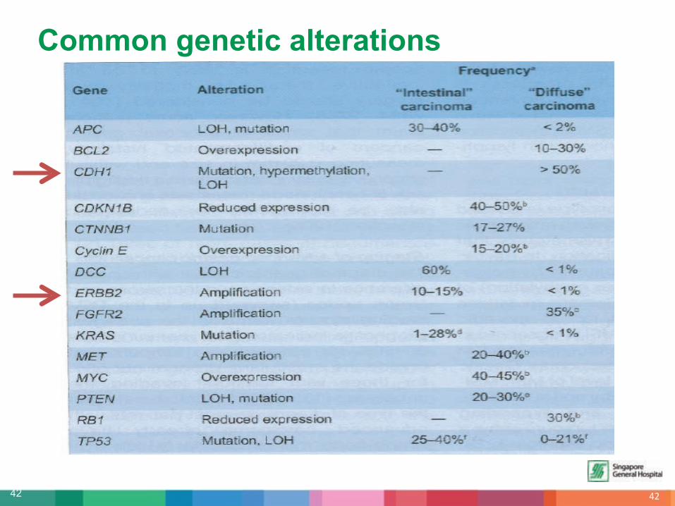
Common genetic alterations

43
• Familial Diffuse Gastric Carcinoma – CDH1 (E-cadherin) gene
• Hereditary Non-polyposis Colorectal Cancer Syndrome – MSI phenotype
• Familial Adenomatous Polyposis Coli – APC
• Li-Fraumeni Syndrome– TP53
• Peutz-Jeghers Syndrome– STK11
Hereditary Tumour Syndromes
#"

44
• Autosomal dominant germline mutation in E-cadherin gene. • Increased risk of diffuse gastric carcinoma and lobular breast
carcinoma. • Average age at diagnosis: 37 years old. • Lifetime risk of gastric carcinoma: 67%-83%.
Familial Diffuse Gastric Carcinoma
##

45
• Epidermal growth factor gene. • Preferentially expressed in the
intestinal type gastric cancers. • HER2 amplification described after its
discovery in breast cancers. • Identification of cases with HER2
amplification presents a therapeutic target for Herceptin therapy.
ERBB2 / HER2 protooncogene amplification
#$#$

46
• 20 nuclei are enumerated. • Her2/Chr17 ratio " 2.0 = Amplified • Her2/Chr17 ratio <2.0 = Non-amplified• If HER2/Chr17 ratio falls between 1.8 – 2.2 on first count, 20 additional
nuclei should be enumerated.
ERBB2 / HER2 protooncogene amplification
#%

47 #&

48
• Neuroendocrine neoplasms
• Gastrointestinal stromal tumours (GISTs)
• Gastrointestinal Lymphomas
Other tumours of the stomach
#'

49
Neuroendocrine neoplasms
50
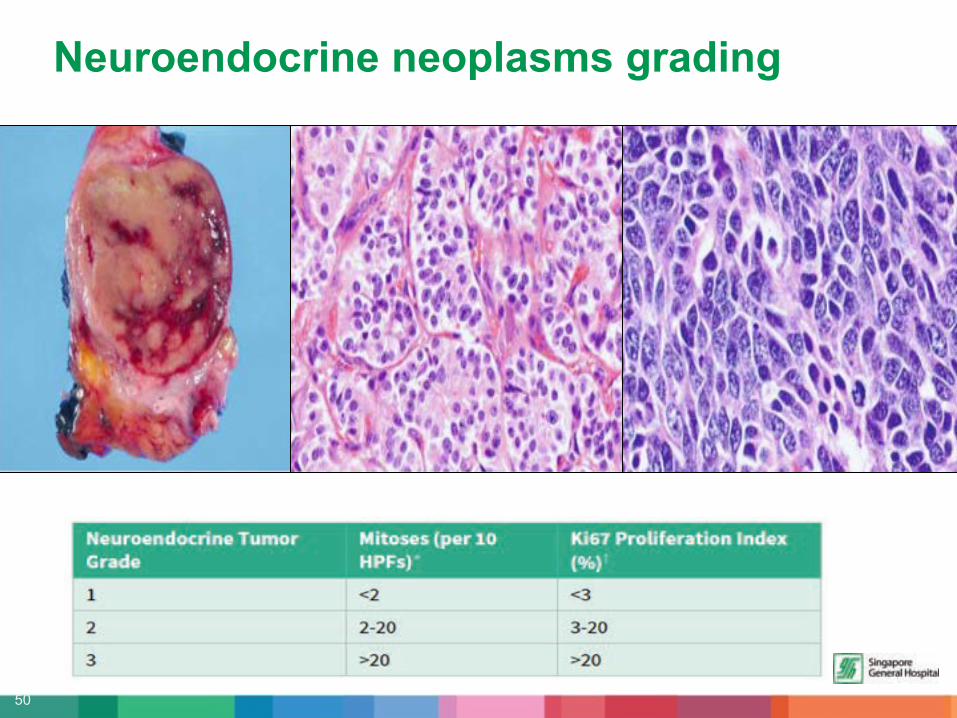
Neuroendocrine neoplasms grading

51
Neuroendocrine proliferations

52
Neuroendocrine neoplasms
$!

53
Gastric NETs: clinical subtypes
$"

54
Mixed adenoneuroendocrine carcinomas (MANECs)
$#
• MANECs are mixed carcinomas with a NET component of at least 30%.
• Rare in the stomach.
• NET component usually high grade, large cell type.

55 $$
Gastrointestinal Stromal Tumours (GISTs)
• Arises from interstital cells of Cajal (pacemaker cells).
• May arise anywhere in the GI tract but stomach is most common (60%).
• Sporadic or occur in connection with tumour syndromes.

56 $%
Gastrointestinal Stromal Tumours (GISTs)
57 $&
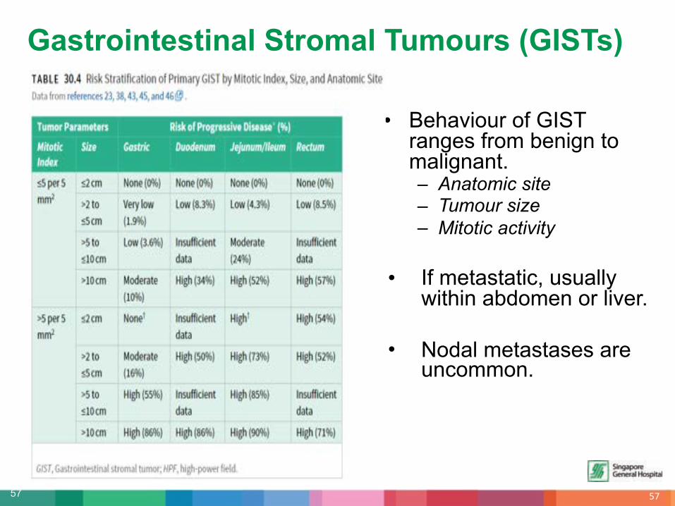
• Behaviour of GIST ranges from benign to malignant.– Anatomic site– Tumour size– Mitotic activity
• If metastatic, usually within abdomen or liver.
• Nodal metastases are uncommon.
Gastrointestinal Stromal Tumours (GISTs)

58 $'
Gastrointestinal Lymphomas• 55-75% of GI lymphomas arises from the
stomach.
• Most common types are: – Extranodal marginal zone lymphoma– Diffuse large B cell lymphoma
• Uncommon types include: – Follicular lymphoma– Mantle cell lymphoma– Burkitt lymphoma– Peripheral T cell lymphoma
59 $(
Extranodal marginal zone lymphoma, MALT type (MZL)
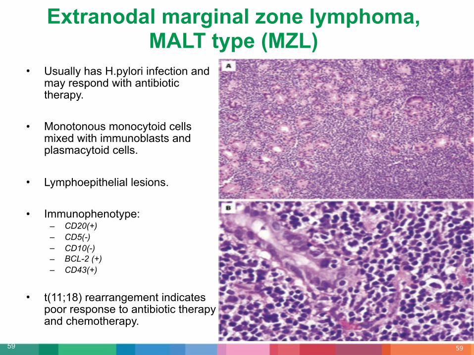
• Usually has H.pylori infection and may respond with antibiotic therapy.
• Monotonous monocytoid cells mixed with immunoblasts and plasmacytoid cells.
• Lymphoepithelial lesions.
• Immunophenotype:– CD20(+)– CD5(-)– CD10(-)– BCL-2 (+)– CD43(+)
• t(11;18) rearrangement indicates poor response to antibiotic therapy and chemotherapy.

60 %*
Diffuse Large B-cell Lymphoma (DLBCL)• May be de novo or arise due to
large cell transformation from MZL.
• Aggressive.
• Diffuse infiltrative large lymphoid cells with high grade cytomorphology.
• Immunophenotype:– CD20(+)– Ki-67 >40%– Variable expression of CD10, BCL-6,
BCL-2 and MUM-1
• May be EBV-associated in immunocompromised patients.

61 %)
The molecular age will bring answers

62 %!

63 %"

64 %#
Thank you for your kind attention.

Additional Slides

66 %%
Goseki’s 4 subtypes

67 %&

68
Tumour Regression Grade (AJCC)
%'

69
Molecular targets in OGJ cancers
%(
Molecular targets in OGJ cancers

70
In-vitro chemosensitivity of cell lines to 5-FU, oxaliplatin and cisplatin
&*

71 &)
72
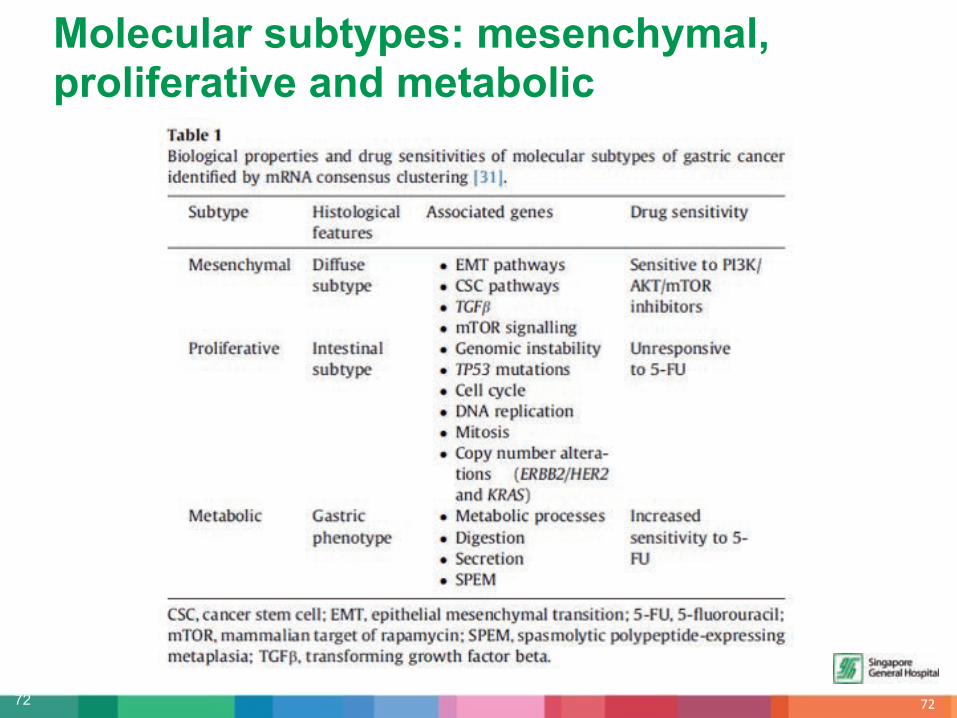
Molecular subtypes: mesenchymal, proliferative and metabolic
&!

73 &"

74 &#

75
PD-1 / PDL-1 (Programmed Death Ligand)
&$


![Approach to Gastric Cancers - Sci Forschen · Gastric carcinoma is the second most prevalent cancer in men, and fourth most prevalent cancer in women worldwide [27]. The incidence](https://img.dokumen.tips/doc/110x75/5fc7a6c4a5f82734a930563a/approach-to-gastric-cancers-sci-forschen-gastric-carcinoma-is-the-second-most.jpg)


