Embed Size (px)
Citation preview

An investigation into the efficacy of strain-counterstrain technique to produce immediate
changes in pressure pain thresholds in symptomatic subjects
James R Hutchinson
A research project submitted in partial fulfillment for the requirements for the degree of Master of Osteopathy at Unitec 2007.

1
Declaration
Name of candidate: James Richard Hutchinson
This Research Project is submitted in partial fulfillment for the requirements for the
Unitec degree of Master of Osteopathy.
The regulations for the degree are set out in the Master of Osteopathy Programme
Schedule and are elaborated in the course handbook.
Candidate’s declaration
I confirm that:
• This Research Project represents my own work;
• The contribution of supervisors and others to this work was consistent with the
Unitec Regulations and Policies.
• Research for this work has been conducted in accordance with the Unitec Research
Ethics Committee Policy and Procedures, and has fulfilled any requirements set for
this project by the Unitec Research Ethics Committee.
Research Ethics Committee Approval Number: 2006.611
Candidate Signature: ……….…………………………………….Date: 10th September 2007
(James Richard Hutchinson)
Student number: 1113979

2
DISSERTATION ABSTRACT
Background and objective: Strain counterstrain (SCS) is an osteopathic technique used by
osteopaths and manual therapists for the relief of musculoskeletal pain and associated
dysfunction. Limited literature exists to support the efficacy of SCS technique. This dissertation is
presented in two sections. Section one is a literature review of SCS technique and the proposed
outcome measures. Section two is the research conducted presented as a manuscript in the style
required by the International Journal of Osteopathic Medicine. The second section is supported
by three appendices of material not intended for publication. The aim of this study was to
investigate the efficacy of SCS technique on subjects with a history of a recreational sports injury
of the upper extremity.
Design: Randomized assessor blinded placebo controlled trial.
Methods: Twenty three subjects (13 males, 10 females; mean age=26.1, SD=6.3) fulfilled the
requirements for the study. Subjects were screened to establish the presence of a primary tender
point (TeP) around the elbow joint. Subjects were randomly assigned into two groups and
received either an SCS intervention or a Sham intervention. The primary outcome measures were
pressure pain threshold (PPT) on the primary TeP, and visual analog scale (VAS) assessing local
pain intensity elicited by the application of approximately 3kg/cm2 of pressure on the primary TeP.
The secondary outcome measure was pain-free grip strength (PFGS).
Results: Within group changes showed a significant improvement in VAS for pain intensity
following the SCS intervention (p<0.001) compared with the Sham intervention (p=0.053). Pre-
post effect sizes for the VAS for pain intensity were ‘large’ in the SCS intervention group (d=1.87)
and ‘moderate’ (d=0.90) for the Sham intervention group. Both groups surpassed the minimal
clinically important difference (MCID) defined as a decrease ≥30% in VAS for pain intensity (SCS
group=55%, Sham group=31%). Within group changes showed a small improvement for PPT at
the TeP following either the SCS intervention (p=0.497) compared with the sham intervention
(p=0.749). Pre-post effect sizes for the TeP were small in the SCS intervention group (d=0.29)
and trivial (d=0.14) for the sham intervention group. No significant differences were found for the
PFGS (SCS: p=0.936 d=0.03, Sham: p=0.989 d=0.01)
Conclusions: The results indicate that SCS technique may be an efficacious technique in
treatment of TePs around the elbow in subjects with a history of a recreational sports injury of the
upper extremity. SCS technique can produce decreases in pain intensity as reported from
mechanical pressure at a primary TeP.

3
Table of Contents
Declaration .................................................................................................................................. 1
DISSERTATION ABSTRACT ..................................................................................................... 2
List of Tables ............................................................................................................................... 4
List of Figures.............................................................................................................................. 4
Acknowledgements ..................................................................................................................... 5
Abbreviations............................................................................................................................... 6
SECTION I – LITERATURE REVIEW ..................................................................7
Introduction.................................................................................................................................. 8
Effectiveness and Efficacy of Strain-counterstrain Technique ............................................. 10
Sports Related Injuries.......................................................................................................... 14
Outcome Measures............................................................................................................... 14 Visual Analogue Scale for Pain Intensity ........................................................................................ 14 Pressure pain threshold.................................................................................................................. 15 Pain-Free Grip Strength.................................................................................................................. 16
Minimal Clinically Important Difference................................................................................. 17
Sham Protocol ...................................................................................................................... 19
Conclusion................................................................................................................................. 20
References ................................................................................................................................ 21
SECTION II - MANUSCRIPT..............................................................................26
ABSTRACT ............................................................................................................................... 28
INTRODUCTION....................................................................................................................... 29
METHODS ................................................................................................................................ 30
Design ................................................................................................................................... 30
Eligibility Criteria ................................................................................................................... 31
Pre-Intervention Examination ............................................................................................... 31
Pre-Intervention Outcomes Assessment .............................................................................. 32
Sample Size.......................................................................................................................... 32
Randomization ...................................................................................................................... 32
Intervention Protocol ............................................................................................................. 33
SCS Intervention............................................................................................................................. 33 Sham Intervention........................................................................................................................... 34
Outcome Measures............................................................................................................... 34
Visual Analogue Scale.................................................................................................................... 35 Pressure Pain Threshold ................................................................................................................ 35 Pain-Free Grip Strength.................................................................................................................. 36 Reliability ........................................................................................................................................ 36
DATA ANALYSIS ...................................................................................................................... 37
RESULTS.................................................................................................................................. 38
Subjects ................................................................................................................................ 38
VAS for Pain Intensity ........................................................................................................... 39 Changes in PPT levels ................................................................................................................... 40 Changes in PFGS levels................................................................................................................. 42 Reliability ........................................................................................................................................ 43
DISCUSSION ............................................................................................................................ 45
VAS for Pain Intensity ........................................................................................................... 46
Changes in PPT levels.......................................................................................................... 46
Changes in PFGS levels....................................................................................................... 47
Reliability............................................................................................................................... 47
Limitations............................................................................................................................. 48
Conclusion ............................................................................................................................ 49
ACKNOWLEDGEMENTS ......................................................................................................... 49
REFERENCES.......................................................................................................................... 50
Appendix A - Figures................................................................................................................. 54
Appendix B - Ethics Resources................................................................................................. 59
Appendix C – Instructions for authors for manuscript submission............................................ 64

4
List of Tables
Table 1 Baseline Characteristics of Subjects ................................................................................ 38
Table 2 Reliability indices .............................................................................................................. 43
Table 3 Group and Individual Changes in Context of Smallest Detectable Differences and Minimal
Clinically Important Difference....................................................................................................... 44
List of Figures
Figure 1 Flowchart of Study Design .............................................................................................. 30
Figure 2 Mean group VAS for pain intensity.................................................................................. 39
Figure 3 Individual mean VAS for pain intensity............................................................................ 40
Figure 4 Group Means for Primary Tender point PPTs................................................................. 41
Figure 5 Group Pain Free Grip Strength. ...................................................................................... 43
Figure 6 Individual mean PPT for tender point. ............................................................................. 54
Figure 7 Group Means for Medial Epicondyle PPTs. .................................................................... 55
Figure 8 Individual mean PPT for medial epicondyle. ................................................................... 56
Figure 9 Group Means for Lateral Epicondyle PPTs..................................................................... 57
Figure 10 Individual mean PPT for lateral epicondyle................................................................... 58

5
Acknowledgements
I wish to thank the following people for their support throughout the process of completing this
research project:
To all the participants who so generously gave up their time to be part of my research.
To my supervisors: Dr Carol Horgan for her support and feedback, and Rob Moran for his
assistance during data collection, support, feedback, and tireless pursuit of accuracy.
To Marshall Gabin who generously gave up his spare time to provide the intervention for this
project.
To my family for their continuous support, encouragement and sacrifices they have made.

6
Abbreviations
SCS Strain Counterstrain
VAS Visual Analog Scale
TeP Tender point
PPT Pressure Pain Threshold
PFGS Pain-Free Grip Strength
SEM Standard Error of Measurement
SDD Smallest Detectable Difference
MCID Minimal Clinically Important Difference
SD Standard Deviation
d Effect Size (Cohen’s d)
OMT Osteopathic Manipulative Therapy
RCT Randomized Controlled Trial
ICC Intra-class Correlation Coefficient

7
SECTION I – LITERATURE REVIEW

8
Introduction
Strain-counterstrain (SCS) is an osteopathic treatment technique first developed and described
by Lawrence Jones in 1964 (L. H. Jones, 1981). Strain-counterstrain, also known as positional
release, is an indirect osteopathic technique, whereby dysfunctional joints and their muscles are
moved away from their restrictive barriers into positions of ease in the treatment of both
musculoskeletal (D'Ambrogio & Roth, 1997; L. H. Jones, 1981; Ward, 2003) and visceral
dysfunctions (Giammatteo & Weiselfish-Giammatteo, 1997). Since Jones’ first description there
has been a variety of anecdotal evidence presented by assorted therapists in support of the
technique but only limited experimental evidence to demonstrate it’s efficacy in the treatment of
musculoskeletal pain and related joint dysfunction (D'Ambrogio & Roth, 1997; Giammatteo &
Weiselfish-Giammatteo, 1997; L. H. Jones, 1981; Ward, 2003).
The application of SCS technique requires a practitioner to first palpate a tender point (TeP) in
the soft tissues, the patient’s limb is then moved in such a way that the pain associated with
pressure on the TeP is relieved by at least 70 percent to find the position of ease (D'Ambrogio &
Roth, 1997; L. H. Jones, 1981; McPartland & Goodridge, 1997; Wong & Schauer-Alvarez, 2004;
Wong & Schauer, 2004). Jones (1981) suggested a minimum period required to hold a position of
ease as 90 seconds. It is theorized that the shortening or “folding-over” of aberrant tissues in SCS
technique achieves its therapeutic modifications via both proprioceptive (Korr, 1975) and
nociceptive mechanisms (Bailey & Dick, 1992), or by the biomechanical principle of
nonsequential motion expounded by McPartland & Klofat (1995).
Bailey & Dick (1992) proposed a nociceptive hypothesis that tissue damage in dysfunctional
muscles can be reduced by the positional release mechanism utilized by SCS. They suggest that
relaxation of the damaged tissues may be achieved by placing patients in a position of ease
which may advance local perfusion of fluids (i.e. blood, and lymph) and enhance the removal of
sensitizing inflammatory mediators.
In order to use SCS technique a practitioner needs to be able to accurately identify a painful TeP.
The TeP can be interpreted using anatomical charts (D'Ambrogio & Roth, 1997; L. H. Jones,
1981) or the “Paracelsus approach” (McPartland & Klofat, 1995), probing a region of patient’s
pain complaint with the practitioners thumb or finger to locate sensitive TeP’s. McPartland &
Goodridge (1997) found the SCS method of palpatory diagnosis to be more reliable compared to
a traditional osteopathic examination (the tests studied included palpation for restriction of motion,
local tissue texture changes and joint capsule tenderness) for subjects with chronic neck pain,
demonstrating a 72.2% (κ=0.45; ‘moderate’) agreement between examiners.
9
The essence of SCS is the relief of a painful TeP by moving the patient’s limb in order to find a
position of ease (L. H. Jones, 1981). Total pain relief is not always the goal, a minimum level of
70% reduction in the (short term) pain associated with the TeP is said to be expected immediately
after treatment (D'Ambrogio & Roth, 1997; McPartland & Klofat, 1995).
In 1981 Jones reported diagnostic correlations between joint dysfunction and palpable myofascial
tissue TePs (L. H. Jones, 1981). D’Ambrogio & Roth (1997) have subsequently published a list of
more than 200 diagnostic TeP locations. The TePs used in SCS are described by Jones (L. H.
Jones, 1981) as:
The tender points used in counterstrain techniques are not located in or just
beneath the skin as are many acupuncture points, but deeper in muscle,
tendon, ligament, or fascia. They measure 1cm across or less, with the most
acute point about 3mm in diameter. They may be multiple for one specific joint
dysfunction, may extend for a few centimeters along a muscle, or may be
arranged in a chain (such as the ones in the muscle and fascia along the
lateral surface of the femur) (p. 28)
Other definitions are similar (D'Ambrogio & Roth, 1997; McPartland & Klofat, 1995). When a
standardized pressure of 2.6kg/cm2 is applied to a TeP it has been found to cause a localized
sensation of pain (L. H. Jones, 1981; McPartland & Klofat, 1995).

10
Effectiveness and Efficacy of Strain-counterstrain Technique
A comprehensive search of peer-reviewed literature analyzing the efficacy or effectiveness of
SCS identified six experimental studies (Blanco et al., 2006; Howell, Cabell, Chila, & Eland, 2006;
Meseguer, Fernández-de-las-Peňas, Navarro-Poza, Rodrìguez-Blanco, & Gandia, 2006; Wong &
Schauer-Alvarez, 2004; Wong & Schauer, 2004; Wynne, Burns, Eland, Conatser, & Howell,
2006). Three additional experimental studies were identified that utilized SCS as part of an
investigation into osteopathic manipulative therapy (Gamber, Shores, Russo, Jimenez, & Rubin,
2002; Noll, Degenhardt, Stuart, McGovern, & Matteson, 2004) or manual therapy (Cleland, Flynn,
& Palmer, 2005). The paucity of peer reviewed randomized controlled trial (RCT) articles related
to SCS necessitated reference to descriptive case studies and osteopathic and manual medicine
texts.
Wong & Schauer (2004) provided evidence of a good correlation between pain reduction (using a
visual analog scale (VAS) as a measurement of pain intensity) and SCS pain reduction at TePs
(using manual palpation of tender points (TePs) as a measurement). The measurement of TeP
sensitivity was performed through manual palpation of TePs to elicit a physical response (visual
cue) from subjects, without the aid of pressure algometry to aid reliability. In a follow up study
Wong & Schauer-Alvarez (2004) demonstrated an increase in strength in hip musculature
following SCS treatment, and a correlated reduction in TeP pain in those muscles.
Meseguer et al. (2006) concluded that the application of SCS technique may be effective in
producing hypoalgesia and decreased reactivity of TePs. In their study Meseguer et al (2006)
reported moderate effect sizes (as described by Hopkins (2002)) for the VAS for pain intensity
between pre –and post- intervention measurement following the application of either the classical
or modified application of SCS technique (p<0.001; Cohen’s d = 1.1). In comparison their control
group did not show any change (p=0.90; Cohen’s d = 0.01). The authors were able to
demonstrate an immediate decrease in the sensitivity of a chosen TeP following the application of
SCS technique (Meseguer et al., 2006). The results in this study were limited by the choice of
outcome measure, the VAS is a valid clinical measure (Reeves, Jaeger, & Graff-Radford, 1986),
but could have become a source of error if the subjects were biased by the ability to remember
their previous scores due to the short time period between pre- and post- recording (Meseguer et
al., 2006). Another potential bias in their group design was the lack of a sham intervention group
to investigate the level of placebo attached to their procedure.

11
Wynne et al. (2006) reported mechanical changes in the peak torque following treatment by SCS
technique for various TePs in the lower leg and foot. The treatment effect was considered by the
authors to be greater than any changes attributed to the process of repeated measures. They
demonstrated a decrease in symptom severity as measured by their adopted four factor ratings
scale. The four factors were: pain, soreness, stiffness, and mobility; each factor was scored
between 0 (no symptoms) to 9 (extreme symptoms/pain), and the researchers then summed the
values for all subjects to give a score out of a maximum range from 0 to 36 (Wynne et al., 2006).
This study was not satisfactorily blinded, as the sham treatment chosen was an oral placebo,
such that most subjects who took them during the course of the cross-over trial were able to
deduce their ineffectiveness.
The data reported by Howell et al. (2006) provides evidence in support of Korr’s (1975)
hypothesis of somatic dysfunction. Korr (1975) hypothesized that the characteristic restriction-of-
motion present in somatic dysfunction was due in part to an alteration in the sensitivity of the
monosynaptic stretch reflex, and could therefore be reset to restore range of motion. Howell et al.
(2006) did not follow a sufficiently rigorous methodology in order to be able to make any
generalized claims as to the effectiveness of SCS for patient’s with Achilles’ tendonitis. Their
procedure compares a symptomatic SCS intervention group with an asymptomatic sham
intervention group.
Blanco et al. (2006) compared a muscle energy technique (post-isometric relaxation) with SCS
technique in asymptomatic subjects presenting with latent myofascial trigger points. The
application of SCS technique was not described as addressing a primary TeP, but rather the
therapist located ‘a trigger point’ in the masseter muscle and proceeded to apply the SCS
technique. It would seem plausible to expect a technique that stretches the whole muscle to be
more effective in an asymptomatic subject when compared to a technique usually employed to
address somatic dysfunction such as SCS. This expectation was upheld with the results reported
by Blanco et al. (2006), where they reported an insignificant ‘small’ effect size (d=0.32; p=0.08)
for the immediate improvements in active mouth opening for asymptomatic subjects treated with
SCS for a trigger point identified within the masseter muscle. Blanco et al. (2006) surmised that
SCS technique applied to a single randomly chosen latent myofascial trigger point did not
improve active mouth opening. The approach employed by Blanco et al. (2006) was a deviation
from the recommended method of application for SCS technique, as defined in the regular
literature which includes a physical examination of an area for the primary TeP (D'Ambrogio &
Roth, 1997; L. H. Jones, 1981; McPartland & Klofat, 1995). Therefore, the findings of this study
should not be considered directly applicable to SCS technique as defined in the literature
(D'Ambrogio & Roth, 1997; L. H. Jones, 1981; McPartland & Klofat, 1995).

12
Two additional experimental studies were identified that utilized SCS as part of an investigation
into osteopathic manipulative therapy (Gamber et al., 2002; Noll et al., 2004). Gamber et al.
(2002) conducted an observer blinded pragmatic randomly assigned clinical trial of osteopathic
manipulative therapy (OMT) in patients diagnosed with fibromyalgia syndrome. The SCS
techniques were consistently applied in combination with other osteopathic techniques to the
TePs the patient identified as most troublesome. The other osteopathic techniques available to
the treating osteopath included myofascial release, muscle energy, soft tissue treatment, and
craniosacral manipulation (Gamber et al., 2002). The amount of time, and number of points
addressed with SCS were not reported. The authors reported significant improvement in pain
thresholds post treatment at three locations (left and right second costochondral junction, and the
left medial epicondyle) for patients receiving SCS in combination with OMT (Gamber et al., 2002).
From the data reported by Gamber et al. (2002) it is not possible to draw any conclusions as to
the efficacy of SCS technique.
A number of descriptive articles on SCS have established it is widely utilized clinically as an
effective osteopathic manipulative technique, although there is limited evidence substantiating the
assumed efficacy of SCS. Several of the descriptive articles have described specific clinical
applications for SCS, such as, Complex Regional Pain Syndrome I (CRPS I) following a Grade II
ankle sprain (Collins, 2007), iliotibial band friction syndrome (Pedowitz, 2005), sacral torsions
(Cislo, Ramirez, & Schwartz, 1991), low back pain (Lewis & Flynn, 2001), chronic myofascial pain
(Dardzinski, Ostrov, & Hamann, 2000), foot disorders (L. H. Jones, 1973), and acute ankle
injuries (Eisenhart, Gaeta, & Yens, 2003).
Collins (2007) reports on the case of a 14 year old with CRPS I following a Grade II ankle sprain,
and the benefits recorded by way of the analgesic effects of SCS and improved function. A
decrease of two points on a numeric pain rating scale was reported for overall pain after two
months, as was, a decrease in tenderness for 10 out of 13 TePs. These analgesic effects were
considered clinically significant, and are suggestive of the need for more formal investigation.
Dardzinski et al. (2000) found in a retrospective review of 20 patients suffering from chronic
localized myofascial pain, the use of the Jones SCS technique could be beneficial in reducing
pain and improving function. Eisenhart et al (2003) found that SCS could be beneficial in the
treatment of acute ankle injury. Cleland et al (2005) produced evidence of increased pain free
grip strength and decreased pain scores after treatment applied to the area of lateral epicondyle
and the cervicothoracic spine. Lewis & Flynn (2001) reported on four case studies of patients with
low back pain treated with SCS protocols. The authors reported improvements in the outcomes

13
measured for disability levels (Oswestry Low Back Pain Disability Questionnaire) and pain (McGill
Pain Questionnaire) in all four cases (Lewis & Flynn, 2001).
Domholdt (2000) has proposed that the primary value of case reports is an effective means for
practitioners to communicate to scientists. This communication stimulates the progression from
theory based on anecdotal evidence to theory based on robust research.
In addition, a number of studies have reported the use of SCS as part of an overall osteopathic
manipulative treatment protocol to treat a variety of disorders including shoulder pain and
repetitive strain injury of the supraspinatus muscle (Jacobson, Lockwood, Hoefner V, Dickey, &
Kuchera, 1989), chronic pelvic pain (Tettambel, 2005), chronic pain (Kuchera, 2005), low back
pain (Licciardone, 2004), arthritis related pain (DeAngelo & Gordin, 2004), and cervicothoracic
pain (Walko & Janouschek, 1994) and acute or chronically ill hospital patients (Schwartz, 1986).
Similarly SCS has been reported beneficial when used in conjunction with massage therapy for
the treatment of temporomandibular joint dysfunction (Eisensmith, 2007).
After reviewing the literature there is a large body of anecdotal and descriptive research in
support of the effectiveness of SCS technique in clinical practice. To date there are only a few
peer reviewed RCT articles that provide substantive evidence as to the efficacy of SCS either in
the short term or long term when compared to either a suitable sham (placebo) or control group.

14
Sports Related Injuries
Recreational sports people offer a broad heterogeneity of age, occupation and socioeconomic
group (Stockard, 2001). Recreational sports injuries to the elbow region often involve either
epicondylalgia or overuse syndromes (Fulcher, Kiefhaber, & Stern, 1998). These injuries are
most frequently the result of activities that require the elbow and forearm to transmit force to a
tool; i.e. racquet sports (Gruchow & Pelletier, 1979), rowing (Fulcher et al., 1998), and golf
(Stockard, 2001). Or to support the body’s weight i.e. cycling (Tucci & Barone, 1988).
The most common injury to amateur golfers is lower back injuries (34.5% of all injuries) followed
by injuries to the elbow (33.1%), and hand and wrist injuries (34.5% combined). In female golfers,
elbow injury (35.5%) is even more common than lower back injury (27.4%) (McHardy & Pollard,
2005; Stockard, 2001). Stockard (2001) reports a wide range of elbow injuries that may be
present in a golf playing population group, such as; lateral epicondylitis, extensor overload
injuries, lateral compression injuries, ulnar neuritis associated with medial epicondylitis (up to
20%), flexor carpi ulnaris tenosynovitis, and possible degenerative change (Stockard, 2001).
Lateral elbow injuries ('tennis elbow') are more common when compared to medial elbow injuries
(‘golfer's elbow’) at a ratio of 5:1 (lateral:medial) (McHardy & Pollard, 2005). Sevier and Wilson
(1999) report that there have been in excess of 40 different treatment methods for lateral
epicondylitis described in the literature. Physical therapy has been recommended when the
condition becomes chronic or does not respond to initial treatment.
Outcome Measures
Visual Analogue Scale for Pain Intensity
The visual analog scale (VAS) can be used to evaluate a subject’s perception of pain level on a
horizontal linear scale; this can be interpreted as pain intensity. The VAS is an unmarked
horizontal line ( generally between 100-130mm in length) with a pain descriptor at each end; “No
Pain” at one end to “Unbearable Pain” at the other (Yeomans & Liebenson, 1996).
The validity of the VAS as an outcome measure of pain intensity is well established (Crossley,
Bennell, Cowan, & Green, 2004; Merskey, 1973; Ostelo & de Vet, 2005; Price, Bush, Long, &
Harkins, 1994; Price, McGrath, Rafii, & Buckingham, 1983; Vicenzino, Collins, Benson, & Wright,
1998; Yeomans & Liebenson, 1997). The VAS has a high level of responsiveness, reliability, and
validity permitting detection of clinically relevant changes – an essential measurement for clinical
trials (Reading, 1980). Bisset, Paungmali, Vicenzino, Beller & Herbet (2005) report that 25 out the

15
28 RCTs accepted in the systematic review and meta-analysis of physical treatments for lateral
epicondylalgia utilized a VAS for pain as an outcome measure. The VAS measured in millimeters
on a 100 mm line has a possible 101 response levels, this increases the sensitivity of the VAS
compared to other measures with more limited response categories (Ostelo & de Vet, 2005).
Pressure pain threshold
Pressure algometry is commonly used to quantify measurement of tissue sensitivity (Fischer,
1987; Fryer, Carub, & McIver, 2004; Fryer & Hodgson, 2005; Reeves et al., 1986). The algometer
is a calibrated force pressure gauge that can be used to assess the pressure pain threshold
(PPT) in an individual. There is a body of evidence (Fischer, 1987; Gold, Punnett, & Katz, 2006;
Maquet, Croisier, Demoulin, & Crielaard, 2004; Ohrbach & Gale, 1989; Reeves et al., 1986)
supporting the reliability of pressure gauges (algometers) when used to determine PPTs on bony
and muscular landmarks. The PPT is highly dependant on anatomical location and gender, as
reported by Maquet et al (2004), for the lateral epicondyle healthy males have a mean PPT = 340
kPa (3.5 kg/cm2) and healthy females a mean PPT = 250 kPa (2.6 kg/cm
2). Kosek, Ekholm &
Hansson (1999) have noted that great inter-individual variability in PPTs exists in healthy
subjects.
Fischer (1987) provided evidence of the reproducibility and validity of PPT measurement, with
identical results obtained from muscles on opposite sides of the body for both male and female
normal subjects. The use of PPT as an outcome measure was reported in six out of the 28 RCTs
accepted in the systematic review and meta-analysis of physical treatments for lateral
epicondylalgia carried out by Bisset et al. (2005). The PPT can be an accurate measure of
change in tissue sensitivity, when sensitivity is defined as the amount of pressure in kg/cm2
needed to elicit a change in sensation from pressure to that of discomfort or pain (Nussbaum &
Downes, 1998; Potter, McCarthy, & Oldham, 2006; Reeves et al., 1986).
Ylinen et al. (2007) reported intra-class correlation coefficients (ICCs) between ‘very large’ and
‘almost perfect’ (0.78-0.93) for the measurement of PPTs of neck muscles. An ICC is considered
large (between 0.5 to 0.7), very large (between 0.7 to 0.9) and almost perfect (between 0.9 and
1.0) (Hopkins, 2002). Similarly Paungmali et al. (2003) showed reliability correlation coefficients
ranging in the ‘very large’ category (0.79 to 0.89) for subjects with lateral epicondylalgia. Jones et
al. (2007) reported a range of ICCs in the ‘almost perfect’ category (0.92 to 0.98) for the
measurement of PPTs in the upper limb and torso of healthy young women. In a study of PPT for
subjects with unilateral shoulder and arm pain Vanderweeën et al. (1996) reported ICCs between
‘large’ to ‘almost perfect’ (0.64 to 0.96).

16
Kosek et al. (1999) showed evidence of lower PPTs over a muscle-nerve site when compared to
PPT measurements from either bony or pure muscle sites. Further they gave evidence that these
relationships remain irrespective of skin hypoesthesia, and thus are more likely to be reflective of
the sensitivity of deeper structures. They conclude that skin pressure pain sensitivity can
influence the PPT (Kosek et al., 1999). Slater, Arendt-Nielsen, Wright, & Graven-Nielsen (2003)
documented that the most sensitive sites to pressure of various tissues in and around the elbow
were the extensor carpi radialis longus origin and the extensor carpi radialis brevis muscle belly.
They hypothesized that this phenomenon can be explained partially by the increased density of
nociceptors in these regions compared with other tissues around the elbow.
While pressure algometry has been commonly used in clinical practice for the quantitative
measurement of tenderness, many variables have been found (such as gender, site of
application, and application rate) that lead to a high degree of inter-individual variability (Maquet
et al., 2004; Paungmali, O’Leary et al., 2003). With adequate operator training the site of
application and application rate can be controlled. Paungmali, O’Leary et al. (2003) demonstrated
treatment effects after application of a manual therapy intervention for lateral epicondylalgia with
positive changes in the PPT. In a later paper Paungmali, Vicenzino & Smith (2003) demonstrated
that repeated application of the manual therapy intervention showed diminished degrees of
improvement.
Pain-Free Grip Strength
Pain-free grip strength (PFGS) is defined as the amount of force a subject is able to generate with
an isometric gripping action before eliciting pain (Paungmali, O’Leary et al., 2003; Pienimäki,
Siira, & Vanharanta, 2002). Pain-free grip strength is commonly used as an outcome measure for
the treatment of forearm pain. Bisset et al. (2005) reported that PFGS was employed as an
outcome measure in 14 out the 28 RCTs accepted in the systematic review and meta-analysis of
physical treatments for lateral epicondylalgia. Pain free grip strength has previously been used as
an outcome measure for physiotherapy RCTs, although it has been noted by Smidt et al. (2002)
that the specification of grip strength is sometimes not properly reported.
Measurement of PFGS has been shown to be highly reliable and is robust as a measure of
functional disability for subjects with elbow pain (Hillman et al., 2005; Paungmali, O’Leary et al.,
2003; Paungmali, Vicenzino et al., 2003; Smidt et al., 2002; Stasinopoulos & Stasinopoulos,
2006). Pienimäki et al. (2002) have shown that increases in grip strength and PFGS correlate well
with measures of treatment effectiveness for both medial and lateral epicondylitis. Studies
investigating the examiner reliability of PFGS have reported high intra-observer reliability
17
coefficients (Smidt et al., 2002). Hillman et al. (2005) reported significant differences in PFGS
between male and female participants in their study.
Minimal Clinically Important Difference
The minimally clinically important change of the VAS score has been approximated between 30-
35 mm (acute subjects) down to 20-25 mm (sub acute to chronic subjects) (Lee, Hobden, Stiell, &
Wells, 2003; Ostelo & de Vet, 2005). This is defined as the minimal clinically important difference
(MCID).
A variety of studies have returned similar figures for the MCID for VAS for pain intensity scores. In
a pragmatic study of chiropractic intervention for low back pain Garner et al. (2007) reported a
MCID of 2.3 (95% CI = 1.9-2.6) on a numeric visual analog scale for pain intensity (p<0.008) for
249 subjects. In a prospective observational study of adult emergency department patients with
acute pain, Lee et al. (2003) reported a mean reduction in VAS score of 30mm (95% CI = 23.6-
36.4) as representing a clinically important difference in pain severity produced by adequate
analgesic control in 81% of the subjects. In an investigation of patient’s with chronic back pain
Mesrian et al. (2007) reported an MCID of 25mm for a VAS for pain intensity. Lee et al. (2003)
reported a mean reduction in VAS of 30mm demonstrative of a patient’s perception of sufficient
pain control in relation to administration of parenteral analgesics. Ries (2005) defined a change in
VAS between 10 to 20mm as the MCID in relation to chronic obstructive pulmonary disease.
Bird & Dickson (2001) developed a variable scale for MCIDs for VAS for pain intensity. Bird &
Dickson (2001), defined ‘a lot better’ as a change in VAS correlated to the initial value recorded.
A pre-intervention VAS of up to 33mm would require a post-intervention reduction in VAS of
>16mm to indicate a perception of ‘a lot better’ for the patient, whereas a pre-intervention VAS of
between 34mm to 66mm would require a post-intervention reduction in VAS of >33mm, likewise
an initial VAS of >67mm would require a reduction of >48mm to demonstrate the same change in
subjectivity (Bird & Dickson, 2001). An issue for consideration in the comparison of a VAS to
other more scalar measures is described by Fosnocht, Chapman, Swanson & Donaldson (2005).
The authors report that a change in pain intensity of 10mm as reported on a VAS for pain may not
be the same subjective experience when the subject decreases a VAS from 40 to 30mm
compared to a change from 90 to 80mm (Fosnocht et al., 2005).

18
Patients with acute pain in a trauma department were reported as being a ‘little better’ having a
mean MCID of 13mm (95% confidence interval, 10 to 17mm) on a 100mm VAS (Todd, Funk,
Funk, & Bonacci, 1996). In their pilot study of acupuncture for low back pain Kennedy et al.
(2007) proposed an MCID of 20mm through post hoc power analysis. An MCID of 20mm was
reported by Crossley et al (2004) in their paper investigating the responsiveness of outcome
measures for the treatment of patellofemoral pain. Kelly (2001) found that there was no statistical
difference in the MCID for VAS for pain regardless of the pain severity. Kelly stated the MCID
numerically as 12 mm (95%CI 9 mm to 15 mm) for adult patients in an urban emergency
department.
The MCID for cancer related break-through pain relief was reported as a 30% decrease in VAS
score (Farrar, Portenoy, Berlin, Kinman, & Strom, 2000) and similarly for chronic pain sufferers
30% (Farrar, Young Jr, LaMoreaux, Werth, & Poole, 2001). In a study of myogenous
temporomandibular disorders van Grootel et al. (2007) reported further evidence in support of an
MCID at a level of 30%. Haas et al. (2004) in a practice based study of interventions for acute or
chronic low back pain chose a MCID of 20%.
O’Leary et al. (2007) defined an MCID for pressure pain threshold (PPT) measurement for
cervical spine muscles as 20%. Their study reported that specific cervical exercises can produce
immediate changes in local mechanical hyperalgesia in the perceived as pain relief by patients
with chronic neck pain (O'Leary et al., 2007). In a previous study of reliability of algometry for the
measurement of PPTs for spinal muscles, Potter et al. (2006) reported that the algometer was a
reliable single pre- and post- intervention measure, with the MCID for spinal muscles between 35-
40%. In a earlier study of lateral epicondylalgia the Paungmali et al. (2003) reported an increase
of 15% in PPT as being therapeutically effective, this could be interpreted as the MCID.

19
Sham Protocol
The blinding of subjects is more likely to be successful if the active treatment closely resembles
the “light touch” sham intervention (Noll et al., 2004). A potential pitfall in using sham techniques
is that the sham could be considered as simply another form of generic manipulation, thus diluting
any treatment effect that may occur with the active intervention. All forms of therapeutic touch
can elicit beneficial physiological effects, therefore reducing the magnitude of the effect size (Noll
et al., 2004). It has been reiterated by McPartland et al. (2005) that the ‘slight application of
human touch and attention’ from the practitioner may induce or contribute beneficial physiological
responses measurable in the subject. In a review of placebo effects found in pain related studies
Licciardone (2004) found that there are small, but consistent effects attributable to placebos.
Noll et al (2004) have previously found that blinding to the treatment protocol can be better
achieved if the sham treatment closely mimics the treatment protocol in application to the same
body areas, duration and similar sequence of manipulation. A subject’s familiarity with other types
of manual therapy will not necessarily make them more difficult to blind (Noll et al., 2004).

20
Conclusion
After reviewing the literature there is a large body of anecdotal and descriptive research in
support of the effectiveness of SCS technique in clinical practice. To date there are only a few
peer reviewed randomized controlled trial articles that provide substantive evidence as to the
efficacy of SCS either in the short term or long term when compared to either a suitable sham
(placebo) or control group.
People with a history of recreational sports injury to the elbow comprise a suitable symptomatic
subject group with injuries to areas of the body readily accessible for the collection of data by
PPT and palpation. When investigating immediate effects of SCS technique it is possible to
consider at least two outcome measures: 1) the subjective level of pain intensity as reported by a
VAS (Price et al., 1983); and 2) the objective level of tissue sensitivity as reported by a PPT
measurement on a TeP (Fischer, 1987).
The VAS is a valid, reliable and highly responsive measure of pain intensity (Price, Harkins, &
Baker, 1987; Reading, 1980), permitting the detection of clinically relevant changes (also known
as MCIDs). The VAS for pain intensity has been frequently utilized in studies investigating
physical treatments for lateral epicondylalgia (Bisset et al., 2005).
Although pressure algometry has been commonly used to quantify changes in tissue sensitivity
(Fryer et al., 2004; Fryer & Hodgson, 2005), it has not been commonly utilized in studies
investigating physical treatments for lateral epicondylalgia (Bisset et al., 2005). Random
allocation of subjects drawn from a heterogeneous pool into intervention and placebo groups may
control for some of the variables that can effect pressure algometry; such as gender, and site of
application. The application rate can be partially controlled with prior operator training (Maquet et
al., 2004; Paungmali, O’Leary et al., 2003).
The reporting of MCIDs for effectiveness and efficacy studies has become more popular, with a
percentage figure for improvement in VAS for pain intensity (a decrease post-intervention)
commonly reported (Farrar et al., 2000; Farrar et al., 2001; van Grootel et al., 2007).
The lack of published RCTs analyzing the efficacy of SCS technique to effect pain sensitivity and
pain intensity of primary TePs in symptomatic subjects makes it difficult to draw any definitive
conclusions from the literature on this topic. Further studies are required investigating the efficacy
of SCS technique applied to various body segments of symptomatic subjects.
21
References
Bailey, M., & Dick, L. (1992). Nociceptive considerations in treating with counterstrain. J Am Osteopath Assoc, 92(3), 334-341.
Bird, S. B., & Dickson, E. W. (2001). Clinically significant changes in pain along the visual analog scale. Ann Emerg Med, 38(6), 639-643.
Bisset, L., Paungmali, A., Vicenzino, B., Beller, E., & Herbert, R. D. (2005). A systematic review and meta-analysis of clinical trials on physical interventions for lateral epicondylalgia. Br J Sports Med, 39(7), 411-422.
Blanco, C. R., Fernández-de-las-Peñas, C., Xumet, J. E. H., Algaba, C. P., Fernández-Rabadán, M., & Lillo-de-la-Quintana, M. C. (2006). Changes in active mouth opening following a single treatment of latent myofascial trigger points in the masseter muscle involving post-isometric relaxation or strain/counterstrain. J Bodywork Mov Ther, 10(3), 197-205.
Cislo, S., Ramirez, M. A., & Schwartz, H. R. (1991). Low back pain: treatment of forward and backward sacral torsions using counterstrain technique (Abstract). J Am Osteopath Assoc, 91(3), 255-.
Cleland, J. A., Flynn, T. W., & Palmer, J. A. (2005). Incorporation of manual therapy directed at the cervicothoracic spine in patients with lateral epicondylalgia: A pilot clinical trial. J Man Manipulative Ther, 13(3), 143-151.
Collins, C. K. (2007). Physical therapy management of complex regional pain syndrome I in a 14-year-old patient using strain counterstrain: A case report. J Man Manipulative Ther, 15(1), 25-41.
Crossley, K. M., Bennell, K. L., Cowan, S. M., & Green, S. (2004). Analysis of outcome measures for persons with patellofemoral pain: which are reliable and valid? Arch Phys Med Rehabil, 85(5), 815-822.
D'Ambrogio, K. J., & Roth, G. B. (1997). Positional Release Therapy:assessment and treatment of musculoskeletal dysfunction. St Louis, Missouri, USA: Mosby.
Dardzinski, J. A., Ostrov, B. E., & Hamann, L. S. (2000). Myofascial Pain Unresponsive to Standard Treatment: Successful Use of a Strain and Counterstrain Technique with Physical Therapy. J Clin Rheumatol, 6(4), 169-174.
DeAngelo, N. A., & Gordin, V. (2004). Treatment of Patients With Arthritis-Related Pain. J Am Osteopath Assoc, 104(11_suppl), 2S-5.
Domholdt, E. (2000). Physical Therapy Research: Principles and Applications (2nd ed.). Philadelphia: WB Saunders Company.
Eisenhart, A. W., Gaeta, T. J., & Yens, D. P. (2003). Osteopathic Manipulative Treatment in the Emergency Department for Patients With Acute Ankle Injuries. J Am Osteopath Assoc., 103(9), 417-421.
Eisensmith, L. P. (2007). Massage therapy decreases frequency and intensity of symptoms related to temporomandibular joint syndrome in one case study. J Bodywork Mov Ther, 11(3), 223-230.
Farrar, J. T., Portenoy, R. K., Berlin, J. A., Kinman, J. L., & Strom, B. L. (2000). Defining the clinically important difference in pain outcome measures. Pain, 88(3), 287-294.
Farrar, J. T., Young Jr, J. P., LaMoreaux, L., Werth, J. L., & Poole, R. M. (2001). Clinical importance of changes in chronic pain intensity measured on an 11-point numerical pain rating scale. Pain, 94(2), 149-158.
Fischer, A. A. (1987). Pressure algometry over normal muscles. Standard values, validity and reproducibility of pressure threshold. Pain, 30(1), 115-126.

22
Fosnocht, D. E., Chapman, R., Swanson, E. R., & Donaldson, G. W. (2005). Correlation of change in visual analog scale with pain relief in the ED. Am J Emerg Med, 23(1), 55-59.
Fryer, G., Carub, J., & McIver, S. (2004). The effect of manipulation and mobilisation on pressure pain thresholds in the thoracic spine. J Osteopath Med, 7(1), 8-14.
Fryer, G., & Hodgson, L. (2005). The effect of manual pressure release on myofascial trigger points in the upper trapezius muscle. J Bodywork Mov Ther, 9(4), 248-255.
Fulcher, S. M., Kiefhaber, T. R., & Stern, P. J. (1998). Upper-Extremity Tendinitis and Overuse Syndromes in the Athlete. Clin Sports Med, 17(3), 433-448.
Gamber, R. G., Shores, J. H., Russo, D. P., Jimenez, C., & Rubin, B. R. (2002). Osteopathic manipulative treatment in conjunction with medication relieves pain associated with fibromyalgia syndrome: results of a randomized clinical pilot project. J Am Osteopath Assoc, 102(6), 321-325.
Garner, M. J., Aker, P., Balon, J., Birmingham, M., Moher, D., Keenan, D., et al. (2007). Chiropractic Care of Musculoskeletal Disorders in a Unique Population Within Canadian Community Health Centers. J Manipulative Physiol Ther, 30(3), 165-170.
Giammatteo, T., & Weiselfish-Giammatteo, S. (1997). Integrative Manual Therapy for the Autonomic Nervous System and Related Disorders: Utilizing Advanced Strain and Counterstrain Technique (Vol. One). Berkeley, California, USA: North Atlantic Books.
Gold, J. E., Punnett, L., & Katz, J. N. (2006). Pressure pain thresholds and musculoskeletal morbidity in automobile manufacturing workers. Int Arch Occup Environ Health, 79(2), 128-134.
Gruchow, H. W., & Pelletier, D. (1979). An epidemiologic study of tennis elbow: Incidence, recurrence, and effectiveness of prevention strategies. Am J Sports Med, 7(4), 234-238.
Haas, M., Goldberg, B., Aickin, M., Ganger, B., & Attwood, M. (2004). A Practice-Based Study of Patients with Acute and Chronic Low Back Pain Attending Primary Care and Chiropractic Physicians: Two-Week to 48-Month Follow-Up. J Manipulative Physiol Ther, 27(3), 160-169.
Hillman, T. E., Nunes, Q. M., Hornby, S. T., Stanga, Z., Neal, K. R., Rowlands, B. J., et al. (2005). A practical posture for hand grip dynamometry in the clinical setting. Clin Nutr, 24(2), 224-228.
Hopkins, W. (2002, 7 August 2006). A Scale of Magnitudes for Effect Statistics. Retrieved 30 June, 2007, from http://sportsci.org/resource/stats/index.html
Howell, J. N., Cabell, K. S., Chila, A. G., & Eland, D. C. (2006). Stretch Reflex and Hoffmann Reflex Responses to Osteopathic Manipulative Treatment in Subjects With Achilles Tendinitis. J Am Osteopath Assoc, 106(9), 537-545.
Jacobson, E. C., Lockwood, M. D., Hoefner V, Jr., Dickey, J. L., & Kuchera, W. L. (1989). Shoulder pain and repetition strain injury to the supraspinatus muscle: etiology and manipulative treatment (Abstract). J Am Osteopath Assoc, 89(8), 1037-.
Jones, D. H., Kilgour, R. D., & Comtois, A. S. (2007). Test-Retest Reliability of Pressure Pain Threshold Measurements of the Upper Limb and Torso in Young Healthy Women. J Pain, 8(8), 650-656.
Jones, L. H. (1973). Foot treatment without hand trauma (Abstract). J Am Osteopath Assoc, 72(5), 481-.
Jones, L. H. (1981). Strain and Counterstrain. Indianapolis, USA: The American Academy of Osteopathy.
Kelly, A. M. (2001). The minimum clinically significant difference in visual analogue scale pain score does not differ with severity of pain. Emerg Med J, 18(3), 205-207.

23
Kennedy, S., Baxter, G. D., Kerr, D. P., Bradbury, I., Park, J., & McDonough, S. M. (2007). Acupuncture for acute non-specific low back pain: A pilot randomised non-penetrating sham controlled trial. Complement Ther Med, In Press, Corrected Proof, doi:10.1016/j.ctim.2007.1003.1001.
Korr, I. (1975). Proprioceptors and somatic dysfunction. J Am Osteopath Assoc, 74(7), 638-650.
Kosek, E., Ekholm, J., & Hansson, P. (1999). Pressure Pain Thresholds in Different Tissues in One Body Region:The Influence of Skin Sensitivity in Pressure Algometry. Scand J Rehabil Med, 31(2), 89-93.
Kuchera, M. L. (2005). Osteopathic Manipulative Medicine Considerations in Patients With Chronic Pain. J Am Osteopath Assoc, 105(suppl_4), S29-36.
Lee, J. S., Hobden, E., Stiell, I. G., & Wells, G. A. (2003). Clinically important change in the visual analog scale after adequate pain control. Acad Emerg Med, 10(10), 1128-1130.
Lewis, C., & Flynn, T. (2001). The use of strain-counterstrain in the treatment of patients with low back pain. J Man Manipulative Ther, 9(2), 92-98.
Licciardone, J. C. (2004). The Unique Role of Osteopathic Physicians in Treating Patients With Low Back Pain. J Am Osteopath Assoc, 104(11_suppl), 13S-18.
Maquet, D., Croisier, J.-L., Demoulin, C., & Crielaard, J.-M. (2004). Pressure pain thresholds of tender point sites in patients with fibromyalgia and in healthy controls. Eur J Pain, 8(2), 111-117.
McHardy, A. J., & Pollard, H. P. (2005). Golf and upper limb injuries: a summary and review of the literature. Retrieved 28 June, 2006, from http://www.chiroandosteo.com/content/13/1/7
McPartland, J. M., Giuffrida, A., King, J., Skinner, E., Scotter, J., & Musty, R. E. (2005). Cannabimimetic Effects of Osteopathic Manipulative Treatment. J Am Osteopath Assoc, 105(6), 283-291.
McPartland, J. M., & Goodridge, J. P. (1997). Counterstrain and traditional osteopathic examination of the cervical spine compared. J Bodywork Mov Ther, 1(3), 173-178.
McPartland, J. M., & Klofat, I. (1995). Strain- und Counterstrain-Technik Kursunterlagen (pp. 52). Baden, Germany: Landesverbände der Deutschen Gesellschaft für Manuelle Medizin.
Merskey, H. (1973). The perception and measurement of pain. J Psychosom Res, 17(4), 251-255.
Meseguer, A. A., Fernández-de-las-Peňas, C., Navarro-Poza, J. L., Rodrìguez-Blanco, C., & Gandia, J. J. B. (2006). Immediate effects of the strain/counterstrain technique in local pain evoked by tender points in the upper trapezius muscle. Clin Chiropr, 9(3), 112-118.
Mesrian, A., Neubauer, E., & Schiltenwolf, M. (2007). Reduction in pain intensity after treatment for chronic back pain: When is it clinically meaningful? Schmerz, 21(3), 212-217.
Noll, D. R., Degenhardt, B. F., Stuart, M., McGovern, R., & Matteson, M. (2004). Effectiveness of a Sham Protocol and Adverse Effects in a Clinical Trial of Osteopathic Manipulative Treatment in Nursing Home Patients. J Am Osteopath Assoc, 104(3), 107-113.
Nussbaum, E. L., & Downes, L. (1998). Reliability of clinical pressure-pain algometric measurements obtained on consecutive days. Phys Ther, 78(2), 160-169.
O'Leary, S., Falla, D., Hodges, P. W., Jull, G., & Vincenzo, B. (2007). Specific Therapeutic Exercise of the Neck Induces Immediate Local Hypoalgesia. J Pain, In Press, Corrected Proof, doi:10.1016/j.jpain.2007.1005.1014.
Ohrbach, R., & Gale, E. (1989). Pressure pain thresholds, clinical assessment, and differential diagnosis: Reliability and validity in patients with myogenic pain. Pain, 39(2), 157-169.

24
Ostelo, R. W. J. G., & de Vet, H. C. W. (2005). Clinically important outcomes in low back pain. Best Pract Res Clin Rheumatol, 19(4), 593-607.
Paungmali, A., O’Leary, S., Souvlis, T., & Vicenzino, B. (2003). Hypoalgesic and sympathoexcitatory effects of mobilization with movement for lateral epicondylalgia. Phys Ther, 83(4), 374-383.
Paungmali, A., Vicenzino, B., & Smith, M. (2003). Hypoalgesia induced by elbow manipulation in lateral epicondylalgia does not exhibit tolerance. J Pain, 4(8), 448-454.
Pedowitz, R. N. (2005). Use of Osteopathic Manipulative Treatment for Iliotibial Band Friction Syndrome. J Am Osteopath Assoc, 105(12), 563-567.
Pienimäki, T. T., Siira, P. T., & Vanharanta, H. (2002). Chronic medial and lateral epicondylitis: A comparison of pain, disability, and function. Arch Phys Med Rehab, 83(3), 317-321.
Potter, L., McCarthy, C., & Oldham, J. (2006). Algometer reliability in measuring pain pressure threshold over normal spinal muscles to allow quantification of anti-nociceptive treatment effects. Int J Osteopath Med, 9(4), 113-119.
Price, D. D., Bush, F. M., Long, S., & Harkins, S. W. (1994). A comparison of pain measurement characteristics of mechanical visual analogue and simple numerical rating scales. Pain, 56(2), 217-226.
Price, D. D., Harkins, S. W., & Baker, C. (1987). Sensory-affective relationships among different types of clinical and experimental pain. Pain, 28(3), 297-307.
Price, D. D., McGrath, P. A., Rafii, A., & Buckingham, B. (1983). The validation of visual analogue scales as ratio scale measures for chronic and experimental pain. Pain, 17(1), 45-56.
Reading, A. E. (1980). A comparison of pain rating scales. J Psychosom Res, 24(3-4), 119-124.
Reeves, J. L., Jaeger, B., & Graff-Radford, S. B. (1986). Reliability of the pressure algometer as a measure of myofascial trigger point sensitivity. Pain, 24(3), 313-321.
Ries, A. L. (2005). Minimally clinically important difference for the UCSD Shortness of Breath Questionnaire, Borg Scale, and Visual Analog Scale. COPD, 2(1), 105-110.
Schwartz, H. R. (1986). The use of counterstrain in an acutely ill in-hospital population (Abstract). J Am Osteopath Assoc, 86(7), 433-.
Sevier, T. L., & Wilson, J. K. (1999). Treating Lateral Epicondylitis. Sports Med, 28(5), 375-380.
Slater, H., Arendt-Nielsen, L., Wright, A., & Graven-Nielsen, T. (2003). Experimental deep tissue pain in wrist extensors--a model of lateral epicondylalgia. Eur J Pain, 7(3), 277-288.
Smidt, N., van der Windt, D. A., Assendelft, W. J., Mourits, A. J., Deville, W. L., de Winter, A. F., et al. (2002). Interobserver reproducibility of the assessment of severity of complaints, grip strength, and pressure pain threshold in patients with lateral epicondylitis. Arch Phys Med Rehab, 83(8), 1145-1150.
Stasinopoulos, D., & Stasinopoulos, I. (2006). Comparison of effects of Cyriax physiotherapy, a supervised exercise programme and polarized polychromatic non-coherent light (Bioptron light) for the treatment of lateral epicondylitis. Clin Rehabil, 20(1), 12-23.
Stockard, A. R. (2001). Elbow injuries in golf. J Am Osteopath Assoc, 101(9), 509-516.
Tettambel, M. A. (2005). An Osteopathic Approach to Treating Women With Chronic Pelvic Pain. J Am Osteopath Assoc, 105(suppl_4), S20-22.
Todd, K. H., Funk, K. G., Funk, J. P., & Bonacci, R. (1996). Clinical Significance of Reported Changes in Pain Severity. Ann Emerg Med, 27(4), 485-489.
Tucci, J. J., & Barone, J. E. (1988). A study of urban bicycling accidents. Am J Sports Med, 16(2), 181-184.

25
van Grootel, R. J., van der Bilt, A., & van der Glas, H. W. (2007). Long-term reliable change of pain scores in individual myogenous TMD patients. Eur J Pain, 11(6), 635-643.
Vanderweeën, L., Oostendorp, R. A. B., Vaes, P., & Duquet, W. (1996). Pressure algometry in manual therapy. Man Ther, 1(5), 258-265.
Vicenzino, B., Collins, D., Benson, H., & Wright, A. (1998). An investigation of the interrelationship between manipulative therapy-induced hypoalgesia and sympathoexcitation. . J Manipulative Physiol Ther, 21(7), 448-453.
Walko, E. J., & Janouschek, C. (1994). Effects of osteopathic manipulative treatment in patients with cervicothoracic pain: pilot study using thermography. J Am Osteopath Assoc, 94(2), 135-141.
Ward, R. (2003). Foundations for Osteopathic Medicine (2nd
ed.). Philadelphia, USA: Lippincott Williams & Wilkins.
Wong, C. K., & Schauer-Alvarez, C. (2004). Effect of strain counterstrain on pain and strength in hip musculature. J Man Manipulative Ther, 12(4), 215-223.
Wong, C. K., & Schauer, C. (2004). Reliability, validity and effectiveness of strain counterstrain techniques. J Man Manipulative Ther, 12(2), 107-112.
Wynne, M. M., Burns, J. M., Eland, D. C., Conatser, R. R., & Howell, J. N. (2006). Effect of Counterstrain on Stretch Reflexes, Hoffmann Reflexes, and Clinical Outcomes in Subjects With Plantar Fasciitis. J Am Osteopath Assoc, 106(9), 547-556.
Yeomans, S. G., & Liebenson, C. (1996). Quantitative Functional Capacity Evaluation: The Missing Link To Outcomes Assessment. Top Clin Chiropr, 3(1), 32-43.
Yeomans, S. G., & Liebenson, C. (1997). Applying Outcomes Management to Clinical Practice. J Neuromusculoskeletal Syst, 5(1).
Ylinen, J., Nykanen, M., Kautiainen, H., & Hakkinen, A. (2007). Evaluation of repeatability of pressure algometry on the neck muscles for clinical use. Man Ther, 12(2), 192-197.

26
SECTION II - MANUSCRIPT
Note: The following manuscript was prepared in accordance with the Instructions for Authors for the International Journal of Osteopathic Medicine [see Appendix C]
27
An investigation into the efficacy of strain-counterstrain technique to produce immediate
changes in pressure pain thresholds in symptomatic subjects
Author: James R Hutchinson Author affiliation: School of Health Science, Unitec New Zealand Correspondence address: School of Health Science Unitec New Zealand Private Bag 92025 Auckland Mail Centre Auckland 1142, NZ Tel: +64 9 8154321 x8642 Email: [email protected]

28
ABSTRACT
Background and objective: Strain counterstrain (SCS) is an osteopathic technique used by
osteopaths and manual therapists for the relief of musculoskeletal pain and associated
dysfunction. Limited literature exists to support the efficacy of SCS technique. The aim of this
study was to investigate the efficacy of SCS technique on subjects with a history of a recreational
sports injury of the upper extremity.
Design: Randomized assessor blinded placebo controlled trial.
Methods: Twenty three subjects (13 males, 10 females; mean age=26.1, SD=6.3) fulfilled the
requirements for the study. Subjects were screened to establish the presence of a primary tender
point (TeP) around the elbow joint. Subjects were randomly assigned into two groups and
received either an SCS intervention or a Sham intervention. The primary outcome measures were
pressure pain threshold (PPT) on the primary TeP, and visual analog scale (VAS) assessing local
pain intensity elicited by the application of approximately 3kg/cm2 of pressure on the primary TeP.
The secondary outcome measure was pain-free grip strength (PFGS).
Results: Within group changes showed a significant improvement in VAS for pain intensity
following the SCS intervention (p<0.001) compared with the Sham intervention (p=0.053). Pre-
post effect sizes for the VAS for pain intensity were ‘large’ in the SCS intervention group (d=1.87)
and ‘moderate’ (d=0.90) for the Sham intervention group. Both groups surpassed the minimal
clinically important difference (MCID) defined as a decrease ≥30% in VAS for pain intensity (SCS
group=55%, Sham group=31%). Within group changes showed a small improvement for PPT at
the TeP following either the SCS intervention (p=0.497) compared with the sham intervention
(p=0.749). Pre-post effect sizes for the TeP were small in the SCS intervention group (d=0.29)
and trivial (d=0.14) for the sham intervention group. No significant differences were found for the
PFGS (SCS: p=0.936 d=0.03, Sham: p=0.989 d=0.01)
Conclusions: The results indicate that SCS technique may be an efficacious technique in
treatment of TePs around the elbow in subjects with a history of a recreational sports injury of the
upper extremity. SCS technique can produce decreases in pain intensity as reported from
mechanical pressure at a primary TeP.
Key words: strain, counterstrain, pressure pain threshold, osteopathy, visual analog scale

29
INTRODUCTION
Strain-counterstrain (SCS) is an osteopathic technique first developed by Lawrence Jones in the
mid 1950s for the treatment of musculoskeletal pain and associated dysfunction.1 The SCS
technique relies on the identification by physical examination of an active TeP (tender point). A
TeP has been defined as a small (between 3mm to 10mm in diameter) tense, tender, and
edematous zone, located deeper in muscle, tendon, ligament, or fascia.1 The SCS technique
requires a practitioner to position a patient through movement of various joints in three planes so
that the primary (or chosen) TeP is placed in a position of ease.1-3
The position of ease was
described as the point whereby TeP pain elicited by mechanical pressure was reduced by at least
70% from its original tenderness.1 The SCS technique offers a modular diagnostic and treatment
system that can be practiced in isolation or incorporated as part of other treatment approaches.
Six experimental studies describing the effects of SCS technique in isolation of other osteopathic
techniques have been reported in the literature. Of these three studies described the immediate
effects of SCS on pain sensitivity. These studies investigated SCS technique for treating
myofascial trigger points in the masseter muscle;4 the analgesic effects of SCS technique in the
treatment of Achilles tendinitis;5 and the analgesic effects of SCS technique in the treatment of
TePs in the upper trapezius muscle.6 The three remaining studies investigated the longer term
effects of SCS technique. These studies investigated SCS technique for the treatment of TePs
found in the hip abductors and adductors,7, 8
and the effectiveness of SCS technique in the
treatment of plantar fasciitis.9
There have been a number of case reports and other forms of uncontrolled investigation reporting
specific clinical applications of SCS in treatment of: foot and ankle disorders;10-12
iliotibial band
friction syndrome;13
sacral torsions;14
low back pain;15
and chronic myofascial pain.16
A number of
studies have reported the use of SCS as part of an overall osteopathic manipulative treatment
protocol to address a variety of disorders including shoulder pain and repetitive strain injury of the
supraspinatus muscle;17
chronic pain;18, 19
low back pain;20
arthritis related pain;21
cervicothoracic
pain;22
and for the treatment of acute or chronically ill hospital patients.23
We were unable to locate any study in the peer-reviewed literature investigating immediate
changes in pressure pain thresholds (PPT) in the soft tissues of the elbow following a single
application of SCS technique to a single TeP located around the elbow joint. The aim of this study
was to investigate the efficacy of SCS technique on subjects with a history of chronic sports
related injury to the upper extremity.

30
METHODS
Design
A randomized, blinded, placebo controlled experiment, comparing the efficacy of a SCS
intervention with a Sham intervention for subjects with an undiagnosed chronic sports related
injury to the upper extremity was conducted in a laboratory setting (refer to figure 1).
Figure 1 Flowchart of Study Design Abbreviations: SCS = Strain Counterstrain; TeP = Tender Point; PPT = Pressure Pain Threshold; PFGS = Pain-Free Grip Strength; VAS = Visual Analog Scale
Pre-Intervention Outcome Assessment Blinded assessment 5 minutes prior to intervention Order of PPT assessment: lateral epicondyle, primary TeP, medial epicondyle Then PFGS for the symptomatic upper limb
Assessed for Eligibility (n=25) Withdrawn (n=1) Not meeting inclusion criteria (n=1)
Randomization (n=24) Allocation disclosed to MG for intervention
SCS Intervention Group (n=12) Received allocated intervention (n=12) Withdrawn (n=0) SCS intervention applied by MG VAS for pain intensity recorded for primary TeP
Analyzed (n=12)
Sham Intervention Group (n=12) Received allocated intervention (n=11) Withdrawn failure to blind (n=1) Sham intervention applied by MG VAS for pain intensity recorded for primary TeP
Pre-Intervention Examination Blinded assessment 5 minutes prior to intervention SCS practitioner (MG) was blinded to group allocation MG identifies and marks primary TeP VAS for pain intensity recorded for primary TeP
Analyzed (n=11)
Withdrawn (n=1) Withdrawn failure to blind
Pre-Intervention Outcome Assessment Blinded assessment 10 minutes post-intervention Order of PPT assessment: lateral epicondyle, primary TeP, medial epicondyle Then PFGS for the symptomatic upper limb
Pre-Intervention Outcome Assessment Blinded assessment 10 minutes post-intervention Order of PPT assessment: lateral epicondyle, primary TeP, medial epicondyle Then PFGS for the symptomatic upper limb

31
Eligibility Criteria
Twenty five volunteers with a history of chronic sports related injury to the upper extremity
responded to written notices distributed at the Unitec NZ Mt Albert campus. Before enrolment all
volunteers completed a general medical questionnaire. Volunteers were excluded if they
exhibited any of the following: (1) they had signs or symptoms of cervical radiculopathy; (2) they
had another medical condition that could interfere with the therapy i.e. cardiovascular disease,
systemic inflammatory disorder, nerve root compression, severe arthritis, or recent surgery; (3)
they had a history of long term steroid usage; (4) they could not read or write in English; (5) they
had used analgesics in the previous 24 hours. All subjects gave written informed consent. The
study was approved by the Unitec Research Ethics Committee.
Pre-Intervention Examination
All subjects were physically examined by the practitioner (MG) to screen for the presence of
palpable TePs around the elbow. During the pre-intervention scanning process the practitioner
was blinded to group allocation (refer to figure 1).
The pre-intervention examination process took approximately 5 minutes, during this time the
subjects lay supine on a standard treatment plinth. To be included subjects needed to exhibit a
TeP(s) in one or more of the following locations around the elbow: the common extensor origin on
the lateral epicondyle; the common flexor origin on the medial epicondyle; the biceps brachii
aponeurosis; the belly of brachioradialis; the insertion of brachialis; and the insertion of triceps
brachii on the olecranon process of the ulna. The primary TeP was identified using the SCS
structural scanning technique for the elbow region, as described by Jones and D’Ambrogio &
Roth.1, 2
The primary TeP was defined as the TeP which elicited the greatest level of pain as
reported by the subject in comparison to all other TePs when pressurized by the practitioner’s
thumb at a level of approximately 3kg/cm.2 The primary TeP was located and marked on the skin
surface with an ink pen by the practitioner. Subjects completed a visual analog scale (VAS) for
pain intensity in the pre-intervention prior to assessment of other outcome measures (refer figure
1).

32
Pre-Intervention Outcomes Assessment
Following the physical examination subjects moved to a separate assessment cubicle where
initial pressure pain thresholds (PPTs) and pain-free grip strength (PFGS) data were collected.
The pre-intervention outcomes assessment took approximately 10 minutes.
The order of anatomical locations for the recording of PPTs was the lateral epicondyle, followed
by the primary TeP, and then the medial epicondyle. The PPTs for the medial and lateral
epicondyles were recorded 1) to provide control data for the assessment of any local or regional
changes in pain sensitivity, and 2) to provide data for the assessment of intra-examiner reliability.
The subject was then assessed for initial PFGS. The assessment of PFGS and PPTs was
undertaken by a research assistant (RM) who was blinded to group allocation. The principal
researcher (JH) recorded all outcomes data. After the initial outcomes measurement the group
allocation was revealed to the practitioner (MG) and the principal researcher (JH).
Sample Size
Previous studies investigating the immediate effects of manual therapy techniques on lateral
epicondylalgia and the upper trapezius muscle as measured by PPT have returned moderate to
large effect sizes.24, 25
The effect size (d=1.20) for this study was based on the work of Fryer &
Hodgson.25
Using G*Power software (v2.0)26
the a priori sample size for a two tailed t-test for the
difference between two independent means (two groups) was calculated. Based on an alpha
error probability of 0.05 and a power (1-β error probability) of 0.80 the minimum sample size
required was 24 subjects.
Randomization
Subjects who met all inclusion criteria were randomly assigned using a computer generated
randomization list to either 1) the SCS intervention group or 2) the Sham intervention group. The
study utilized assessor-blinded outcomes measurement,27
with the research assistant blinded to
intervention group assignment for the duration of data collection.

33
Intervention Protocol
Following pre-intervention measurements, the practitioner (MG), blinded to pre-intervention data,
applied the allocated intervention to the subject. The practitioner was a trained manual therapist
with over 10 years experience using SCS in a clinical environment, and is completing a
postgraduate pre-registration qualification in osteopathy.
SCS Intervention
The SCS intervention was based on the work of D’Ambrogio & Roth2 and is described for the
primary TeP as used in this study below:
1. Pressure was applied to the marked primary TeP site with either one fingertip or the
thumb to determine the degree of tissue tension; subjects were asked to confirm the
presence of tenderness. The patient was instructed to relax throughout the intervention.
2. The practitioner combined movements of the upper limb in several planes while
monitoring the primary TeP site with either one fingertip or the thumb for relaxation of the
myofascial tissues. The range of movements employed to effect the elbow joint consisted
of a selection from the following: compression or distraction, flexion or extension,
supination or pronation, translation (anterior and posterior), and wrist flexion or wrist
extension. The practitioner sought verbal confirmation of a reduction in pain intensity
when applying approximately 3kg/cm2 pressure with either one fingertip or the thumb at
the primary TeP.
3. With minimal movements in all directions the practitioner refined the subject’s positioning
to maximize the reduction of pain as reported by the subject at the primary TeP. The
subject’s arm was held in this position for a period of approximately 90 seconds.
4. The subject was instructed to “remain relaxed and not try to help” as the practitioner
slowly returned the upper limb to a neutral position.
5. After the intervention the subject then completed another VAS for pain intensity at the
primary TeP before returning to the assessment cubicle for post-intervention outcomes
measurement.

34
Sham Intervention
The Sham intervention was based around the work of D’Ambrogio & Roth2 and is described for
the primary TeP as used in this study below:
1. Pressure was applied to the marked primary TeP site with either one fingertip or the
thumb to determine the degree of tissue tension; subjects were asked to confirm the
presence of tenderness. The patient was instructed to relax throughout the intervention.
2. The practitioner combined movements of the upper limb in several planes while
monitoring the primary TeP site with either one fingertip or the thumb in order to maintain
tension of the myofascial tissues. The range of movements employed to effect the elbow
joint consisted of a selection from the following: compression or distraction, flexion or
extension, supination or pronation, translation (anterior and posterior), and wrist flexion or
wrist extension. The practitioner sought verbal confirmation that the pain intensity
remained unchanged when applying approximately 3kg/cm2 pressure with either one
fingertip or the thumb at the primary TeP.
3. With minimal movements in all directions the practitioner refined the subject’s positioning
to maximize the tissue tension and maintain the original level of pain intensity at the
primary TeP, when approximately 3kg/cm2 pressure was applied. The subject’s arm was
held in this position for a period of approximately 90 seconds.
4. The subject was instructed to “remain relaxed and not try to help” as the practitioner
slowly returned the upper limb to a neutral position.
5. After the intervention the subject then completed another VAS for pain intensity at the
primary TeP before returning to the assessment cubicle for post-intervention outcomes
measurement.
There was a 10 minute interval between the intervention phase (SCS or Sham) and post-
intervention outcomes assessment.
Outcome Measures
The immediate effects of SCS were investigated using two primary outcome measures: 1) visual
analog scale (VAS) for pain intensity; 2) algometry for pressure pain threshold (PPT). A strain
gauge hand dynamometer was used to measure the secondary outcome measure of pain-free
grip strength (PFGS). The combination of the VAS for pain intensity and PPT for the primary TeP
offered both a subjective measure and an objective measure of tissue sensitivity.

35
Visual Analogue Scale
The VAS used was a 130mm horizontal line marked ‘no pain’ to ‘worst pain possible’. Previous
studies have demonstrated the reliability and construct validity of the VAS by demonstrating
strong correlations with other self-reported measures of pain intensity.28-30
Subjects were asked to indicate by marking the line at the point that best represented their pain
intensity when pressure of approximately 3kg/cm2 was applied to their primary TeP by the
practitioner’s thumb. The subjects were blinded to the result of their pre-intervention VAS when
completing their post-intervention VAS. The distance from the ‘no pain’ end to the mark made by
the patient was measured to the nearest millimeter, converted to a score out of 100 and recorded
as their VAS score of pain intensity.
Pressure Pain Threshold
The mechanical pressure algometer (Activator Methods Algometer, Activator Methods
International Ltd, Phoenix AZ) used in this study consisted of a round rubber disk (area 1cm2)
attached to a pressure (force) gauge. The gauge displays values in kilograms but, the readings
are expressed in kg/cm2. The range of values of the pressure algometer is 0 to 10 kg/cm
2, with
0.1 kg/cm2 divisions. The rate of pressure increase was maintained at approximately 1kg/cm
2/s,
as described by Fischer31
for mechanical analogue algometers. Downwards pressure was applied
via the head of the algometer which was perpendicular to the surface of measurement (either the
marked primary TeP, or the medial and lateral epicondyles).
Subjects were instructed to say ‘now’ or ‘stop’ when they “first began to feel the sensation of
pressure change to a sensation of discomfort or pain”. The research assistant ceased applying
pressure at this moment and the principal researcher recorded the pressure value (kg/cm2). Prior
to measurement the algometer display was covered so that the research assistant could not
inadvertently see the value; it was uncovered and re-covered by the principal researcher after
each measurement. The PPT was recorded three times at each point under investigation, with a
15 second interval between each measurement. The mean PPT was calculated for each of the
three measurement points in the symptomatic arm and used for analysis.
The PPT measurement was performed 5 minutes prior to the intervention and 10 minutes after
the intervention by the research assistant. The PPT measurements were recorded while the
subjects lay in a lateral recumbent position on a standard treatment plinth with their elbows in 90°
of flexion.32
The examination sequence began with the lateral epicondyle while the subject was
positioned with their upper limb supported above their flank by a pillow. After measurement of the

36
lateral epicondyle, PPTs were recorded over the primary TeP site, followed by the medial
epicondyle. To expose the medial surface of the arm the subject was instructed to lie on the side
of the symptomatic arm.
Pain-Free Grip Strength
The strain gauge hand dynamometer (Smedley’s Dynamometer, TTM, Tokyo) selected for this
study measured a range from 0 to 100 kg with 0.5kg divisions. The subject was instructed to lie
supine with the symptomatic arm at rest on the treatment plinth, when prompted they were asked
to maximally squeeze the strain gauge hand dynamometer. The symptomatic arm was positioned
with elbow extended and forearm pronated so that the grip dynamometer could be held
perpendicular to the treatment table. The research assistant stabilized the dynamometer while the
principal researcher recorded the maximum effort for PFGS in kg. Subjects received verbal
encouragement at a consistent level to perform at their maximum effort as described by Martel et
al.33
Three consecutive measurements were recorded for the symptomatic arm, with a 30 second
rest period between each. The mean PFGS was calculated and used for analysis.
Reliability
Intra-class correlation coefficients (ICC) were used to examine the relative reliability of PPT
measurement for the single blinded assessor, and as a measure of reproducibility.34
The pre-
intervention PPT values from the primary TeP and the medial and lateral epicondyles were
considered viable data for the calculation of individual ICCs.
ICCs were required for the calculation of the standard error of measurement (SEM) and the
smallest detectable difference (SDD). The SEM reflects the variability of measurements due to
repetition and random error, and gives an indication of the absolute reliability of the measures
used.32, 35
The SDD for the measurements reflects the smallest valid change between the two
independent measurements that can be detected in a subject.35, 36
The SDD is a clinically
relevant measure that represents the change that might be expected because of an intervention
rather than sampling error at the 0.05 level of statistical significance.

37
DATA ANALYSIS
All VAS for pain intensity data was converted to percentages. Mean and standard deviations (SD)
were calculated for each variable. Baseline and within-group differences were assessed with a
paired t-test. Within-group and between group effect sizes (Cohen’s d) were calculated using
standardized differences in means. The standardized differences in means were calculated by
taking the difference in the means divided by the mean standard deviation of the groups.34
Effect
sizes were interpreted according to the following guidelines: d>2.0 was considered ‘very large’; d
between 1.2 and 2.0 was considered ‘large’; d between 0.6 and 1.2 was considered ‘moderate’; d
between 0.2 and 0.6 was considered ‘small’; and d<0.2 was considered ‘trivial’.34
The ICCs were calculated using a two-way mixed effects model with a single measure, as all
subjects were examined by the same assessor (ICC model (3,1)).37
The ICCs were interpreted
according to the following guidelines: an ICC between 0.0 to 0.1 was considered ‘trivial’; an ICC
between 0.1 to 0.3 was considered ‘small’; an ICC between 0.3 to 0.5 was considered ‘moderate’;
an ICC between 0.5 to 0.7 was considered ‘large’; an ICC between 0.7 to 0.9 was considered
‘very large’; and between 0.9 to 1.0 ‘almost perfect’.34
The standard error of measurement (SEM) was calculated as the square root of the absolute
error variance ( )( ICCSDSEM −•= 1 ).35, 36
The smallest detectable difference (SDD) was
calculated using the formula ( SEMSDD ••= 296.1 ).
Based on earlier studies, it was decided that a MCID≥30% would be likely to demonstrate a
therapeutically important change in the VAS for pain intensity.38-41
The MCID for the VAS for pain
intensity was compared at a group level and on an individual subject level.
An MCID≥20% was selected for changes in PPT measurement. This is similar to the level of
change reported by Paungmali et al.42
for changes in PPT for subjects with lateral epicondylalgia.
It was considered that the sensitivity of the tissues around the elbow more closely reflected the
responsiveness of cervical spine musculature described by O’Leary et al,43
compared to the
larger spinal muscle groups assessed by Potter et al.35
Microsoft Office Excel 2003 was used to tabulate raw data, calculate means, SD, effect size,
SEM and SDD. The ICCs were calculated using SPSS statistical software (SPSS Inc. v 14.0,
Chicago, Illinois, USA). A p-value less than 0.05 was considered statistically significant.

38
RESULTS
Subjects
One volunteer was withdrawn from the study before allocation due to recent anti-inflammatory
medication use. One subject was withdrawn after allocation to the sham intervention group as a
result of failure to blind. There were 12 subjects in the SCS intervention group and 11 subjects in
the sham intervention group (refer to table 1). No significant differences were found between the
groups for the pre-intervention measures; VAS (p=0.737), PPT for the TeP (p=0.866), PFGS
(p=0.851). Therefore, it could be assumed that all groups were comparable at the start of the
study.
Table 1 Baseline Characteristics of Subjects
SCS Intervention Sham Intervention p-value
Number of subjects (n) 12 11
Age range: years 18-39 18-40
Mean (SD) age years 28 (6) 25 (6) 0.285
Gender: Female 5 5
Gender: Male 7 6
Height range: cm 161-189 163.5-179
Mean (SD) height cm 175.3 (8.8) 172.1 (5.3) 0.296
Weight range: kg 47-81.5 60-100
Mean (SD) weight kg 69.6 (10.8) 72.8 (13.4) 0.542
Dominant Arm Right 12 9
Dominant Arm Left 0 2
Symptomatic Arm Right 5 7
Symptomatic Arm Left 7 4
VAS between group pre-intervention 0.737
PPT for Primary TeP pre-intervention 0.866
PFGS pre-intervention 0.851
Notes
VAS = Visual Analog Scale
PPT = Pressure Pain Threshold
PFGS = Pain-Free Grip Strength

39
VAS for Pain Intensity
When the data was compiled by group (SCS or Sham intervention), both groups showed a
reduction in pain intensity according to the VAS for pain intensity (refer figure 2). Both groups
satisfied the reduction in VAS≥30% to achieve a MCID (the SCS group showed a 55% reduction
and the Sham group a 31% reduction). There was a significant and large effect for the SCS group
(p<0.001; d=1.87) and a less significant but moderate effect for the Sham group (p=0.053;
d=0.90). At baseline the two groups showed no difference in VAS (p=0.737), whereas the SCS
group showed a significant lower post-intervention VAS compared to the Sham group (p=0.047;
d=0.89). Thus, the SCS intervention reduced pain intensity significantly more than the sham
intervention.
0
10
20
30
40
50
60
70
80
90
100
VAS Pre-
Intervention
VAS Post-
Intervent ion
VAS Pre-
Intervent ion
VAS Post-
Intervent ion
SCS Group Sham Group
Vis
ual A
nalo
g S
cale
(perc
enta
ge)
p<0.001;d =1.87 p=0.053;d =0.90
p=0.737; d =0.14
p=0.047;d =0.89
60
27
64
44
Figure 2 Mean group VAS for pain intensity. Within group mean scores and the within group effect sizes for both interventions are shown. Both groups showed a mean change in VAS for pain Intensity that met the criteria for MCID≥30% change. The SCS group showed a mean reduction in VAS for pain intensity of 55% (p<0.001; d=1.87). The Sham group showed a mean reduction in VAS for pain intensity of 31% (p=0.053; d=0.90). P values were generated using a two-tailed paired t-test, and effect size (d) was generated by standardized difference in means.
34
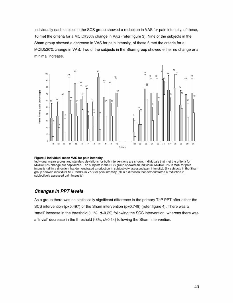
40
Individually each subject in the SCS group showed a reduction in VAS for pain intensity, of these,
10 met the criteria for a MCID≥30% change in VAS (refer figure 3). Nine of the subjects in the
Sham group showed a decrease in VAS for pain intensity, of these 6 met the criteria for a
MCID≥30% change in VAS. Two of the subjects in the Sham group showed either no change or a
minimal increase.
0
10
20
30
40
50
60
70
80
90
100
T1 T2 T3 T4 T5 t6 T7 T8 T9 T10 T11 t12 S1 s2 s3 S4 S5 s6 S7 s8 s9 S10 S11
Subjects
Vis
ua
l An
alo
g S
ca
le (
perc
enta
ge)
3537
45
74
86
62
57
37
95
6562
72
13
25
78
72 72 72
69
76
7892
54
7
0
13
41
37
47
18 22
17
37 37
52
7
25
62
31
39
69
45
79
48
35
42
Figure 3 Individual mean VAS for pain intensity. Individual mean scores and standard deviations for both interventions are shown. Individuals that met the criteria for MCID≥30% change are capitalized. Ten subjects in the SCS group showed an individual MCID≥30% in VAS for pain intensity (all in a direction that demonstrated a reduction in subjectively assessed pain intensity). Six subjects in the Sham group showed individual MCID≥30% in VAS for pain intensity (all in a direction that demonstrated a reduction in subjectively assessed pain intensity).
Changes in PPT levels
As a group there was no statistically significant difference in the primary TeP PPT after either the
SCS intervention (p=0.497) or the Sham intervention (p=0.749) (refer figure 4). There was a
‘small’ increase in the threshold (11%; d=0.29) following the SCS intervention, whereas there was
a ‘trivial’ decrease in the threshold (-3%; d=0.14) following the Sham intervention.

41
0
1
2
3
4
5
6
Tender Point Pre-
Intervent ion M ean
Tender Point Post-
Intervention M ean
Tender Point Pre-
Intervent ion M ean
Tender Point Post-
Intervention M ean
SCS Group Sham Group
Pre
ssure
Pain
Thre
shold
(kg/c
m2)
p=0.497; d =0.29 p=0.749; d =0.14
p=0.866; d =0.07
p=0.365; d =0.41
3.7
4.1
3.8
3.7
Figure 4 Group Means for Primary Tender point PPTs. Within group mean scores and the within group effect sizes for both interventions are shown. The SCS group showed a mean increase in PPT for TeP of 11% (p=0.497; d=0.29). The Sham group showed a mean reduction in PPT for TeP of 3% (p=0.749; d=0.14). P values were generated using a two-tailed paired t-test, effect size (d) was generated by standardized difference in means.
34 On a group level neither group met the criteria for MCID≥20% change.
Individually 5 out of 12 subjects in the SCS group showed a mean change in PPT at the primary
TeP meeting the criteria of the MCID≥20%, all showed an increase in PPT following the
intervention [refer figure 6, appendix A]. In the Sham group 2 out of 11 subjects showed a mean
change in PPT at the primary TeP meeting the criteria of the MCID≥20%, of these one showed an
increase in PPT, and the other a decrease.
As a group there was no statistically significant difference in the PPT for the medial epicondyle
after either the SCS intervention (p=0.803; d=0.10) or the Sham intervention (p=0.924; d=0.13)
[refer figure 7, appendix A]. Individually 2 out of 12 subjects in the SCS group showed a mean
change in PPT at the medial epicondyle meeting the criteria of the MCID≥20%, one showed an
increase in PPT following the intervention, the other a decrease [refer figure 8, appendix A]. In the
Sham group 1 out of 11 subjects showed a mean change in PPT at the medial epicondyle
meeting the criteria of the MCID≥20%, recording an increase in PPT following the intervention.

42
As a group there was no statistically significant difference in the PPT for the lateral epicondyle
after either the SCS intervention (p=0.568) or the Sham intervention (p=0.778). There was a
modest decline of 8% (d=0.24) following the SCS intervention, whereas a trivial improvement of
4% was noted following the Sham intervention (d=0.12) [refer figure 9, appendix A]. Individually, 6
out of 12 subjects in the SCS group showed a mean change in PPT at the lateral epicondyle
meeting the criteria of the MCID≥20%. Two showed an increase in PPT following the intervention;
the other four a decrease [refer figure 10, appendix A]. In the Sham group 1 out of 11 subjects
showed a mean change in PPT at the primary TeP meeting the criteria of the MCID≥20%,
recording an increase in PPT following the intervention.
When all subjects were considered, three subjects allocated to the SCS intervention group
(subjects T3, T4, and T8) and one subject allocated to the Sham intervention group (subject S7)
recorded a clinically significant (as defined by the MCID) improvement in both pain intensity and
PPT for the primary TeP.
Changes in PFGS levels
As a group (refer figure 5) there was no statistically significant difference in the PFGS after either
the SCS intervention (p=0.936; d=0.03) or the Sham intervention (p=0.989; d=0.01). With no
significant difference recorded after intervention between the groups (p=0.778).

43
0
5
10
15
20
25
30
35
40
45
50
Pre-Intervention
M ean Pain Free
Grip Strength
Post-Intervention
M ean Pain Free
Grip Strength
Pre-Intervent ion
M ean Pain Free
Grip Strength
Post-Intervent ion
M ean Pain Free
Grip Strength
SCS Group Sham Group
Pain
-Fre
e G
rip S
trength
(kg)
p=0.936; d =0.03 p=0.989; d =0.01
p=0.851; d =0.08
p=0.778; d =0.12
30 30 29 29
Figure 5 Group Pain Free Grip Strength. Within group mean scores and the within group effect sizes for both interventions are shown. Neither the SCS nor the Sham group showed any change in PFGS, SCS group (p=0.936; d=0.03), and Sham group (p=0.989; d=0.01). P values were generated using a two-tailed paired t-test; the effect size (d) was generated by standardized difference in means.
34
Reliability
The ICCs, SEM and SDD for the PPT measurements of the primary TeP, and the medial and
lateral epicondyles are displayed in table 2. The reliability of the research assistant using the
pressure algometer was ‘very high’.34
Table 2 Reliability indices
Measure ICC3,1 (LCL-UCL) SEM SDD
PPT medial epicondyle (kg/cm2) 0.841 (0.712-0.923) 0.806 2.2
PPT lateral epicondyle (kg/cm2) 0.851 (0.730-0.928) 0.886 2.5
PPT Primary TeP (kg/cm2) 0.839 (0.709-0.922) 0.519 1.4
Notes
PPT = Pressure Pain Threshold
ICC3,1 = Intra-class Correlation Coefficient
SEM = Standard Error of Measurement ( )( ICCSDSEM −•= 1)
SDD = Smallest Detectable Difference ( SEMSDD ••= 296.1 )

44
Table 3 Group and Individual Changes in Context of Smallest Detectable Differences and Minimal Clinically Important Difference
SDD SCS Intervention Sham Intervention
Measure by Group
PPT medial epicondyle (kg/cm2) 2.2 0.1 * -0.1 *
PPT lateral epicondyle (kg/cm2) 2.5 -0.6 * 0.3 *
PPT Primary TeP (kg/cm2) 1.4 0.4 * -0.1 *
Number individuals Surpass SDD from Improved(Total) Improved(Total)
PPT medial epicondyle 0 0
PPT lateral epicondyle 1(5) 1(1)
PPT Primary TeP 2(2) 0(1)
Number individuals Surpass MCID from Improved(Total) Improved(Total)
PPT medial epicondyle 1(2) 1(1)
PPT lateral epicondyle 2(6) 1(1)
PPT Primary TeP 5(5) 1(2)
Notes
PPT = Pressure Pain Threshold
SDD = Smallest Detectable Difference ( SEMSDD ••= 296.1 )
MCID = Minimal Clinically Important Difference for PPT defined as a difference in means (Pre to Post) ≥20%
Improved(Total) = number of subjects who show beneficial clinical affect (total number of subjects surpassing either
SDD or MCID whether beneficial or not).
* Difference in group means to enable comparison with SDD (post-intervention mean – pre-intervention mean)
No group surpassed the SDD required to indicate a clinically significant change in PPT after
either intervention (refer to table 3). All the individual subjects who surpassed the SDD for PPT at
either the primary TeP, or medial and lateral epicondyles also surpassed the pre-selected level of
20% for the MCID (by between 5-58%).

45
DISCUSSION
In this study we measured an immediate decrease in pain intensity (using VAS) for both groups,
and an increase in PPT for the SCS group. Effect sizes for the SCS were ‘very large’ for the
change in pain intensity and ‘small’ for the change in PPT, whereas the effect sizes of the Sham
group were ‘moderate’ for the change in pain intensity and ‘trivial’ for the change in PPT.
Many previous studies have described the use of SCS in combination with other osteopathic
techniques for the treatment of a variety of disorders.5, 15, 16, 44, 45
However, only one paper has
assessed the immediate effects of SCS on TeP sensitivity.6 Meseguer et al.
6 dealt with a specific
muscle, the upper trapezius, in subjects with mechanical neck pain. In the present study, the
application of the SCS technique was not to a specific muscle, but rather was to the primary TeP
which was found in a variety of locations around the elbow joint between the different
symptomatic subjects. The diagnostic TePs in the upper extremity are well suited to experimental
investigation since they are readily accessible for PPT measurement. Three outcomes measures
were selected as a means of decreasing the likelihood of a single outcome measure giving rise to
misleading impressions, as well providing the ability to assess different aspects of the subject’s
pain experience.46
The position adopted for delivery of the SCS intervention concurs with previous descriptions of
SCS application where the relevant body segments around the TeP are arranged so as to
maximize relaxation and reduce tenderness.1, 2
The moderate improvement in pain intensity
reported by the subjects in the Sham intervention group may be attributed to the effectiveness of
the Sham intervention producing a placebo effect. Well designed Sham interventions in manual
therapy should yield only marginal clinical benefits, and should be inactive and nonspecific in
comparison to the intervention.47
Due to the nonspecific beneficial effects of touch, the change in
pain intensity for the Sham group could be attributed to an otherwise unquantified treatment
effect. Fernández-de-las-Peňas et al.48
reported findings consistent with an earlier review by Hey
and Helewa49
that no reviewed manual therapy intervention had been more effective than the
placebo intervention.

46
VAS for Pain Intensity
Both SCS and Sham groups surpassed the minimal clinically important difference (MCID) of 30%
for change in VAS for pain intensity, suggesting a strong clinical effect. Several studies have
reported evidence in support of an MCID for pain intensity (via VAS) as ≥30%.38, 40, 41, 50, 51
Our results agree with those of Meseguer et al.6 who found that SCS was effective in reducing
tenderness represented by an increase in pressure pain thresholds of TePs in the upper
trapezius muscle of subjects with mechanical neck pain. Our data is also consistent with the
findings of Wong & Schauer-Alvarez,8 who found that SCS reduced pain intensity in response to
manual palpation of TePs in hip musculature.
Changes in PPT levels
The application of SCS is thought to decrease tissue tenderness by altering nociceptor activity in
the soft tissues.52
An increase in PPT is synonymous with a decrease in tissue sensitivity,35, 53, 54
thus the increase in PPT in response to SCS technique provides a measure of the analgesic
effect of SCS.35, 55
Based on previous literature and our current findings, it appears that SCS
techniques have the capacity to provide immediate relief of tenderness and local pain provoked
by TePs.
Other authors investigating the effectiveness of manual therapy techniques to increase PPT
levels at the elbow have reported improvements ranging from 10% to 20%.24, 42, 56
Such findings
provide evidence that manual therapy directed to the cervical spine, or to the elbow joint, can
both result in analgesic effects at the elbow.
Bailey & Dick52
proposed a nociceptive hypothesis that tissue damage in dysfunctional muscles
can be reduced by the positional release mechanism utilized by SCS. They suggest that
relaxation of the damaged tissues may be achieved by placing patients in a position of ease
which may advance local perfusion of fluids (i.e. blood, and lymph) and enhance the removal of
sensitizing inflammatory mediators.

47
Jones et al.57
reported a decrease in PPT levels when assessing repeated outcome
measurements on the same anatomical locations over successive days. They reported no
significant change in PPT levels after repeated pressure algometry measurements on the same
anatomical location on the same day.57
It is therefore reasonable to conclude that an
improvement in PPT demonstrated immediately after an intervention can be attributed to a
treatment effect rather than an effect of the testing process.
Changes in PFGS levels
In our study the maximal grip strength, as represented by the pain free grip strength, showed no
significant changes between pre- and post- intervention measurement. This could be a reflection
of the limited sensitivity of the measuring instrument, or the level of discomfort reported by the
subjects before reaching what they felt was a true maximum effort.
Our results concur with the findings of Slater et al.58
who were unable to demonstrate an increase
in maximal force post treatment in subjects with experimentally induced lateral epicondylalgia.
Authors of previous studies have been able to demonstrate a clinically important improvement in
PFGS after treatment for subjects with lateral epicondylalgia.59-61
Reliability
The intra-class correlation coefficient (ICC) for our baseline data recorded for both the medial and
lateral epicondyles showed good test re-test reliability for our single blinded assessor. The ICCs
compare favorably with those described in earlier studies utilizing PPT measurement.42, 55, 57, 62
The smallest detectable difference (SDD) for the lateral and medial epicondyles was similar to
that reported by Smidt et al.,36
and the SDD for the primary TeP was higher than the MCID≥15%
reported by Paungmali et al.42
for lateral epicondylalgia which may reflect the level of sensitivity
associated with our choice of subjects.
All the individual subjects who surpassed the SDD for PPT at either the primary TeP, or medial
and lateral epicondyles also surpassed the pre-selected level of MCID. This would suggest that
the MCID was at a suitable level for the sensitive tissues around the elbow in subjects with a
chronic sports related injury to the upper extremity.

48
Limitations
This study has several limitations. Only the TePs pertaining to the elbow joint were included for
treatment. Interestingly, there has been evidence of beneficial effects in treating the cervical
spine in relation to elbow pain.61
Therefore, further studies are needed to examine the distal
effects of SCS interventions when applied to TePs found in the spine.
It was sometimes difficult to accurately apply the algometer to match the angle of the TeP
palpated in soft tissues; this was due in part to the shape of the algometer applicator head but
was also related to difficulty localizing the algometer to precisely the same point. The increase of
pressure for the measurement of PPT was maintained at an approximate rate. The research
assistant had received prior training in the application of the algometer at approximately
1kg/cm2/s. Future studies utilizing finger tip flexible pressure meters as described by Marcotte et
al.63
may circumvent these problems as: 1) the application of a finger tip sensor allows the
assessor a greater degree of sensitivity to accurately locate the pressure sensor, and 2) an
electronic device can accurately display the rate of increase in pressure.
There is not yet a strong enough body of evidence to rule out the likelihood the perception of TeP
pain can be changing over time for symptomatic subjects. It follows that in order to rule out the
negative changes for pressure algometry reported in this study as being due to possible irritation
caused by the of the testing process a third group is required. Future studies could include a third
group to compare against the sham and SCS intervention groups. This group could act as a
control receiving no intervention, so as to quantify the placebo effect attributable to the sham
intervention.
The results in this study may have been limited by the choice of outcome measure; the VAS is a
valid clinical measure,54
but could have become a source of error if the subjects were biased by
the ability to remember their previous scores due to the short time period between pre- and post-
recording.6 Future studies could investigate the longer-term effects of this intervention, so that
outcome measurement would occur after greater time periods.

49
Conclusion
We have demonstrated a significant decrease (54%) in VAS for pain intensity with an associated
increase in PPT for the SCS group in our study. The improvement in PPT for the SCS and Sham
groups was below the MCID. Our results suggest that SCS was efficacious in reducing
tenderness of TePs around the elbow in subjects presenting with mechanical pain related to a
sports related injury.
ACKNOWLEDGEMENTS
The author thanks Robert Moran, School of Health Science, Unitec New Zealand and Dr Carol
Horgan, School of Health Science, Unitec New Zealand for their critical review of this manuscript.
The author thanks Marshall Gabin, fifth year osteopathic student, School of Health Science,
Unitec New Zealand for his expertise in the application of the SCS technique.

50
REFERENCES
1 Jones LH. Strain and Counterstrain. Indianapolis, USA: The American Academy of Osteopathy, 1981
2 D'Ambrogio KJ, Roth GB. Positional Release Therapy:assessment and treatment of musculoskeletal dysfunction. St Louis, Missouri, USA: Mosby, 1997
3 Giammatteo T, Weiselfish-Giammatteo S. Integrative Manual Therapy for the Autonomic Nervous System and Related Disorders: Utilizing Advanced Strain and Counterstrain Technique. Integrative Manual Therapy volume One. Berkeley, California, USA: North Atlantic Books, 1997
4 Blanco CR, Fernández-de-las-Peñas C, Xumet JEH, Algaba CP, Fernández-Rabadán M, Lillo-de-la-Quintana MC. Changes in active mouth opening following a single treatment of latent myofascial trigger points in the masseter muscle involving post-isometric relaxation or strain/counterstrain. J Bodywork Mov Ther 2006; 10: 197-205
5 Howell JN, Cabell KS, Chila AG, Eland DC. Stretch Reflex and Hoffmann Reflex Responses to Osteopathic Manipulative Treatment in Subjects With Achilles Tendinitis. J Am Osteopath Assoc 2006; 106: 537-45
6 Meseguer AA, Fernández-de-las-Peňas C, Navarro-Poza JL, Rodrìguez-Blanco C, Gandia JJB. Immediate effects of the strain/counterstrain technique in local pain evoked by tender points in the upper trapezius muscle. Clin Chiropr 2006; 9: 112-18
7 Wong CK, Schauer C. Reliability, validity and effectiveness of strain counterstrain techniques. J Man Manipulative Ther 2004; 12: 107-12
8 Wong CK, Schauer-Alvarez C. Effect of strain counterstrain on pain and strength in hip musculature. J Man Manipulative Ther 2004; 12: 215-23
9 Wynne MM, Burns JM, Eland DC, Conatser RR, Howell JN. Effect of Counterstrain on Stretch Reflexes, Hoffmann Reflexes, and Clinical Outcomes in Subjects With Plantar Fasciitis. J Am Osteopath Assoc 2006; 106: 547-56
10 Collins CK. Physical therapy management of complex regional pain syndrome I in a 14-year-old patient using strain counterstrain: A case report. J Man Manipulative Ther 2007; 15: 25-41
11 Jones LH. Foot treatment without hand trauma (Abstract). J Am Osteopath Assoc 1973; 72: 481-
12 Eisenhart AW, Gaeta TJ, Yens DP. Osteopathic Manipulative Treatment in the Emergency Department for Patients With Acute Ankle Injuries. J Am Osteopath Assoc. 2003; 103: 417-21
13 Pedowitz RN. Use of Osteopathic Manipulative Treatment for Iliotibial Band Friction Syndrome. J Am Osteopath Assoc 2005; 105: 563-67
14 Cislo S, Ramirez MA, Schwartz HR. Low back pain: treatment of forward and backward sacral torsions using counterstrain technique (Abstract). J Am Osteopath Assoc 1991; 91: 255-
15 Lewis C, Flynn T. The use of strain-counterstrain in the treatment of patients with low back pain. J Man Manipulative Ther 2001; 9: 92-98
16 Dardzinski JA, Ostrov BE, Hamann LS. Myofascial Pain Unresponsive to Standard Treatment: Successful Use of a Strain and Counterstrain Technique with Physical Therapy. J Clin Rheumatol 2000; 6: 169-74

51
17 Jacobson EC, Lockwood MD, Hoefner V, Jr., Dickey JL, Kuchera WL. Shoulder pain and repetition strain injury to the supraspinatus muscle: etiology and manipulative treatment (Abstract). J Am Osteopath Assoc 1989; 89: 1037-
18 Tettambel MA. An Osteopathic Approach to Treating Women With Chronic Pelvic Pain. J Am Osteopath Assoc 2005; 105: S20-22
19 Kuchera ML. Osteopathic Manipulative Medicine Considerations in Patients With Chronic Pain. J Am Osteopath Assoc 2005; 105: S29-36
20 Licciardone JC. The Unique Role of Osteopathic Physicians in Treating Patients With Low Back Pain. J Am Osteopath Assoc 2004; 104: 13S-18
21 DeAngelo NA, Gordin V. Treatment of Patients With Arthritis-Related Pain. J Am Osteopath Assoc 2004; 104: 2S-5
22 Walko EJ, Janouschek C. Effects of osteopathic manipulative treatment in patients with cervicothoracic pain: pilot study using thermography. J Am Osteopath Assoc 1994; 94: 135-41
23 Schwartz HR. The use of counterstrain in an acutely ill in-hospital population (Abstract). J Am Osteopath Assoc 1986; 86: 433-
24 Fernández-de-las-Peňas C, Pérez-de-Heredia M, Brea-Rivero M, Miangolarra-Page JC. Immediate Effects on Pressure Pain Threshold Following a Single Cervical Spine Manipulation in Healthy Subjects. J Ortho Sports Phys Ther 2007; 37: 325-29
25 Fryer G, Hodgson L. The effect of manual pressure release on myofascial trigger points in the upper trapezius muscle. J Bodywork Mov Ther 2005; 9: 248-55
26 Faul F, Erdfelder E. GPOWER: A priori, post-hoc, and compromise power analyses for MS-DOS [Computer Program]. Bonn, FRG: Bonn University, Dep. of Psychology, 1992; Computer Program
27 Bisset L, Russell T, Bradley S, Ha B, Vicenzino B. Bilateral sensorimotor abnormalities in unilateral lateral epicondylalgia. Arch Phys Med Rehab 2006; 87: 490-95
28 Ostelo RWJG, de Vet HCW. Clinically important outcomes in low back pain. Best Pract Res Clin Rheumatol 2005; 19: 593-607
29 Hagg O, Fritzell P, Nordwall A. The clinical importance of changes in outcome scores after treatment for chronic low back pain. Eur Spine J 2003; 12: 12-20
30 Price DP, McGrath PA, Rafii A, Buckingham B. The validation of visual analogue scales as ratio scale measures for chronic and experimental pain. Pain 1983; 17: 45-56
31 Fischer AA. Pressure algometry over normal muscles. Standard values, validity and reproducibility of pressure threshold. Pain 1987; 30: 115-26
32 Smidt N, van der Windt DA, Assendelft WJ, Mourits AJ, Deville WL, de Winter AF, Bouter LM. Interobserver reproducibility of the assessment of severity of complaints, grip strength, and pressure pain threshold in patients with lateral epicondylitis. Arch Phys Med Rehab 2002; 83: 1145-50
33 Martel GF, Roth SM, Ivey FM, Lemmer JT, Tracy BL, Hurlbut DE, Metter EJ, Hurley BF, Rogers MA. Age and sex affect human muscle fibre adaptations to heavy-resistance strength training. Exp Physiol 2006; 91: 457-64
34 Hopkins WG. A scale of magnitudes for effect statistics. In: A New View of Statistics: Internet Society for Sport Science, 2002
35 Potter L, McCarthy C, Oldham J. Algometer reliability in measuring pain pressure threshold over normal spinal muscles to allow quantification of anti-nociceptive treatment effects. Int J Osteopath Med 2006; 9: 113-19

52
36 Smidt N, van der Windt D, Assendelft WJJ, Mourits AJ, Deville WL, de Winter AF, Bouter LM. Interobserver reproducibility of the assessment of severity of complaints, grip strength, and pressure pain threshold in patients with lateral epicondylitis. Arch Phys Med Rehab 2002; 83: 1145-50
37 Shrout PE, Fleiss JL. Intraclass Correlations: Uses in Assessing Rater Reliability. Psychol Bull 1979; 86: 420-28
38 van Grootel RJ, van der Bilt A, van der Glas HW. Long-term reliable change of pain scores in individual myogenous TMD patients. Eur J Pain 2007; 11: 635-43
39 Haas M, Goldberg B, Aickin M, Ganger B, Attwood M. A Practice-Based Study of Patients with Acute and Chronic Low Back Pain Attending Primary Care and Chiropractic Physicians: Two-Week to 48-Month Follow-Up. J Manipulative Physiol Ther 2004; 27: 160-69
40 Farrar JT, Young Jr JP, LaMoreaux L, Werth JL, Poole RM. Clinical importance of changes in chronic pain intensity measured on an 11-point numerical pain rating scale. Pain 2001; 94: 149-58
41 Farrar JT, Portenoy RK, Berlin JA, Kinman JL, Strom BL. Defining the clinically important difference in pain outcome measures. Pain 2000; 88: 287-94
42 Paungmali A, O’Leary S, Souvlis T, Vicenzino B. Hypoalgesic and sympathoexcitatory effects of mobilization with movement for lateral epicondylalgia. Phys Ther 2003; 83: 374-83
43 O'Leary S, Falla D, Hodges PW, Jull G, Vincenzo B. Specific Therapeutic Exercise of the Neck Induces Immediate Local Hypoalgesia. J Pain 2007; In Press, Corrected Proof: doi:10.1016/j.jpain.2007.05.014
44 Gamber RG, Shores JH, Russo DP, Jimenez C, Rubin BR. Osteopathic manipulative treatment in conjunction with medication relieves pain associated with fibromyalgia syndrome: results of a randomized clinical pilot project. J Am Osteopath Assoc 2002; 102: 321-25
45 Radjieski JM, Lumley MA, Cantieri MS. Effect of osteopathic manipulative treatment of length of stay for pancreatitis: a randomized pilot study. J Am Osteopath Assoc 1998; 98: 264-
46 Reading AE. A comparison of pain rating scales. J Psychosom Res 1980; 24: 119-24
47 Hawk C, Long CR, Rowell RM, Gudavalli MR, Jedlicka J. A Randomized Trial Investigating a Chiropractic Manual Placebo: A Novel Design Using Standardized Forces in the Delivery of Active and Control Treatments. J Altern Complement Med 2005; 11: 109-17
48 Fernández-de-las-Peňas C, Campo MS, Carnero JF, Miangolarra-Page JC. Manual therapies in myofascial trigger point treatment: a systematic review. J Bodywork Mov Ther 2005; 9: 27-34
49 Hey LR, Helewa A. Myofascial pain syndrome: a critical review of the literature. Physiother Can 1994; 46: 28-36
50 Beaton DE, Boers M, Wells GA. Many faces of the minimal clinically important difference (MICD): A literature review and directions for future research. Curr Opin Rheumatol 2002; 14: 109-14
51 Lee JS, Hobden E, Stiell IG, Wells GA. Clinically important change in the visual analog scale after adequate pain control. Acad Emerg Med 2003; 10: 1128-30
52 Bailey M, Dick L. Nociceptive considerations in treating with counterstrain. J Am Osteopath Assoc 1992; 92: 334-41

53
53 Nussbaum EL, Downes L. Reliability of clinical pressure-pain algometric measurements obtained on consecutive days. Phys Ther 1998; 78: 160-69
54 Reeves JL, Jaeger B, Graff-Radford SB. Reliability of the pressure algometer as a measure of myofascial trigger point sensitivity. Pain 1986; 24: 313-21
55 Vanderweeën L, Oostendorp RAB, Vaes P, Duquet W. Pressure algometry in manual therapy. Man Ther 1996; 1: 258-65
56 Vicenzino B, Paungmali A, Buratowski S, Wright A. Specific manipulative therapy treatment for chronic lateral epicondylalgia produces uniquely characteristic hypoalgesia. Man Ther 2001; 6: 205-12
57 Jones DH, Kilgour RD, Comtois AS. Test-Retest Reliability of Pressure Pain Threshold Measurements of the Upper Limb and Torso in Young Healthy Women. J Pain 2007; 8: 650-56
58 Slater H, Arendt-Nielsen L, Wright A, Graven-Nielsen T. Effects of a manual therapy technique in experimental lateral epicondylalgia. Man Ther 2006; 11: 107-17
59 Pienima¨ki TT, Siira PT, Vanharanta H. Chronic medial and lateral epicondylitis: A comparison of pain, disability, and function. Arch Phys Med Rehabil 2002; 83: 317-21
60 Vicenzino B, Collins D, Benson H, Wright A. An investigation of the interrelationship between manipulative therapy-induced hypoalgesia and sympathoexcitation. . J Manipulative Physiol Ther 1998; 21: 448-53
61 Vicenzino B, Collins D, Wright A. The initial effects of a cervical spine manipulative physiotherapy treatment on the pain and dysfunction of lateral epicondylalgia. Pain 1996; 68: 69-74
62 Ylinen J, Nykanen M, Kautiainen H, Hakkinen A. Evaluation of repeatability of pressure algometry on the neck muscles for clinical use. Man Ther 2007; 12: 192-97
63 Marcotte J, Normand MC, Black P. Measurement of the Pressure Applied During Motion Palpation and Reliability for Cervical Spine Rotation. J Manipulative Physiol Ther 2005; 28: 591-96

54
Appendix A - Figures
0.0
1.0
2.0
3.0
4.0
5.0
6.0
7.0
8.0
T1 t2 T3 T4 t5 t6 t7 T8 t9 t10 t11 T12 s1 s2 s3 s4 s5 s6 S7 s8 S9 s10 s11
Subjects
Pre
ssure
Pain
Thre
shold
(kg/c
m2)
3.3
4.5
5.9
1.3
2.2
3.6
5.3
3.43.8
4.9
4.5
1.6
3.3
2.8
3.6
4.7
4.9
3.5
2.7
4.1
5.9
3.3
3.0
5.3
4.1 4.3
7.4
1.9
2.4
3.5
5.1
3.9
4.44.6
2.5
3.4
2.7
3.8
5.3
4.1
3.3 3.53.7 4.0
3.6
3.1
Figure 6 Individual mean PPT for tender point. Individual mean scores and standard deviations for both interventions are shown. Individuals that met the criteria for MCID≥20% change are capitalized. Five subjects in the SCS group showed an individual MCID≥20% in PPT for the primary TeP (all in a direction that demonstrated a reduction in objectively assessed pain intensity). Two subjects in the Sham group showed individual MCID≥20% in PPT for the primary TeP (one in a direction that demonstrated a reduction in objectively assessed pain intensity, the other showed an objective increase in assessed pain intensity).
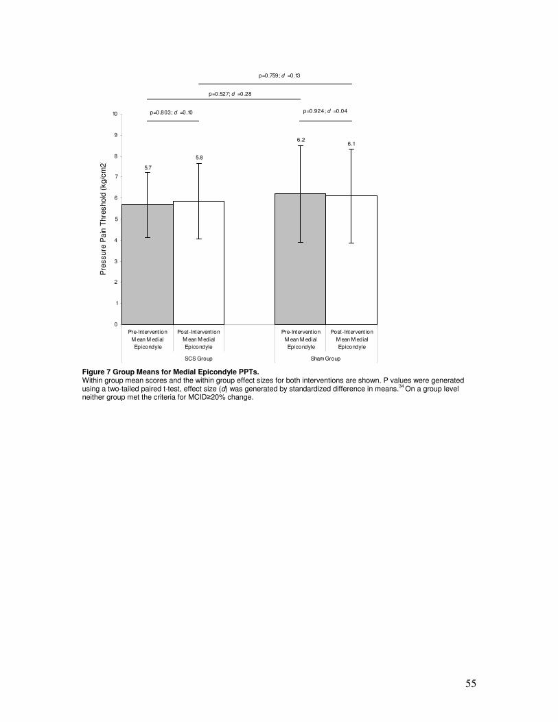
55
0
1
2
3
4
5
6
7
8
9
10
Pre-Intervent ion
M ean M edial
Epicondyle
Post-Intervent ion
M ean M edial
Epicondyle
Pre-Intervent ion
M ean M edial
Epicondyle
Post-Intervent ion
M ean M edial
Epicondyle
SCS Group Sham Group
Pre
ssure
Pain
Thre
shold
(kg/c
m2)
p=0.803; d =0.10 p=0.924; d =0.04
p=0.527; d =0.28
p=0.759; d =0.13
5.7
5.8
6.26.1
Figure 7 Group Means for Medial Epicondyle PPTs. Within group mean scores and the within group effect sizes for both interventions are shown. P values were generated using a two-tailed paired t-test, effect size (d) was generated by standardized difference in means.
34 On a group level
neither group met the criteria for MCID≥20% change.
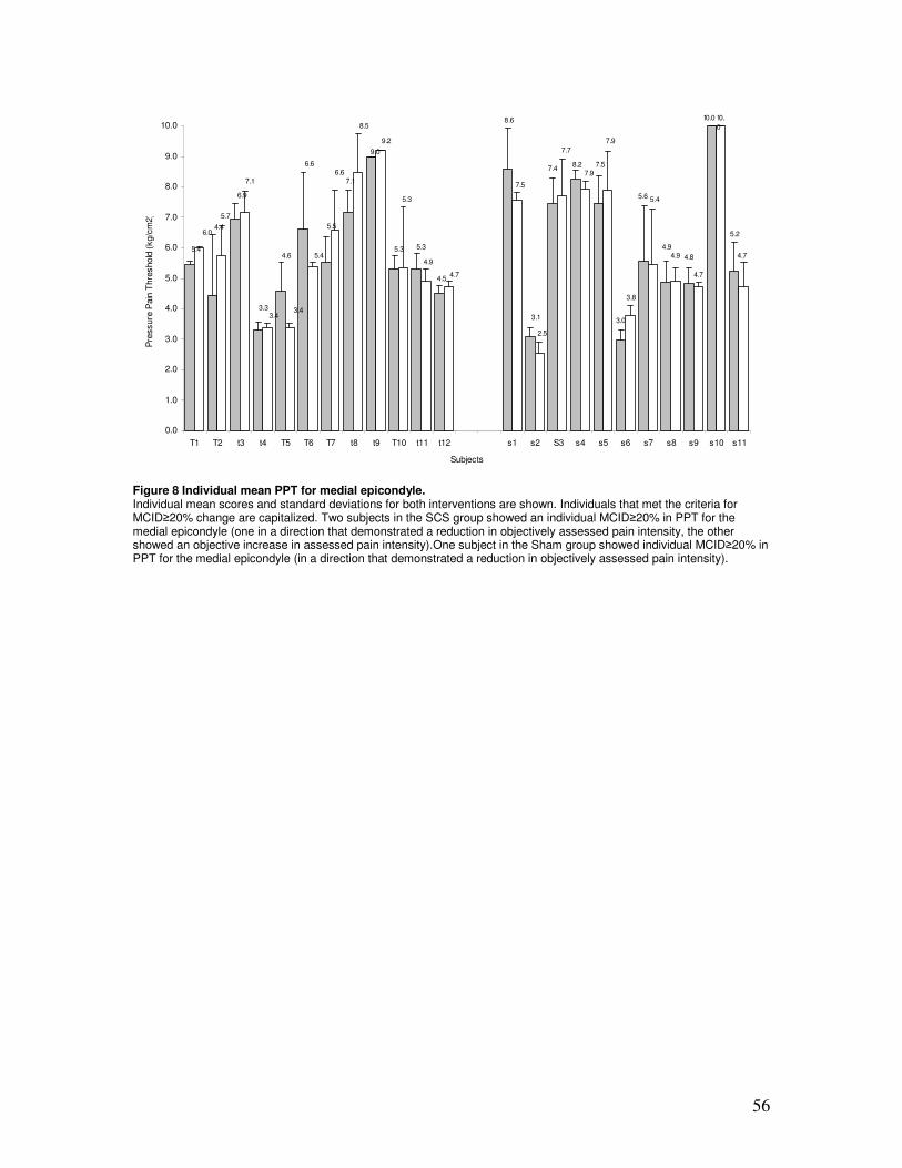
56
0.0
1.0
2.0
3.0
4.0
5.0
6.0
7.0
8.0
9.0
10.0
T1 T2 t3 t4 T5 T6 T7 t8 t9 T10 t11 t12 s1 s2 S3 s4 s5 s6 s7 s8 s9 s10 s11
Subjects
Pre
ssure
Pain
Thre
shold
(kg/c
m2)
5.4
4.4
6.9
3.3
4.6
6.6
5.5
7.1
9.0
5.3 5.3
4.5
8.6
3.1
7.48.2 7.5
3.0
5.6
4.9
4.8
10.0
5.2
6.6
6.0
5.7
7.1
3.43.4
5.4
8.5
9.2
5.3
4.9
4.7
7.5
2.5
7.7
7.9
7.9
3.8
5.4
4.9
4.7
10.
0
4.7
Figure 8 Individual mean PPT for medial epicondyle. Individual mean scores and standard deviations for both interventions are shown. Individuals that met the criteria for MCID≥20% change are capitalized. Two subjects in the SCS group showed an individual MCID≥20% in PPT for the medial epicondyle (one in a direction that demonstrated a reduction in objectively assessed pain intensity, the other showed an objective increase in assessed pain intensity).One subject in the Sham group showed individual MCID≥20% in PPT for the medial epicondyle (in a direction that demonstrated a reduction in objectively assessed pain intensity).
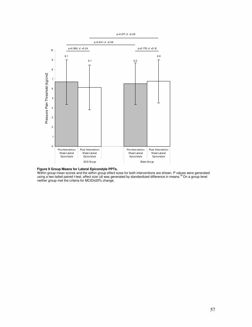
57
0
1
2
3
4
5
6
7
8
9
10
Pre-Intervent ion
M ean Lateral
Epicondyle
Post-Intervent ion
M ean Lateral
Epicondyle
Pre-Intervention
M ean Lateral
Epicondyle
Post-Intervent ion
M ean Lateral
Epicondyle
SCS Group Sham Group
Pre
ssure
Pain
Thre
shold
(kg/c
m2)
p=0.568; d =0.24 p=0.778; d =0.12
p=0.841; d =0.08
p=0.517; d =0.28
6.7
6.1 6.5
6.8
Figure 9 Group Means for Lateral Epicondyle PPTs. Within group mean scores and the within group effect sizes for both interventions are shown. P values were generated using a two-tailed paired t-test, effect size (d) was generated by standardized difference in means.
34 On a group level
neither group met the criteria for MCID≥20% change.

58
0.0
1.0
2.0
3.0
4.0
5.0
6.0
7.0
8.0
9.0
10.0
T1 T2 t3 t4 T5 T6 T7 t8 t9 T10 t11 t12 s1 s2 S3 s4 s5 s6 s7 s8 s9 s10 s11
Subjects
Pre
ssure
Pain
Thre
shold
(kg/c
m2)
6.0
7.7
5.6
3.2
6.5
10.0 10.0
8.7
9.0
4.9
4.8
3.9
7.6
3.4
5.1
10.0
5.7
4.0
7.1
5.4
5.9
10.0
7.4
7.2
7.8
4.7
5.3
2.9
2.9
6.7
9.0
9.8 8.1
5.7
3.6
7.9
3.5
9.0
10.
0
5.9
3.7
7.1
5.4 5.8
10.0
6.1
Figure 10 Individual mean PPT for lateral epicondyle. Individual mean scores and standard deviations for both interventions are shown. Individuals that met the criteria for MCID≥20% change are capitalized. Six subjects in the SCS group showed an individual MCID≥20% in PPT for the lateral epicondyle (two in a direction that demonstrated a reduction in objectively assessed pain intensity, four showed an objective increase in assessed pain intensity).One subject in the Sham group showed individual MCID≥20% in PPT for the lateral epicondyle (in a direction that demonstrated a reduction in objectively assessed pain intensity).

59
Appendix B - Ethics Resources
An Investigation into the Efficacy of Strain Counterstrain Technique to Produce Hypoalgesic Effects in Symptomatic Subjects
Information Sheet
About the research You are invited to take part in a research project being undertaken as part of the Masters of Osteopathy Degree. This information sheet is designed to inform you as to the nature of the research and what will happen should you choose to take part. The purpose of this research is to investigate how well an osteopathic technique called Strain-Counterstrain can reduce pain and improve the functioning of a forearm injury sustained by a recreational sports person. We are recruiting subjects who have sustained such a sports related injury. It is hoped that the information gained from this research will help osteopaths when making technique decisions in the treatment of patients. Strain-Counterstrain is an indirect technique, this means that the patient’s arm will be moved by a trained practitioner into a position of comfort, and held in this position for 90 seconds and returned to a rest position. During the Strain-Counterstrain technique the participant is only required to relax.
The researchers The researcher is James Hutchinson. Dr Carol Horgan and Dr Graham Fordy are supervising the research project.
What will participation involve?
• To currently have a forearm injury sustained while playing some form of recreational sports.
• Read and complete a screening questionnaire on General Health and Musculoskeletal Injuries that may prevent you from receiving a Strain-Counterstrain technique.
• Be available for one data collection session lasting up to 1 ½ hours.
• The research will require you to grip a measurement instrument to test your grip strength before you feel pain. A trained assessor will also apply pressure to an identified tender point on your forearm until you experience discomfort. This testing process will be repeated before and after treatment in order to assess the treatment’s effectiveness. Data will be collected from both of your arms for comparison.
• As this is an experimental design research you may be randomly assigned to either a treatment group to receive the Strain-Counterstrain technique or another osteopathic technique. You will not be informed of the type of technique you received until the end of data collection, as the experimental method requires that this information is concealed from you until after treatment and data collection has finished.
• All participants will receive one complementary follow-up treatment session with the principal researcher, one week after data collection or at a later date that suits.
• In order to participate all subjects are required to sign a consent form.
• The research requires a participant to remove any superficial clothing covering the upper body (shirts/blouses etc), from the waist up [NOT underwear].

60
• For the purpose of data collection three to five anatomical landmarks will be marked with coloured non-permanent (non-toxic) felt tipped pens on various parts of your forearms.
• There will be no sudden movements and your comfort will be monitored. You will be allowed rest breaks, as you require them. You are free to withdraw from the sessions at any point and do not need to state a reason for doing so.
• Consent to the research team’s use of the research data in preparing both a research project dissertation and an article for publication (all data will be anonymous).
• Consent to the storage of your anonymous research data indefinitely for future research.
Getting help Please contact either one of us should you require help with this research project.
James Hutchinson Email: [email protected] Phone: 027 3498919 Dr Carol Horgan E- mail [email protected] Dr Graham Fordy Email: [email protected] Phone: (09) 815 4321 ext 7908
Potential risks to research participants There is no known published data indicating any risks associated with this research. However, the researcher accepts that it is possible there may be some undetermined risks involved in the research process. In the case that any potential risk of harm should arise for any research participant, it will be treated on an individual basis. In any such case the research process will be halted immediately.
Confidentiality
Confidentiality and your anonymity will be protected in the following ways:
• Only the researchers will see completed questionnaires and consent forms.
• All forms will be stored in a locked file. Only the researchers will have access to this file.
• Any data derived from the research will be anonymous and your identity will be kept confidential.
You have the right not to participate, or withdraw from this research project at any time up until the point of data analysis (2 weeks after the last session). Contact James Hutchinson or Dr Carol Horgan that you no longer wish to participate by telephone or email, or by telling us when we contact you. A copy of the final report will be available at the Unitec New Zealand library. All participants are welcome to view this. Summaries and recommendations may be published in research journals.
Information and concerns If you want further information about the project, you can call or email the above addresses. At anytime if you are concerned or confused about the research project you may contact James Hutchinson, the primary researcher on the details above.
If you have concerns about the way in which the research is being conducted you can contact the following:
Health Advocates: Advocates Network Services Trust, Phone (09) 623 5799, 0800 205 555, Fax (09) 623 5798, PO Box 9983, Newmarket, Auckland.
Finally, we would like to thank you for your valuable contribution to this research. This study has been approved by the Unitec Research Ethics Committee from March 2007 to December 2007. If you have any complaints or reservations about the ethical conduct of this research, you may contact the Committee through the Secretary (ph: 09 815-4321 ext 8041). Any issues you raise will be treated in confidence and investigated fully, and you will be informed of the outcome.

61
Screening Questionnaire for General Health and Musculoskeletal Injury History
1. Are you between the ages of 20 to 70 years old?
2. Have you currently or are you receiving treatment for another musculoskeletal disorder that is more “troublesome” than your upper limb pain?
3. Have you experienced any neck pain that travels past the tip of your shoulder?
Have you experienced any pins & needles or numbness in your arm since the injury?
4. Are you currently taking any prescription pain medication or unable to refrain from
using any over the counter analgesics?
5. Have you been diagnosed, treated and/or medicated for any circulatory, blood or heart conditions in the last 10 years? Including any of the following: Arteriosclerosis/Atherosclerosis Coronary Artery Disease / Angina / Aneurysm High Blood Pressure (Hypertension), if controlled by diet leave blank Pericarditis / Rheumatic Fever Blood Clots / Deep Vein Thrombosis (exclude single attack)
6. Have you been diagnosed, treated and/or medicated for any serious digestive
conditions in the last 10 years?
7. Have you been diagnosed, treated and/or medicated for endocrine, lymphatic or metabolic conditions in the last 10 years? Including the long term use of steroids.
8. Are you currently receiving treatment or have received treatment within the last
month for any muscle, skeletal or skin conditions? Including any of the following: Arthritis (chronic or severe osteo- or rheumatoid arthritis includes Ankylosing Spondylitis, Degenerative Disk Disease, and Psoriatic Arthritis; includes conditions that are controlled by aspirin or over-the-counter non-steroidal anti-inflammatory agents) Bone Disorders (includes Osteoporosis, hip dysplasia, fractures, or bone spurs) Cartilage/Ligament/Tendon Conditions (includes Chondritis, or Dupuytren’s Contracture (in hands) Synovitis (which is a joint lining inflammation, includes Tendonitis or Tendinitis; Tendonitis of Thumb (De Quervain’s Disease) Surgery
9. Have you been diagnosed, treated and/or medicated for any non-psychiatric
nervous system conditions in the last 10 years? Including any of the following: Carpal Tunnel Syndrome Thoracic Outlet Syndrome
10. Have you been diagnosed, treated and/or medicated for any respiratory system
conditions in the last 10 years?

62
An Investigation into the Efficacy of Strain Counterstrain Technique to Produce Hypoalgesic Effects in Symptomatic Subjects
Consent Form
This research project investigates the efficacy of strain counterstrain technique to improve function and pain-free status of the forearm in a symptomatic population. The research is being undertaken by James
Hutchinson from Unitec New Zealand, and will be supervised by Dr Carol Horgan and Robert Moran. Name of Participant:…………………………………………………………………………. I have seen the Information Sheet dated October 2006 for people wishing to participate in the project investigating the efficacy of Strain Counterstrain technique to produce hypoalgesia in symptomatic subjects. I have had the opportunity to read the contents of the information sheet and to discuss the project with the
researchers and I am satisfied with the explanations I have been given. I understand that taking part in this project is voluntary (my choice) and that I may withdraw up until the point at which data analysis is started and this will in no way affect my access to the services provided by Unitec New Zealand or any other support service. I understand that I can withdraw from the experiment if, for any reason, I want this. I understand that my participation in this project is confidential and that no material that could identify me will be used in any reports on this project. I have had enough time to consider whether I want to take part. I know whom to contact if I have any questions or concerns about the project The principal researcher and first contact for this project is: James Hutchinson 027 349 8919 mailto:[email protected]
Signature………………………………………………….participant ………. (Date) Project explained by…………………………………… Signature………………………………………………. …………... (Date) The participant should retain a copy of this consent form. This study has been approved by the Unitec Research Ethics Committee from March 2007 to December 2007. If you have any complaints or reservations about the ethical conduct of this research, you may contact the Committee through the Secretary (ph: 09 815-4321 ext 8041). Any issues you raise will be treated in confidence and investigated fully, and you will be informed of the outcome.
63

64
Appendix C – Instructions for authors for manuscript
submission
INTERNATIONAL JOURNAL OF OSTEOPATHIC MEDICINE Former title: Journal of Osteopathic Medicine
Guide for Authors The journal Editors welcome contributions for publication from the following categories: Letters to the Editor, Reviews and Original Articles, Commentaries and Clinical Practice case studies with educational value.
Online Submission Submission to this journal proceeds totally online.( http://ees.elsevier.com/ijom) you will be guided stepwise through the creation and uploading of the various files. The system automatically converts source files to a single Adobe Acrobat PDF version of the article, which is used in the peer-review process. Please note that even though manuscript source files are converted to PDF at submission for the review process, these source files are needed for further processing after acceptance. All correspondence, including notification of the Editor's decision and requests for revision, takes place by e-mail and via the Author's homepage, removing the need for a hard-copy paper trail. The above represents a very brief outline of this form of submission. It can be advantageous to print this "Guide for Authors" section from the site for reference in the subsequent stages of article preparation. Submission of an article implies that the work described has not been published previously (except in the form of an abstract or as part of a published lecture or academic thesis), that it is not under consideration for publication elsewhere, that its publication is approved by all authors and tacitly or explicitly by the responsible authorities where the work was carried out, and that, if accepted, it will not be published elsewhere in the same form, in English or in any other language, without the written consent of the Publisher. Types of contributions Letters to the Editor As is common in biomedical journals the editorial board welcomes critical response to any aspect of the journal. In particular, letters that point out deficiencies and that add to, or further clarify points made in a recently published work, are welcomed. The Editorial Board reserves the right to offer authors of papers the right of rebuttal, which may be published alongside the letter. Reviews and Original Articles These should be either i) reports of new findings related to osteopathic medicine that are supported by research evidence. These should be original, previously unpublished works. The report will normally be divided into the following sections: abstract, introduction, materials and methods, results, discussion, conclusion, references. Or ii) critical or systematic review that seeks to summarise or draw conclusions from the established literature on a topic relevant to osteopathic medicine. Short review The drawing together of present knowledge in a subject area, in order to provide a background for the reader not currently versed in the literature of a particular topic. Shorter in length than and not intended to be as comprehensive as that of the literature review paper. With more emphasis on outlining areas of deficit in the current literature that warrant further investigation.

65
Research Note Findings of interest arising from a larger study but not the primary aim of the research endeavour, for example short experiments aimed at establishing the reliability of new equipment used in the primary experiment or other incidental findings of interest, arising from, but not the topic of the primary research. Including further clarification of an experimental protocol after addition of further controls, or statistical reassessment of raw data. Preliminary Findings Presentation of results from pilot studies which may establish a solid basis for further investigations. Format similar to original research report but with more emphasis in discussion of future studies and hypotheses arising from pilot study. Commentaries Include articles that do not fit into the above criteria as original research. Includes commentary and essays especially in regards to history, philosophy, professional, educational, clinical, ethical, political and legal aspects of osteopathic medicine. Clinical Practice Authors are encouraged to submit papers in one of the following formats: Case Report, Case Problem, and Evidence in Practice. Case Reports usually document the management of one patient, with an emphasis on presentations that are unusual, rare or where there was an unexpected response to treatment eg. an unexpected side effect or adverse reaction. Authors may also wish to present a case series where multiple occurrences of a similar phenomenon are documented. Preference will be given to reports that are prospective in their planning and utilise Single System Designs, including objective measures. The aim of the Case Problem is to provide a more thorough discussion of the differential diagnosis of a clinical problem. The emphasis is on the clinical reasoning and logic employed in the diagnostic process. The purpose of the Evidence in Practice report is to provide an account of the application of the recognised Evidence Based Medicine process to a real clinical problem. The paper should be written with reference to each of the following five steps: 1. Developing an answerable clinical question. 2. The processes employed in searching the literature for evidence. 3. The appraisal of evidence for usefulness and applicability. 4. Integrating the critical appraisal with existing clinical expertise and with the patient's unique biology, values, and circumstances. 5. Reflect on the process (steps 1-4), evaluating effectiveness, and identifying deficiencies. Presentation of Typescripts Your article should be typed on A4 paper, double-spaced with margins of at least 3cm. Number all pages consecutively beginning with the title page. To facilitate anonymity, the author's names and any reference to their addresses should only appear on the title page. Please check your typescript carefully before you send it off, both for correct content and typographic errors. It is not possible to change the content of accepted typescripts during production. Papers should be set out as follows, with each section beginning on a separate page: Title page To facilitate the peer-review process, two title pages are required. The first should carry just the title of the paper and no information that might identify the author or institution. The second should contain the following information: title of paper; full name(s) and address(es) of author(s) clearly indicating who is the corresponding author; you should give a maximum of four degrees/qualifications for each author and the current relevant appointment only; institutional affiliation; name, address, telephone, fax and e-mail of the corresponding author; source(s) of support in the form of funding and/or equipment.

66
Keywords Include three to ten keywords. These should be indexing terms that may be published with the abstract with the aim of increasing the likely accessibility of your paper to potential readers searching the literature. Therefore, ensure keywords are descriptive of the study. Refer to http://www.nlm.nih.gov/mesh/meshhome.html for the MeSH thesaurus. Abstract Both qualitative and quantitative research approaches should be accompanied by a structured abstract. Commentaries and Essays may continue to use text based abstracts of no more than 150 words. All original articles should include the following headings in the abstract as appropriate: Background, Objective, Design, Setting, Methods, Subjects, Results, and Conclusions. As an absolute minimum: Objectives, Methods, Results, and Conclusions must be provided for all original articles. Abstracts for reviews of the literature (in particular systematic reviews and meta-analysis) should include the following headings as appropriate: Objectives, Data Sources, Study Selection, Data Extraction, Data Synthesis, Conclusions. Abstracts for Case Studies should include the following headings as appropriate: Background, Objectives, Clinical Features, Intervention and Outcomes, Conclusions. Text The text of observational and experimental articles is usually, but not necessarily, divided into sections with the headings; introduction, methods, results, results and discussion. In longer articles, headings should be used only to enhance the readability. Three categories of headings should be used: •major ones should be typed in capital letter in the centre of the page and underlined •secondary ones should be typed in lower case (with an initial capital letter) in the left hand margin and underlined •minor ones typed in lower case and italicised Do not use 'he', 'his' etc. here the sex of the person is unknown; say 'the patient' etc. Avoid inelegant alternatives such as 'he/she'. Avoid sexist language. References Responsibility for the accuracy of bibliographic citations lies entirely with the Authors. Citations in the text: Please ensure that every reference cited in the text is also present in the reference list (and vice versa). Avoid using references in the abstract. Unpublished results and personal communications are not recommended in the reference list, but may be mentioned in the text. If these references are included in the reference list they should follow the standard reference style of the journal and should include a substitution of the publication date with either "Unpublished results" or "Personal communication" Citation of a reference as "in press" implies that the item has been accepted for publication. Text: Indicate references by superscript numbers in the text. The actual Authors can be referred to, but the reference number(s) must always be given. List: Number the references in the list in the order in which they appear in the text. Examples: Reference to a journal publication: 1. Van der Geer J, Hanraads JAJ, Lupton RA. The art of writing a scientific article. J Sci Commun 2000;163:51-9. Reference to a book: 2. Strunk Jr W, White EB. The elements of style. 3rd ed. New York: Macmillan; 1979.

67
Reference to a chapter in an edited book: 3. Mettam GR, Adams LB. How to prepare an electronic version of your article. In: Jones BS, Smith RZ, editors. Introduction to the electronic age. New York: E-Publishing Inc; 1999, p. 281-304 Note shortened form for last page number. e.g., 51-9, and that for more than 6 Authors the first 6 should be listed followed by "et al." For further details you are referred to "Uniform Requirements for Manuscripts submitted to Biomedical Journals" (J Am Med Assoc 1997;277:927-934) (see also http://www.nejm.org/general/text/requirements/1.htm) Citing and listing of Web references. As a minimum, the full URL should be given. Any further information, if known (Author names, dates, reference to a source publication, etc.), should also be given. Web references can be listed separately (e.g., after the reference list) under a different heading if desired, or can be included in the reference list. Tables, Illustrations and Figures A detailed guide on electronic artwork is available on our website: http://elsevier.com/authors Preparation of supplementary data. Elsevier now accepts electronic supplementary material (e-components) to support and enhance your scientific research. Supplementary files offer the Author additional possibilities to publish supporting applications, movies, animation sequences, high-resolution images, background datasets, sound clips and more. Supplementary files supplied will be published online alongside the electronic version of your article in Elsevier Web products, including ScienceDirect: http://www.sciencedirect.com. In order to ensure that your submitted material is directly usable, please ensure that data is provided in one of our recommended file formats. Authors should submit the material in electronic format together with the article and supply a concise and descriptive caption for each file. For more detailed instructions please visit our artwork instruction pages at http://www.elsevier.com/authors. Illustrations and tables that have appeared elsewhere must be accompanied by written permission to reproduce them from the original publishers. This is necessary even if you are an author of the borrowed material. Borrowed material should be acknowledged in the captions in the exact wording required by the copyright holder. If not specified, use this style: `Reproduced by kind permission of . . . (publishers) from . . . (reference).' Identifiable clinical photographs must be accompanied by written permission from the patient. The text of original research for a quantitative or qualitative study is typically subdivided into the following sections: Introduction State the purpose of the article. Summarise the rationale for the study or observation. Give only strictly pertinent references and do not review the subject extensively. Do not include data or conclusions from the work being reported. Materials and Methods Describe your selection of observational or experimental subjects (including controls). Identify the methods, apparatus (manufacturer's name and address in parenthesis) and procedures in sufficient detail to allow workers to reproduce the results. Give references and brief descriptions for methods that have been published but are not well known; describe new methods and evaluate limitations. Indicate whether procedures followed were in accordance with the ethical standards of the institution or regional committee responsible for ethical standards. Do not use patient names or initials. Take care to mask the identity of any subjects in illustrative material.

68
Results Present results in logical sequence in the text, tables and illustrations. Do not repeat in the text all the data in the tables or illustrations. Emphasise or summarise only important observations. Discussion Emphasise the new and important aspects of the study and the conclusions that follow from them. Do not repeat in detail data or other material given in the introduction or the results section. Include implications of the findings and their limitations, include implications for future research. Relate the observations to other relevant studies. Link the conclusion with the goals of the study, but avoid unqualified statements and conclusions not completely supported by your data. State new hypothesis when warranted, but clearly label them as such. Recommendations, when appropriate, may be included. Acknowledgments In the appendix one or more statements should specify (a) contributions that need acknowledging, but do not justify authorship (b) acknowledgments of technical support (c) acknowledgments of financial and material support, specifying the nature of the support. Persons named in this section must have given their permission to be named. Authors are responsible for obtaining written permission from those acknowledged by name since readers may infer their endorsement of the data and conclusions. IJOM Author Contribution Statement All manuscripts submitted to the journal should be accompanied by an Author Contribution Statement. The purpose of the Statement is to give appropriate credit to each author for their role in the study. All persons listed as authors should have made substantive intellectual contributions to the research. To qualify for authorship each person listed should have made contributions in each of the following; 1) Contributions to conception and design; data acquisition; data analysis and interpretation; 2) Drafting of manuscript, or critical revision for important intellectual content; 3) All authors must have given approval to the final version of the manuscript submitted for consideration to publish. Acquisition of funding; provision of resources; data collection; or general supervision, alone, is not sufficient justification for authorship. Contributors who do not meet the criteria for authorship as outlined above should be listed in the Acknowledgements section. Acknowledgements may include contributions of technical assistance, proof reading and editing, or assistance with resources and funding. The statement may be published in the paper as appropriate. Example of suggested format. Note the use of author initials. AB conceived the idea for the study. AB and CD contributed to the design and planning of the research. All authors were involved in data collection. AB and EF analysed the data. AB and CD wrote the first draft of the manuscript. EF coordinated funding for the project. All authors edited and approved the final version of the manuscript. Copyright Upon acceptance of an manuscript, authors will be asked to sign a "Journal Publishing Agreement'" (for more information on this and copyright see http://www.elsevier.com/copyright. Acceptance of the agreement will ensure the widest possible dissemination of information. An e-mail (or letter) will be sent to the corresponding author confirming receipt of the manuscript together with a 'Journal Publishing Agreement' form or a link to the online version of this agreement. If excerpts from other copyrighted works are included, the author(s) must obtain written permission from the copyright owners and credit the source(s) in the article. Elsevier has preprinted forms for use by authors in these cases: contact Elsevier's Rights Department, Philadelphia, PA, USA: phone (+1) 215 239 3804, fax (+1) 215 239 3805, [email protected] . Requests may also be completed online via the Elsevier

69
homepage http://www.elsevier.com/locate/permissions. Page Proofs Proofs will be sent to the author (first named author if no corresponding author is identified of multi-authored papers) by PDF wherever possible and should be returned within 48 hours of receipt, preferably by e-mail. Corrections should be restricted to typesetting errors; any others may be charged to the author. Any queries should be answered in full. Elsevier will do everything possible to get your article corrected an published as quickly and accurately as possible. Therefore, it is important to ensure that all of your corrections are returned to us in one all-inclusive e-mail or fax. Subsequent additional corrections will not be possible, so please ensure that your first communication is complete. Should you choose to mail your corrections, please return them to: Log-in Department, Elsevier, Stover Court, Bampfylde Street, Exeter, Devon EX1 2AH, UK. Author Enquiries For enquiries relating to the submission of articles (including electronic text and artwork) please visit http://www.elsevier.com/authors. The Author Gateway also provides the facility to track accepted articles and set up e-mail alerts to inform you of when an article's status has changed, as well as detailed artwork guidelines, copyright information, frequently asked questions and more. Contact details for questions arising after acceptance of an article, especially those relating to proofs, are provided when an article is accepted for publication. Language Polishing. Authors who require information about language editing and copyediting services pre- and post-submission please visit ( http://www.elsevier.com/wps/find/authorshome.authors/languagepolishing or contact [email protected] for more information. Please note Elsevier neither endorses nor takes responsibility for any products, goods or services offered by outside vendors through our services or in any advertising. For more information please refer to our Terms & Conditions http://www.elsevier.com/wps/find/termsconditions.cws_home/termsconditions Offprints The corresponding author, at no cost, will be provided with a PDF file of the article via e-mail or, alternatively, 25 free paper offprints. The PDF file is a watermarked version of the published article and includes a cover sheet with the journal cover image and a disclaimer outlining the terms and conditions of use. Additional paper offprints can be ordered by the authors. An order form with prices will be sent to the corresponding author. Checklist Please check your typescript carefully before you send it off to the Editorial Office, both for correct content and typographical errors, as it is not possible to change the content of accepted typescripts during the production process. •One copy of typescript and illustrations •Reference list in correct style •Written Permission from original publishers and authors to reproduce any borrowed any borrowed material