Embed Size (px)
Citation preview

Copyright © 2010 Pearson Education, Inc.
C h a p t e r
1
An Introduction to Anatomy and
Physiology
PowerPoint® Lecture Slides
prepared by Jason LaPres
Lone Star College - North Harris
Copyright © 2010 Pearson Education, Inc.

Copyright © 2010 Pearson Education, Inc.
1-1 The common functions
of all living things include
responsiveness, growth,
reproduction,movement,
and metabolism

Copyright © 2010 Pearson Education, Inc.
Living Things
• All living organisms share the following
characteristics
– Responsiveness
– Growth
– Reproduction
– Movement
– Metabolism

Copyright © 2010 Pearson Education, Inc.
1-2 Anatomy is structure,
and physiology is function

Copyright © 2010 Pearson Education, Inc.
Anatomy
• Describes the structures of the body
– What they are made of
– Where they are located
– Associated structures

Copyright © 2010 Pearson Education, Inc.
Anatomy
• Gross anatomy, or macroscopic
anatomy, examines large, visible
structures
– Surface anatomy: exterior features
– Regional anatomy: body areas
– Systemic anatomy: groups of organs working
together
Copyright © 2010 Pearson Education, Inc.
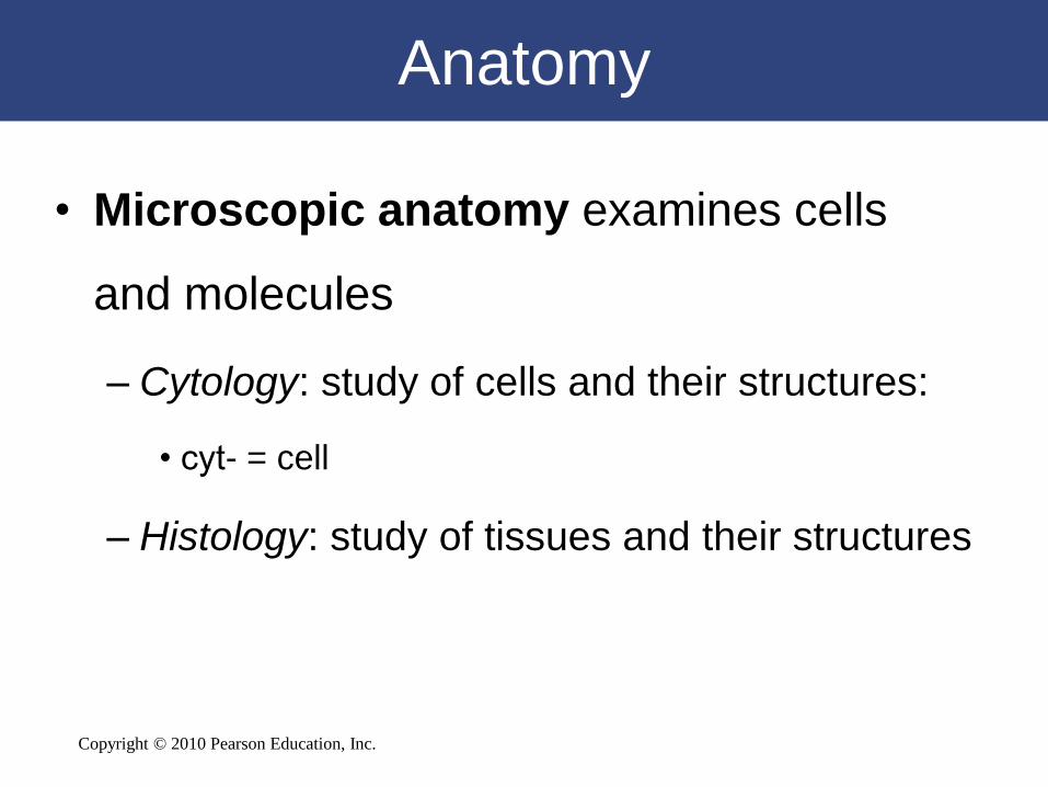
Anatomy
• Microscopic anatomy examines cells
and molecules
– Cytology: study of cells and their structures:
• cyt- = cell
– Histology: study of tissues and their structures

Copyright © 2010 Pearson Education, Inc.
Physiology
• Is the study of
– Functions of anatomical structures
– Individual and cooperative functions
Copyright © 2010 Pearson Education, Inc.
Physiology
• Cell physiology: processes within and between
cells
• Special physiology: functions of specific
organs
• Systemic physiology: functions of an organ
system
• Pathological physiology: effects of diseases

Copyright © 2010 Pearson Education, Inc.
1-3 Levels of organization
progress from molecules to a
complete organism

Copyright © 2010 Pearson Education, Inc.
Levels of Organization
• The Chemical (or Molecular) Level
– Atoms are the smallest chemical units
– Molecules are a group of atoms working together
• The Cellular Level
– Cells are a group of atoms, molecules, and organelles
working together
• The Tissue Level
– Tissues are a group of similar cells working together
• The Organ Level
– An organ is a group of different tissues working
together

Copyright © 2010 Pearson Education, Inc.
Levels of Organization
• The Organ System Level
– Organ systems are a group of organs working
together
– Humans have 11 organ systems
• The Organism Level
– A human is an organism
Levels of Organization
Organ Systems

Copyright © 2010 Pearson Education, Inc.
Levels of Organization
Figure 1-1

Copyright © 2010 Pearson Education, Inc.
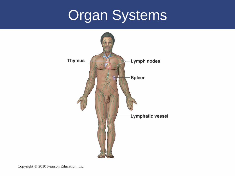
1-4 The human body consists
of 11 organ systems

Copyright © 2010 Pearson Education, Inc.
Organ Systems

Copyright © 2010 Pearson Education, Inc.
Organ Systems

Copyright © 2010 Pearson Education, Inc.
Organ Systems

Copyright © 2010 Pearson Education, Inc.
Organ Systems

Copyright © 2010 Pearson Education, Inc.
Organ Systems

Copyright © 2010 Pearson Education, Inc.
Organ Systems
Copyright © 2010 Pearson Education, Inc.
Organ Systems

Copyright © 2010 Pearson Education, Inc.
Organ Systems

Copyright © 2010 Pearson Education, Inc.
Organ Systems

Copyright © 2010 Pearson Education, Inc.
Organ Systems

Copyright © 2010 Pearson Education, Inc.
Organ Systems

Copyright © 2010 Pearson Education, Inc.
Organ Systems

Copyright © 2010 Pearson Education, Inc.
1-5 Homeostasis is the
tendency toward internal
balance

Copyright © 2010 Pearson Education, Inc.
Homeostasis
• Homeostasis: all body systems working
together to maintain a stable internal
environment
– Systems respond to external and internal
changes to function within a normal range
(body temperature, fluid balance)

Copyright © 2010 Pearson Education, Inc.
Homeostasis
• Receptor
– Receives the stimulus
• Control Center
– Processes the signal and sends instructions
• Effector
– Carries out instructions

Copyright © 2010 Pearson Education, Inc.
Figure 1-3
The Control of Room Temperature

Copyright © 2010 Pearson Education, Inc.
1-6 Negative feedback
opposes variations from
normal, whereas positive
feedback exaggerates them

Copyright © 2010 Pearson Education, Inc.
Negative Feedback
• The Role of Negative Feedback
– The response of the effector negates the
stimulus
– Body is brought back into homeostasis
• Normal range is achieved

Copyright © 2010 Pearson Education, Inc.
Negative Feedback in
Thermoregulation
Figure 1-4
Copyright © 2010 Pearson Education, Inc.
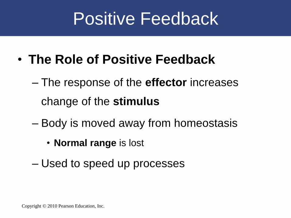
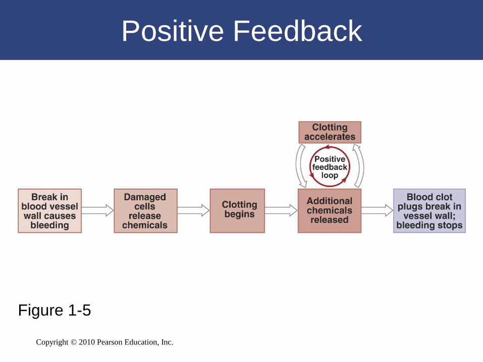
Positive Feedback
• The Role of Positive Feedback
– The response of the effector increases
change of the stimulus
– Body is moved away from homeostasis
• Normal range is lost
– Used to speed up processes
Copyright © 2010 Pearson Education, Inc.
Positive Feedback
Figure 1-5

Copyright © 2010 Pearson Education, Inc.
The Big Picture
• Systems integration
– Systems work together to maintain
homeostasis
• Homeostasis is a state of equilibrium
– Opposing forces are in balance
• Physiological systems work to restore
balance
– Failure results in disease or death

Copyright © 2010 Pearson Education, Inc.
1-7 Anatomical terms describe
body regions, anatomical
positions and directions, and
body sections

Copyright © 2010 Pearson Education, Inc.
Surface Anatomy
• Anatomical Landmarks
– Anatomical position: hands at sides, palms forward
– Supine: lying down, face up
– Prone: lying down, face down
• Anatomical Regions
– Body regions
– Abdominopelvic quadrants
– Abdominopelvic regions
• Anatomical Directions
– Reference terms based on subject

Copyright © 2010 Pearson Education, Inc.
Anatomical Landmarks. Anterior
Figure 1-6

Copyright © 2010 Pearson Education, Inc.
Anatomical Landmarks. Anterior
Figure 1-6

Copyright © 2010 Pearson Education, Inc.
Anatomical Landmarks. Posterior
Figure 1-6

Copyright © 2010 Pearson Education, Inc.
Anatomical Landmarks. Posterior
Figure 1-6

Copyright © 2010 Pearson Education, Inc.

Copyright © 2010 Pearson Education, Inc.

Copyright © 2010 Pearson Education, Inc.
Abdominopelvic Quadrants
Figure 1-7

Copyright © 2010 Pearson Education, Inc.
Abdominopelvic Regions
Figure 1-7
Copyright © 2010 Pearson Education, Inc.
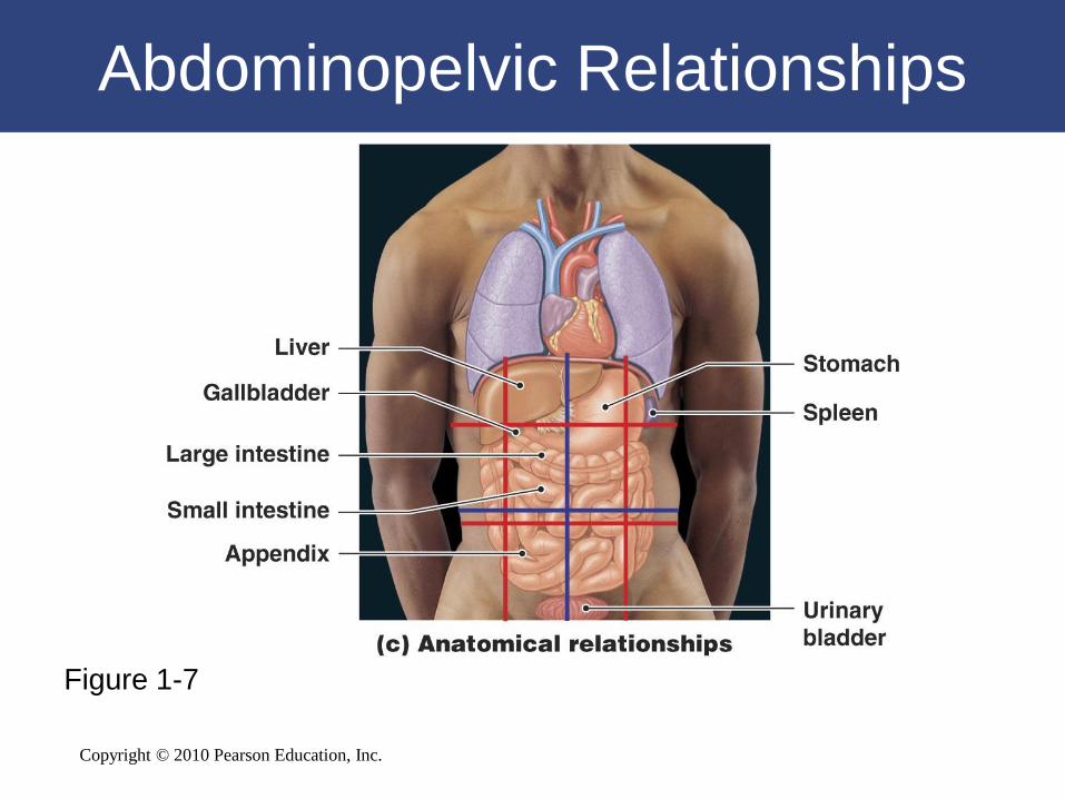
Abdominopelvic Relationships
Figure 1-7
Copyright © 2010 Pearson Education, Inc.
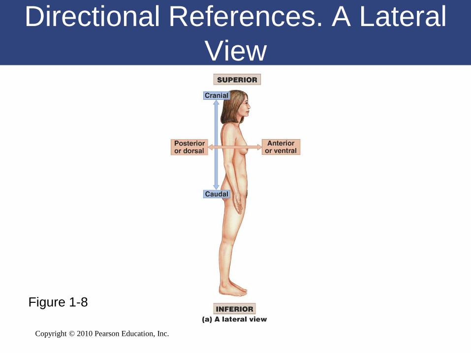
Directional References. A Lateral
View
Figure 1-8
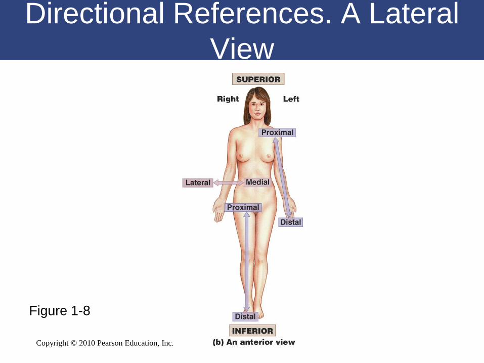
Copyright © 2010 Pearson Education, Inc.
Figure 1-8
Directional References. A Lateral
View

Copyright © 2010 Pearson Education, Inc.

Copyright © 2010 Pearson Education, Inc.
Sectional Anatomy
• Planes and sections
– Plane: a three-dimensional axis
– Section: a slice parallel to a plane
– Used to visualize internal organization and
structure
– Important in radiological techniques:
• MRI
• PET
• CT

Copyright © 2010 Pearson Education, Inc.
Plane of Section
Figure 1-9

Copyright © 2010 Pearson Education, Inc.

Copyright © 2010 Pearson Education, Inc.
1-8 Body cavities protect
internal organs and allow
them to change shape

Copyright © 2010 Pearson Education, Inc.
Body Cavities
• Body cavities have two essential functions
– Protect organs from accidental shocks
– Permit changes in size and shape of internal organs
• Ventral body cavity (coelom)
– Divided by the diaphragm:
• Thoracic cavity
• Abdominopelvic cavity

Copyright © 2010 Pearson Education, Inc.
Body Cavities
• Serous Membranes
– Line body cavities and cover organs
– Consist of parietal layer and visceral layer
• Parietal layer — lines cavity
• Visceral layer — covers organ

Copyright © 2010 Pearson Education, Inc.
The Thoracic Cavity
• Separated into regions
– Right and left pleural cavities:
• Contain right and left lungs
– Mediastinum
• Upper portion filled with blood vessels,
trachea, esophagus, and thymus
• Lower portion contains pericardial cavity:
– The heart is located within the pericardial
cavity

Copyright © 2010 Pearson Education, Inc.
The Ventral Body Cavity and Its
Subdivisions
Figure 1-10

Copyright © 2010 Pearson Education, Inc.
The Abdominopelvic Cavity
• Peritoneal cavity — chamber within
abdominopelvic cavity
– Parietal peritoneum lines the internal body
wall
– Visceral peritoneum covers the organs
Copyright © 2010 Pearson Education, Inc.
The Abdominopelvic Cavity
• Abdominal cavity — superior portion
– Diaphragm to top of pelvic bones
– Contains digestive organs
• Pelvic cavity — inferior portion
– Within pelvic bones
– Contains reproductive organs, rectum, and bladder
Copyright © 2010 Pearson Education, Inc.
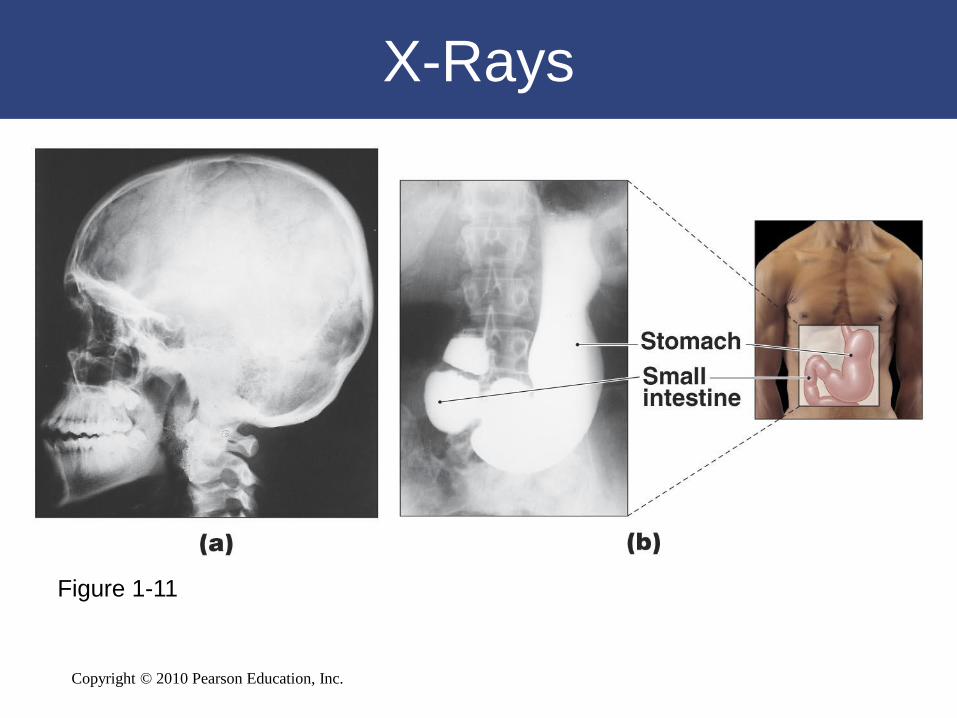
X-Rays
Figure 1-11

Copyright © 2010 Pearson Education, Inc.
Figure 1-12