Embed Size (px)
Citation preview

J. med. Genet. (1967). 4, 295.
An Infant with Multiple Congenital Anomalies and aRing Chromosome in Group C (X-6-12)
L. J. BUTLER, N. E. FRANCE, and N. M. JACOBYFrom Queen Elizabeth Hospital for Children, Hackney Road, London E.2, and Pembury Hospital, Kent
The occurrence in man of a ring chromosome re-placing an autosome has been reported only rarely.Although three examples have involved a Group D(13-15) chromosome (Wang, Melnyk, McDonald,Uchida, Carr, and Goldberg, 1962; Bain and Gauld,1963; Smith-White, Peacock, Turner, and DenDulk, 1963) and four an 'E' Group (16-18)chromosome (Wang et al., 1962; Lucas, Kemp,Ellis, and Marshall, 1963; Genest, Leclerc, andAuger, 1963; Gropp, Jussen, and Ofteringer, 1964),they had few clinical features in common except formental retardation. Partial deletion ofchromosomeNo. 1 due to ring formation has been described in amicrocephalic dwarf (Gordon and Cooke, 1964),while Rohde and Tompkins (1965) have reported aring 5 chromosome in a case of 'cri-du-chat' syn-drome. Two cases with a C-group ring autosomewere recently described (Atkins, Pant, Hazard, andOuellette, 1966), one showing an associated B/Ctranslocation.Ring X chromosomes appear to be equally rare,
the only seven patients recorded having an XO/XXRconfiguration (Lindsten and Tillinger, 1962; Luers,Struck, and Nevinny-Stickel, 1963; Hustinx andStoelinga, 1964; Pfeiffer and Buchner, 1964;Fisher, 1965; Bain, Gauld, and Farquhar, 1965;Bishop, Blank, Simpson, and Dewhurst, 1966).Most of them showed features of Tumer's syn-drome. A further case (Turner, 1963) had a ringchromosome in group C (X-6-12) which wasprobably not an X.
Case ReportThis female child was the result of a normal preg-
nancy and was delivered spontaneously at term weighing2605 g. (5 lb. 12 oz.). There were four apparentlynormal sibs and no family historv of congenital malfor-mations.
She had unusual facies with marked hirsutes of theforehead (Fig. 1). The eyebrows were thick and joined
above the bridge of the nose, as in de Lange's syndrome,and the eyes were prominent but otherwise normal.The ears were rather large and low set, there was somehypertrophy of the gums, particularly of the upper jaw,and the hard palate was high arched with a cleft of theentire soft palate. The neck was short with slightwebbing. The extemal genitalia were those of a normalfemale. There were no flexion deformities of the digitsbut all limbs were very hypotonic.
She failed to gain weight and a loud praecordialsystolic murmur appeared on the 12th day and persistedthereafter. She finally died aged 5 months 4 days.
Necropsy. The child was emaciated and weighedonly 1950 g. (4 lb. 5 oz.). The crown-rump length was34 cm. and crown-heel 52 cm., while the head cir-cumference was 32 cm.
FIG. 1. Facies showing marked hirsutes,295
Received March 8, 1967.
on 28 May 2018 by guest. P
rotected by copyright.http://jm
g.bmj.com
/J M
ed Genet: first published as 10.1136/jm
g.4.4.295 on 1 Decem
ber 1967. Dow
nloaded from

Butler, France, and JacobyThe heart showed an ostium secundum defect
measuring 0-8 x 04 cm. and a high ventricular septaldefect measuring 0-6 x 04 cm. The pulmonary valvewas bicuspid, but all other valves were normal. Theductus arteriosus was closed.Each kidney weighed only 7 g. and showed mild dilata-
tion of pelvis and ureter. On histological examinationoccasional cysts lined by flattened, cuboidal, or colum-nar ephithelium were demonstrated in the outer part ofthe cortex. The uterus was small with normal Fallopiantubes. The gonads were only about 0-1 cm. wide andmeasured 1-6 cm. (right) and 1-2 cm. (left) in length.Microscopically they showed numerous small collec-tions of cells resembling granulosa cells clearly demar-cated from the apparently normal ovarian stroma whichformed the bulk of both gonads. In spite of the pre-sence of these cells no oocytes could be recognized in theright gonad, and only a few were present in the leftovary.The pylorus showed moderate hypertrophy of the
circular fibres to produce a pyloric tumour measuring1-3 cm. long and 1-0 cm. diameter. No abnormalitiesof the central nervous system were found. Death wasdue to bronchopneumonia.
Dermatoglyphs. The quality of palm printstaken at necropsy was too poor for detailed analysis, butno unusual features were noted in the main patterns andthe palmar creases were normal.
Cytogenetic Investigations.(a) Sex chromatin observations. (i) Buccal smear
(cresyl fast violet stain): A total of 47 out of 223 (21 %)nuclei in oral mucosal cells contained a typical sexchromatin body. This proportion is only slightlybelow the normal female range (25-70%), as determinedin this laboratory. (ii) Skin fibroblasts (aceto-orceinstain): 36% chromatin-positive cells were present afterculture for 20 days, 37% after 34 days, and 3300 after69 days. These values do not differ significantly fromeach other or from those in the normal female range (15-60%). Three biopsies of normal female skin culturedand processed at approximately the same time produced19%h, 28%, and 40% chromatin-positive cells.
In both oral mucosal cells and fibroblasts the chro-matin mass was rather more variable in size than in thenormal females but was never unduly small or large.
(b) Chromosome studies. Using standard techniques(Butler, 1965), chromosome preparations were obtainedfrom peripheral blood leucocytes and skin fibroblasts
TABLECHROMOSOME COUNTS
<45 45WithRing
45 46Without WithRing Ring
>46 NearTetra-ploid
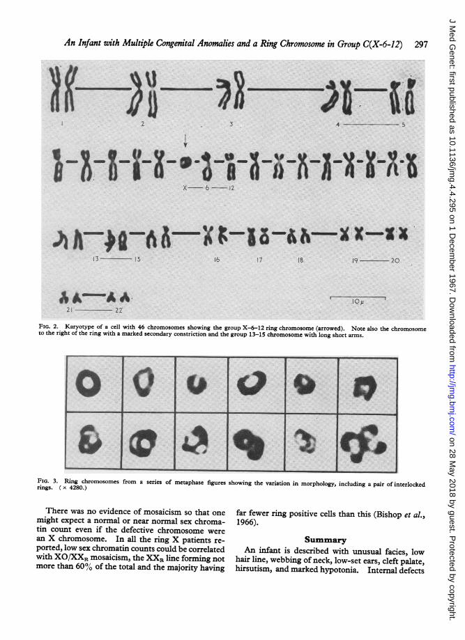
(Table). Cells with less than 45 chromosomes andthose with 45 including the ring showed random loss andwere considered to be broken cells. Karyotypes of cellswith 46 chromosomes (Fig. 2) contained normal groupsexcept for C(X-6-12) and D(13-15). Group C(X-6-12)contained 15 normal members and, in addition, a chro-mosome with a monocentric ring configuration. Varia-tion in the appearance of the ring is shown in Fig. 3.Its size was estimated to be approximately 85% of thelength of chromosome No. 6, thus indicating only a smalldeletion of an X or an autosome of similar size.
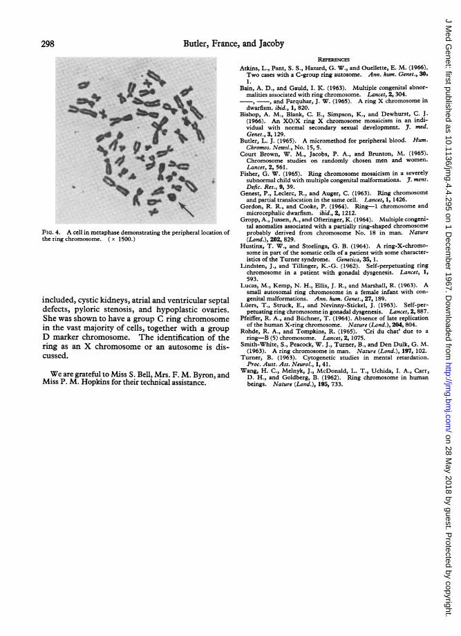
Observations were made on the position of the ringchromosome in a series of metaphase figures. Onaverage 12 chromosomes were peripherally situated ineach metaphase so that the expected random frequencywas 26%. The ring chromosome was truly peripheralin 52% of all plates (Fig. 4), showing that its distributionwas non-random. One Group D chromosome hadabnormally long short arms terminating in large satellites.
These investigations were carried out before the use ofautoradiographic techniques in this laboratory. Wewere unable to enlist the co-operation of the parents forfurther studies.
DiscussionWith one exception (Fisher, 1965) patients with
ring X chromosomes show few congenital malfor-mations. The formation of XO cells by secondaryloss of the ring leads to XO/XXR mosaicism andconsequently a significant proportion show featuresof Turner's syndrome (Bishop et al., 1966). Theovaries were only examined in one patient (Lindstenand Tillinger, 1962) and were found to be hypo-plastic with reduced numbers of primordialfollicles.Our patient had minor features suggesting Tur-
ner's syndrome, including a low hair line and slightwebbing of the neck. In addition the ovaries werehypoplastic with a few recognizable oocytes but werequite different from those usually found in cases ofgonadal dysgenesis. It is therefore possible thatthe ring chromosome was derived from an X chro-mosome, but it could equally represent one of theautosomes in group C (7, 8, or 9). The effectivedeletion in the latter case might then be correlatedwith the presence of cleft palate, cardiac and renalanomalies, and pyloric stenosis. Unfortunately, inthe absence of autoradiographic studies, it was notpossible to prove the true identity of the ring chro-mosome. She possessed a structural variant ingroup D(13-15) somewhat similar to that found inthe patient of Lindsten and Tillinger (1962).Since approximately 3% of the general populationshow minor karyotype variations without phenotypeabnormality (Court Brown, Jacobs, and Brunton,1965), this marker chromosome was probably un-connected with the congenital defects present.
Blood 1 3 2 47 - .
Skin 3 2 3 21 4
296
on 28 May 2018 by guest. P
rotected by copyright.http://jm
g.bmj.com
/J M
ed Genet: first published as 10.1136/jm
g.4.4.295 on 1 Decem
ber 1967. Dow
nloaded from
An Infant with Multiple Congenital Anomalies and a Ring Chromosome in Group C(X-6-12)
FIG. 2. Karyotype of a cell with 46 chromosomes showing the group X-6-12 ring chromosome (arrowed). Note also the chromosometo the right of the ring with a marked secondary constriction and the group 13-15 chromosome with long short arms.
FIG. 3. Ring chromosomes from a series of metaphase figures showing the variation in morphology, including a pair of interlockedrings. ( x 4280.)
There was no evidence of mosaicism so that one far fewer ring positive cells than this (Bishop et al.,might expect a normal or near normal sex chroma- 1966).tin count even if the defective chromosome werean X chromosome. In all the ring X patients re- Summaryported, low sex chromatin counts could be correlated An infant is described with unusual facies, lowwith XO/XXR mosaicism, the XXR line forming not hair line, webbing of neck, low-set ears, cleft palate,more than 60% of the total and the majority having hirsutism, and marked hypotonia. Internal defects
OQoQegr __- ----- - - --------- ------------.-1:e at
297
on 28 May 2018 by guest. P
rotected by copyright.http://jm
g.bmj.com
/J M
ed Genet: first published as 10.1136/jm
g.4.4.295 on 1 Decem
ber 1967. Dow
nloaded from
Butler, France, and Jacoby
FIG. 4. A cell in metaphase demonstrating the peripheral location ofthe ring chromosome. ( x 1500.)
included, cystic kidneys, atrial and ventricular septaldefects, pyloric stenosis, and hypoplastic ovaries.She was shown to have a group C ring chromosomein the vast majority of cells, together with a group
D marker chromosome. The identification of thering as an X chromosome or an autosome is dis-cussed.
We are grateful to Miss S. Bell, Mrs. F. M. Byron, andMiss P. M. Hopkins for their technical assistance.
REFERENCESAtkins, L., Pant, S. S., Hazard, G. W., and Ouellette, E. M. (1966).Two cases with a C-group ring autosome. Ann. hum. Genet., 30.1.
Bain, A. D., and Gauld, I. K. (1963). Multiple congenital abnor-malities associated with ring chromosome. Lancet, 2, 304.-, -, and Farquhar, J. W. (1965). A ring X chromosome in
dwarfism. ibid., 1, 820.Bishop, A. M., Blank, C. E., Simpson, K., and Dewhurst, C. J.
(1966). An XO/X ring X chromosome mosaicism in an indi-vidual with normal secondary sexual development. J. med.Genet., 3, 129.
Butler, L. J. (1965). A micromethod for peripheral blood. Hum.Chromos. Newsl., No. 15, 5.
Court Brown, W. M., Jacobs, P. A., and Brunton, M. (1965).Chromosome studies on randomly chosen men and women.Lancet, 2, 561.
Fisher, G. W. (1965). Ring chromosome mosaicism in a severelysubnormal child with multiple congenital malformations. J. ment.Defic. Res., 9, 39.
Genest, P., Leclerc, R., and Auger, C. (1963). Ring chromosomeand partial translocation in the same cell. Lancet, 1, 1426.
Gordon, R. R., and Cooke, P. (1964). Ring-1 chromosome andmicrocephalic dwarfism. ibid., 2, 1212.
Gropp, A., Jussen, A., and Ofteringer, K. (1964). Multiple congeni-tal anomalies associated with a partially ring-shaped chromosomeprobably derived from chromosome No. 18 in man. Nature(Lond.), 202, 829.
Hustinx, T. W., and Stoelinga, G. B. (1964). A ring-X-chromo-some in part of the somatic cells of a patient with some character-istics of the Turner syndrome. Genetica, 35, 1.
Lindsten, J., and Tillinger, K.-G. (1962). Self-perpetuating ringchromosome in a patient with gonadal dysgenesis. Lancet, 1,593.
Lucas, M., Kemp, N. H., Ellis, J. R., and Marshall, R. (1963). Asmall autosomal ring chromosome in a female infant with con-genital malformations. Ann. hum. Genet., 27, 189.
Luers, T., Struck, E., and Nevinny-Stickel, J. (1963). Self-per-petuating ring chromosome in gonadal dysgenesis. Lancet, 2, 887.
Pfeiffer, R. A., and Buchner, T. (1964). Absence of late replicationof the human X-ring chromosome. Nature (Lond.), 204, 804.
Rohde, R. A., and Tompkins, R. (1965). 'Cri du chat' due to a
ring-B (5) chromosome. Lancet, 2, 1075.Smith-White, S., Peacock, W. J., Turner, B., and Den Dulk, G. M.
(1963). A ring chromosome in man. Nature (Lond.), 197, 102.Turner, B. (1963). Cytogenetic studies in mental retardation.
Proc. Aust. Ass. Neurol., 1, 41.Wang, H. C., Melnyk, J., McDonald, L. T., Uchida, I. A., Carr,D. H., and Goldberg, B. (1962). Ring chromosome in humanbeings. Nature (Lond.), 195, 733.
298
on 28 May 2018 by guest. P
rotected by copyright.http://jm
g.bmj.com
/J M
ed Genet: first published as 10.1136/jm
g.4.4.295 on 1 Decem
ber 1967. Dow
nloaded from