Embed Size (px)
Citation preview

AN IMMUNOFLUORESCENCE APPROACH TO THE DIAGNOSIS OF ACUTE LYMPHOBLASTIC LEUKEMIA*
Albert0 M. Marmont, Eugenio E. Damasio, Gin0 Santini, Andrea Bacigalupo, and Domenico Giordano
Division of Hematology Civil Hospital Genoa, Italy
Although the cytomorphologic diagnosis of acute lymphoblastic leukemia (ALL) is comparatively simple and may be aided further by cytochemical methods, there are occasional cases in which the identification of the leukemic cells may be controversial. Thus, from a practical viewpoint and in order to detect, if possible, antigenic diversities between apparently similar varieties of acute leukemias, an immunomorphologic approach was attempted.
For this purpose, an indirect immunofluorescence (IF) test that employed antilymphocytic globulin (ALG) was devised. ALG has recently been demon- strated on canine,' murine,2 and human3 lymphocytes by an in vitro indirect IF test; fixed and dried lymphocyte preparations were employed by Thomas et ~ 1 . ~ 9 ~ It has also been shown that ALG uptake by human peripheral lymphocytes is the same when ALG is reacted with either T or B lymphocytes5 and that only when an antithymocyte antiserum has been previously absorbed with B lymphocytes from chronic lymphatic leukemia (CLL) is it subsequently able to react with T lymphocytes.6
Although some of these studies were performed with fixed lymphocyte preparation^,^^^ and others with fresh suspension^,^^^^^ a transmitted BV illumination was used during all of them.
MATERIALS AND METHODS
Because this presentation is intended to be brief, only a simple outline of the technical procedures will be given. Methodologic steps and details have been published elsewhere.
Fixed and fresh lymphoblast preparations were studied, but the latter were preferred, because they offered higher titers and better observation conditions. These preparations were incubated with horse and rabbit antihuman ALG in progressive dilutions; in addition to the commercial preparations, Behringwerke kindly furnished us with antisera raised in horses by utilizing as antigens thoracic duct, thymus, lymph nodes, spleen, and tonsil lymphocytes, and also lympho- cytes from CLL and membranes of cultured lymphoblasts, according to methods specified elsewhere.' After incubation with ALG for 1 hr a t 4°C and the usual washes, the preparations were reacted with the appropriate FITC-conjugated antisera and examined both with transmitted excitation that utilized a short-wave pass interference filter (KP 500) with a sharp cut-off at 490-500 pm and with incident illumination furnished through an interference dividing plate fitted with the same interference filter.
* Supported by Grant 73,00652,04 from the Consiglio Nazionale delle Ricerche.
618

Marmont et al. : Acute Lymphoblastic Leukemia 619
FIGURE 1 . Positive indirect IF test of absorbed commercial ALG at a dilution of 1 : 50,000.
Myeloid-absorbed ALG was prepared by performing two subsequent in- cubations of 1 : 50 dilutions of ALG in PBS with buffy coats obtained from patients in the acute blastic transformation stage of chronic myelogenous leukemia (CML) and by then utilizing the clear centrifuged supernatant.
RESULTS
Although a bright fluorescence that involved the entire cytoplasmic ring was seen when the fixed preparations were inspected, background staining was often present, although it was not particularly troublesome. This was not t rue with the fresh preparations, which displayed a brilliant membrane fluorescence on a uniformly dark background. With increasing dilution, the fluorescent rings gradually became less brilliant, breaking into crescents and spots before total extinction. A difference of not less than three logs could be found between fixed and fresh cell titers. Because of the above reasons, the latter procedure was preferred.
N o difference in appearance and titers was found between CLL and ALL preparations. However, we soon discovered that cells from AML also reacted vigorously with ALG, which process produced strong cross reactions. To prevent this occurrence, a systemic preabsorption with leukocytes from patients in the blastic transformation of stage of CML was performed. After this procedure, cross reactions were markedly diminished, as is shown in TABLES 1 and 2.

620 Annals New York Academy of Sciences
TABLE 1 IF TITERS WITH COMMERCIAL MYELOID-ABSORBED ALG
Leukemia Type Cases Titers (reciprocal of dilution)
CLL 10 2 x 10⁵ ALL 8 2 105 S6zary’s syndrome 1 2 105 AML 10 1-2 x 10⁴ CML-B1.Cr. 5 1-5 104
Whereas a continuous equatorial fluorescence pattern was the commonest finding, especially a t lower ALC dilutions, with increasingly high dilutions, typical polar crescent-shaped or caplike fluorescence figures appeared at room temperature; the latter was more notably characteristic of myeloid than of lymphoid cells. When capped cells were inspected by phase contrast microscopy, the capped region invariably corresponded to the part of the cell that contained the Golgi complex and was in opposition t o the nucleus, which, in these cells, had always assumed an eccentric location.
TABLE 2 IF TITERS WITH THORACIC DUCT MYELOID-ABSORBED ALG
Leukemia Type Cases Titers (reciprocal of dilution)
CLL 4 2-5 104 ALL 4 2-5 104 AM L 4 5 x 10
COMMENTS
We have demonstrated that antihuman ALG, when reacted in an indirect IF test, is capable of reacting not only with mature lymphoid cells but also with the lymphoblasts of ALL. Such reactions may occur with fixed and dried preparations, which thus display cytoplasmic fluorescence; they are much more brilliant and definite, however, when membrane fluorescence is investigated on fresh preparations, where titers of five logs may be attained. It would accordingly appear that similar, if not identical, “lymphoid” antigens are recognized by ALC both deep in the cytoplasm and on the surface of these cells.
Cross reactions with immature myeloid cells, especially the basophilic agranular varieties, were practically constant and are in agreement with the finding that ALG interferes with hematopoietic stem cells.* It has also been shown that the relevant antigens may not be possessed exclusively by lymphoid cells.g Changes in membrane antigen expression may also occur as a function of time in culture. Nevertheless, careful preabsorption with immature myeloid cells was capable of preventing cross reactions, which result thus heralds the introduction of the indirect IF test into the routine hematology laboratory. Although a difference in titer of only one log was obtained by absorbing
Marmont er al. : Acute Lymphoblastic Leukemia 62 1
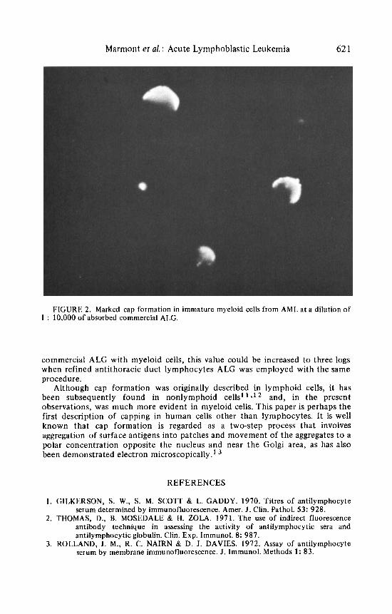
FIGURE 2. Marked cap formation in immature myeloid cells from AML at a dilution of 1 : 10,000 of absorbed commercial ALG.
commercial ALG with myeloid cells, this value could be increased t o three logs when refined antithoracic duct lymphocytes ALG was employed with the same procedure.
Although cap formation was originally described in lymphoid cells, it has been subsequently found in nonlymphoid cells' ' i1 * and, in the present observations, was much more evident in myeloid cells. This paper is perhaps the first description of capping in human cells other than lymphocytes. It is well known that cap formation is regarded as a two-step process that involves aggregation of surface antigens into patches and movement of the aggregates t o a polar concentration opposite the nucleus and near the Golgi area, as has also been demonstrated electron microscopically.'
REFERENCES
1. GILKERSON, S. W., S. M. SCOTT & L. CADDY. 1970. Titres of antilymphocyte serum determined by immunofluorescence. Amer. J . Clin. Pathol. 53: 928.
2. THOMAS, D., B. MOSEDALE & H. ZOLA. 1971. The use of indirect fluorescence antibody technique in assessing the activity of antilymphocytic sera and antilymphocytic globulin. Clin. Exp. Immunol. 8: 987.
3. ROLLAND, J . M., R. C. NAIRN & D. J. DAVIES. 1972. Assay of antilymphocyte serum by membrane immunofluorescence. J . Immunol. Methods 1: 83.

622 Annals New York Academy of Sciences
4. THOMAS, D., J . G. WOODROOFF, B. MOSEDALE, P. D. WARD, H. ZOLA, D. C. EDWARDS & H. BALNER. 1972. Antibody titres of antilymphocytic sera and globulins determined by immunofluorescence and their correlation with immunosuppressive activity. Clin. Exp. Immunol. 11: 569.
5. BRAIN, P. & R. H. MARSTON. 1973. Uptake of antilymphocyte globulin by human peripheral lymphocytes. Clin. Exp. Immunol. 13: 423.
6. AISENBERG, A. C., K. J. BLOCH, J. C. LONG & R. B. COLVIN. 1973. Reaction of normal human lymphocytic leukemic cells with an anti-thymocyte antiserum. Blood 41: 417.
7. MARMONT. A. M., E. E. DAMASIO, G. SANTINI, D. GIORDANO & A. BACIGALUPO. 1974. An immunohistochemical approach to the cytological diagnosis of acute lymphoblastic leukemia. I n 1st International Meeting on Acute Leukemias, Rome, Dec. 6-8. In press.
8. FIELD, E. 0. & J. E. GIBBS. 1968. Cross-reaction of anti-lymphocyte serum with haemopoietic stem cells. Nature (London) 217: 561.
9. LANCE, E. M., P. B. MEDAWAR & R. N. TAUB. 1973. Antilymphocyte serum. Advan. Immunol. 17: 1.
10. ROSENFELD, C., J. F. DORE, C. CHOQUET, A. M. VfiNUAT, L. MARHOLEV, J. P. WASTIAUX, C. GUIBOUT & J. L. PICO. 1973. Increase in membrane antigen expression of human leucocytes traced by ALG as a function of time in culture. Biomedicine 19: 541.
11. EDIDIN, M. & A. WEISS. 1972. Antigen cap formation in cultured fibroblasts: a reflection of membrane fluidity and of cell motility. Proc. Nat. Acad. Sci. USA 69: 2456.
12. FERRARINI, M., A. MUNRO & A. B. WILSON. 1973. Cytophilic antibody: correlation of its distribution with activation of basophils and macrophages. Eur. J. Immunol. 3: 364.
13. DE PETRIS, S. & M. C. RAFF. 1972. Distribution of immunoglobulin on the surface of mouse lymphoid cells as determined by immunoferritin electron microscopy. Antibody-induced, temperature-dependent redistribution and its implications for membrane structure. Eur. J. Immunol. 2: 253.