Embed Size (px)
Citation preview

An “Epidemic” of Chemical Meningitis*
WINFREY W. GOLDMAN, JR., M.D. and JAY P. SANFORD, M.D.
Dallas, Texas
M ENINGITIS following the administration of a spinal anesthetic may be the consequence
of a number of factors: trauma from the pro- cedure itself, aggravation of an underlying disease of the central nervous system, contamina- tion with viable bacteria, inflammatory response to the anesthetic agent or medication, or an inflammatory response to a foreign irritant (chemical) which has contaminated the equip- ment used during the anesthetic procedure.
During an eleven-month period we observed five patients in whom an acute purulent “aseptic” meningitis developed shortly following
Saddle Clinical Block Meningitis
~~%.+ti
T E i03*- -.
M P 102’- E R ioi’-
/c .
A ,
&* ‘\I T ioo
.
U R 99’ E I __-_---__-d---Y
98’: i
./
I \ \ . \/
----_-_ J i
I _-
3
spinal anesthesia which seemed to be a result of the chance use of syringes contaminated with trace amounts of a phenolic disinfectant. Based on these observations and review of previous clinical and experimental studies, the patho- genesis, clinical features and means of preven- tion of this syndrome are summarized.
CASE REPORTS
The following five women were admitted to Parkland Memorial Hospital for delivery of un- complicated full-term pregnancies. None dem- onstrated signs or symptoms of acute or chronic
Asymptomatic M. 5.
5 6
I 24 HOUR PERIODS ]
I ANT/B/O7/CS * v jz? a’ap
+‘+I “,,d&cSF CSF: WBC count: CSF:
17,200 cells/mm3 24,950/mm3 2,90O~/mm’ i4,400/mm3 NGZcelle
98% PMN 82% PMN 100% Lymphocytes 80% PMN Protein not obtoinea
Sugar 40mgsA $e% Lyniphocytes Protein 44 mgs% 070 Bands Sugar not obtained
Smear negative Sugar 63 mgs% iZ’/o Lymphocytes
19% Lymphocytes
FIG. 1.
* From the Department of Internal Medicine, The University of Texas Southwestern Medical School, Dallas, Texas.
94 AMERICAN JOURNAL OF MEDICINE
Chemical Meningitis-Goldman, Sanford
Saddle CLinicaL Block Meningitis Asymptomatic
I %4 hrs.+, -
ioB
T E 103~
M I-----
,-.-.- -.-.- ,
I I 24 HOUR PERIODS 1
AN7/5/07/C.S *
4 I
C_ CSF: WBC count: CsF: WBC count: Opening pressure Openin~ssure i8,800/mm3 Opening pressure 8,050/mm3 -
300 mmCSF i80 mm CSF 77 70 PMN 150 mm CSF 54% PMN 5,000 cells/mm3 7,200 cells/mm’ 2% Bands 15 celk./mm3 6% Bands
90% PMN 90% PMN 20% Lymphocytes All lymphocytes 37% Lymphocytes Protein 200 mgs9. Protein 48 mgs% 1% Eosinophils Protein 35 mgss 1% Eas’kutphils Sugar 6emgs”/o Sugar 58 mgs% Sugar 65mgs% Smear negative Blood sugar 75mgs% Blood sugar 86mgsK for organisms
*C~+t*/rri?e penicilllh 20 fo 3007. u. dffr./~y Ch/oramphent>o/ 2 gms. c~&/Yq
Po/4//77fJxti3 5 1.5 grr;s. dc7CZ~
FIG. 2.
disease prior to delivery. These cases are sporadic, considering the number of spinal anesthetics administered during this period by the Department of Obstetrics and Gynecology.
CASE I (PMH No. 145156) (Fig. 1). A seventeen year old gravida 2, para 1 housewife was admitted in September 1957 in active labor. A low spinal anesthetic (saddle block) was administered using 4 mg. tetracaine hydrochloride. Delivery was un- eventful. Four hours after administration of the spinal anesthetic her temperature was 100.4”~. orally. Thirteen and a half hours after administration of the anesthetic her temperature was 101”~. and she com- plained of moderate headache. She was found to have nuchal rigidity. No residual motor or sensory effects of the anesthetic were detected and there were no neurological deficits. Lumbar puncture was per- formed and the cerebrospinal fluid (CSF) was purulent, containing 17,200 cells,/cu. mm., 98 per cent of which were polymorphonuclear leukocytes. No organisms were demonstrated on stained smears of
JULY 1960
CSF sediment. Blood and CSF cultures were negative. The CSF sugar was 40 mg. per cent. A peripheral white blood cell count obtained twenty-four hours following the onset of meningitis was 15,6OO/cu. mm. with a shift to the left. Results of serologic tests for syphilis were negative. On the assumption that this represented meningitis secondary to bacterial con- tamination, the patient was treated with chlor- amphenicol, penicillin and polymyxin B. Within four days after onset of meningitis she was afebrile and had clinically recovered. Seven days after onset the CSF contained no cells and she was discharged shortly thereafter. She was examined eleven months after this episode and no neurological abnormalities were detected.
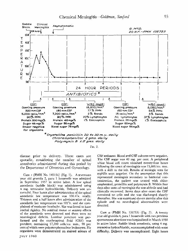
CASE II (PMH No. 135753) (Fig. 2). A twenty year old gravida 4, para 1 housewife with two previous spontaneous abortions was hospitalized in March 1958
in active labor. Saddle block anesthesia, using 4 mg. tetracaine hydrochloride, was accomplished with some
difficulty, Delivery was uncomplicated. Eight hours
Chemical Meningitis-Goldman, Sanford
Asymptomatic
f
I 24 HOUR PERIODS
A/1/77/B/O 7-/G’S * I I I
- ---- _- _----___ ---OS\,O~
6
.-- b
CSF: -----, w0c :ount: CSF: Opening pressure WBC count: 9, 200/mm3
i3El mm CSF 2,600 cells/mm’
ii, 500/mm3 57% PMN i8 cells/mm’
78% Lymphocytes 06% PMN 2070 Bands
S6 70 PMN Protein 27mgs%
4% Bands 23% Lymphocytes Sugar not obtained Protein 465 mgr% iO% Lymphocytes Smear negative for organisms x
C!g.stia//r;le Sugar not obtained
penckc;/lci, 10 m. U. durrirg +?>.5t 24 AH. Proccrfir e pen&Y/A I. B M. u . dur’lq/ 2% erea*er
Chlorpmp~e/3c~od Zgms. da,Xy
FIG. 3.
after administration of the anesthetic her oral tem- perature was 101.4”~. rising td 101.8’~. Six hours later severe headache, backache and nuchal rigidity were noted. There were no residual effects of the anes- thetic and no neurological changes were detected. On lumbar puncture, the CSF pressure was increased and the CSF contained 5,000 cells/cu. mm. with 90 per cent polymorphonuclear cells, protein 200 mg. per cent and sugar 68 mg. per cent. No organisms were seen on stained smears of the CSF sediment. Blood and CSF cultures were negative. Results of serologic tests for syphilis were negative. Again, bacterial con- tamination was considered and the patient was treated with penicillin, chloramphenicol and poly- myxin B. She had clinically recovered and was afebrile in less than twenty-four hours after onset of the illness. The patient was examined five months after the meningitis and was found to have no neurological abnormalities.
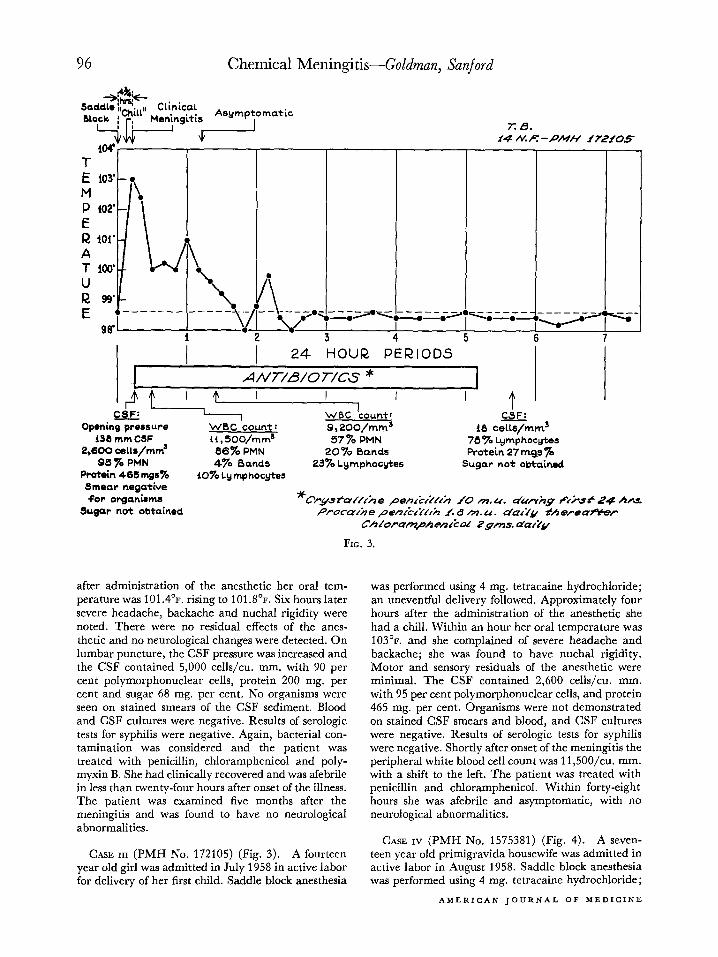
CASE III (PMH No. 172105) (Fig. 3). A fourteen year old girl was admitted in July 1958 in active labor for delivery of her first child. Saddle block anesthesia
was performed using 4 mg. tetracaine hydrochloride; an uneventful delivery followed. Approximately four hours after the administration of the anesthetic she had a chill. Within an hour her oral temperature was 103’~. and she complained of severe headache and backache; she was found to have nuchal rigidity. Motor and sensory residuals of the anesthetic were minimal. The CSF contained 2,600 cells/cu. mm. with 95 per cent polymorphonuclear cells, and protein 465 mg. per cent. Organisms were not demonstrated on stained CSF smears and blood, and CSF cultures were negative. Results of serologic tests for syphilis were negative. Shortly after onset of the meningitis the peripheral white blood cell count was 11,5OO/cu. mm. with a shift to the left. The patient was treated with penicillin and chloramphenicol. Within forty-eight hours she was afebrile and asymptomatic, with no neurological abnormalities.
CASE IV (PMH No. 1575381) (Fig. 4). A seven- teen year old primigravida housewife was admitted in active labor in August 1958. Saddle block anesthesia was performed using 4 mg. tetracaine hydrochloride;
AMERICAN JOURNAL OF MEDICINE

Chemical Meningitis-Goldman, Sanford
Saddle Clinical Block Meningitis
Asymptomatic
T E 103’
M P IO?’ E
24 HOUR PERIODS
A NT/B/O T/CS *
,r--J@-----’ ’ ’ t
.-
6
_--_
* Opening pressure
145 mm C5F 2,730 cells/mm3 94% Lymphocytes Protein 172 mgss
Sugar 63 rnw% Smear negative for organisms
WBC count: CSF:
i6,050/mm3 6 Lymphocytes
78 % PMN i PMN
5% Bands 17% Lymphocytes
* CrystffCfihe penibi’//rir 20 m.u. dur/ig +f+sf 24 hrs. Proc~l,h,e pen&Y/fir f.2 m.u. olcrr& thereafter
Tetracycyche 1’gm. dQf’Cy
FIG. 4.
an uneventful delivery followed. Thirteen and a half hours after administration of the anesthetic her oral temperature was 101’~. and she complained of head- ache and backache. There was nuchal rigidity but no other neurological findings. The CSF contained 2,730 cellsjcu. mm.. 94 per cent of which were reported as “lymphocytes” (this was not checked by the medical staff). The CSF protein was 172 mg. per cent and the sugar was 63 mg. per cent. No organisms were seen on stained smears of the CSF. Blood and CSF cultures were sterile. Results of serologic tests for syphilis were negative. The peripheral white blood cell count shortly after onset of meningitis was 16,05O/cu. mm. with a shift to the left. The patient was treated with penicillin and tetracycline. Twenty-four hours after onset of the illness she was asymptomatic and afebrile. There were no neurological changes at the time of discharge.
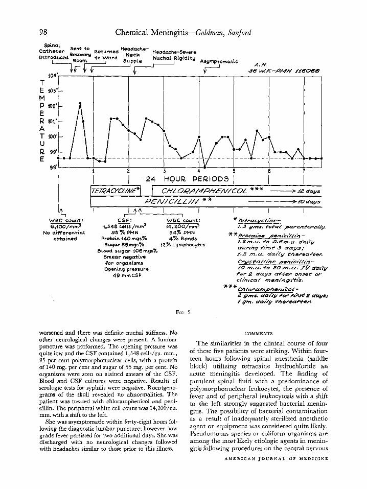
CASE v (PMH No. 116066) (Fig. 5). The fifth patient differed somewhat from the preceding pa- tients. A thirty-six year old gravida 9, para 7 house- wife was hospitalized in August 1958 for Caesarean section and hysterectomy. She had previously under- gone Caesarean section for cephalopelvic dispropor- tion and was being treated for iron deficiency anemia
JULY 1960
attributable to multiple pregnancies and menorrhagia. For some time she had complained of vague headaches for which her attending physician and various con- sultants had been unable to detect an organic basis. The preoperative peripheral white blood cell count was 6,1OO/cu. mm.
Two days after admission a Caesarean section and hysterectomy were performed under continuous spinal anesthesia using 3.3 per cent procaine with a catheter introduced intrathecally through the third and fourth lumbar interspace. Surgery was completed three hours after initiation of the spinal anesthetic and the patient was moved to the recovery room. Six and a half hours after the beginning of spinal anesthesia her rectal temperature was 102.2”~. Examination of the chest and legs was described as being within normal limits. Penicillin and tetracycline were given paren- terally. Twelve hours after initiation of spinal anes- thesia she was returned to her room. At this time she complained of headache. Her oral temperature was recorded as 98.6’~. The neck was “supple.” The patient persistently complained of “stabbing” frontal headache and, twenty-seven hours after administra- tion of the anesthetic, her temperature was 101.2”F. orally. Approximately forty-eight hours after admin- istration of the spinal anesthetic her headache
Chemical Meningitis-Goldman, Sanford
Spinal Catheter Sent to Returned Headache- Headache-Severe Introduced RecQvery to Ward Syppe
L-lYrnr=L__I.
Nuchal Rigidity Asymptomatic
I A./t!
WBC Aunt: CSF: 6,100/mm3 1,548 cell5 /mm3
WBC ‘count: 14,2OO/mm”
No differential obtained
95% PMN 64% PMN Protein 140 mgs% 4% Bands
Sugar 55 mgs% i2% Lymphocytes Blood sugar i06mgsX
Smear negative for organisms Opening pressure
49 mmCSF
durfhg f/i.sf 3 days; f.2 m.u. dat2y therea;roCer
for 2 day.s after onset of clhr caf monc’ngr’t&.
FIG. 5.
worsened and there was definite nuchal stiffness. No other neurological changes were present. A lumbar puncture was performed. The opening pressure was quite low and the CSF contained 1,548 cells/cu. mm., 9.5 per cent polymorphonuclear cells, with a protein of 140 mg. per cent and sugar of 55 mg. per cent. No organisms were seen on stained smears of the CSF. Blood and CSF cultures were negative. Results of serologic tests for syphilis were negative. Roentgeno- grams of the skull revealed no abnormalities. The patient was treated with chloramphenicol and peni- cillin. The peripheral white cell count was 14,2OO/cu. mm. with a shift to the left.
She was asymptomatic within forty-eight hours fol- lowing the diagnostic lumbar puncture; however, low grade fever persisted for two additional days. She was discharged with no neurological changes followed with headaches similar to those prior to this illness.
COMMENTS
The similarities in the clinical course of four of these five patients were striking. Within four- teen hours following spinal anesthesia (saddle block) utilizing tetracaine hydrochloride an acute meningitis developed. The finding of purulent spinal fluid with a predominance of polymorphonuclear leukocytes, the presence of fever and of peripheral leukocytosis with a shift to the left strongly suggested bacterial menin- gitis. The possibility of bacterial contamination as a result of inadequately sterilized anesthetic agent or equipment was considered quite likely. Pseudomonas species or coliform organisms are among the most likely etiologic agents in menin- gitis following procedures on the central nervous
AMERICAN JOURNAL OF MEDICINE

Chemical Meningitis-Goldman, Sanford 99
system, including spinal anesthesia [I]. How- ever, in no instance were organisms demon- strated on stained smears of the cerebrospinal fluid sediment and the spinal fluid sugar con- centrations were normal. Despite this discrep- ancy and because of our early failure to appre- ciate the possibility of purulent sterile meningitis, intensive antibacterial therapy directed against these organisms was initiated in each instance. The failure to culture organisms from either the cerebrospinal fluid or blood suggested the likelihood of a non-bacterial form of postspinal anesthetic meningitis. Another consideration was that the trauma of the procedure had caused bleeding into the intrathecal space. The cerebro- spinal fluid was neither bloody nor xantho- chromic, and the magnitude of the pleocytosis without frank blood in the cerebrospinal fluid militated against this view. Aggravation of an underlying disease of the central nervous system by spinal anesthesia has been reported, especially in patients with pernicious anemia, multiple sclerosis, tabes dorsalis, general paresis, and metastatic and primary neoplasms of the central nervous system [&lo]. None of our patients exhibited the clinical features of these illnesses and follow-up examination have failed to detect such diseases.
Thus, an inflammatory response to a foreign irritant or “chemical meningitis” was suggested. The occurrence of untoward reactions to the intrathecal injection of foreign substances is well documented.
Early in the use of spinal anesthesia, aseptic meningeal reactions manifested principally by a cerebrospinal fluid pleocytosis frequently were reported. Merritt and Fremont-Smith [3] re- viewed several reports published between 1928 and 1934 in which over one hundred and fifty patients were studied following spinal anes- thesia; slight to marked cerebrospinal fluid pleocytosis was found within twelve to twenty- four h6urs in from 65 per cent to approximately 100 per cent of patients. A predominance of polymorphonuclear cells was demonstrated in some patients, in others the response was lymphocytic. In roughly one-third, there was elevation of the cerebrospinal fluid protein. However, in 1936 Orkin [d] summarized the re- ports of approximately 46,000 administrations of spinal anesthetics and reported an incidence of aseptic meningitis of only 0.26 per cent. Sub- sequent and more recent studies have not demonstrated a significant incidence of inflam-
JULY 1960
matory response after spinal anesthesia [5,77]. Kamsler [A reported no cells in the cerebrospinal fluid in nine of twelve patients given a single injection of an anesthetic agent; 5 cells,/cu. mm. was the highest number found in the other three [5].
Yet there can be little question that severe neurological complications still may occur fol- lowing spinal anesthesia [6X?]. Two general syn- dromes have been encountered following spinal anesthesia using procaine or one of its deriva- tives. One is a destructive neurological process which either may begin promptly, the patient failing to recover fully from the effects of the anesthetic agent or may begin after weeks or months. Such complications are usually progres- sive and are not associated with fever or overt clinical meningitis. The lesions consist of pro- gressive demyelinization of the spinal roots beginning with the cauda equina, chronic adhesive arachnoiditis, hyperplastic pia with marginal dem) elinization of the cord, or, in some cases, progression of the process to a meningo- encephalitis and sometimes to internal hydro- cephalus. A fatal outcome is not uncommon and residual motor and sensory deficits are frequent [2,77-731. Winkleman [ 721 studied eleven such cases and found that the syringes used for administering the anesthetic agents were cleansed in a “mild detergent solution” with only casual rinsing in tap water before autoclaving. He demonstrated that “injection of a similar mild detergent solution into an animal produced paralysis of the hind legs.” Paddison and Alpers [ 731 also reported a fatal case of adhesive arachnoiditis, nerve root and spinal cord de- generation which they attributed to detergent remaining in the spinal anesthesia equipment following washing. Additional experimental support came from the observations of Denson and co-workers [74,75]. Using syringes which had been soaked in various detergent solutions, then autoclaved without washing, they injected spinal anesthetic solutions into the subarach- noid spaces of monkeys; most showed varying degrees of arachnoiditis and nerve tissue damage when sacrificed.
The second syndrome encountered following spinal anesthesia is an acute meningitis which is apparently benign. Livingstone and co-workers [76] reported two cases and reviewed the earlier case reports. In their first case headache, stiff neck, nausea, vomiting and photophobia devel- oped within fourteen hours following spinal

100 Chemical Meningitis-Goldman, Sanford
anesthesia. There was marked lymphocytosis and elevation of the protein content of the cerebro- spinal fluid. On the fourth day the patient was asymptomatic. Their second case was similar. Cultures of the cerebrospinal fluid including guinea pig and mouse inoculations, were sterile in both cases. The authors called this complication “chemical meningitis” but suggested no specific chemical agent. Rendell
[771 encountered seven similar cases, in six of which stiff neck, headache, vomiting and leth- argy developed within twenty-four hours follow- ing the administration of spinal anesthesia, with recovery in two to three days. A poly- morphonuclear leukocyte response in the cere- brospinal fluid was observed in all seven patients. On the premise that inadequately washed syringes contaminated by disinfectants were a prime cause of postspinal anesthesia aseptic meningitis, she obtained syringes which had been stored in a phenolic disinfectant solution, washed them with “exaggerated care,” and injected a spinal anesthetic in two patients. In one patient headache and low grade fever developed with no other symptoms or signs; however, the cerebrospinal fluid showed ‘<a large number of polys.” Symptoms and fever were not recorded in the other patient but the cerebro- spinal fluid contained over 2,000 “polys” and the protein was elevated.
Hurst [ 783 has reported experimental studies in which monkeys were injected intrathecally with different concentrations of various chemi- cals including cationic quarternary ammonium compounds, anionic detergents, non-ionic de- tergents and various disinfectants including phenol. He produced pathological changes simi- lar to those described by others following the use of detergents. However, he reported that such changes occurred only when rather high con- centrations of the chemicals were injected. Despite the failure to produce an experimental acute meningitis in monkeys with trace amounts of phenolic disinfectants, the introduction of trace quantities of such agents into the sub- arachnoid space of human subjects seems to produce an acute benign purulent meningitis which is not followed by progressive neurological changes.
Because of the striking similarity between the patients we observed and the findings of Rendell, the technics in the care of spinal anes- thetic equipment and anesthetic agents which were used in our patients were reviewed. Four
of the patients were given 4 mg. tetracaine hydrochloride in 6 per cent dextrose solution distributed in 2 ml. ampoules. The ampoules were kept in their original cartons prior to being placed on the anesthetic trays which were then wrapped and autoclaved. At no time were the ampoules placed in disinfectant solutions. How- ever, until shortly after the appearance of the first case, the syringes used for “saddle blocks” were cleansed according to technic utilized for ‘Youtine” syringes in the hospital. This consists of soaking syringes in a phenolic disinfectant solution (2.5 per cent AmphyP), followed by washing and rinsing. In the Department of Anesthesia syringes for spinal anesthesia were kept entirely separate from other syringes and cleansed only in distilled water, alcohol and ether. This procedure was instituted by the Department of Obstetrics and Gynecology fol- lowing the first episode of “chemical meningitis.” However, many of the trays prepared by the older method were kept and periodically re- autoclaved without opening the trays and re- washing. Also, it was possible that sporadic “breaks” in the procedure allowed a disinfect- ant-soaked routine hospital syringe to be included in the trays. In several of these patients the spinal anesthesia was technically difficult and it was quite certain that additional “rou- tine” syringes were utilized. After reviewing the clinical and experimental observations relating to meningeal irritation following spinal anes- thesia, we believe that the five patients herein described clearly represent chemical meningitis secondary to the intrathecal injection of phenolic disinfectant which contaminated the syringes used in the procedures.
If the problem of chemically contaminated syringes as used in various procedures is con- sidered: the implications extend beyond spinal anesthesia. The seemingly unpredictable and capricious reactions to many intrathecal medica- tions, including antibiotics, dyes and other agents may be explained in part by the use of chemically contaminated equipment. It is essential that equipment which is to be used for the administration of intrathecal materials must be handled according to procedures which are designed to eliminate completely exposure to chemical cleansing or disinfecting agents.
SUMMARY
1. Five patients in whom aseptic meningitis developed following spinal anesthesia are de-
AMERICAN JOURNAL OF MEDICINE

Chemical Meningitis-Goldman, Sanford 101
scribed. Initially, each patient appeared to have an acute bacterial meningitis. In the majority, the onset of clinical meningitis occurred within fourteen hours following the administration of the spinal anesthetic. Recovery was prompt and no neurologic changes were encountered.
2. Review of the clinical and experimental observations relating to meningeai irritation following spinal anesthesia supports the impres- sion that these patients had a chemical meningi- tis secondary to the intrathecal injection of phenolic disinfectants remaining on syringes following a cleansing procedure employing a phenolic disinfectant.
3. The necessity for more stringent procedures in preparing syringes which are to be used for the administration of intrathecal medication is emphasized.
Acknowledgment: We wish to express our ap- preciation to Dr. J. A. Pritchard for allowing us to follow and report these cases.
REFERENCES
1. BIEHL, J. P. and HAMBURGER, M. Polymyxin B therapy of meningitis following procedures on central nervous system. Arch. Int. Med., 93: 367, 1954.
2. NICHOLSON, M. J. and EVERSOLE, U. H. Neurological complications of spinal anesthesia. J. A. M. A., 132: 679, 1946.
3. MERRITT, H. H. and FREMONT-SMITH, F. The Cere- brospinal Fluid, pp. 220-223. Philadelphia, 1937. W. B. Saunders Co.
4. ORKIN, L. D. Reported mortality and morbidity following spinal anesthesia. American Society Re- gional Anesthesia, Scientific Session, March 3, 1936.
5. KAMSLER, P.-M. Study of changes in spinal fluid cell count during spinal anesthesia. Current Res. Anesth.
@ Am&., 30: 103, 1951. 6. KENNEDY, F., SOMBERG, H. M. and GOLDBERG,
B. R. Arachnoiditis and paralysis following spinal anesthesia. J. A. M. A., 129: 664, 1945.
7. YASKIN, H. E. and ALPERS, B. 3. Neuropsychiatric complication following spinal anesthesia. Ann. Int. Med., 23: 184, 1945.
8. THORSEN, G. Neurological complications after spinal anesthesia and results from 2,493 follow-up cases. Acta chir. scandinau. (supp. 121), 95: 1, 1947.
9. KENNEDY, F., EFFRON, A. S. and PERRY, G. The grave spinal cord paralyses caused by spinal anesthesia. Surg., G_ynec. @ Obst., 91: 385, 1950.
10. REYNOLDS, K. E. and WILSON, G. J. Aseptic menin- gitis following diagnostic lumbar puncture. J. A. M. A., 102: 1460, 1934.
11. BERGNER, R. P., ROSEMAN, E., JOHNSON, H. and SMITH, R. W. Severe neurologic complications following spinal anesthesia. Anesthesiology, 12: 717, 1951.
12. WINKLEMAN, N. W. Neurological symptoms follow- ing accidental intraspinal detergent injection. ,~eueurology, 2: 284, 1952.
13. PADDISON, R. M. and ALPERS, B. J. Role of intra- thecal detergents in pathogen&s of adhesive arachnoiditis. Arch. Neural. G Pqchiat., 71: 87, 1954.
14. DENSON, J. S., JOSEPH, S. E., KOONS, R. A., MURRY, W. E. and BISONNETTE, H. W. Effects of detergents intrathecally. Anesthesiology, 18: 143, 1957.
15. JOSEPH, S. I. and DENSON, J. S. Spinal anesthesia, arachnoiditis. and Daranleeia. J. A. M. A., 168: 1330, 1958. ’ - - -
16. LIVINGSTONE, H., WELLMAN, V., CLARK, D. and LAUBROS, V. So called “aseptic or chemical meninzitis.” Sure.. Gvnec. & Obst.. 77: 216. 1943.
” I
17. RENDELL, C. M. Chemical meningitis due to syringes stored in lysol. Anesthesia, 9: 281, 1954.
18. HURST, E. W. Adhesive arachnoiditis and vascular blockage caused by detergents and other chemical irritants: An experimental study. J. Path. G3 Ract., 70: 167, 1955.
JULY 1960