Embed Size (px)
Citation preview

Clinical Oncology (2007) jj: jjjejjj
doi:10.1016/j.clon.2007.05.002
ARTICLE IN PRESS
Original Article
An Atlas of the Pelvic Lymph Node Regions to AidRadiotherapy Target Volume Definition
A. Taylor*, A. G. Rockally, M. E. B. Powell*
*Department of Radiotherapy, St Bartholomew’s Hospital, London, UK; yDepartment of Radiology,St Bartholomew’s Hospital, London, UK
ABSTRACT:Aims: The implementation of advanced three-dimensional radiotherapy planning techniques requires accurate targetvolume localisation. We have previously developed guidelines to aid definition of the pelvic lymph node regions, and theaim of this study was to produce a CT atlas.Materials and methods: The guidelines were applied to a CT scan of a patient to receive adjuvant radiotherapy.Results: Reference CT images of the pelvis were generated, illustrating the nodal regions and a typical target volume foradjuvant pelvic radiotherapy for gynaecological cancer.Conclusion: These images can be used as an aid for target volume definition of the pelvic nodal regions. Taylor, A. et al.(2007). Clinical Oncology jj, jej
ª 2007 Published by Elsevier Ltd on behalf of The Royal College of Radiologists.
Key words: Clinical target volume, conformal radiotherapy, intensity-modulated radiotherapy, iron oxide particles, pelvic lymph nodes,radiotherapy planning
Introduction
Accurate and reproducible delineation of target volumes isessential for effective three-dimensional radiotherapy.Pelvic lymph node irradiation has an important role in themanagement of many pelvic malignancies. Planning studiescomparing intensity-modulated radiotherapy (IMRT) withconventional approaches have shown significant dosimetricadvantages in gynaecological and urological cancer [1e3].However, one of the factors limiting widespread imple-mentation of the technique has been a lack of consensus onthe target volume.
The probability of lymph nodes containing metastases iscurrently assessed using size criteria with computedtomography or magnetic resonance imaging (MRI). A nodalmaximal short axis diameter of greater than 10 mmindicates a high risk of metastatic involvement, but thesensitivity of this method is only 40e70% [4e6]. Unenlargednodes may still contain tumour deposits and it is thereforenecessary to include all lymph nodes within the drainingregions of the tumour in the clinical target volume (CTV).Most ‘normal size’ lymph nodes are too small to be directlyvisualised with standard imaging and delineation of thenodal CTV depends on their relationship to other pelvicstructures.
Anatomical studies have shown that pelvic lymph nodeslie adjacent to the major pelvic blood vessels. These arerelatively well visualised on conventional imaging and can,with an appropriate margin, be used as a surrogate target
0936-6555/07/000000þ09 $35.00/0 ª 2007 Pub
Please cite this article in press as: Taylor A et al., An Atlas of the PelvClin Oncol (2007), doi:10.1016/j.clon.2007.05.002
for lymph nodes. Ultra-small particles of iron oxide (USPIO)are a novel class of MRI contrast agent that make lymphnodes more readily visible [7,8]. We have previouslyreported a study with USPIO that assessed the position ofthe pelvic lymph nodes in relation to the blood vessels, anddeveloped guidelines for outlining the lymph node regions[9]. These guidelines have been independently applied toa further series of patients and found to be effective inensuring target volume coverage for O99% of nodes [10].
The purpose of this study was to use the guidelines todevelop a generic computed tomography atlas showing theposition of pelvic lymph nodes that could then be appliedfor three-dimensional radiotherapy planning techniques.
Materials and Methods
Radiotherapy planning computed tomography was used ina patient who was to receive postoperative radiotherapy tothe pelvis, having previously undergone a simple abdominalhysterectomy and bilateral salpingo-oophorectomy for uter-ine cancer. The patient was scanned in the supine positionwith a full bladder, with 5 mm slice intervals from 2 cm abovethe aortic bifurcation to the lower limit of the inguinalregion. Intravenous contrast was given to aid visualisation ofthe blood vessels. The images were transferred to a radio-therapy planning system (Eclipse v6.5, Varian).
Using pelvic blood vessels as a surrogate target for lymphnodes, our guidelines for delineation were applied (Table 1).First, a 7 mm margin was drawn around the pelvic blood
lished by Elsevier Ltd on behalf of The Royal College of Radiologists.
ic Lymph Node Regions to Aid Radiotherapy Target Volume Definition,

2 CLINICAL ONCOLOGY
ARTICLE IN PRESS
vessels. The following modifications were then made: in thecommon iliac region, the contour was extended posteriorlyto the vertebral body, passing around the psoas muscle; toencompass the obturator region, the contours around theexternal and internal iliac vessels were joined to createa single volume on each side of the pelvis, ensuring thatthe width was at least 17 mm from the pelvic side wall;the upper pre-sacral region was covered with a 10 mm stripover the anterior sacral prominence; where the marginpassed over muscle or bone, these structures were deletedfrom the CTV; and finally, to cover the distal lateral externaliliac nodes, the volume was extended anterolaterally bya further 1 cm along the line of the iliopsoas muscle. Theparametria and vaginal vault were also outlined as a separatestructure.
Reference images were produced showing the typicaltarget volume for adjuvant pelvic radiotherapy, includingthe common iliac, anterior and medial external iliac,internal iliac, obturator and the subaortic pre-sacral lymphnode groups.
Results
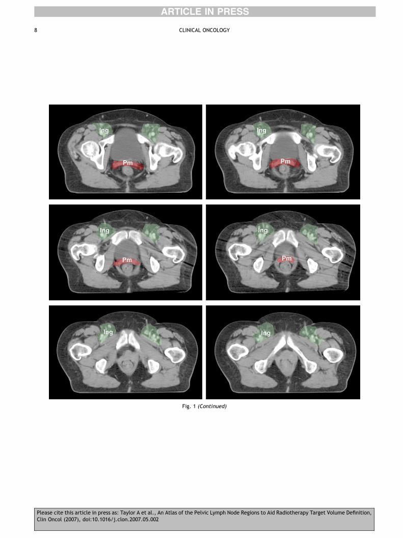
The reference images produced by applying the guidelinesare shown in Fig. 1. The typical target volume for adjuvantpelvic radiotherapy in gynaecological cancer has beenoutlined, and the nodal groups are indicated. The additionalmargin required for including the distal lateral external iliacnodes is shown in blue. The parametrium and the uppervagina are shown in red, and the inguinal, pre-sacral andlower para-aortic nodal regions are also indicated.
Discussion
Conformal radiotherapy is increasingly recognised asoffering benefit in normal tissue sparing in whole pelvicirradiation. IMRT offers even greater potential for normaltissue protection and dose escalation. The CTV usually
Table 1 e Summary of the guidelines for delineating nodalregions
Lymph node group Recommended margins*
Common iliac 7 mm margin around vessels. Extend posteriorand lateral borders topsoas and vertebral body
External iliac 7 mm margin around vessels. Extend anteriorborder by a further 10 mm anterolaterallyalong the iliopsoas muscle to include thelateral external iliac nodes
Internal iliac 7 mm margin around vessels. Extend lateralborders to pelvic side wall
Obturator Join external and internal iliac regions witha 17 mm wide strip along the pelvic side wall
Pre-sacral Subaortic: 10 mm strip over anterior sacrumMesorectal: cover entire mesorectal space
*Also include any visible nodes.
Please cite this article in press as: Taylor A et al., An Atlas of the PelvClin Oncol (2007), doi:10.1016/j.clon.2007.05.002
comprises the primary tumour, or tumour bed, structures atrisk of direct tumour spread, such as the parametrium, andthe draining lymph node regions. The pelvic lymph nodes,however, are difficult to delineate as most cannot bevisualised on computed tomography or MRI, but still maycontain metastases. Because of greater conformity in alldimensions, consistent and accurate target volume defini-tion is particularly important with IMRT, as salvagetreatment for relapsed disease due to a geographical missis rarely successful [11].
With the complexity of dose pattern that can be achievedwith IMRT, it is now possible to select which nodal groupsneed to be covered depending on tumour site and stage. Theproposed guidelines have been applied to generate an atlasof reference images defining each of the pelvic lymph noderegions and enabling standardisation of the target volumesfor IMRT. It must be emphasised that there are nopathologically enlarged nodes on these images, and it is stillimportant to ensure that all visible nodes are fully encom-passed by the CTV, as nodes with a diameter greater than8 mm are readily identified with computed tomography.
For gynaecological cancers, the nodal CTV wouldtypically include the external iliac, internal iliac andobturator nodes. Treatment of the common iliac region isindicated for tumours involving the cervix, or when there islymphadenopathy in another pelvic region [12e15]. Thedistal lateral external iliac nodes often lie distant from thevessels, and studies have shown that despite being missedby conventional fields in 34e45% of cases, this region isa rare site of recurrence [16e18]. Therefore, these nodesdo not need to be routinely encompassed and our practicewould be to include them only if there is other externaliliac nodal involvement, or if the target volume alsoincludes the inguinal regions. The lower pre-sacral nodesare also included if there is tumour extension along theuterosacral ligaments or if there is rectal involvement.Although the images show the CTV for gynaecologicalcancer, it would not be unreasonable to extrapolate theresults to male pelvic anatomy, and to use a similar targetvolume when treating pelvic nodes for urological cancer.
The selection of the appropriate nodal groups dependson existing data from surgical and autopsy series, but therewill increasingly be more information available fromfunctional imaging and from sentinel lymph node studies,which could help to refine or individualise the targetvolume. It will also be essential to collect data pro-spectively on the sites of pelvic recurrence in all patientstreated with pelvic IMRT to assess whether future modifi-cation of the target volume is necessary.
Acknowledgements. This research was supported by the X-Appeal fund, Royal College of Radiologists and the BUPA Founda-tion, UK.
Author for correspondence: A. Taylor, Department ofRadiotherapy, Hammersmith Hospital, Du Cane Road, London W120HS, UK. Tel: þ44-20-8383-8132; E-mail: [email protected]
Received 6 April 2007; accepted 2 May 2007
ic Lymph Node Regions to Aid Radiotherapy Target Volume Definition,

3PELVIC LYMPH NODE ATLAS
ARTICLE IN PRESS
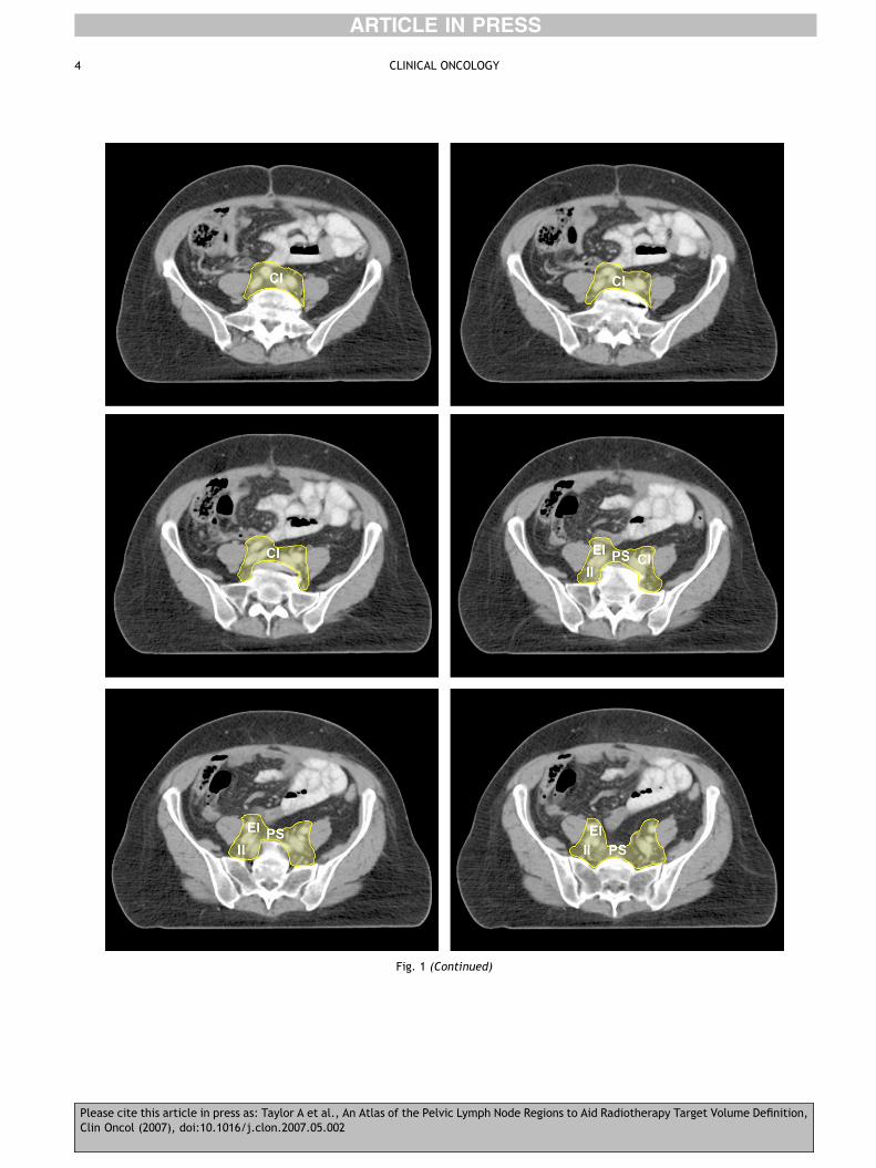
Fig. 1 e The typical clinical target volume (CTV) for adjuvant pelvic radiotherapy in gynaecological cancer contoured in yellow, andmodifications shown to include lateral external iliac nodes (blue), inguino-femoral nodes (green) and the parametria and upper vagina (red).The nodal regions indicated are para-aortic (PA), common iliac (CI), pre-sacral (PS), internal iliac (II), obturator (Obt), lateral (EIl), medial(EIm) and anterior (EIa) external iliac, parametrial and paravaginal (Pm), and inguino-femoral (Ing) lymph nodes.
Please cite this article in press as: Taylor A et al., An Atlas of the Pelvic Lymph Node Regions to Aid Radiotherapy Target Volume Definition,Clin Oncol (2007), doi:10.1016/j.clon.2007.05.002
4 CLINICAL ONCOLOGY
ARTICLE IN PRESS
Fig. 1 (Continued)
Please cite this article in press as: Taylor A et al., An Atlas of the Pelvic Lymph Node Regions to Aid Radiotherapy Target Volume Definition,Clin Oncol (2007), doi:10.1016/j.clon.2007.05.002

5PELVIC LYMPH NODE ATLAS
ARTICLE IN PRESS
Fig. 1 (Continued)
Please cite this article in press as: Taylor A et al., An Atlas of the Pelvic Lymph Node Regions to Aid Radiotherapy Target Volume Definition,Clin Oncol (2007), doi:10.1016/j.clon.2007.05.002

6 CLINICAL ONCOLOGY
ARTICLE IN PRESS
Fig. 1 (Continued)
Please cite this article in press as: Taylor A et al., An Atlas of the Pelvic Lymph Node Regions to Aid Radiotherapy Target Volume Definition,Clin Oncol (2007), doi:10.1016/j.clon.2007.05.002
7PELVIC LYMPH NODE ATLAS
ARTICLE IN PRESS
Fig. 1 (Continued)
Please cite this article in press as: Taylor A et al., An Atlas of the Pelvic Lymph Node Regions to Aid Radiotherapy Target Volume Definition,Clin Oncol (2007), doi:10.1016/j.clon.2007.05.002
8 CLINICAL ONCOLOGY
ARTICLE IN PRESS
Fig. 1 (Continued)
Please cite this article in press as: Taylor A et al., An Atlas of the Pelvic Lymph Node Regions to Aid Radiotherapy Target Volume Definition,Clin Oncol (2007), doi:10.1016/j.clon.2007.05.002

9PELVIC LYMPH NODE ATLAS
ARTICLE IN PRESS
References
1 Mundt AJ, Lujan AE, Rotmensch J, et al. Intensity-modulatedwhole pelvic radiotherapy in women with gynecologicmalignancies. Int J Radiat Oncol Biol Phys 2002;52:1330e1337.
2 Portelance L, Chao KS, Grigsby PW, et al. Intensity-modulatedradiation therapy (IMRT) reduces small bowel, rectum, andbladder doses in patients with cervical cancer receiving pelvicand para-aortic irradiation. Int J Radiat Oncol Biol Phys 2001;51:261e266.
3 Nutting CM, Convery DJ, Cosgrove VP, et al. Reduction ofsmall and large bowel irradiation using an optimizedintensity-modulated pelvic radiotherapy technique in patientswith prostate cancer. Int J Radiat Oncol Biol Phys 2000;48:649e656.
4 Scheidler J, Hricak H, Yu KK, et al. Radiological evaluation oflymph node metastases in patients with cervical cancer. Ameta-analysis. JAMA 1997;278:1096e1101.
5 Williams AD, Cousins C, Soutter WP, et al. Detection of pelviclymph node metastases in gynecologic malignancy: a comparisonof CT, MR imaging, and positron emission tomography. Am JRoentgenol 2001;177:343e348.
6 Bipat S, Glas AS, van der Velden J, et al. Computed tomographyand magnetic resonance imaging in staging of uterine cervicalcarcinoma: a systematic review. Gynecol Oncol 2003;91:59e66.
7 Mack MG, Balzer JO, Straub R, et al. Superparamagnetic ironoxide-enhanced MR imaging of head and neck lymph nodes.Radiology 2002;222:239e244.
8 Rockall AG, Sohaib SA, Harisinghani MG, et al. Diagnosticperformance of nanoparticle-enhanced magnetic resonanceimaging in the diagnosis of lymph node metastases in patientswith endometrial and cervical cancer. J Clin Oncol 2005;23:2813e2821.
Please cite this article in press as: Taylor A et al., An Atlas of the PelviClin Oncol (2007), doi:10.1016/j.clon.2007.05.002
9 Taylor A, Rockall AG, Reznek RH, et al. Mapping pelvic lymphnodes: guidelines for delineation in intensity-modulated radio-therapy. Int J Radiat Oncol Biol Phys 2005;63:1604e1612.
10 Vilarino-Varela MJ, Taylor A, Rockall AG, et al. Whole pelvisIMRT: verification of guidelines for lymph node delineation. ClinOncol 2005;17(Suppl. 1):3e4.
11 Hong JH, Tsai CS, Lai CH, et al. Recurrent squamous cellcarcinoma of cervix after definitive radiotherapy. Int J RadiatOncol Biol Phys 2004;60:249e257.
12 Benedetti-Panici P, Maneschi F, Scambia G, et al. Lymphaticspread of cervical cancer: an anatomical and pathological studybased on 225 radical hysterectomies with systematic pelvic andaortic lymphadenectomy. Gynecol Oncol 1996;62:19e24.
13 Sakuragi N, Satoh C, Takeda N, et al. Incidence and distributionpattern of pelvic and paraaortic lymph node metastasis inpatients with stages IB, IIA, and IIB cervical carcinoma treatedwith radical hysterectomy. Cancer 1999;85:1547e1554.
14 Mariani A, Webb MJ, Keeney GL, et al. Routes of lymphaticspread: a study of 112 consecutive patients with endometrialcancer. Gynecol Oncol 2001;81:100e104.
15 Matsumoto K, Yoshikawa H, Yasugi T, et al. Distinct lymphaticspread of endometrial carcinoma in comparison with cervicaland ovarian carcinomas. Cancer Lett 2002;180:83e89.
16 Pendlebury SC, Cahill S, Crandon AJ, et al. Role of bipedallymphangiogram in radiation treatment planning for cervixcancer. Int J Radiat Oncol Biol Phys 1993;27:959e962.
17 Bonin SR, Lanciano RM, Corn BW, et al. Bony landmarks are notan adequate substitute for lymphangiography in defining pelviclymph node location for the treatment of cervical cancer withradiotherapy. Int J Radiat Oncol Biol Phys 1996;34:167e172.
18 Greer BE, Koh WJ, Figge DC, et al. Gynecologic radiotherapyfields defined by intraoperative measurements. Gynecol Oncol1990;38:421e424.
c Lymph Node Regions to Aid Radiotherapy Target Volume Definition,