Embed Size (px)
Citation preview
RESEARCH Revista Mexicana de Fısica60 (2014) 222–226 MAY-JUNE 2014
An approximation of tribological behavior of Ti 1−xAlxN coatingsagainst animal bone in ringer’s solution
A. Esguerra-Arcea, N. A. Arteagaa, C. Amayab, L. Ipaza, N. Alba de Sanchezc, and Y. AguilaraaGrupo de investigacion en Tribologıa-Polımeros-Metalurgia de Polvos y Residuos Solidos,
TPMR, Universidad del Valle, Calle 13 No. 100-00, Cali, Colombia,e-mail: [email protected]
bGrupo de investigacion en Desarrollo de Materiales y Productos, GIDEMP,Centro ASTIN, SENA, Calle 52 No.2 Bis-15, Cali, Colombia,
cGrupo de investigacion Ciencia e Ingenierıa de Materiales, GCIM,Universidad Autonoma de Occidente, Calle 25 No.115-85, Cali, Colombia.
Received 22 November 2013; accepted 18 March 2014
Due to their excellent properties, Ti-Al-N coatings have become attractive for biomedical applications. In this paper, friction and wear prop-erties of Ti1−xAlxN films having various aluminum contents,x, have been studied. Adhesion was measured by the scratch test technique;friction was carried out by a pin-on-disk tribometer using an animal bone-pin as counterpart and Ringer’s solution as simulated body fluid;and wear mechanisms were identified by SEM and EDS. It was found that the coating withx = 0.41 exhibited the highest COF, conserves itsintegrity as a coating, and causes the lowest wear on the bone in Ringer’s solution.
Keywords:Sliding wear; PVD coatings; corrosion wear; biological aspects
PACS: 81.15.-z; 81.40.PQ
1. Introduction
Several kinds of alloys are available for biomedical ap-plications based on titanium, nickel, aluminum, vanadium,chromium, zirconium, etc., approved by ASTM. However,some disadvantages are encountered with these alloys, be-cause they mechanically destroy the normal bone with whichthey come in contact, causing allergy and inflammation dueto abrasive particles and leached toxic ions [1]. Taking intoaccount that the surface properties govern the material per-formance in this kind of applications, the modification of thesurface of biomedical alloys to improve tribological proper-ties could be considered as a solution. Ti-Al-N coatings seemto be an excellent material for this field, due to its excellentin vivo [2] andin vitro [3] biocompatibility, desirable electro-chemical properties in simulated body fluids [2,4] and goodmechanical behavior [5].
Considering that the application of this coating onto thesurface of implants fixtures needs further investigation, thepurpose of this work is to study the effect of atomic alu-minum content,x, in Ti1−xAlxN coatings deposited by re-active magnetron co-sputtering, in the tribological properties,such as friction coefficient and wear mechanisms, when sub-mitted to conditions close to those found in the human body,using Ringer’s solution and animal bone as counterpart. Thisresearch seeks to study the viability of using these coatingsin orthopedic applications, by relating the tribological perfor-mance with mechanical properties and adhesion of the coat-ings.
2. Materials and Methods
Ti1−xAlxN films were deposited on Si (100) and AISI 304sssubstrates by reactive magnetron co-sputtering at 250◦C with
high purity titanium and aluminum targets. The substrateswere ultrasonically cleaned in isopropyl alcohol and acetonesequentially and then dried in air jet before being placedinto the vacuum chamber. The base pressure in the cham-ber was 2.1×10⁻⁴ mBar and the working pressure was set at2.0×10−2 mBar during deposition. A titanium buffer-layerwas deposited on the substrate in presence of Ar. The powerapplied to the Ti target was 400 W. Nitrogen gas was injectedafterwards (Ar flow: 50 sccm; N2 flow: 3.7 sccm) into thedeposition chamber and the power applied to the Al target(10.16 cm in diameter) was varied in 200, 250 and 350 Wfor reactive deposition of TiAlN. Due to the variation in alu-minum sputter yielding, the deposition time varied in 2.0, 1.5and 1.2 hours, respectively. The substrates were rotated ata speed of 21 rev/min in order to obtain homogeneous filmcomposition and the r. f. bias voltage at the substrate was putat -20 V.
EDS analysis was performed to determine the contents ofTi, Al and N. An X-ray diffraction (XRD) study was carriedout using an X’pert HighScore Plus diffractometer with Cu-Kα radiation (α = 1.5406A) at grazing angle of 0.5◦. Themorphological characterization of the coatings (grain size)was obtained using an atomic force microscope (AFM) fromAsylum Research MFP-3DR©using a cantilever silicon tip innon-contact mode and calculated by a Scanning Probe ImageProcessor (SPIPR©) which has the standard program for pro-cessing and presenting AFM data. Thickness was evaluatedby SEM (JEOL JSM-649 OLV).
A cortical bone specimen was prepared from a pighumerus obtained from a local market. The bone was im-mersed in water at 100◦C for 2 h to remove soft tissue at-tached to the bone. In order to prevent changes in the bone’smechanical properties, the humerus was never allowed totouch the heat source while being slowly heated. After this

AN APPROXIMATION OF TRIBOLOGICAL BEHAVIOR OF Ti1−xAlxN COATINGS AGAINST ANIMAL. . . 223
process, the remnant soft tissue attached to the bone was me-chanically removed by hand and then the bone was cleaned,dried in ambient air and mechanized to spherical form.
Adherence was studied by using a Scratch Test MicrotestMTR2 system, with a 6 mm scratch length and a raising loadof 0–90 N. To identify the different adherence failures anoptical microscope was used. To identify the different ad-herence failures, the adhesion properties of coatings can beanalyzed by the following two terms, Lc1, the lower criticalload defined as the load where the first cracks occurred (co-hesive failure) and the Lc2, detachment and separation of acoating from the substrate with cracking and de-bonding atthe coating-substrate interface (adhesion failure) [6].
Each sample was indented 24 times using a Berkovich
pyramidal indenter, varying the maximum load applied from479.6 to 5995.8µN (10 seconds of loading and 10 seconds ofunloading). The elastic properties were calculated for eachindent and the reduced modulus was calculated by the Pharr–Oliver relationship [7].
To evaluate the tribological properties of the Ti1−xAlxNcoatings, sliding wear tests were carried out by using a Mi-croTest pin-on-disc tribometer (two replicates). The testswere performed at a normal load of 10 N using an animal-bone ball with 6 mm in diameter, as the wear counterpart,and Ringer’s solution as simulated body fluid. The slidinglinear speed and total sliding distance were set at 8 mm/s and400 m, respectively. The wear tracks were studied by EDSand SEM (JEOL JSM-649 OLV).
TABLE I. Characteristics of the samples.
Sample Hardness (GPa) Young’s Modulus (GPa) Roughness (nm) Grain diameter (nm) Thickness (nm)
304ss 5.3± 0.7 234± 12 37.5± 2
x = 0.24 28.5± 0.3 260± 13 92.5± 5 85± 4 566
x= 0.41 29.6± 0.4 304± 15 35.0± 2 53± 3 557
x= 0.60 22.6± 0.5 278± 13 71.3± 4 80± 4 883
Bone 6.0± 1.0 18.8± 2
FIGURE 1. EDS spectra of the coatings.
Rev. Mex. Fis.60 (2014) 222–226
224 A. ESGUERRA-ARCE, N. A. ARTEAGA, C. AMAYA, L. IPAZ, N. ALBA DE SANCHEZ, AND Y. AGUILAR
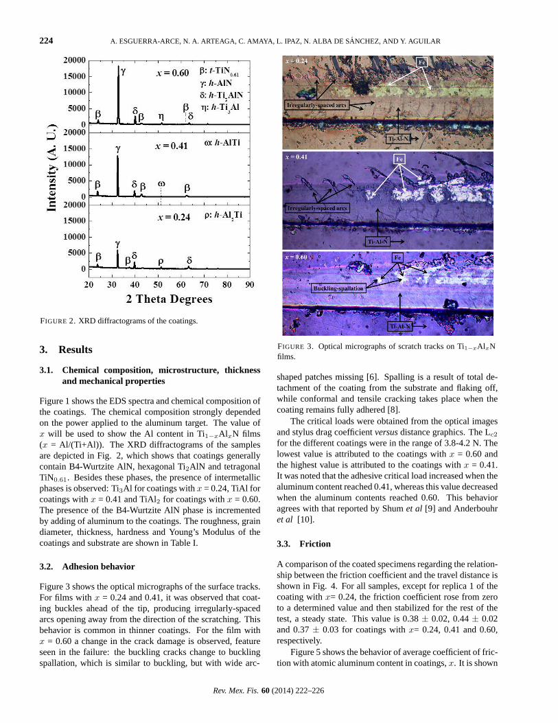
FIGURE 2. XRD diffractograms of the coatings.
3. Results
3.1. Chemical composition, microstructure, thicknessand mechanical properties
Figure 1 shows the EDS spectra and chemical composition ofthe coatings. The chemical composition strongly dependedon the power applied to the aluminum target. The value ofx will be used to show the Al content in Ti1−xAlxN films(x = Al/(Ti+Al)). The XRD diffractograms of the samplesare depicted in Fig. 2, which shows that coatings generallycontain B4-Wurtzite AlN, hexagonal Ti2AlN and tetragonalTiN0.61. Besides these phases, the presence of intermetallicphases is observed: Ti3Al for coatings withx = 0.24, TiAl forcoatings withx = 0.41 and TiAl2 for coatings withx = 0.60.The presence of the B4-Wurtzite AlN phase is incrementedby adding of aluminum to the coatings. The roughness, graindiameter, thickness, hardness and Young’s Modulus of thecoatings and substrate are shown in Table I.
3.2. Adhesion behavior
Figure 3 shows the optical micrographs of the surface tracks.For films withx = 0.24 and 0.41, it was observed that coat-ing buckles ahead of the tip, producing irregularly-spacedarcs opening away from the direction of the scratching. Thisbehavior is common in thinner coatings. For the film withx = 0.60 a change in the crack damage is observed, featureseen in the failure: the buckling cracks change to bucklingspallation, which is similar to buckling, but with wide arc-
FIGURE 3. Optical micrographs of scratch tracks on Ti1−xAlxNfilms.
shaped patches missing [6]. Spalling is a result of total de-tachment of the coating from the substrate and flaking off,while conformal and tensile cracking takes place when thecoating remains fully adhered [8].
The critical loads were obtained from the optical imagesand stylus drag coefficientversusdistance graphics. The Lc2
for the different coatings were in the range of 3.8-4.2 N. Thelowest value is attributed to the coatings withx = 0.60 andthe highest value is attributed to the coatings withx = 0.41.It was noted that the adhesive critical load increased when thealuminum content reached 0.41, whereas this value decreasedwhen the aluminum contents reached 0.60. This behavioragrees with that reported by Shumet al [9] and Anderbouhret al [10].
3.3. Friction
A comparison of the coated specimens regarding the relation-ship between the friction coefficient and the travel distance isshown in Fig. 4. For all samples, except for replica 1 of thecoating withx= 0.24, the friction coefficient rose from zeroto a determined value and then stabilized for the rest of thetest, a steady state. This value is 0.38± 0.02, 0.44± 0.02and 0.37± 0.03 for coatings withx= 0.24, 0.41 and 0.60,respectively.
Figure 5 shows the behavior of average coefficient of fric-tion with atomic aluminum content in coatings,x. It is shown
Rev. Mex. Fis.60 (2014) 222–226

AN APPROXIMATION OF TRIBOLOGICAL BEHAVIOR OF Ti1−xAlxN COATINGS AGAINST ANIMAL. . . 225
FIGURE 4. Comparison of the coated specimens regarding the re-lationship between the friction coefficient (COF) and the travel dis-tance.
FIGURE 5. Comparison of COF and coating hardness values foreach sample.
that the coating withx = 0.41 exhibits the highest frictioncoefficient, likely related to the highest hardness value exhib-ited by this coating, because a correlation between frictioncoefficient values and the hardness of the discs can be seen:films with higher hardness values exhibit higher friction withthe bone in Ringer’s solution.
Considering that the friction coefficient is an importantvalue in biomaterials design, given that the friction coefficientaffects the relative micromotion in the biomaterial-bone inter-face (for example, increasing the friction coefficient causes
FIGURE 6. Wear mechanism of the coating withx = 0.24.
FIGURE 7. Wear mechanism of the coating withx = 0.41.
the peak micromotion between cup-pelvis pair to be reduced[11]), the coating withx = 0.41 would be the more appropri-ate for this application.
3.4. Wear
SEM inspection of the scars generated using the bone coun-terpart was made to verify the actual predominant wear mode.For the coatings withx= 0.24 (Fig. 6) and 0.41 (Fig. 7) theadhesive wear mechanism is observed. EDS analysis, whichshowed the presence of phosphorus, calcium and oxygen inthis adhered film, confirmed that this is a bone layer adheredto the coatings surface. Also it was observed, by the EDSanalysis, that coatings conserve their integrity and do not suf-fer detachment, because it were not observed the presenceof iron or chromium atoms at the surface, but titanium, alu-minum and nitrogen were observed, indicating the steel wascovered and protected by the Ti-Al-N coating.
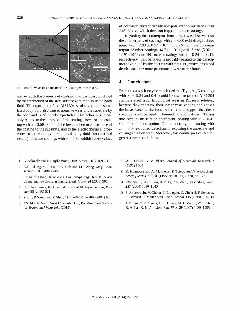
On the contrary, coatings withx = 0.60 (Fig. 8) exhibiteddetachment, as can be seen in SEM micrographs. The latteris evidenced in EDS analysis of the surface, given that thesurface exhibit the presence of iron and chromium, principalcomponents of the stainless steel. Besides this, the surface
Rev. Mex. Fis.60 (2014) 222–226
226 A. ESGUERRA-ARCE, N. A. ARTEAGA, C. AMAYA, L. IPAZ, N. ALBA DE SANCHEZ, AND Y. AGUILAR
FIGURE 8. Wear mechanism of the coating withx = 0.60.
also exhibits the presence of oxidized iron particles, producedby the interaction of the steel surface with the simulated bodyfluid. The exposition of the AISI 304ss substrate to the simu-lated body fluid also caused abrasive wear of the substrate bythe bone and Ti-Al-N debris particles. This behavior is prob-ably related to the adhesion of the coatings, because the coat-ing with x = 0.60 exhibited the lower adherence resistance ofthe coating to the substrate, and to the electrochemical prop-erties of the coatings in simulated body fluid (unpublishedresults), because coatings withx = 0.60 exhibit lower values
of corrosion current density and polarization resistance thanAISI 304 ss, which does not happen in other coatings.
Regarding the counterpart, bone pins, it was observed thatthe counterparts of coatings withx = 0.60 exhibit eight timesmore wear, (3.99± 0.57)×10−3 mm3/N×m, than the coun-terpart of other coatings, (4.71± 0.11)×10−4 and (5.02±1.19)×10−4 mm3/N×m, via coatings withx = 0.24 and 0.41,respectively. This behavior is probably related to the detach-ment exhibited by the coating withx = 0.60, which produceddebris cause the more pronounced wear of the bone.
4. Conclusions
From this study it may be concluded that Ti1−xAlxN coatingswith x = 0.24 and 0.41 could be used to protect AISI 304stainless steel from tribological wear in Ringer’s solution,because they conserve their integrity as coating and causesthe lesser wear to the bone, which could suggest that thesecoatings could be used in biomedical applications. Takinginto account the friction coefficient, coating withx = 0.41should be the best option. On the contrary, the coating withx = 0.60 exhibited detachment, exposing the substrate andcausing abrasion wear. Moreover, this counterpart causes thegreatest wear on the bone.
1. G. Schmalz and P. Garphammer,Dent. Mater. 18 (2002) 396
2. K.H. Chung, G.T. Liu, J.G. Duh and J.H. Wang,Surf. Coat.Technol.188(2004) 745
3. Chia-Chi Chien, Kuan-Ting Liu, Jenq-Gong Duh, Kuo-WeiChang and Kwok-Hung Chung,Dent. Mater.24 (2008) 986
4. B. Subramanian, R. Ananthakumar and M. Jayachandran,Vac-uum85 (2010) 601
5. Z. Liu, P. Shum and Y. Shen,Thin Solid Films468(2004) 161
6. ASTM C1624-05, West Conshohocken, PA,American Societyfor Testing and Materials, (2010)
7. W.C. Oliver, G. M. Pharr,Journal of Materials Research7(1992) 1564
8. K. Holmberg and A. Matthews,Tribology and Interface Engi-neering Series, 2nd ed. (Elsevier, Vol. 56, 2009), pp. 128.
9. P.W. Shum, W.C. Tam, K.Y. Li, Z.F. Zhou, Y.G. Shen,Wear.257(2004) 1030–1040
10. S. Anderbouhr, V. Ghetta, E. Blanquet, C. Chabrol, F. Schuster,C. Bernard, R. Madar,Surf. Coat. Technol.115(1999) 103–110
11. J. T. Hsu, C. H. Chang, H. L. Huang, M. E. Zobitz, W. P. Chen,K. A. Lai, K. N. An, Med. Eng. Phys.29 (2007) 1089–1095
Rev. Mex. Fis.60 (2014) 222–226


![3. Tribological Behavior of Thermal Spray Coatings ... · material to the substrate [4‐6]. The NiCr 80/20 alloy is also used as metal matrix to produce composite coatings reinforced](https://img.dokumen.tips/doc/110x75/607da147439d931e17263d25/3-tribological-behavior-of-thermal-spray-coatings-material-to-the-substrate.jpg)