Embed Size (px)
Citation preview

E. Mark Haacke, PhD, [email protected], Magnetic Resonance Innovations, Inc.Director, MR Research Facility, Wayne State UniversityDetroit, Michigan
An Academic Life in Imaging: Teaching, Insight and Research

YOUR MISSION, IF YOU SHOULD DECIDE TO ACCEPT IT,

IS TO BOLDLY GO WHERE NO MAN HAS GONE BEFORE

Magnetic Resonance PIONEERS
By 1946 Bloch and Purcell and co-workers captured thethe behavior of an atom with a non-zero magneticmoment (like the proton, for example) situated in amagnetic field. (Nobel Prize in Physics in 1952.)Their contributions to the field from these first papers wasprecocious and even 70 years later their work carriesmodern significance.Although quantum physics was needed to formulate theexperimental and theoretical results, the final impactrests on the very simple relationship given by the Larmorequation relating the frequency of rotation or precessionfrequency () and the local magnetic field (B) via B.

Visualizing Quantum Mechanics
Thin slice T1 and T2 weighted 3D images of the brain.Nobel prize in Medicine or Physiology 2003.
Lauterbur
Mansfield

An Academic Life in Imaging: Teaching, Insight and Research
E. MARK HAACKE, PHD
PRESIDENT, MAGNETIC RESONANCE INNOVATIONS, INC.
DIRECTOR, MR RESEARCH FACILITY, WAYNE STATE UNIVERSITY
DETROIT, MICHIGAN 48202
HOW LUCKY TO ENJOY ONE’SJOB AS PART OF EVERYDAY LIFE.
1

Education
• Take good students under your wing and help guide and nurture precocious students at an early age.
• Prof. John Moffat was my advisor and mentor.
• Summertime provided me the opportunity to teach (statistics/math) and earn some money.
2

Education
• Clear lectures, get the main points across• Use examples that demonstrate the key principles• Promote students to ask questions• Get students to work together to attack problems• Sit with groups of 5 or 6 and review in detail a problem• Get the students to present at the blackboard• Once a principle is learned it will never be forgotten
3

Education and early collaborations• Have more senior students help tutor new students• Have regular informal lectures at the blackboard• Have monthly meetings giving an overview of research• Create a team environment early; have weekly journal club meetings• Engage them outside work on
occasion with sports or outings
4
Have office hours, be accessible (Ramesh)

Closing thoughts on education
• “It’s easy to show that …”
• Create a text that carries the student step by step through the basic to advanced concepts without once using that phrase!
• The result of this 28 man-year effort was “The Green Bible of MRI”.
5

Collaborations and team science• Offers new scientific and cultural experiences • Increases your group’s knowledge• Provides more data and much faster!
6
East China Normal University, Shanghai, China with the physics group of Prof. Yang Guang.
NeusoftMR laboratory, Shenyang, China

When it comes to research:be determined
Research does not always come with the infamous “AHA” but often follows the adage: 10% inspiration and 90% perspiration
This applies to:abstracts (rf)papers (MRA)grants (mannitol)
7

SWI
Haacke EM et al.Susceptibility weighted imaging. MRM, 52: 612; 2004.
Susceptibility Weighted Imaging
Enhances the presence of ferritin, hemosiderin and deoxyhemoglobin.
Exquisite images from which brain damage, microbleeding and increases in deoxyhemoglobin can be diagnosed.
One President mused: “Beautiful but what’s it good for?”
8
Wizard of Id

Scientific Creativity• Ideas take time to gestate and mature and become useful like SWI
• Everyone has a different perspective.
• Think out of the box (Zhipei).
• Advisors have to be open andflexible to new ideas.
• Reviewers need to have the insight as tonot only the fact that these new ideashave potential but that a group’s historyis key to their future success and not toget stuck on minutia.
ReviewerAdvisor
Student
9

Eye anatomy / Our MRI Image
Human Retina, Jan 27 2016, Courtesy of Yongsheng Chen
10

7T SWI215µ x 215µ x 1000µTE = 16ms; TR = 45msFA = 25°; 8 slice mIP
15 minute scan
Image courtesy of Yulin Ge, NYU
3T SWI200µ x 400µ x 1000µTE = 15ms; TR = 30msFA = 12°; 4 slice mIP
9 minute scan
Imagine brain functional imaging (fMRI) at this resolution!
11

The Dream of MR angiography
Small arteries around 250μ are now visible. Ahhh, at last, high resolution MRA!
Or is it? Close but no cigar! It takes far too long to be useful clinically and still fails to show 50μ vessels.
SWI is so beautiful, showing veins down to 50μ to 100μ.
How can we make arteries look like veins to get the same quality MRA as we do with SWI?
AHA!! Modify the susceptibility of the arteries!!
250μ x 250μ x 500μ
12

MICRO“Microvascular In-vivo Contrast Revealed Origins”
MICRO imaging: 50μx200μ in 15 minUsing the iron based USPIO agent called Ferumoxytol.
13

Comparison of MRAV using SWI and cadaver brain dye injection.
a) Georges Salamon’s image of arteries in the human brain. b,c,d) Data from TE = 8mswith 0.1mm x 0.2mm x 1.25mm at 7T. b) MRA pre-contrast, c) mIP post-contrast SWI for4mg ferumoxytol and d) mIP pre-contrast. Note that c) is a mixture of arteries andveins. However, the thalamic arteries are clearly highlighted in the post-contrast image.Image d) can be used as a guide as to where the veins are since it is a pre-contrastSWI.
b
c d
14

Ferumoxytol enhanced MRAV
Image courtesy of Yulin Ge and NYU.
A
15

Future Directions• Imaging materials
• Creating an image for something that itself has no signal, possible or impossible?
• Distortion free fMRI• The perfect PhD demonstrating that we can create
an image with no signal that has value!!!• Until that is you begin thinking, when the light bulb
comes on and the function of the brain comes to life. • Micro magnets
• Creating an imaging system with unheard of gradient strengths and possibly enhancing signal in cells by a factor of 1000 or more.
16

The Anatomy Lesson of Dr. NicolaesTulp: Rembrandt 1632
Until very recently we had to cut the body open from the outside, now we have inverted the problem and we can view the body from the inside out!
17

Strategically Acquired Gradient Echo (STAGE) Imaging
Chen, Yongsheng PhD student WSU, Detroit, USA
Wang, Yu MS student ECNU, Shanghai, China
18

Strategically Acquired Gradient Echo (STAGE) Imaging: Complete 3D coverage of the brain in 5 min.
19

T1 Weighted Images
A: T1 MP RAGE, the reference T1W;B: T1W from 1st echo of B2; C: STAGE Enhanced T1W from B1 and B2.
A B C
Richard Ernst, Nobel Prize in
Chemistry 1991
20

Presenting the first visualization of the fetal venous anatomy of the brain using SWI
Pilot scan on the left, effective transverse SWI on the right: 37 weeks 1 day
21

GA 37 weeksFetus
Placenta
22

Non contrast enhanced MR angiography images of the heart and the major vessels in human fetus.
scale~ 4 mm
23

Venous Thrombosis: before treatment and after thrombolysis
Guangbin Wang M.D. Shandong Medical Imaging Research Institute
T1 T2
PRE POST
24

Sturge Weber Syndrome
3D-T1 Post Gd 3D T1 SWI (no contrast agent)
Czabo Juhasz, Yang Xuan and Dr. E. Haacke, Wayne State University
25

Motorcycle trauma: medullary vein involvement
SWIT1 PCn
26

SWI minIP image projected over 16mm
The First Clinical Applications of SWIM in Traumatic Brain Injury (TBI)
Corresponding MaxIPsusceptibility map image
projected over 16mm
Cerebral Microbleeds: Pathophysiology to Clinical Practice (Cambridge Medicine) Editor, David J. Werring, 2011, ISBN-13: 9780521198455
27

Normal volunteer
Iterative SWIM single slice
Iterative SWIM single slice
SWIM single slice with boundaries
SWIM single slice with boundaries
MIP Iterative SWIM over 4 slices
MIP Iterative SWIM over 4 slices
28

Substantia Nigra (SN) Region II Iron content in PD and Controls from SWIM
The plots show region II (high iron region) content plotted as a function of age for 15 PD (red) and 174 controls1
(green) for right (upper) and left (lower) SN.
The regression line for the PD group shows a higher slope which may indicate an increased rate of deposition from the onset.
1. Liu M, Liu S, Ghassaban K, Zheng W, Dicicco D, Miao Y, et al. Assessing global and regional iron content in deep gray matter as a function of age using susceptibility mapping. J Magn Reson Imaging. 2016 Jul;44(1):59-71.
Right substantia nigra
Left substantia nigra
29

Tumor draining/feeding vessels
T1 subtraction maximum intensity projection image
Flash 3D T1weighted pre-Gadolinium series
Flash 3D T1 weighted post-Gadolinium series
The 3D sequence allows better coverage of the tumor volume and pre-post contrast imaging allows exact subtraction revealing enhanced regions and in some cases even feeding/draining vessels.
Contrast Enhanced Tumor Imaging3D T1 weighted
30

two cups of coffee and you will have a major change of blood flow to the brain
maybe we should approach Starbucks for funding
at least it is a relatively harmless contrast agent to use to study the brain and a heck of a lot cheaper
31Using caffeine decreases blood flow to the brain

MinIP of caffeine/Gd over 28 slices with 4 phase multiplications
SWI PRE CAFFEINE
SWI POST CAFFEINE
SWI as a high resolution BOLD imaging method32
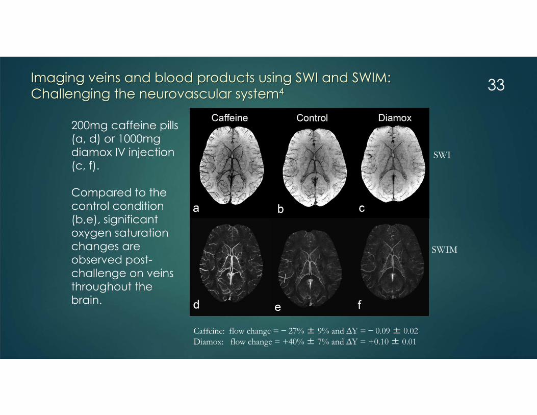
200mg caffeine pills (a, d) or 1000mg diamox IV injection (c, f).
Compared to the control condition (b,e), significant oxygen saturation changes are observed post-challenge on veins throughout the brain.
SWI
SWIM
Imaging veins and blood products using SWI and SWIM:Challenging the neurovascular system4
Caffeine: flow change = − 27% ± 9% and ΔY = − 0.09 ± 0.02 Diamox: flow change = +40% ± 7% and ΔY = +0.10 ± 0.01
33

MRI scan date: 2013.01.04
MRI scan date: 2013.01.11
Two scans from same stroke patientMTT
SWI
SWIMTT
34

Cerebral amyloid angiopathy
50µ objects can manifest as 1mm3 objects
35

Time to go sailing
Black dots count
8
16
29
45
05
101520253035404550
1 - 4/29/03 2 - 5/27/04 3 - 6/9/05 4 - 3/2/06
Scan no
No
of c
ount
s
36

Informational Websites
Clinical applications of SWI and SWIM See www.swim-mri.com The role of abnormal venous flow in neurodegenerative
diseases: MS as an example See www.ms-mri.com Our work in Detroit at Wayne State University See www.mrc.wayne.edu Business website See www.mrinnovations.com
37

CommunicationCommunication oral: Talking, yelling, fire/light, land line phones, cell phones, skype on computers
Data storage: Stone, sheepskin, paper, floppy disks, CDs, DVDs, thumb drives, cloud storage
Communication written: Hand written, typed letters, books, fax, optic cables for file transfer, computer word files, internet transfer
Math and Science: Mental calculations, abacus, calculator, computer
The future: All of the above integrated into a single entity = BCI
38

Brain Computer Interfacing (BCI) and functional MRI (fMRI)
39

Neuroscience advances with imaging The goal is to understand how the brain works using cognitive
neuroscience. Imaging plays a major role. In fact it has been said that “Imaging is one of the most important inventions in the last 1000 years!”
40

Studying the brain with fMRI brain connectivity and DTI fiber tracking
41

A Revolution in Evolution
Where are we going? Computers are faster than us and can store more data than us. But they lack our creativity and art and still need our guidance. But what if our minds are already quantum computers? What if we use BCI to grow our connections and our brain evolves
a special ability to grow related to our connection and learning via the link to the computer?
And what if we now link all 6 billion people with all the computer power in the world?
42

And imaging will have helped us get there.
We will in fact create a new being “Mother earth” where we as individuals will be like the cells in our body and the integrated whole will be our new super-being “Mother earth”.
43