Embed Size (px)
Citation preview

Amino Acids, Peptides, and
Proteins ( Chapter 3)
Dr. Rula Abdul-Ghani
Dr.Rula
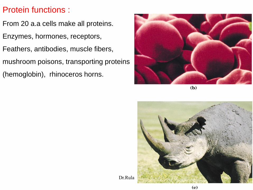
Protein functions :
From 20 a.a cells make all proteins.
Enzymes, hormones, receptors,
Feathers, antibodies, muscle fibers,
mushroom poisons, transporting proteins
(hemoglobin), rhinoceros horns.
Dr.Rula

First a.a. asparagine 1806, Last threonine 1938.
Names derived from source of isolation:
Asparagine = Asparagus
Tyrosine = cheese ‘tyros’ in greek
Glycine = ‘glycos’ in greek = sweet taste.
General structure of a.a
(except proline):
Differ in R group affect
structure, size, electric
charge, solubility.
α- carbon
Dr.Rula

Two conventions used to identify the carbons in amino acids:
[---------------------R group-------------------]
Dr.Rula
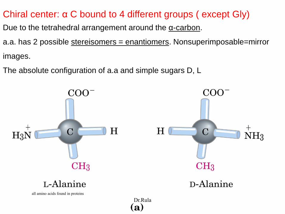
Chiral center: α C bound to 4 different groups ( except Gly)
Due to the tetrahedral arrangement around the α-carbon.
a.a. has 2 possible stereisomers = enantiomers. Nonsuperimposable=mirror
images.
The absolute configuration of a.a and simple sugars D, L
all amino acids found in proteins
Dr.Rula
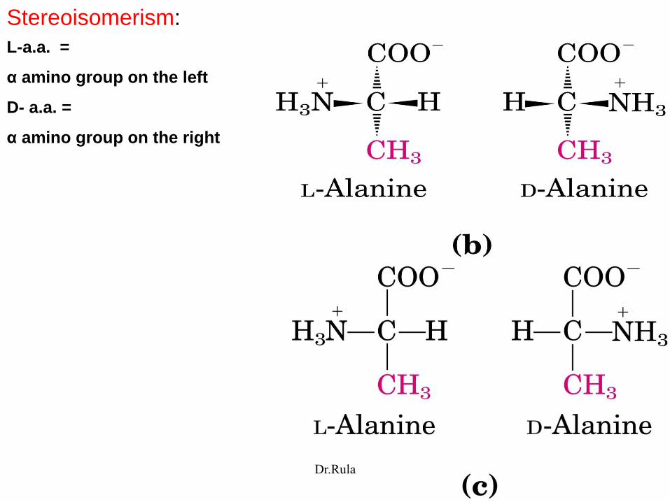
Stereoisomerism:
L-a.a. =
α amino group on the left
D- a.a. =
α amino group on the right
Dr.Rula

This configuration based on the reference molecule 3C sugar glyceraldehyde
configuration.
Carbons lined up vertically
with chiral atom in center
Terminal aldehyde/
carboxyl as number 1
The R group below the
chiral carbon.
Dr.Rula
- All a.a. in proteins are exclusively L stereoisomers.
D- stereoisomers only in small peptides (peptide antibiotics,
peptides of bacterial cell wall.
-Cells synthesize the L- isomer of a.a because the active sites of
enzymes are asymmetric the rxns they catalyze are
sterospecific.
Dr.Rula
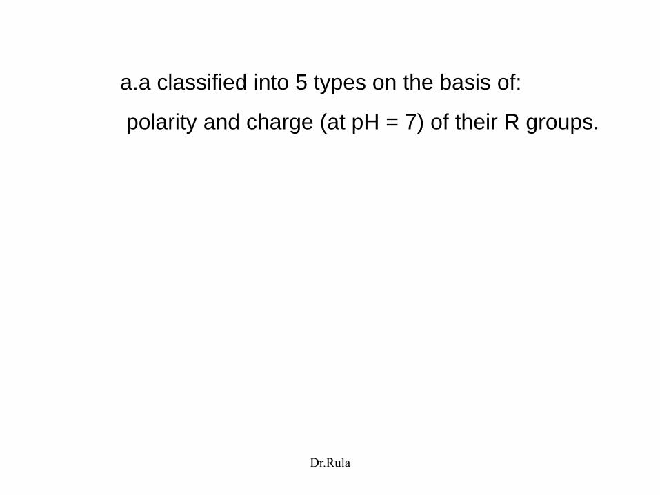
a.a classified into 5 types on the basis of:
polarity and charge (at pH = 7) of their R groups.
Dr.Rula

/ symbol
Dr.Rula
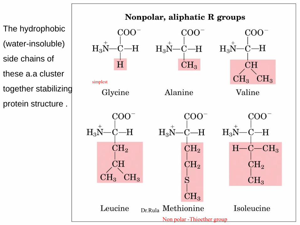
The hydrophobic
(water-insoluble)
side chains of
these a.a cluster
together stabilizing
protein structure .
simplest
Non polar -Thioether group
Dr.Rula
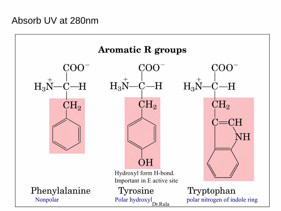
Absorb UV at 280nm
Hydroxyl form H-bond.
Important in E active site
Nonpolar Polar hydroxyl polar nitrogen of indole ringDr.Rula

Absorption of UV light by
aromatic a.a :
Maximum absorbance at 280nm
Tryptophan 4 times>> tyrosine
Dr.Rula
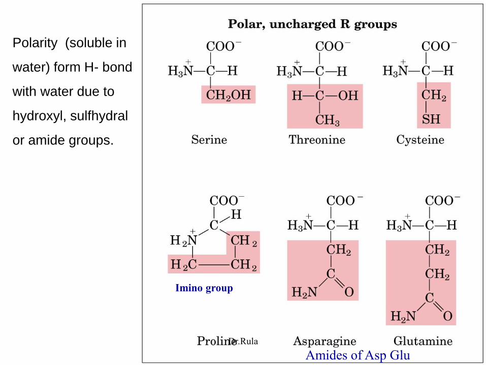
Polarity (soluble in
water) form H- bond
with water due to
hydroxyl, sulfhydral
or amide groups.
Imino group
Amides of Asp GluDr.Rula

Cysteine oxidized to form a covalently linked dimeric a.a.
Cystine from a disulfide bond joining 2 Cysteine molecules.
Strongly
hydrophobic
nonpolar
Dr.Rula
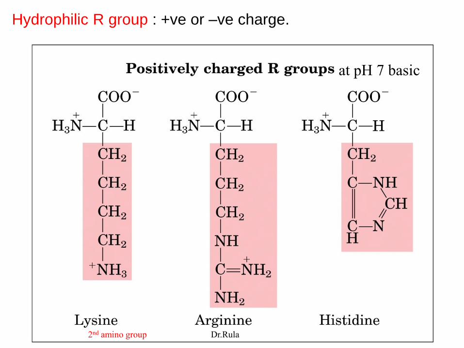
Hydrophilic R group : +ve or –ve charge.
2nd amino group
at pH 7 basic
Dr.Rula

Dr.Rula
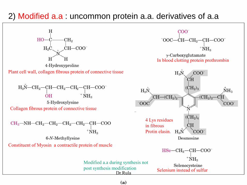
2) Modified a.a : uncommon protein a.a. derivatives of a.a
Plant cell wall, collagen fibrous protein of connective tissue
Collagen fibrous protein of connective tissue
Constituent of Myosin a contractile protein of muscle
4 Lys residues
in fibrous
Protin elasin.
In blood clotting protein prothrombin
Selenium instead of sulfur
Modified a.a during synthesis not
post synthesis modificationDr.Rula

3) Non-protein a.a :
Around 300 additional a.a
Important metabolites for pathways such as urea cycle .
Dr.Rula
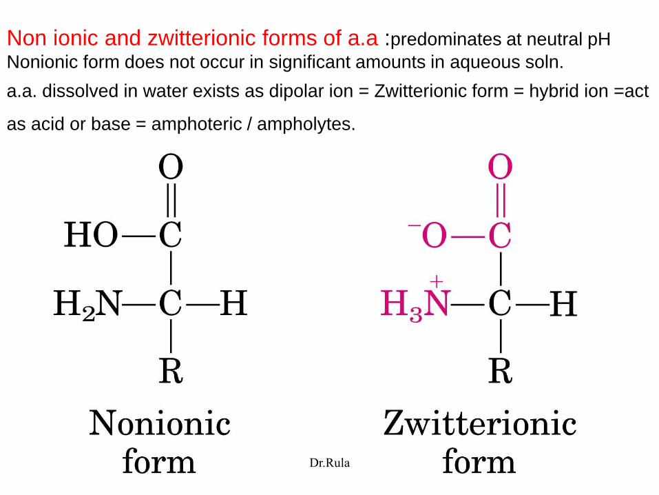
Non ionic and zwitterionic forms of a.a :predominates at neutral pH
Nonionic form does not occur in significant amounts in aqueous soln.
a.a. dissolved in water exists as dipolar ion = Zwitterionic form = hybrid ion =act
as acid or base = amphoteric / ampholytes.
Dr.Rula

Absorption of light by molecules:
Lambert Beer law: Spectrophotometer:
The fraction of incident light absorbed by a soln at a given wavelength is related
to the thickness of the absorbing layer and conc. of absorbing species.
Absorbance = A = Log Io / I = εcl
Io =intensity of incident light, I =intensity of transmitted light, ε =molar extinction
coefficient , c =conc of absorbing species, l =path length
Single wavelength
Dr.Rula
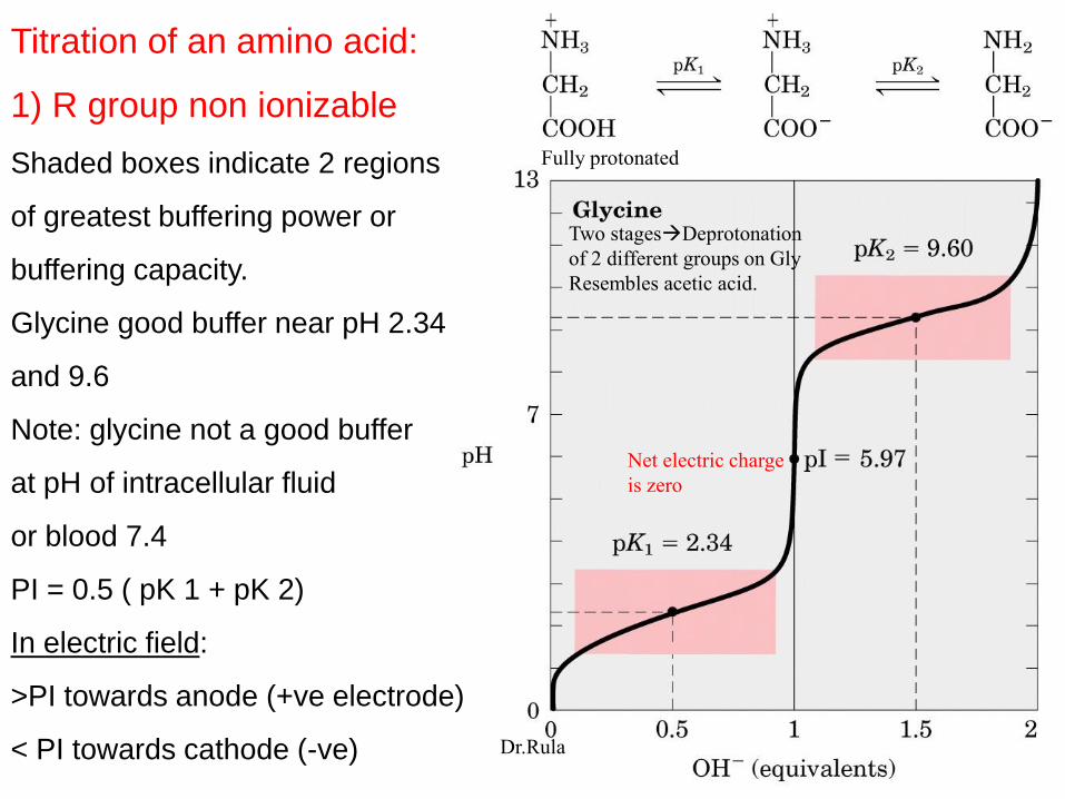
Titration of an amino acid:
1) R group non ionizable
Shaded boxes indicate 2 regions
of greatest buffering power or
buffering capacity.
Glycine good buffer near pH 2.34
and 9.6
Note: glycine not a good buffer
at pH of intracellular fluid
or blood 7.4
PI = 0.5 ( pK 1 + pK 2)
In electric field:
>PI towards anode (+ve electrode)
< PI towards cathode (-ve)
Net electric charge
is zero
Fully protonated
Two stagesDeprotonation
of 2 different groups on Gly
Resembles acetic acid.
Dr.Rula
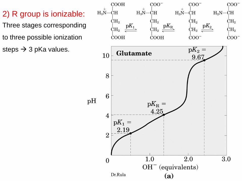
2) R group is ionizable:
Three stages corresponding
to three possible ionization
steps 3 pKa values.
Dr.Rula

Peptides and proteins: Polymers of a.a
Range in size from 2 / 3 to thousounds of a.a
a.a. covalently joined
by an amide linkage
= peptide bond
Dehydration=
removal of water =
condensation rxn.
Dipeptide , tripeptide,
tetrapeptide,pentapeptide
oligopeptide, polypeptide.
M wt <10000 =polypeptide
> protein-
α-aminoα-carboxyl
Dr.Rula

Pentapeptide:
a.a. unit in a peptide = residue
Ser-Gly-Tyr-Ala-Leu or serylglycyltyrosylalanylleucine
By convention left right .
N-terminal
Peptide bond
C-terminal
R group
Dr.Rula

Tetrapeptide:
Alanylglutamylglycyllysine
Peptides have only one
free amino termial and one
free carboxyl group.
Acid - base behavior of a
peptide depend on the R
groups and free amino free
carboxyl ends (not
nonterminal ends).
Two ionizable R groups
Dr.Rula

Many small peptides exert their effect at low conc.
1) Hormones:
Oxytocin (9 a.a) uterine contraction , milk secretion.
Bradykinin (9 a.a) inhibits tissue inflamation
Some Hormones are oligopeptides:
insulin two chains 30+21 a.a,
Glucagon 29 a.a
2) Toxins : mushroom poisons.
3) Antibiotics.
Dr.Rula

Length and number of polypeptide chains vary from one protein to another.
Single polypeptide chain or multisubunit protein (2/more polypeptides identical/different
associated non covalently).
If at least 2 identical, protein called oligomeric, the identical unit called protomer.
Hemoglobin 4 polypeptide subunits 2α 2β = tetramer/ dimer of αβ protemer.
Insulin 2 polypeptide chains (disulfide bond) not subunits.
*
Dr.Rula
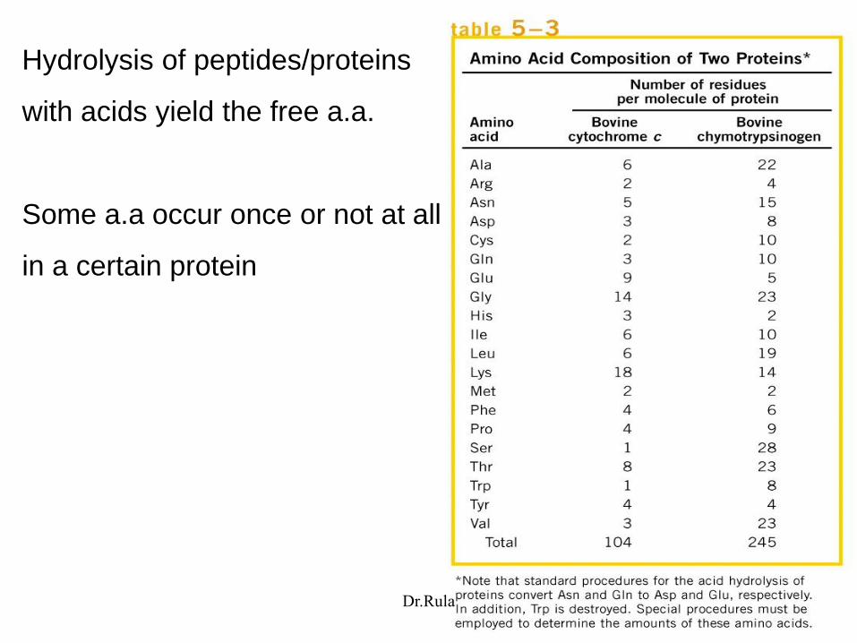
Hydrolysis of peptides/proteins
with acids yield the free a.a.
Some a.a occur once or not at all
in a certain protein
Dr.Rula

Conjugated proteins:
proteins that contain chemical groups other than a.a.
Prosthetic group: non-amino acid part of the conjugated protein.
Conjugated proteins classified according to the prosthetic group.
Dr.Rula

Several levels of protein structure:
Primary structure is the sequence of a.a linked together by peptide
bonds and includes any disulfide bonds.
Dr.Rula


![[Gly-X-Y]n ‘R’ US](https://img.dokumen.tips/doc/110x75/56815658550346895dc3fda5/gly-x-yn-r-us.jpg)


![modulo8 14 COLLAG.ppt [modalit compatibilit ]) · STRUTTURA 1a: regione ripetitive -(Gly-Ala-Gly-Ala-Gly-Ser-)n-intercalate a regioni non organizzate. STRUTTURA 2a: βββ - foglietti](https://img.dokumen.tips/doc/110x75/5c68828b09d3f29b758b976e/modulo8-14-modalit-compatibilit-struttura-1a-regione-ripetitive-gly-ala-gly-ala-gly-ser-n-intercalate.jpg)