Embed Size (px)
Citation preview

1
American Gastroenterological Association Technical Review on the Use of Transient Elastography in Chronic Liver Diseases
Siddharth Singh, MD, MS1; Andrew J. Muir, MD2; Douglas T. Dieterich, MD3;
Yngve T. Falck-Ytter, MD4 1Division of Gastroenterology, University of California San Diego, La Jolla, CA; 2Division of Gastroenterology, Duke University School of Medicine, Durham, NC; 3Division of Liver Disease, Icahn School of Medicine, New York, NY; 4Division of Gastroenterology and Hepatology, Cleveland VA Medical Center and University Hospitals, Case Western Reserve University, Cleveland, Ohio Corresponding Author: Chair, Clinical Guidelines Committee, American Gastroenterological Association National Office, 4930 Del Ray Avenue, Bethesda, Maryland 20814. E-mail: [email protected] Telephone: (301) 941-2618. Manuscript Word Count: 9929 References: 118 Tables and Figures: 11+0 eTables and eFigures (in the supplement): 7+7 Disclosures: Dr. Singh is partly supported by the National Library of Medicine training grant T15LM011271.
Acknowledgements: We sincerely thank Kellee Kaulback, Medical Information Officer, Health Quality Ontario, for helping in the literature search for this technical review.

2
INTRODUCTION
Globally over 1.75 million deaths are attributed to chronic liver diseases
(CLDs), which are an important source of health and economic burden.1 In the
United States, nearly 150,000 persons are diagnosed with CLDs annually (of which
20% are diagnosed with cirrhosis), and 36,000 patients die of CLDs, primarily
attributable to complications of decompensated cirrhosis and/or hepatocellular
cancer (HCC).2, 3 Annually, these generate approximately 5.9 million CLD-related
ambulatory care visits and 759,000 CLD-related hospitalizations, with healthcare
costs exceeding $1.5 billion.3 HCC is the second leading cause of cancer-related
death worldwide, and most patients with HCC will have underlying CLDs.4
Globally, it is estimated that over 185 million and 248 million persons may be living
with chronic hepatitis C virus (HCV) infection and chronic hepatitis B virus (HBV)
infection, respectively; corresponding rates in the United States are approximately
4.7 million, and 2 million, respectively.5-7 Nonalcoholic fatty liver disease (NAFLD)
is rapidly increasing cause of CLDs, with estimated 13.5% to 31.8% affected
globally and 24.1% of adults in North America.8 The burden of alcoholic liver
disease is more difficult to determine, but one report estimated that alcohol-
attributable liver cirrhosis was responsible for 493,300 deaths globally in 2010.9
Early identification of patients at high risk of progression to decompensated
cirrhosis may help direct high value care, and decrease the morbidity and mortality
attributed to CLDs. One of the key determinants of progression to CLD-related
complications is degree of liver fibrosis, and is often factored in making treatment
and surveillance decisions (for HCC and/or esophageal variceal screening).
Historically, liver biopsy has been the gold standard for diagnosis and staging of
fibrosis, besides its role in identifying etiology of abnormal liver enzymes and
assessing degree of inflammation. However, this procedure has several
limitations. It is invasive and associated with an estimated morbidity (including
severe pain) and mortality rate of 3% and 0.01%, respectively; in the Hepatitis C
Antiviral Long-Term Treatment against Cirrhosis trial, serious adverse events
occurred in 29 of the 2740 (1.1%) biopsies performed and included 16 (0.6%)
bleeding cases.10, 11 Liver biopsy is prone to sampling error resulting in

3
misclassification of fibrosis stage in up to 25% of cases, and there is also
considerable intra- and inter-observer variability in interpretation of histology,
especially at lower stages of fibrosis.12
To overcome these limitations and inconvenience of an invasive test, non-
invasive, serum and imaging-based methods of staging fibrosis have been
developed. While several proprietary and non-proprietary serum-based markers
have been developed, they are non-specific for the liver and may have inferior
performance characteristics to imaging-based tests.13 Among imaging tests,
ultrasound-based vibration-controlled transient elastography (TE) has been most
extensively studied and validated with high intra- and inter-observer reproducibility
and can be performed quickly, potentially at point-of-care.14 In this technique, a
piston vibrator placed in the intercostal space generates a shear wave, and then
the velocity is measured in a region 25 to 65 millimeters below the skin surface
with the standard adult M probe and 35 to 75 mm with the XL probe for larger
patients. The unit of measurement is kilopascals (kPa), and the device readings
range from 2.5 to 75kPa.
With recent recommendations for universal screening for HCV, availability
of highly effective but expensive newer direct acting agents against HCV, and
rising prevalence of NAFLD, an increasing number of patients are seeking
evaluation for CLDs, and fibrosis staging through non-invasive means has become
increasingly important and appealing for physicians.15, 16 Patients also have a
strong preference for TE over liver biopsy. In a Canadian survey of 422 patients
of whom 205 had undergone liver biopsy, approximately 95% patients preferred
TE over liver biopsy, with the majority reporting no discomfort (84%), no feelings
of anxiety (78%), short test duration and short time to result.17 In its recent
guidelines, the European Association for the Study of Liver Diseases and the Latin
American Association for the Study of the Liver have recommended TE as a
validated non-invasive standard for assessment of liver fibrosis, in patients with
HCV and HBV, with >90% negative predictive value in ruling out cirrhosis.18
However, these guidelines offer limited guidance on the diagnostic performance of
specific cut-offs of TE-identified liver stiffness, in clinical contexts of high- and low-

4
risk populations of patients with CLD, and its potential impact on downstream
patient-important outcomes. Identifying specific cut-offs for liver stiffness
corresponding to cirrhosis and advanced fibrosis could guide management
decisions, including treatment for HCV and HBV and triage for preventive cirrhosis
care. Hence, the AGA prioritized this topic for generation of clinical guidelines.
Objectives of this review
This technical review addresses focused clinical questions on the diagnostic
performance of TE in patients with HCV, HBV, NAFLD and alcoholic liver disease,
focusing specifically on: (a) overall performance relative to non-proprietary, serum-
based fibrosis markers and (b) implications of specific liver stiffness cut-offs on
downstream patient-important outcomes. Additionally, in this review we sought to
evaluate the performance of specific liver stiffness cut-offs to assess portal
hypertension to either triage patients with compensated cirrhosis with low
likelihood of high-risk esophageal varices (EV), as well as its role in pre-surgical
risk stratification of patients with CLD.19 Given limited validation and availability of
other imaging-based tests for fibrosis assessment, we did not include other
ultrasound-based shear wave modalities and magnetic resonance in this review.

5
METHODS
Formulation of Clinical Questions
The participants (including SS, AJM, DTD, and YFY) for this technical
review were selected by the AGA Clinical Guidelines Committee based on their
clinical content and guidelines methodological expertise, and went through a
thorough vetting process for potential conflicts of interest. Through an iterative
process, the participants developed focused clinical questions deemed relevant for
clinical practice that the guideline would address, related to the diagnostic
performance and utility of TE in 5 different populations: adults with HCV, HBV,
NAFLD, chronic alcoholic liver disease, and adults with CLD suspected to have
portal hypertension. From these focused questions, well-defined statements in
terms of patients, intervention, comparator and outcome (PICO) were defined, and
these formed the framework for formulating the study inclusion and exclusion
criteria and guided the literature search. The AGA Governing Board approved the
final set of questions and statements. The final focused and PICO questions are
shown in Table 1.
There were two broad themes for our focused questions. The first set of
questions for each population of interest (HCV, HBV, NAFLD and alcoholic liver
diseases) were centered around the overall diagnostic performance (across a
broad range of cut-offs) of TE in relation to commonly used, non-proprietary, non-
invasive serum biomarkers of fibrosis in these conditions (APRI, FIB-4) (PICO# 1,
4, 6).13, 20 Though proprietary serum-based fibrosis markers may have slightly
higher diagnostic accuracy as compared to non-proprietary markers, the latter are
inexpensive, easy to calculate and widely available.18 The second set of focused
questions were focused on identifying reliable TE-derived liver stiffness cut-offs to
either diagnose cirrhosis (PICO# 2, 4, 6, 8), or rule out advanced fibrosis (PICO#
3) or rule out high-risk esophageal varices (EV) [defined as any medium/large EV,
or small varices with high risk stigmata for bleeding] (PICO# 9) or clinically
significant portal hypertension (CSPH, defined as presence of any EV) (PICO#
10).

6
For PICOs# 1-8, TE was considered as a test replacement strategy for
detection of cirrhosis, i.e., in patients with valid results, TE would replace routine
use of liver biopsy and limit its use to cases with inconclusive TE results or
diagnostic equipoise. For PICO# 9, TE was considered as a triage (screening)
strategy for upper endoscopy for ruling out high-risk EV, i.e., in patients with liver
stiffness below TE-identified threshold, the likelihood of high-risk EVs is sufficiently
low to avoid routine upper endoscopy; however, in patients with liver stiffness at or
above TE-identified threshold, upper endoscopy is warranted to confirm diagnosis
before treatment is considered. Likewise for PICO# 10, TE was considered a
triage strategy, i.e., patients with liver stiffness below TE-identified threshold,
clinically significant portal hypertension may be ruled out in risk stratification for
elective, non-hepatic surgery; however, in patients with liver stiffness at or above
TE-identified threshold, further testing (with upper endoscopy or HVPG) to
evaluate CSPH may be warranted before a patient is deemed high risk for elective
surgery.
Search Strategy, Study Selection, Data Abstraction and Quality Assessment
Details of the search strategy, study selection, data abstraction and risk of
bias assessment is reported in the online supplement.
Outcomes of Interest
For the first set of PICO statements pertaining to overall diagnostic
performance of TE as compared to other commonly used, non-proprietary, non-
invasive fibrosis biomarkers, primary outcome of interest was the overall diagnostic
performance (true positives [TP], false positives [FP], true negatives [TN] and false
negatives [FN] rates) for detection of cirrhosis, in different illustrative clinical
scenarios, corresponding to variable observed prevalence of cirrhosis depending
on practice setting and population in which the test was applied.
For the second set of PICO statements pertaining to reliable TE-derived
liver stiffness cut-offs to either diagnose (PICO# 2, 4, 6, 8), or rule out (PICO# 3)
cirrhosis or rule out high-risk EVs or any EVs (PICO# 9, 10), the preferred outcome

7
was direct consequences on patient-important outcomes (i.e., implications of TP,
FP, TN, FN results for patients – see below). However, none of the studies
assessed these outcomes directly, and hence, we used TP, FP, TN, FN rates as
surrogate outcomes and inferred downstream consequences on patient-important
outcomes. For questions focusing on ruling out cirrhosis, high-risk or any EV, our
outcome was minimizing rates of FN (i.e. patients incorrectly being labeled as not
having the condition, when they actually have the condition) to a level <3% in
general, preferably lower, with reasonable rates of TP, FP and TN. For questions
focusing on diagnosing patients with cirrhosis, our outcome was a balance of FN
and FP (i.e., patients incorrectly labeled as having condition, when they actually
do not have the condition). This was also estimated in different clinical scenarios,
as detailed above.
Consequences of diagnostic test results on patient-important outcomes
Corresponding to each possible outcome (TP, FP, TN, FN), presumed
downstream consequences on patient-important outcomes were considered. For
example, for PICOs# 1-8 on detection of cirrhosis,
True positives (patients correctly diagnosed as having cirrhosis) would be
eligible to receive preventive cirrhosis care (such as HCC surveillance,
screening for EV), may receive treatment prioritization (HBV patients with
compensated cirrhosis who may not have qualified for treatment) or
different treatment regimen (HCV patients may receive 12-week therapy
instead of 8-week therapy with direct anti-viral agents), all of which may
eventually decrease morbidity and mortality, without being subject to risks
and invasive testing with liver biopsy.
False positives (patients incorrectly labeled as having cirrhosis based on
TE, when actually they do not) may receive unnecessary testing (HCC
surveillance, screening for EV) and treatment (longer treatment for HCV,
etc.) and have avoidable anxiety, potential testing- or treatment-related
complications and excessive resource utilization.

8
True negatives (patients correctly diagnosed as not having cirrhosis based
on TE) would be reassured and obviate the need for invasive testing with
liver biopsy, although they may need to undergo serial assessment of liver
stiffness at periodic intervals.
False negatives (patients incorrectly labeled as not having cirrhosis based
on TE, when actually they have cirrhosis) would be falsely reassured, and
may not receive appropriate preventive cirrhosis, may receive inappropriate
treatment (shorter HCV treatment course, etc.), potentially leading to
increased morbidity and mortality.
In using specific TE-derived liver stiffness cut-offs either as a test
replacement or triage strategy, healthcare providers and patients need to be aware
of test performance, and be comfortable with potential FN and FP rate with
attending downstream consequences. Such downstream consequences of test
results for each PICO statement and scenario have been discussed in detail in
each evidence profile. For both test replacement and triage questions, the
technical review team decided to focus on optimizing FN rate, with a reasonable
FP trade-off (depending on downstream consequences).
A pre-meeting questionnaire was administered to both the content experts
in the technical review team and the guideline panel to determine their a priori
maximal tolerable false negative rate for each PICO (i.e., what level of FN rate
would they be willing to accept for a particular test, for their patient). As the
maximally tolerable rates of false negative tests for any diagnostic strategy is
highly context sensitive, we devised different clinical scenarios with corresponding
downstream consequences for each PICO to arrive at fully contextualized
estimates of false negative thresholds (see online supplement).
Data Synthesis and Statistical Analysis
Details of data synthesis and statistical analysis are reported in the online
supplement. Specifically, for PICOs focusing on identifying reliable cut-offs, we a
priori sought to identify TE cut-off maximizing sensitivity (to rule out cirrhosis, high-
risk EV or CSPH), or maximizing specificity (to diagnose cirrhosis). However,

9
during the data abstraction process, we recognized that variable cut-offs were not
consistently reported in included studies; moreover, most studies did not
prospectively study a particular cut-off, but rather retrospectively applied the cut-
off corresponding to AUROC. Hence, to identify reliable cut-offs, we used the most
commonly reported cut-off in studies, confirmed their clinical use with content
experts (and use in clinical trials which recruited patients with cirrhosis based on
TE cut-offs), and calculated sensitivity, specificity, positive and negative predictive
value corresponding to these.
Quality of Evidence
We rated the quality of evidence using the GRADE approach for diagnostic
tests and strategies.21 In this approach, all evidence from randomized controlled
trials (comparing different diagnostic tests or cut-offs of same test) and
observational diagnostic accuracy studies start at high-quality, but can be rated
down for any of the following factors:
Risk of bias in included studies (inferred based on QUADAS instrument),22
Indirectness (e.g., deemed present if (a) there are important differences in
population studied and those for whom recommendation is being is
intended, (b) if cut-offs for TE for cirrhosis detection were not pre-specified
but obtained post-hoc corresponding to AUROC, (c) if TP, FP, TN and FN
rates are used as surrogates for presumed downstream consequences on
patient-important outcomes),
Inconsistency (e.g., deemed present if there were considerable differences
in the accuracy estimates),
Imprecision (deemed present if there were wide CIs for true and false
positive and true and false negative rates), and
Publication bias.
In the absence of direct patient-important outcomes from observational
diagnostic accuracy studies, surrogate outcomes including TP, FP, TN, and FN
were all rated as critical outcomes, and included in evidence profiles.
10
RESULTS
Appropriate Interpretation of Transient Elastography
For optimal interpretation of TE, the following are required: at least 10
validated measurements, a success rate (the ratio of valid measurements to the
total number of measurement) above 60%, and an interquartile range (reflects
variations among measurements) of less than 30% of the median value (IQR/M,
≤30%) is required.13, 14 In a prospective study of 13,369 TE examinations in
Europe, liver stiffness measurement was unsuccessful (no valid shots) could be
obtained in 3% of patients, and in another 16% patients, the results were
unreliable;23 corresponding rates in an Asian population were 2.5% unsuccessful
measurement, and 0.9% with unreliable liver stiffness measurement,
respectively.24 Primary factors associated with unsuccessful or unreliable
measurements are: obesity (body mass index >30kg/m2), in particular increased
waist circumference, ascites, narrow inter-rib spaces, advanced age, female sex
and operator inexperience (<500 examinations).23-25. Besides fibrosis, factors that
influence viscoelastic properties of the liver may also result in increased liver
stiffness, such as the presence by severe hepatic inflammation, extrahepatic
arteriovenous or biliary obstruction and congestive heart failure. Hence, caution
should be exercised in interpreting TE results in patients with significant elevation
in liver enzymes (aminotransferases >5x upper limit of normal) or excessive
alcohol consumption.26-30 Recent studies have identified that non-fasting state may
also significantly influence liver stiffness, and hence, TE should ideally be
undertaken when the patient has been fasting for at least 2 hours.31
Illustrative Prevalence of Cirrhosis
The diagnostic accuracy of any test in terms of rates of TP, TN, FP and FN
depends on pre-test probabilities and prevalence of condition, which in turn
depends on practice setting (higher prevalence of cirrhosis in referral liver clinic as
compared to community primary care practice as compared to), patient-level
characteristics (higher prevalence of cirrhosis in patients with concomitant viral

11
infections like HIV, obesity, diabetes, excessive alcohol use) and physician
suspicion (which often encompasses practice setting and patient characteristics,
including clinical history, physical examination, laboratory features, etc.). While we
acknowledge that this baseline pre-test probability of cirrhosis varies along a
continuum of these factors, for ease of interpretation of data in day-to-day practice,
we anchored the baseline prevalence of cirrhosis into two categories – low-risk
(5% prevalence of cirrhosis) and high-risk (30% prevalence of cirrhosis). To
illustrate this concept, patients with high-risk of having prevalent cirrhosis may be
asymptomatic patients with HCV, HBV, NAFLD or alcoholic liver disease with
associated obesity, diabetes mellitus, excessive alcohol use and/or concomitant
viral infections (like HIV), who are often seen in referral centers, and the estimated
risk of cirrhosis in this population would be approximately 30%.32-35 Patients with
low-risk of having prevalent cirrhosis would be those who are asymptomatic, seen
by community primary care practitioners with HCV, HBV, NAFLD or alcoholic liver
disease, without clear factors associated with presence of cirrhosis, and the
estimated risk of cirrhosis of cirrhosis in this population would be approximately
5%. Using this illustrative prevalence of outcome, and sensitivity/specificity of liver
stiffness cut-offs in different scenarios, positive and negative predictive value of
each cut-off is summarized in Table 2.
Question 1. In adults with chronic HCV, is the overall diagnostic performance
of TE superior to other non-invasive markers of liver fibrosis (APRI, FIB-4)
for detection of cirrhosis?
Key Message. In adults with chronic HCV, TE has superior sensitivity and
specificity, and lower false positive and false negative rates, suggesting
better diagnostic performance, as compared to APRI and FIB-4 for
detection of cirrhosis. (Moderate quality of evidence).
Effect Estimates: We used an existing well-conducted systematic review on the
diagnostic performance of non-invasive methods for assessment and monitoring
of liver fibrosis and cirrhosis in patients with chronic liver diseases published in
January 2015.36 This systematic review included 36 studies in patients with HCV

12
which reported on the diagnostic performance of TE for detection of cirrhosis using
liver biopsy as a reference standard. In these studies, the liver stiffness cut-off
corresponding to AUROC ranged from 9.2-17.3 kPa. The summary sensitivity and
specificity for detection of cirrhosis across this range of cut-offs was 0.89 (95% CI,
0.84-0.92) and 0.91 (0.89-0.93), respectively. The evidence profiles are
summarized in Tables 3A and B. Based on this, TE classified more patients
correctly as having cirrhosis (TP) or not having cirrhosis (TN) compared to APRI
and FIB-4, and had lower rates of misclassification (FP, FN), both in a low- and
high-prevalence population.
Quality of Evidence: All included studies were cross-sectional diagnostic
accuracy studies, required <6m interval between performance of diagnostic test
(TE, APRI or FIB-4) and gold standard (liver biopsy) minimizing disease
progression bias, and generally of fair quality and not at serious risk of bias
(spectrum bias, disease progression bias, partial or differential verification bias,
adequate blinding of outcome assessors). For comparison of diagnostic
performance of TE with other non-invasive, serum-based fibrosis markers, rates
of TP, FP, TN and FN were directly relevant outcomes in a comparable population,
and though there were limited head-to-head comparisons of TE and other
measures, the overall body of evidence was not deemed to be at serious evidence
of indirectness. Considerable heterogeneity was observed in pooled sensitivity
and specificity, and there was a wide range of ‘ideal’ cut-offs for TE (corresponding
to AUROC), rather than pre-specified cut-offs for detection of cirrhosis, and hence,
evidence was rated down for inconsistency. There was no evidence of serious
imprecision, and diagnostic performance of TE was superior to APRI and to FIB-4
in both low- and high-risk populations, even in worst-performance scenarios (using
lower limit of 95% CI for diagnostic accuracy of TE, and upper limit of 95% CI of
APRI or FIB-4). To summarize, using the GRADE approach for diagnostic
accuracy studies, the quality of evidence supporting the use of TE over APRI or
FIB-4 for detection of cirrhosis, was rated as moderate quality.
Discussion: Pre-treatment assessment of fibrosis stage is important to facilitate
appropriate HCV treatment decisions and determine need for additional measures

13
for managing cirrhosis, such as HCC surveillance.37 While non-proprietary,
inexpensive, serum-based fibrosis markers like APRI and FIB-4 are readily
available, their diagnostic performance was sub-optimal in both low- and high-
prevalence scenarios, with high FP rates for detection of cirrhosis. This may result
in avoidable patient anxiety and unnecessary testing and treatment. There was
moderate certainty in the observation that TE has superior diagnostic performance
in identifying cirrhosis in patients with HCV, with lower rates of misclassification of
patients. We did not factor in cost-effectiveness of TE vs. serum-based fibrosis
markers, given rapidly changing prices of anti-viral therapy, which may offset cost-
benefit assessments.
Question 2. In adults with chronic HCV undergoing TE, at what liver stiffness
cut-off, can we accurately diagnose cirrhosis (and initiate downstream
management), obviating the need for liver biopsy?
Key Message. In adults with chronic HCV, we can accurately diagnose
cirrhosis (and initiate downstream management) with TE-defined liver
stiffness of ≥12.5 (±1) kPa, with acceptable FP and FN rates. (Low quality
of evidence).
Effect Estimates: We updated an existing systematic review, to identify a range
of liver stiffness cut-offs (9.2-26.5kPa) corresponding to optimal sensitivity and
specificity for diagnosis of cirrhosis in adults with HCV. From this, we identified a
narrow range of liver stiffness cut-off, 12.5 (±1) kPa, which corresponded to the
most commonly observed value in included studies (17 studies, 5812 patients),
and corresponding to value most commonly applied in clinical trials and practice.38-
53 eTable 1 describes the characteristics of these included studies, and eFigures
1A and B report the sensitivity and specificity of this cut-off. The performance of
this cut-off in low- and high-risk populations is shown in Table 4. In an illustrative
low-risk population (5% prevalence of cirrhosis), for example, patients with HCV
detected in primary care clinics during routine age-appropriate screening, using a
cut-off of 12.5 (±1) kPa may misclassify 0.7% patients as not having cirrhosis (FN)
and 8.6% patients as having cirrhosis (FP). In an illustrative high-risk population

14
(30% prevalence of cirrhosis), for example, HCV patients with obesity, diabetes,
excessive alcohol use or co-infection with HIV or HBV, using a cut-off of 12.5 (±1)
kPa may misclassify 4.2% as not having cirrhosis (FN) and 6.3% patients as having
cirrhosis (FP).
Quality of Evidence: The evidence supporting the use of this cut-off was derived
from cross-sectional diagnostic accuracy studies, and there was no data on
comparing different cut-offs and their effect on downstream patient-important
outcomes related to impact of cirrhosis diagnosis (or misdiagnosis). Hence, due
to use of FP and FN as surrogates for presumed downstream consequences, and
because the cut-off was largely obtained from post-hoc analysis corresponding to
AUROC, the overall body of evidence was rated down for indirectness. Since we
selectively included only studies which identified a cut-off of 12.5 (±1) kPa, and
excluded studies in which the optimal cut-off was higher or lower (in which the
diagnostic performance corresponding to a cut-off of 12.5 kPa was not reported
and conceivably poorer), and since considerable heterogeneity was observed in
the pooled sensitivity and specificity corresponding to the identified cut-off, we
rated down further for inconsistency. The diagnostic accuracy studies were
generally of fair quality, and there was no serious risk of bias. In addition, there
was no evidence serious imprecision and no evidence of publication bias was
detected. To summarize, using the GRADE approach for using diagnostic
accuracy studies for patient management, the quality of evidence supporting the
use of TE-defined liver stiffness of ≥12.5 (±1) kPa for diagnosis of cirrhosis in adults
with HCV, was rated as low quality.
Discussion: In the evaluation of patients with HCV, the stage of disease and the
ability to detect cirrhosis is critical. While fibrosis stage at which antiviral therapy
should be initiated is still in flux with the introduction of highly effective but
expensive direct acting antiviral agents, patients with advanced fibrosis/
compensated cirrhosis definitely require antiviral treatment to prevent progression
(and potential fibrosis regression); additionally, the presence of cirrhosis may
extend treatment duration with some regimens.37 Patients with cirrhosis will also
need close surveillance for complications of portal hypertension and HCC even

15
after cure of HCV cirrhosis.54 As mentioned earlier, in using TE as strategy to
replace liver biopsy, healthcare providers and patients need to be aware of test
performance, and be comfortable with potential FN and FP rate with attending
downstream consequences. A priori, the maximal tolerable FN rate accepted upon
by the technical review and guideline content expert panel was 5-10%, i.e., the test
threshold would be acceptable if <10% of patients are misclassified as not having
cirrhosis. With the use of this TE-defined liver stiffness cut-off of 12.5 (±1) kPa,
we estimated that over 85% of patients would be able to avoid liver biopsy with
correct classification of either having or not having cirrhosis. Importantly, we
observed that with this cut-off, <1% and <5% of low- and high-risk patients,
respectively may be falsely reassured (of not having cirrhosis; FN rate), potentially
may not receive adequate duration of treatment course and be at-risk of treatment
failure, may not receive supportive cirrhosis and consequently, may be at
increased risk of hepatic decompensation. Additionally, with this cut-off, <10% of
patients without cirrhosis, in both low- and high-risk populations, may be falsely
diagnosed as having cirrhosis, and receive unnecessary tests (like surveillance for
HCC) and treatment (longer antiviral therapy), and have anxiety, testing- and
treatment-related complications, and lead to excessive burden on resource
utilization. Due to the convenience of a non-invasive test, serial testing on a
periodic basis may improve the classification of patients with HCV. Liver biopsy
may be needed in case there is discrepancy between physician gestalt (based on
clinical scenario, imaging such as CT or ultrasound and biochemical markers) and
TE findings.
Question 3. In adults with chronic HCV who have achieved sustained
virologic response with antiviral therapy undergoing TE, at what liver
stiffness cut-off, can we accurately rule out advanced fibrosis (F3 or F4) and
consider discharging patients from a dedicated liver clinic?
Key Message. In adults with chronic HCV who have achieved sustained
virologic response with antiviral therapy, we can accurately rule out
advanced fibrosis (F3 and F4) and consider discharging from dedicated liver

16
clinic with TE-defined liver stiffness of ≤9.5 (±1) kPa, with acceptable FN
rates. (Very low quality of evidence).
Effect Estimates: We updated an existing systematic review, to identify a narrow
range of liver stiffness cut-off, 9.5 (±1) kPa, which corresponded to the most
commonly observed liver stiffness value for detection of advanced fibrosis or
cirrhosis (13 studies, 4106 patients), and corresponding to value most commonly
applied in clinical practice.39, 42, 48, 50, 51, 53, 55-61 eTable 2 describes the
characteristics of these included studies, and eFigures 2A and B report the
sensitivity and specificity of this cut-off. The performance of this cut-off in low- and
high-risk populations is shown in Table 5. In an illustrative low-risk population
(5% prevalence of advanced fibrosis), for example, patients with HCV who achieve
sustained virologic response (SVR) and have no ongoing risk factors for chronic
liver diseases, using a cut-off of ≤9.5 (±1) kPa may misclassify 1.1% patients as
not having advanced fibrosis (FN). Although the FP rate will be high with such a
sensitive cut-off, the goal is exclude significant fibrotic liver disease and serial
examinations over time may reduce this false positive rate. In a high-risk
population (30% prevalence of advanced fibrosis), for example, HCV patients
who achieve SVR, but either had cirrhosis (liver stiffness >12.5 kPa) prior to
therapy or continue to have other risk factors for chronic liver diseases such as
obesity, diabetes, excessive alcohol use or co-infection with HIV or HBV, using a
cut-off of ≤9.5 (±1) kPa may misclassify 6.6% patients as not having advanced
fibrosis (FN).
Quality of Evidence: As with most evidence on diagnostic performance of
different TE-derived liver stiffness cut-offs, the evidence supporting the use of this
cut-off was derived from cross-sectional diagnostic accuracy studies in all patients
with HCV, as opposed to studies specifically conducted in patients who achieve
SVR after antiviral therapy. Additionally, there was no data on comparing different
cut-offs (which were derived from post-hoc analysis corresponding to AUROC) and
their effect on downstream patient-important outcomes related to impact of
advanced fibrosis diagnosis (or misdiagnosis). Hence, the overall body of
evidence was rated down twice for very serious indirectness. Since we selectively

17
included only studies, which identified a cut-off of 9.5 (±1) kPa, and since
considerable heterogeneity was observed in the pooled sensitivity and specificity
corresponding to the identified cut-off, we rated down further for inconsistency.
There was no evidence of serious risk of bias, serious imprecision and no evidence
of publication bias was observed. To summarize, using the GRADE approach for
using diagnostic accuracy studies for patient management, the quality of evidence
supporting the use of TE-defined liver stiffness of ≤9.5 (±1) kPa for ruling out
advanced fibrosis in adults with HCV who have achieved SVR, was rated as very
low quality.
Discussion: With recommendations for universal screening for hepatitis C for
persons born between 1945-65, availability of effective antiviral therapies,
tremendous numbers of patients are seeking care and being cured of HCV. For
these cured patients, healthcare providers will need to decide whether or not they
need ongoing care for their liver. This decision to discharge patients from hepatitis
C care can be very meaningful to patients (who can put HCV behind them) and
healthcare providers (to improve access to other patients to receive care for HCV).
The AASLD/IDSA guidance recommends ongoing care and surveillance for
complications of portal hypertension and hepatocellular carcinoma in patients with
advanced fibrosis (F3-4), and no further follow-up for patients for early fibrosis (F0-
F2).37 The technical review team decided that it may be appropriate to discharge
patients from the liver clinic if there was no evidence of advanced fibrosis on liver
biopsy after SVR. However, since repeat liver biopsy after achieving SVR is not
feasible or universally acceptable, TE-defined liver stiffness may help make
decisions regarding discharging patients after treatment for HCV. The maximal
tolerable predefined FN rate accepted upon by the technical review and guideline
content expert panel was 1-5%, i.e., the test threshold would be acceptable if <5%
of patients are misclassified as not having advanced fibrosis and are discharged
from clinic. With a cut-off of ≤9.5 (±1) kPa, >80% and >60% of low-risk and high-
risk patients, respectively without advanced fibrosis may be considered for
discharge from a dedicated liver clinic after achieving SVR with anti-viral therapy,
without increased risk of patient morbidity and mortality and decreasing healthcare

18
utilization and burden. While approximately 1% of low-risk patients may be
misclassified as not having advanced fibrosis using this cut-off, approximately 7%
of high-risk patients (more than the maximal tolerable FN rate) may be falsely
reassured and be discharged from a dedicated liver clinic, and not receive
appropriate post-treatment supportive care, putting them at increased risk of
hepatic morbidity and mortality. However, despite this higher rate, it is expected
that most misclassifications will occur by missing some patients with stage F3
fibrosis, but likely very few or no patients with cirrhosis will be discharged. Any
decision to discharge patients from a dedicated liver clinic would require
consideration of other factors such as co-existing liver diseases or ongoing
abnormal liver tests. It is important to note that quality of evidence supporting this
observation was very low and further research is needed in this area.
Question 4. In adults with chronic HBV, is the overall diagnostic performance
of TE superior to other non-invasive markers of liver fibrosis (APRI, FIB-4)
for detection of cirrhosis?
Key Message. In adults with chronic HBV, TE has superior sensitivity and
specificity, and lower FP and FN rates, suggesting superior diagnostic
performance, as compared to APRI and FIB-4 for detection of cirrhosis.
(Low quality of evidence).
Effect Estimates: We used an existing well-conducted systematic reviews on the
diagnostic performance of non-invasive methods for assessment and monitoring
of liver fibrosis and cirrhosis in patients with CLD.36, 62 This systematic review
included 19 studies in patients with HBV, which reported on the diagnostic
performance of TE for detection of cirrhosis using liver biopsy as a reference
standard. In these studies, the liver stiffness cut-off corresponding to AUROC
ranged from 9.4-16.0 kPa. The summary sensitivity and specificity for detection of
cirrhosis across this range of cut-offs was 0.86 (95% CI, 0.79-0.91) and 0.85 (0.78-
0.89), respectively. The evidence profiles are summarized in Tables 6A and B.
Based on this, TE classified more patients correctly as compared to APRI with
higher rates of TP and TN, and lower rates of FP and FN, though these estimates

19
were imprecise in worst-performance scenarios. TE’s diagnostic performance was
comparable to FIB-4 for diagnosing cirrhosis, but was superior to FIB-4 in ruling
out cirrhosis.
Quality of Evidence: Similar to studies on diagnostic performance of TE in HCV,
studies were not deemed to be at serious risk of bias and there was no evidence
of indirectness. Considerable heterogeneity was observed, and there was a wide
range ‘ideal’ cut-offs for TE (corresponding to AUROC), rather than pre-specified
cut-offs for detection of cirrhosis, and hence, evidence was rated down for
inconsistency. In the comparison of TE vs. APRI, there was evidence of serious
imprecision both for ruling in and ruling cirrhosis, whereas in the comparison of TE
vs. FIB-4, there was evidence of serious imprecision only for ruling in, but not ruling
out cirrhosis, in worst-performance scenarios. To summarize, using the GRADE
approach for diagnostic accuracy studies, the overall quality of evidence
supporting the use of TE over APRI or FIB-4 for detection of cirrhosis, was rated
as low quality.
Discussion: While several management decisions in patients with HBV are
determined by host or virus-related characteristics, fibrosis assessment may be
important for a subset of patients who do not meet criteria for antiviral treatment
based on other characteristics.63 However, given the risks and burden of liver
biopsies, overall adherence to perform biopsy where indicated is low both due to
physician- and patient-related factors;64 non-invasive testing may help overcome
this barrier. Among non-invasive tests we identified that low-quality evidence
supported the use of TE over other non-invasive serum-based fibrosis markers
with lower rates of false positivity (i.e., risk of falsely classifying patients as having
cirrhosis, and initiating lifelong therapy), although rates of FN were comparable to
FIB-4.
Question 5. In adults with chronic HBV undergoing TE, at what liver stiffness
cut-off, can we accurately diagnose cirrhosis (and initiate downstream
management), obviating the need for liver biopsy?

20
Key Message. In adults with chronic HBV, we can accurately diagnose
cirrhosis (and initiate downstream management) with TE-defined liver
stiffness of ≥11.0 (±1) kPa, with acceptable FP and FN rates. (Low quality
of evidence).
Effect Estimates: We updated an existing systematic review, to identify a range
of liver stiffness cut-offs (8.4-18.2kPa) corresponding to optimal sensitivity and
specificity for diagnosis of cirrhosis in adults with HBV. From this, we identified a
narrow range of liver stiffness cut-off, 11.0 (±1) kPa, which corresponded to the
most commonly observed value in included studies (17 studies, 4864 patients).39,
57, 65-79 eTable 3 describes the characteristics of these included studies, eFigures
3A and B report the sensitivity and specificity of this cut-off. The performance of
this cut-off in low- and high-risk populations is shown in Table 7. In an illustrative
low-risk population (5% prevalence of cirrhosis), for example, patients with HBV
detected during routine screening with low HBV viral load, using a cut-off of ≥11.0
(±1) kPa may misclassify 0.9% patients as not having cirrhosis (FN), and 16.1%
patients as having cirrhosis (FP). In an illustrative high-risk population (30%
prevalence of cirrhosis), for example, HBV patients with obesity, diabetes,
excessive alcohol use or co-infection with HIV or HCV, using a cut-off of ≥11.0 (±1)
kPa may misclassify 5.7% patients as not having cirrhosis (FN) and 11.9% patients
as having cirrhosis (FP).
Quality of Evidence: Similar to studies related to TE in HCV management, the
evidence supporting the use of this cut-off was derived from cross-sectional
diagnostic accuracy studies. FP and FN rates were used as surrogates for
presumed patient-important downstream consequences, and cut-offs were largely
obtained from post-hoc analysis corresponding to AUROC, and selective cut-off of
≥11.0 (±1) kPa was chosen as being most representative. For these reasons,
evidence was rated down for imprecision and inconsistency. There was no
evidence of serious risk of bias, serious imprecision and no evidence of publication
bias was observed. Hence, using the GRADE approach, the quality of evidence
supporting the use of TE-defined liver stiffness of ≥11.0 (±1) kPa for diagnosis of
cirrhosis in adults with HBV, was rated as low quality.

21
Discussion: Liver stiffness threshold corresponding to cirrhosis seems to vary
across the diseases, which could be truly related to differences in underlying
disease processes, or may be an artifact of limited prospective research using
predefined liver stiffness thresholds to define cirrhosis. While HCC surveillance is
required for the majority of patients with HBV regardless of cirrhosis status, the
diagnosis of compensated cirrhosis may be useful in identifying patients for
antiviral therapy who would do not meet other criteria for receipt of therapy. A
priori, the maximal tolerable FN rate accepted upon by the technical review and
guideline content expert panel was 5-10%, i.e., the test threshold would be
acceptable if <10% of patients are misclassified as not having cirrhosis. With a cut-
off of ≥11.0 (±1) kPa, we estimated that over 80% of patients would be able to
avoid liver biopsy with correct classification of either having or not having cirrhosis.
Approximately 1% and 5% of low- and high-risk patients, respectively may be
falsely reassured (of not having cirrhosis), and be ineligible to receive antiviral
therapy which can decrease risk of decompensation, below the predefined
maximal tolerable FN rate of 10%; hypothetically, in a setting where the prevalence
of cirrhosis in HBV patients is >50%, the threshold FN rate of 10% would be
exceeded. In contrast, this threshold carries a FP rate of 16% and 12% for low-
and high-risk patients, respectively, wherein these patients without cirrhosis may
be falsely diagnosed as having cirrhosis, and receive unnecessary tests and
treatment (HBV-related therapy, if there are no other indications for treatment),
have anxiety, testing- and treatment-related complications, and lead to excessive
burden on resource utilization. Due to the convenience of a non-invasive test,
serial testing on a periodic basis may improve the classification of patients with
HBV. Liver biopsy may be needed in case there is discrepancy between physician
gestalt (based on clinical scenario, imaging such as CT or ultrasound and
biochemical markers) and TE findings.
Question 6. In adults with NAFLD, is the overall diagnostic performance of
TE (M-mode) superior to other non-invasive markers of liver fibrosis (APRI,
FIB-4) for detection of cirrhosis?

22
Key Message. In adults with NAFLD, TE (M-mode) has superior sensitivity
and specificity, and lower FP and FN rates, suggesting superior diagnostic
performance, as compared to APRI and FIB-4 for detection of cirrhosis.
(Very low quality of evidence).
Effect Estimates: We updated an existing well-conducted systematic review on
the diagnostic performance of non-invasive tests in patients with chronic liver
diseases, and identified 11 studies on 1266 patients with NAFLD, which reported
on the diagnostic performance of TE for detection of cirrhosis using liver biopsy as
a reference standard (eTable 4).48, 80-89 In these studies, the liver stiffness cut-off
corresponding to AUROC ranged from 10.3-22.3 kPa, and the corresponding
summary sensitivity and specificity for detection of cirrhosis across this range of
cut-offs was 0.90 (95% CI, 0.82-0.95) and 0.87 (0.85-0.89), respectively (eFigure
4A and B). TE was compared with performance of FIB4 and APRI for detection
of cirrhosis in NAFLD, derived from another recent systematic review.90 This is
summarized in Table 8A and B. Based on this, TE classified more patients
correctly as compared to APRI and FIB-4, with higher rates of TP and TN, and
lower rates of FP and FN, though these estimates were imprecise in worst-
performance scenarios.
Quality of Evidence: Studies on diagnostic performance of TE in NAFLD were at
high risk of bias due to analysis of only patients with successful TE, and not an
intention-to-diagnose analysis. Unsuccessful or unreliable liver stiffness
measurement is high in patients with obesity, in particular those with central
adiposity, a population at high-risk for NAFLD. Additionally, several studies on
performance of TE in patients with suspected NAFLD either excluded obese
patients (BMI ≥30 kg/m2), or performed only per-protocol diagnosis (excluding
patients with unreliable TE) contributing to artificially high sensitivity and specificity.
Similar to other TE studies in HCV and HBV, evidence was rated down for
inconsistency. In the comparison of TE vs. APRI, there was evidence of serious
imprecision both for ruling in and ruling out cirrhosis, whereas in the comparison
of TE vs. FIB-4, there was evidence of serious imprecision only for ruling in, but
not ruling out cirrhosis, in worst-performance scenarios (using lower limit of 95%

23
CI for diagnostic accuracy of TE, and upper limit of 95% CI of APRI or FIB-4).
Hence, using the GRADE approach for diagnostic accuracy studies, the quality of
evidence supporting the use of TE over APRI or FIB-4 for detection of cirrhosis,
was rated as very low quality.
Discussion: NAFLD is estimated to affect about 24% of Americans, and a small
proportion of them can progress to cirrhosis.8, 91 Through a systematic review, we
identified that, though TE had superior diagnostic performance as compared to
APRI and FIB-4, there were limitations in the literature, particularly high rates of
unsuccessful or unreliable TE readings with M-probe in obese patients and
selection bias in studies, excluding obese patients at high-risk for NAFLD. In a
prospective study, XL probe was able to overcome some limitations of M-probe,
with higher rates of successful (95% vs. 81%) and reliable (90% vs. 74%) liver
stiffness measurement.86 However, even with the XL probe, reliable liver stiffness
measurements could be obtained in only 65% of obese patients. In another study
of 169 patients with NAFLD, BMI and waist circumference negatively impacted the
diagnostic accuracy of TE; in their intention-to-diagnose analysis, the area under
the receiver-operator curve for diagnosis of advanced fibrosis or cirrhosis with TE
was 0.65.92 Magnetic resonance elastography also has superior diagnostic
performance as compared to TE, detect fibrosis in patients with NAFLD.81 With
several novel pharmacologic therapies in development for patients with NAFLD,
significant advances are required in non-invasive assessment of fibrosis in these
patients to identify treatment candidates and assess response to therapy.
Question 7. In adults with NAFLD undergoing TE, at what liver stiffness cut-
off, can we accurately diagnose cirrhosis (and initiate downstream
management), obviating the need for liver biopsy?
Key Message: Given the inherent limitations of the literature on the use of
TE for fibrosis assessment in patients with NAFLD, the guideline panel and the
technical review team decided not to provide pooled estimates, as the evidence
would not sufficiently support clinical decision making.

24
Question 8. In adults with chronic alcoholic liver disease undergoing TE, at
what liver stiffness cut-off, can we accurately diagnose cirrhosis (and initiate
downstream management), obviating the need for liver biopsy?
Key Message. In adults with chronic alcoholic liver disease, we can
accurately diagnose cirrhosis (and initiate downstream management) with
TE-defined liver stiffness of ≥12.5 (±1) kPa, with acceptable FP and FN
rates. (Low quality of evidence)
Effect Estimates: We updated an existing systematic review, to identify a range
of liver stiffness cut-offs (7.2-34.9kPa) corresponding to optimal sensitivity and
specificity for diagnosis of cirrhosis in adults with NAFLD based on 14 studies (834
patients).93 From this, we identified a narrow range of liver stiffness cut-off, ≥12.5
(±1) kPa, which corresponded to the most commonly observed value in included
studies (7 studies, 330 patients).94-100 eTable 5 describes the characteristics of
these included studies, and eFigures 5A and B report the sensitivity and
specificity of this cut-off. The performance of this cut-off in low- and high-risk
populations is shown in Table 9. In an illustrative low-risk population (5%
prevalence of cirrhosis), for example, patients with chronic excessive alcohol use
without any other high-risk factors, using a cut-off of ≥12.5 (±1) kPa may
misclassify 0.2% patients as not having cirrhosis (FN), and 27.5% patients as
having cirrhosis (FP). In an illustrative high-risk population (30% prevalence of
cirrhosis), for example, patients with alcoholic liver disease with advanced age,
obesity, diabetes and co-infection with HIV/HBV/HCV, using a cut-off of ≥12.5 (±1)
kPa may misclassify 1.5% as not having cirrhosis (FN), and 20.3% patients as
having cirrhosis (FP).
Quality of Evidence: Similar to prior questions pertaining to TE cut-offs for
diagnosis of cirrhosis in patients with HCV, the overall body of evidence was rated
down for indirectness and inconsistency. Hence, using the GRADE approach for
using diagnostic accuracy studies for patient management, the quality of evidence
supporting the use of TE-defined liver stiffness of ≥12.5 (±1) kPa for diagnosis of
cirrhosis in adults with chronic alcoholic liver disease, was rated as low quality.
25
Discussion: Non-proprietary, serum-based fibrosis markers like APRI and FIB-4
have limited utility in diagnosing cirrhosis in patients with chronic alcoholic liver
disease. The number of studies is small, and among these, the performance of
these markers was poor. Even for TE, timing of assessment of liver stiffness is
very important – in the presence acute alcoholic hepatitis, inflammation would
increase liver stiffness, and a false elevation in TE readings. For patients who
have been treated for alcoholic hepatitis or have had a sustained period of sobriety
with resulting reduction in inflammation, non-invasive assessment of cirrhosis
would be helpful in counseling patients and facilitating appropriate cirrhosis
supportive care. A priori, the maximal tolerable FN rate accepted upon by the
technical review and guideline content expert panel was 5-10%, i.e., the test
threshold would be acceptable if <10% of patients are misclassified as not having
cirrhosis. With a cut-off of ≥12.5 (±1) kPa, we estimated that over 70%
asymptomatic patients would be able to avoid liver biopsy with correct
classification of either having or not having cirrhosis. Less than 2% of patients with
cirrhosis, in both low- and high-risk populations, may be falsely reassured (of not
having cirrhosis), and may not receive supportive cirrhosis care. Around 20-30%
of patients without cirrhosis, in both low- and high-risk populations, may be falsely
diagnosed as having cirrhosis, and receive unnecessary tests (like surveillance for
HCC), and have anxiety and testing-related complications, and lead to excessive
burden on resource utilization. In the absence of effective directed therapy against
alcoholic liver disease, the detection of cirrhosis would not necessarily impact
treatment decisions.
Question 9. In adults with suspected compensated cirrhosis undergoing TE,
at what liver stiffness cut-off, can we accurately rule out high-risk
esophageal varices, obviating the need for routine endoscopic screening?
Key Message. In adults with suspected compensated cirrhosis due to any
etiology, we can accurately rule out presence of high-risk esophageal
varices (at high risk of bleeding) with TE-defined liver stiffness of ≤19.5 (±2)
kPa, with acceptable FN rates. (Low quality of evidence)

26
Effect Estimates: From a range of liver stiffness cut-offs (14.6-47.2kPa) reported
in 15 studies corresponding to optimal sensitivity and specificity to detect high-risk
esophageal varices (EVs), we identified a narrow range of liver stiffness cut-off,
19.5 (±2) kPa, which was the most commonly observed value to rule out high-risk
EVs (8 studies, 964 patients),101-108 and similar to cut-offs identified in the recent
Baveno VI consensus conference.109 eTable 6 describes the characteristics of
these included studies, and eFigures 6A and B report the sensitivity and
specificity of this cut-off. The performance of this cut-off in low- and high-risk
populations is shown in Table 10. In an illustrative low-risk population (5%
prevalence of high-risk EVs), for example, patients with newly diagnosed
compensated cirrhosis based on imaging (liver nodularity on CT, coarse
echotexture on ultrasound, or based on TE), with platelet count >150,000/μL, using
a cut-off of ≤19.5 (±2) kPa may misclassify 0.6% patients as not having high-risk
EVs (FN), and 41.8% patients as having high-risk EVs (FP). In an illustrative high-
risk population (20% prevalence of high-risk EVs), for example, patients with
known compensated cirrhosis with platelet count <150,000/μL, using this cut-off
may misclassify 2.2% patients as not having high-risk EVs (FN) and 35.2%
patients as having high-risk EVs (FP).
Quality of Evidence: The observed prevalence of high-risk EVs was much higher
in included studies, which included the entire spectrum of patients with chronic liver
diseases (including patients with decompensated cirrhosis) as compared to values
used as illustrative examples (restricted to patients with compensated cirrhosis).
Due to use of FP and FN as surrogates for presumed downstream consequences,
differences in patient population, post-hoc ascertainment of cut-off, selective use
of studies (with reported a cut-off of 19.5 kPa) and considerable heterogeneity in
estimates, evidence was rated down for imprecision and indirectness. Hence,
using the GRADE approach, the quality of evidence supporting the use of TE-
defined liver stiffness of ≤19.5 (±2) kPa to rule out high-risk EVs in adults with
compensated cirrhosis, was rated as low quality.
Discussion: Current AASLD guidelines recommend upper endoscopy in all
patients with a new diagnosis of cirrhosis to evaluate for the presence of

27
gastroesophageal varices.110 While the risk of variceal bleeding is very low in
patients with no or small varices, the risk increases significantly in patients with
high-risk EVs such that intervention (primary prophylaxis with non-selective beta-
blockers) is recommended in these patients. With this knowledge, triaging patients
at low risk of harboring high-risk EVs through non-invasive liver stiffness
measurement is very appealing, especially as number of patients diagnosed with
‘cirrhosis’ increases with rising prevalence of NAFLD, new diagnoses of HCV and
increasing uptake of TE to assess liver stiffness and classify patients as having
‘cirrhosis’ or not. However, healthcare providers and patients need to be aware of
test performance, and be comfortable with potential FN and FP rate with attending
downstream consequences. A priori, the maximal tolerable FN rate accepted upon
by the technical review and guideline content expert panel was 1-5%, i.e., the test
threshold would be acceptable if <5% of patients are misclassified as not having
high-risk EVs. With a cut-off of ≤19.5 (±2) kPa, we estimate that approximately
55% of low-risk patients and 45% of high-risk patients without high-risk EVs, may
be able to avoid invasive routine testing for high-risk EVs. With this cut-off, <1%
of low-risk and <3% of high-risk patients with compensated cirrhosis may be falsely
reassured (of not having high-risk EVs), leading to delayed diagnosis and
increasing risk of variceal bleeding and associated morbidity and mortality;
hypothetically, in a setting where the prevalence of high-risk EVs in patients with
compensated cirrhosis is >45%, the maximal tolerable FN rate of 5% would be
exceeded. Since TE is being proposed as a triage test (to minimize use of an
invasive test like upper endoscopy) and not as a test-replacement strategy to EGD,
patients with liver stiffness >19.5 kPa (both TP and FP) would undergo a
confirmatory upper endoscopy to verify diagnosis of high-risk EVs. Hence, a high
FP rate of approximately 40% does not add any additional patient or provider
burden or anxiety, since routine EGD would have been recommended even if TE
were not performed. It is important to note that these observations do not apply to
patients with decompensated cirrhosis or known EV and portal hypertension. It
should also be mentioned that since patients with no varices or low-risk EV
28
progress to high-risk EVs gradually over time, serial TE assessment at periodic
intervals for change in liver stiffness, may improve the triage accuracy.
Question 10. In adults with suspected chronic liver diseases undergoing
elective non-hepatic surgery, at what TE-identified liver stiffness cut-off, can
we accurately rule out clinically significant portal hypertension (identified
here by presence of any esophageal varices), potentially minimizing the risk
of portal hypertension-related bleeding, obviating the need for routine
invasive testing for portal hypertension.
Key Message. In adults with suspected chronic liver diseases undergoing
elective non-hepatic surgery, we can accurately rule out presence of
clinically significant portal hypertension (absence of esophageal varices)
with TE-defined liver stiffness of ≤17.0 (±2) kPa, with acceptable FN rates.
(Low quality of evidence).
Effect Estimates: From a range of liver stiffness cut-offs (12.0-27.3 kPa) reported
in 17 studies corresponding to optimal sensitivity and specificity to detect any EVs,
we identified a narrow range of liver stiffness cut-off, 17.0 (±2) kPa, which was the
most commonly observed value to rule out any EVs (8 studies, 895 patients).101,
106, 111-116 eTable 7 describes the characteristics of these included studies, and
eFigures 7A and B report the sensitivity and specificity of this cut-off. The
performance of this cut-off in very low-, low- and high-risk populations is shown in
Table 11. In an illustrative very low-risk population (0.5% prevalence of any
EVs), for example, in patients with very low risk of cirrhosis but with a recorded
diagnosis of CLDs, patients with prior non-cirrhotic HCV who have achieved
sustained virologic response, non-cirrhotic hepatitis B in chronic inactive stage,
fatty liver disease based on imaging with normal transaminases and absence of
diabetes, etc., using a cut-off of ≤17.0 (±2) kPa may misclassify 0.1% patients as
not having any EVs (FN) and 47.8% patients as having any EVs (FP). In an
illustrative low-risk population (5% prevalence of any EVs), for example, patients
at risk for cirrhosis, but without any imaging/biochemical evidence supporting
portal hypertension, such as patients with untreated hepatitis C or hepatitis B,

29
excessive alcohol use, using a cut-off of ≤17.0 (±2) kPa may misclassify 0.8%
patients as not having any EVs (FN) and 45.6% patients as having any EVs (FP).
In an illustrative high-risk population (40% prevalence of any EVs), for example,
patients with known or suspected compensated cirrhosis, using a cut-off of ≤17.0
(±2) kPa may misclassify 6.8% patients as not having any EVs (FN) and 28.8%
patients as having any EVs (FP).
Quality of Evidence: Similar to the PICO on liver stiffness cut-off for triaging high-
risk EVs, the quality of evidence for this question was rated down for indirectness
and inconsistency, and the overall evidence supporting the use of TE-defined liver
stiffness of ≤17.0 (±2) kPa for triaging patients undergoing elective, extra-hepatic
surgery to minimize the risk of portal hypertensive bleeding was rated as low
quality.
Discussion: Pre-operative surgical risk stratification is a common consultation for
hepatologists and gastroenterologists prior to elective, extra-hepatic surgeries.
Besides Child-Pugh score and MELD score (which have been validated in patients
with known cirrhosis) and specific surgical risk stratification scores, presence or
absence of portal hypertension also influences risk of bleeding and post-operative
hepatic decompensation.117, 118 For patients and surgeons considering these
procedures, the ability to rule out clinically significant portal hypertension is an
important issue, and currently, upper endoscopy or measurement of the hepatic
venous pressure gradient is considered. In this technical review, we identified a
TE-defined liver stiffness threshold of 17.0 (±2) kPa corresponding to presence or
absence of EVs. A priori, the maximal tolerable FN rate accepted upon by the
technical review and guideline content expert panel was 1-5%, i.e., the test
threshold would be acceptable if <5% of patients are misclassified as not having
EVs prior to elective surgery. With this cut-off, 0.1% of very low and <1% of low-
risk patients may be falsely reassured (of not having clinically significant portal
hypertension), which may lead to increased risk of portal hypertensive bleeding
and hepatic decompensation after elective surgery; in contrast, among high-risk
patients (with known compensated cirrhosis), approximately 7% may be falsely
reassured of not having clinically significant portal hypertension, which is above

30
the maximal tolerable FN rate of 5% identified by the expert panel. Since TE is
being proposed as a triage test (to minimize use of an invasive test like upper
endoscopy or hepatic venous wedge pressure measurement) and not as a test-
replacement strategy to EGD, patients with liver stiffness >17.0 kPa (both TP and
FP) would undergo a confirmatory upper endoscopy to verify diagnosis of any EVs,
before being deemed high risk candidates. Hence, a high FP rate of approximately
30-50% does not necessarily add any additional patient or provider burden or
anxiety, since routine EGD would have been recommended even if TE were not
performed. With such high FP rate, In addition, Child-Pugh score, MELD score
and other pre-surgical risk stratification scores may continue to be used as part of
this clinical evaluation.
Key Aspects in Interpreting the Technical Review
This technical review provides a somewhat different approach to analyzing
TE performance in clinical practice when compared to narrative reviews in the field
so far. Firstly, we do not provide a table that aligns the kPa values with fibrosis
stage (F0 to F4). The reason is that by doing so, we would ignore that a specific
kPa value / fibrosis stage pair is associated with defined false negative and false
positive rates, and while lowering kPa thresholds decreases the false negative
rate, it will invariable increase the false positives. Rather, clinical goals should
guide the threshold setting procedure.
Secondly, we provide thresholds for maximal tolerable false negative rates
in different patient management scenarios, and these were based on pre-defined,
best consensus judgment of clinical content experts. However, acceptable
thresholds for maximal tolerable FN rates may vary from practitioner to practitioner
when discussing diagnostic approaches with patients (some may have lower
tolerance of false negatives, and others higher tolerance), depending on patients’
values and preferences, and subtle variations in clinical scenarios. Hence, eliciting
patient’s values and preferences and tolerance for test inaccuracies are important
and requires appropriate contextualization depending on clinical practice to enable
best utilization of observations in this technical review.

31
Lastly, by rating our confidence in the evidence of downstream clinical
consequences of true test positives and negatives, but also the potentially
detriment of false negatives (missing a diagnosis) and false positives (over-
diagnosing), our goal was to increase transparency in the process and enable
clinicians to provide optimal shared decision making.
Limitations of Current Evidence and Future Directions
This review of the literature for TE in patients with liver disease revealed a
number of limitations. Although a variety of modalities have been developed, only
the vibration-controlled TE technology has been studied extensively and is
available in clinical practice, to warrant critical synthesis. Studies used a wide
range of cut-offs to define fibrosis stages in CLDs, mostly identified post-hoc
corresponding to the AUROC, and this variability subsequently impacted the
quality of evidence. Future studies need to evaluate the performance of standard
predefined cut-offs for the different liver diseases. The strength of the TE literature
is in HCV but is generally limited to the initial assessment. Many patients with
advanced fibrosis and cirrhosis have been or will soon be cured of their HCV, and
there is hope that many will see improvement in their fibrosis in time. Their long-
term care is currently expected to include surveillance for the complications of
portal hypertension and liver cancer for many years. Studies are needed to
establish ongoing assessment and determine if fibrosis (or early cirrhosis) has
regressed to the point where ongoing surveillance will no longer be required.
However, this will likely require correlation with liver biopsies as the decrease over
time in kPa values alone may, or may not, be related to fibrosis regression as other
factors, such as degree of inflammation or fatty infiltration, may influence liver
stiffness. The other major finding of this review was the dearth of high quality
evidence in patients with NAFLD. As more patients present for evaluation and
therapies are developed for the treatment of NAFLD, clinicians will require effective
diagnostic tools to identify patients with progressive fibrosis at risk for
complications.

32

33
REFERENCES
1. Mortality GBD, Causes of Death C. Global, regional, and national age-sex specific all-cause and cause-specific mortality for 240 causes of death, 1990-2013: a systematic analysis for the Global Burden of Disease Study 2013. Lancet 2015;385:117-71.
2. Asrani SK, Larson JJ, Yawn B, et al. Underestimation of liver-related mortality in the United States. Gastroenterology 2013;145:375-82 e1-2.
3. Everhart JE, Ruhl CE. Burden of digestive diseases in the United States Part III: Liver, biliary tract, and pancreas. Gastroenterology 2009;136:1134-44.
4. Jemal A, Bray F, Center MM, et al. Global cancer statistics. CA Cancer J Clin 2011;61:69-90.
5. Messina JP, Humphreys I, Flaxman A, et al. Global distribution and prevalence of hepatitis C virus genotypes. Hepatology 2015;61:77-87.
6. Schweitzer A, Horn J, Mikolajczyk RT, et al. Estimations of worldwide prevalence of chronic hepatitis B virus infection: a systematic review of data published between 1965 and 2013. Lancet 2015;386:1546-55.
7. Ioannou GN. Hepatitis B virus in the United States: infection, exposure, and immunity rates in a nationally representative survey. Ann Intern Med 2011;154:319-28.
8. Younossi ZM, Koenig AB, Abdelatif D, et al. Global epidemiology of nonalcoholic fatty liver disease-Meta-analytic assessment of prevalence, incidence, and outcomes. Hepatology 2016;64:73-84.
9. Rehm J, Samokhvalov AV, Shield KD. Global burden of alcoholic liver diseases. J Hepatol 2013;59:160-8.
10. Bravo AA, Sheth SG, Chopra S. Liver biopsy. N Engl J Med 2001;344:495-500.
11. Seeff LB, Everson GT, Morgan TR, et al. Complication rate of percutaneous liver biopsies among persons with advanced chronic liver disease in the HALT-C trial. Clin Gastroenterol Hepatol 2010;8:877-83.
12. Regev A, Berho M, Jeffers LJ, et al. Sampling error and intraobserver variation in liver biopsy in patients with chronic HCV infection. Am J Gastroenterol 2002;97:2614-8.
13. Castera L. Noninvasive methods to assess liver disease in patients with hepatitis B or C. Gastroenterology 2012;142:1293-1302.e4.
14. Tapper EB, Castera L, Afdhal NH. FibroScan (vibration-controlled transient elastography): Where does it stand in the United States practice. Clinical Gastroenterology and Hepatology 2015;13:27-36.
15. Smith BD, Morgan RL, Beckett GA, et al. Hepatitis C virus testing of persons born during 1945-1965: recommendations from the Centers for Disease Control and Prevention. Ann Intern Med 2012;157:817-22.
16. Sebastiani G, Ghali P, Wong P, et al. Physicians' practices for diagnosing liver fibrosis in chronic liver diseases: a nationwide, Canadian survey. Can J Gastroenterol Hepatol 2014;28:23-30.

34
17. Kan VY, Marquez Azalgara V, Ford JA, et al. Patient preference and willingness to pay for transient elastography versus liver biopsy: A perspective from British Columbia. Can J Gastroenterol Hepatol 2015;29:72-6.
18. European Association for Study of L, Asociacion Latinoamericana para el Estudio del H. EASL-ALEH Clinical Practice Guidelines: Non-invasive tests for evaluation of liver disease severity and prognosis. J Hepatol 2015;63:237-64.
19. Castera L, Pinzani M, Bosch J. Non invasive evaluation of portal hypertension using transient elastography. Journal of Hepatology 2012;56:696-703.
20. Rockey DC, Bissell DM. Noninvasive measures of liver fibrosis. Hepatology 2006;43:S113-20.
21. Schunemann HJ, Oxman AD, Brozek J, et al. Grading quality of evidence and strength of recommendations for diagnostic tests and strategies. BMJ 2008;336:1106-10.
22. Whiting PF, Rutjes AW, Westwood ME, et al. QUADAS-2: a revised tool for the quality assessment of diagnostic accuracy studies. Ann Intern Med 2011;155:529-36.
23. Castera L, Foucher J, Bernard PH, et al. Pitfalls of liver stiffness measurement: A 5-year prospective study of 13,369 examinations. Hepatology 2010;51:828-835.
24. Ji D, Shao Q, Han P, et al. The frequency and determinants of liver stiffness measurement failure: a retrospective study of "real-life" 38,464 examinations. PLoS ONE [Electronic Resource] 2014;9:e105183.
25. Sirli R, Sporea I, Bota S, et al. Factors influencing reliability of liver stiffness measurements using transient elastography (M-probe)-monocentric experience. European journal of radiology 2013;82:e313-e316.
26. Arena U, Vizzutti F, Corti G, et al. Acute viral hepatitis increases liver stiffness values measured by transient elastography. Hepatology 2008;47:380-4.
27. Sagir A, Erhardt A, Schmitt M, et al. Transient elastography is unreliable for detection of cirrhosis in patients with acute liver damage. Hepatology 2008;47:592-5.
28. Millonig G, Reimann FM, Friedrich S, et al. Extrahepatic cholestasis increases liver stiffness (FibroScan) irrespective of fibrosis. Hepatology 2008;48:1718-23.
29. Millonig G, Friedrich S, Adolf S, et al. Liver stiffness is directly influenced by central venous pressure. J Hepatol 2010;52:206-10.
30. Bardou-Jacquet E, Legros L, Soro D, et al. Effect of alcohol consumption on liver stiffness measured by transient elastography. World J Gastroenterol 2013;19:516-22.
31. Arena U, Platon ML, Stasi C, et al. Liver stiffness is influenced by a standardized meal in patients with chronic hepatitis C virus at different stages of fibrotic evolution. Hepatology 2013;58:65-72.

35
32. Rinella ME. Nonalcoholic fatty liver disease: a systematic review. JAMA 2015;313:2263-73.
33. Thein HH, Yi Q, Dore GJ, et al. Estimation of stage-specific fibrosis progression rates in chronic hepatitis C virus infection: a meta-analysis and meta-regression. Hepatology 2008;48:418-31.
34. Yim HJ, Lok AS. Natural history of chronic hepatitis B virus infection: what we knew in 1981 and what we know in 2005. Hepatology 2006;43:S173-81.
35. Schwartz JM, Reinus JF. Prevalence and natural history of alcoholic liver disease. Clin Liver Dis 2012;16:659-66.
36. Crossan C, Tsochatzis EA, Longworth L, et al. Cost-effectiveness of non-invasive methods for assessment and monitoring of liver fibrosis and cirrhosis in patients with chronic liver disease: Systematic review and economic evaluation. Health Technology Assessment 2015;19:1-458.
37. AASLD-IDSA. Recommendations for testing, managing, and treating hepatitis C. Volume 2016. http://www.hcvguidelines.org, 2016.
38. Bota S, Sporea I, Peck-Radosavljevic M, et al. The influence of aminotransferase levels on liver stiffness assessed by Acoustic Radiation Force Impulse Elastography: A retrospective multicentre study. Digestive and Liver Disease 2013;45:762-768.
39. Cardoso AC, Carvalho-Filho RJ, Stern C, et al. Direct comparison of diagnostic performance of transient elastography in patients with chronic hepatitis B and chronic hepatitis C. Liver International 2012;32:612-621.
40. Castera L, Winnock M, Pambrun E, et al. Comparison of transient elastography (FibroScan), FibroTest, APRI and two algorithms combining these non-invasive tests for liver fibrosis staging in HIV/HCV coinfected patients: ANRS CO13 HEPAVIH and FIBROSTIC collaboration. HIV Medicine 2014;15:30-39.
41. D'Ambrosio R, Aghemo A, Fraquelli M, et al. The diagnostic accuracy of Fibroscan for cirrhosis is influenced by liver morphometry in HCV patients with a sustained virological response. Journal of Hepatology 2013;59:251-256.
42. Lupsor Platon M, Stefanescu H, Feier D, et al. Performance of unidimensional transient elastography in staging chronic hepatitis C. Results from a cohort of 1,202 biopsied patients from one single center. Journal of Gastrointestinal & Liver Diseases 2013;22:157-66.
43. Verveer C, Zondervan PE, Ten Kate FJW, et al. Evaluation of transient elastography for fibrosis assessment compared with large biopsies in chronic hepatitis B and C. Liver International 2012;32:622-628.
44. Carrion JA, Navasa M, Bosch J, et al. Transient elastography for diagnosis of advanced fibrosis and portal hypertension in patients with hepatitis C recurrence after liver transplantation. Liver Transplantation 2006;12:1791-1798.
45. Castera L. Transient elastography and other noninvasive tests to assess hepatic fibrosis in patients with viral hepatitis. Journal of Viral Hepatitis 2009;16:300-314.
36
46. Degos F, Perez P, Roche B, et al. Diagnostic accuracy of FibroScan and comparison to liver fibrosis biomarkers in chronic viral hepatitis: A multicenter prospective study (the FIBROSTIC study). Journal of Hepatology 2010;53:1013-1021.
47. De Ledinghen V, Douvin C, Kettaneh A, et al. Diagnosis of hepatic fibrosis and cirrhosis by transient elastography in HIV/hepatitis C virus-coinfected patients. Journal of Acquired Immune Deficiency Syndromes 2006;41:175-179.
48. Gaia S, Carenzi S, Barilli AL, et al. Reliability of transient elastography for the detection of fibrosis in Non-Alcoholic Fatty Liver Disease and chronic viral hepatitis. Journal of Hepatology 2011;54:64-71.
49. Gara N, Zhao X, Kleiner DE, et al. Discordance Among Transient Elastography, Aspartate Aminotransferase to Platelet Ratio Index, and Histologic Assessments of Liver Fibrosis in Patients With Chronic Hepatitis C. Clinical Gastroenterology and Hepatology 2013;11:303-308.e1.
50. Liu CH, Liang CC, Huang KW, et al. Transient elastography to assess hepatic fibrosis in hemodialysis chronic hepatitis C patients. Clinical Journal of the American Society of Nephrology 2011;6:1057-1064.
51. Nitta Y, Kawabe N, Hashimoto S, et al. Liver stiffness measured by transient elastography correlates with fibrosis area in liver biopsy in patients with chronic hepatitis C. Hepatology Research 2009;39:675-684.
52. Patel K, Friedrich-Rust M, Lurie Y, et al. FibroSURE and FibroScan in relation to treatment response in chronic hepatitis C virus. World Journal of Gastroenterology 2011;17:4581-9.
53. Sporea I, Sirli R, Deleanu A, et al. Liver stiffness measurements in patients with HBV vs HCV chronic hepatitis: A comparative study. World Journal of Gastroenterology 2010;16:4832-4837.
54. Aleman S, Rahbin N, Weiland O, et al. A risk for hepatocellular carcinoma persists long-term after sustained virologic response in patients with hepatitis C-associated liver cirrhosis. Clin Infect Dis 2013;57:230-6.
55. Cassinotto C, Lapuyade B, Mouries A, et al. Non-invasive assessment of liver fibrosis with impulse elastography: Comparison of Supersonic Shear Imaging with ARFI and FibroScan. Journal of Hepatology 2014;61:550-557.
56. Crisan D, Radu C, Grigorescu MD, et al. Prospective non-invasive follow-up of liver fibrosis in patients with chronic hepatitis C. Journal of Gastrointestinal and Liver Diseases 2012;21:375-382.
57. Seo YS, Kim MY, Kim SU, et al. Accuracy of transient elastography in assessing liver fibrosis in chronic viral hepatitis: A multicentre, retrospective study. Liver International 2015;35:2246-2255.
58. Castera L, Vergniol J, Foucher J, et al. Prospective comparison of transient elastography, Fibrotest, APRI, and liver biopsy for the assessment of fibrosis in chronic hepatitis C. Gastroenterology 2005;128:343-350.

37
59. Cho HJ, Seo YS, Lee KG, et al. Serum aminotransferase levels instead of etiology affects the accuracy of transient elastography in chronic viral hepatitis patients. Journal of Gastroenterology and Hepatology (Australia) 2011;26:492-500.
60. Obara N, Ueno Y, Fukushima K, et al. Transient elastography for measurement of liver stiffness measurement can detect early significant hepatic fibrosis in Japanese patients with viral and nonviral liver diseases. Journal of Gastroenterology 2008;43:720-728.
61. Ziol M, Handra-Luca A, Kettaneh A, et al. Noninvasive assessment of liver fibrosis by measurement of stiffness in patients with chronic hepatitis C. Hepatology 2005;41:48-54.
62. Xiao G, Yang J, Yan L. Comparison of diagnostic accuracy of aspartate aminotransferase to platelet ratio index and fibrosis-4 index for detecting liver fibrosis in adult patients with chronic hepatitis B virus infection: a systemic review and meta-analysis. Hepatology 2015;61:292-302.
63. Terrault NA, Bzowej NH, Chang KM, et al. AASLD guidelines for treatment of chronic hepatitis B. Hepatology 2016;63:261-83.
64. Wu Y, Johnson KB, Roccaro G, et al. Poor adherence to AASLD guidelines for chronic hepatitis B Management and treatment in a large academic medical center. Am J Gastroenterol 2014;109:867-75.
65. Castera L, Bernard PH, Le Bail B, et al. Transient elastography and biomarkers for liver fibrosis assessment and follow-up of inactive hepatitis B carriers. Alimentary Pharmacology and Therapeutics 2011;33:455-465.
66. Chan HLY, Wong GLH, Choi PCL, et al. Alanine aminotransferase-based algorithms of liver stiffness measurement by transient elastography (Fibroscan) for liver fibrosis in chronic hepatitis B. Journal of Viral Hepatitis 2009;16:36-44.
67. Chen YP, Liang XE, Dai L, et al. Improving transient elastography performance for detecting hepatitis B cirrhosis. Digestive and Liver Disease 2012;44:61-66.
68. Goyal R, Mallick SR, Mahanta M, et al. Fibroscan can avoid liver biopsy in Indian patients with chronic hepatitis B. Journal of Gastroenterology and Hepatology (Australia) 2013;28:1738-1745.
69. Jia J, Hou J, Ding H, et al. Transient elastography compared to serum markers to predict liver fibrosis in a cohort of Chinese patients with chronic hepatitis B. Journal of Gastroenterology and Hepatology (Australia) 2015;30:756-762.
70. Kongtawelert P, Chanmee T, Pothacharoen P, et al. Diagnostic accuracy of liver stiffness measurement and serum hyaluronic acid for detecting liver fibrosis in chronic hepatitis B with respect to ALT levels. Asian Biomedicine 2013;7:609-617.
71. Marcellin P, Ziol M, Bedossa P, et al. Non-invasive assessment of liver fibrosis by stiffness measurement in patients with chronic hepatitis B. Liver International 2009;29:242-247.
72. Kumar M, Rastogi A, Singh T, et al. Analysis of discordance between transient elastography and liver biopsy for assessing liver fibrosis in

38
chronic hepatitis B virus infection. Hepatology International 2013;7:134-143.
73. Lee HW, Kang W, Kim BK, et al. Red cell volume distribution width-to-platelet ratio in assessment of liver fibrosis in patients with chronic hepatitis B. Liver International 2016;36:24-30.
74. Papatheodoridis GV, Manolakopoulos S, Margariti A, et al. The usefulness of transient elastography in the assessment of patients with HBeAg-negative chronic hepatitis B virus infection. Journal of Viral Hepatitis 2014;21:517-524.
75. Trembling PM, Lampertico P, Parkes J, et al. Performance of Enhanced Liver Fibrosis test and comparison with transient elastography in the identification of liver fibrosis in patients with chronic hepatitis B infection. Journal of Viral Hepatitis 2014;21:430-438.
76. Wong VWS, Lampertico P, de Ledinghen V, et al. Probability-based interpretation of liver stiffness measurement in untreated chronic hepatitis B patients. Digestive diseases and sciences 2015;60:1448-1456.
77. Zhang D, Chen M, Wang R, et al. Comparison of acoustic radiation force impulse imaging and transient elastography for non-invasive assessment of liver fibrosis in patients with chronic hepatitis B. Ultrasound in Medicine & Biology 2015;41:7-14.
78. Ogawa E, Furusyo N, Murata M, et al. Longitudinal assessment of liver stiffness by transient elastography for chronic hepatitis B patients treated with nucleoside analog. Hepatology Research 2011;41:1178-1188.
79. Wang JH, Changchien CS, Hung CH, et al. FibroScan and ultrasonography in the prediction of hepatic fibrosis in patients with chronic viral hepatitis. Journal of Gastroenterology 2009;44:439-446.
80. Chan WK, Nik Mustapha NR, Mahadeva S. A novel 2-step approach combining the NAFLD fibrosis score and liver stiffness measurement for predicting advanced fibrosis. Hepatology International 2015;9:594-602.
81. Imajo K, Kessoku T, Honda Y, et al. Magnetic Resonance Imaging More Accurately Classifies Steatosis and Fibrosis in Patients With Nonalcoholic Fatty Liver Disease Than Transient Elastography. Gastroenterology 2016;150:626-637 e7.
82. Myers RP, Elkashab M, Ma M, et al. Transient elastography for the noninvasive assessment of liver fibrosis: A multicentre Canadian study. Canadian Journal of Gastroenterology 2010;24:661-670.
83. Kumar R, Rastogi A, Sharma MK, et al. Liver stiffness measurements in patients with different stages of nonalcoholic fatty liver disease: Diagnostic performance and clinicopathological correlation. Digestive Diseases and Sciences 2013;58:265-274.
84. Mahadeva S, Mahfudz AS, Vijayanathan A, et al. Performance of transient elastography (TE) and factors associated with discordance in non-alcoholic fatty liver disease. Journal of Digestive Diseases 2013;14:604-610.

39
85. Myers RP, Pomier-Layrargues G, Kirsch R, et al. Discordance in fibrosis staging between liver biopsy and transient elastography using the FibroScan XL probe. Journal of Hepatology 2012;56:564-570.
86. Wong VWS, Vergniol J, Wong GLH, et al. Liver stiffness measurement using XL probe in patients with nonalcoholic fatty liver disease. American Journal of Gastroenterology 2012;107:1862-1871.
87. Wong VWS, Vergniol J, Wong GLH, et al. Diagnosis of fibrosis and cirrhosis using liver stiffness measurement in nonalcoholic fatty liver disease. Hepatology 2010;51:454-462.
88. Yoneda M, Mawatari H, Fujita K, et al. Noninvasive assessment of liver fibrosis by measurement of stiffness in patients with nonalcoholic fatty liver disease (NAFLD). Digestive and Liver Disease 2008;40:371-378.
89. Yoneda M, Suzuki K, Kato S, et al. Nonalcoholic fatty liver disease: US-based acoustic radiation force impulse elastography. Radiology 2010;256:640-647.
90. Sun W, Cui H, Li N, et al. Comparison of FIB-4 index, NAFLD fibrosis score and BARD score for prediction of advanced fibrosis in adult patients with non-alcoholic fatty liver disease: A meta-analysis study. Hepatol Res 2016;46:862-70.
91. Singh S, Allen AM, Wang Z, et al. Fibrosis progression in nonalcoholic fatty liver vs nonalcoholic steatohepatitis: a systematic review and meta-analysis of paired-biopsy studies. Clin Gastroenterol Hepatol 2015;13:643-54 e1-9; quiz e39-40.
92. Petta S, Di Marco V, Camma C, et al. Reliability of liver stiffness measurement in non-alcoholic fatty liver disease: The effects of body mass index. Alimentary Pharmacology and Therapeutics 2011;33:1350-1360.
93. Pavlov CS, Casazza G, Nikolova D, et al. Systematic review with meta-analysis: diagnostic accuracy of transient elastography for staging of fibrosis in people with alcoholic liver disease. Aliment Pharmacol Ther 2016;43:575-85.
94. Bardou-Jacquet E, Legros L, Soro D, et al. Effect of alcohol consumption on liver stiffness measured by transient elastography. World Journal of Gastroenterology 2013;19:516-522.
95. Boursier J, Vergniol J, Sawadogo A, et al. The combination of a blood test and Fibroscan improves the non-invasive diagnosis of liver fibrosis. Liver International 2009;29:1507-1515.
96. De Ledinghen V, Wong VWS, Vergniol J, et al. Diagnosis of liver fibrosis and cirrhosis using liver stiffness measurement: Comparison between M and XL probe of FibroScan. Journal of Hepatology 2012;56:833-839.
97. Dolman GE, Nieboer D, Steyerberg EW, et al. The performance of transient elastography compared to clinical acumen and routine tests - what is the incremental diagnostic value? Liver International 2013;33:172-179.

40
98. Lannerstedt H, Konopski Z, Sandvik L, et al. Combining transient elastography with FIB4 enhances sensitivity in detecting advanced fibrosis of the liver. Scandinavian Journal of Gastroenterology 2013;48:93-100.
99. Mueller S, Millonig G, Sarovska L, et al. Increased liver stiffness in alcoholic liver disease: Differentiating fibrosis from steatohepatitis. World Journal of Gastroenterology 2010;16:966-972.
100. Kim SG, Kim YS, Jung SW, et al. [The usefulness of transient elastography to diagnose cirrhosis in patients with alcoholic liver disease]. Korean J Hepatol 2009;15:42-51.
101. Calvaruso V, Bronte F, Conte E, et al. Modified spleen stiffness measurement by transient elastography is associated with presence of large oesophageal varices in patients with compensated hepatitis C virus cirrhosis. Journal of Viral Hepatitis 2013;20:867-874.
102. Castera L, Bail BL, Roudot-Thoraval F, et al. Early detection in routine clinical practice of cirrhosis and oesophageal varices in chronic hepatitis C: Comparison of transient elastography (FibroScan) with standard laboratory tests and non-invasive scores. Journal of Hepatology 2009;50:59-68.
103. Kazemi F, Kettaneh A, N'Kontchou G, et al. Liver stiffness measurement selects patients with cirrhosis at risk of bearing large oesophageal varices. Journal of Hepatology 2006;45:230-235.
104. Nguyen-Khac E, Saint-Leger P, Tramier B, et al. Noninvasive diagnosis of large esophageal varices by fibroscan: Strong influence of the cirrhosis etiology. Alcoholism: Clinical and Experimental Research 2010;34:1146-1153.
105. Pineda JA, Recio E, Camacho A, et al. Liver stiffness as a predictor of esophageal varices requiring therapy in HIV/hepatitis C virus-coinfected patients with cirrhosis. Journal of Acquired Immune Deficiency Syndromes 2009;51:445-449.
106. Pritchett S, Cardenas A, Manning D, et al. The optimal cut-off for predicting large oesophageal varices using transient elastography is disease specific. Journal of Viral Hepatitis 2011;18:e75-e80.
107. Vermehren J, Polta A, Zimmermann O, et al. Comparison of acoustic radiation force impulse imaging with transient elastography for the detection of complications in patients with cirrhosis. Liver International 2012;32:852-858.
108. Wang HM, Lo GH, Chen WC, et al. Efficacy of transient elastography in screening for large esophageal varices in patients with suspicious or proven liver cirrhosis. Journal of Digestive Diseases 2012;13:430-438.
109. de Franchis R, Baveno VIF. Expanding consensus in portal hypertension: Report of the Baveno VI Consensus Workshop: Stratifying risk and individualizing care for portal hypertension. J Hepatol 2015;63:743-52.
110. Garcia-Tsao G, Sanyal AJ, Grace ND, et al. Prevention and management of gastroesophageal varices and variceal hemorrhage in cirrhosis. Hepatology 2007;46:922-38.
41
111. Al-Dahshan M. Clinical application of transient elastography in prediction of portal hypertension related complication in patients with chronic liver diseases. Journal of the Egyptian Society of Parasitology 2012;42:79-88.
112. Bintintan A, Chira RI, Bintintan VV, et al. Value of hepatic elastography and Doppler indexes for predictions of esophageal varices in liver cirrhosis. Medical Ultrasonography 2015;17:5-11.
113. Colecchia A, Montrone L, Scaioli E, et al. Measurement of spleen stiffness to evaluate portal hypertension and the presence of esophageal varices in patients with HCV-related cirrhosis. Gastroenterology 2012;143:646-654.
114. Fraquelli M, Giunta M, Pozzi R, et al. Feasibility and reproducibility of spleen transient elastography and its role in combination with liver transient elastography for predicting the severity of chronic viral hepatitis. Journal of viral hepatitis 2014;21:90-98.
115. Stefanescu H, Grigorescu M, Lupsor M, et al. Spleen stiffness measurement using fibroscan for the noninvasive assessment of esophageal varices in liver cirrhosis patients. Journal of Gastroenterology and Hepatology (Australia) 2011;26:164-170.
116. Vizzutti F, Arena U, Romanelli RG, et al. Liver stiffness measurement predicts severe portal hypertension in patients with HCV-related cirrhosis. Hepatology 2007;45:1290-1297.
117. Telem DA, Schiano T, Goldstone R, et al. Factors that predict outcome of abdominal operations in patients with advanced cirrhosis. Clin Gastroenterol Hepatol 2010;8:451-7, quiz e58.
118. Teh SH, Nagorney DM, Stevens SR, et al. Risk factors for mortality after surgery in patients with cirrhosis. Gastroenterology 2007;132:1261-9.

42
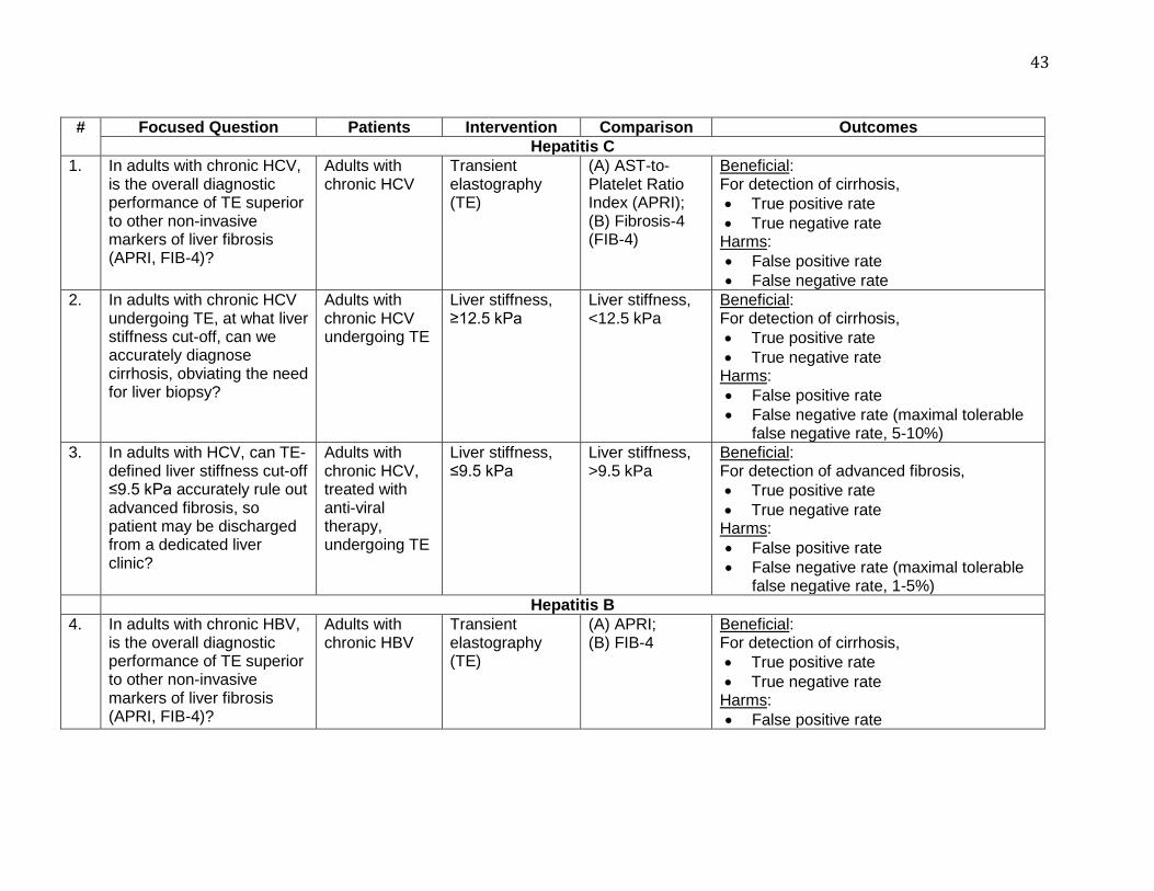
43
# Focused Question Patients Intervention Comparison Outcomes
Hepatitis C
1. In adults with chronic HCV, is the overall diagnostic performance of TE superior to other non-invasive markers of liver fibrosis (APRI, FIB-4)?
Adults with chronic HCV
Transient elastography (TE)
(A) AST-to-Platelet Ratio Index (APRI); (B) Fibrosis-4 (FIB-4)
Beneficial: For detection of cirrhosis,
True positive rate
True negative rate Harms:
False positive rate
False negative rate
2. In adults with chronic HCV undergoing TE, at what liver stiffness cut-off, can we accurately diagnose cirrhosis, obviating the need for liver biopsy?
Adults with chronic HCV undergoing TE
Liver stiffness, ≥12.5 kPa
Liver stiffness, <12.5 kPa
Beneficial: For detection of cirrhosis,
True positive rate
True negative rate Harms:
False positive rate
False negative rate (maximal tolerable false negative rate, 5-10%)
3. In adults with HCV, can TE-defined liver stiffness cut-off ≤9.5 kPa accurately rule out advanced fibrosis, so patient may be discharged from a dedicated liver clinic?
Adults with chronic HCV, treated with anti-viral therapy, undergoing TE
Liver stiffness, ≤9.5 kPa
Liver stiffness, >9.5 kPa
Beneficial: For detection of advanced fibrosis,
True positive rate
True negative rate Harms:
False positive rate
False negative rate (maximal tolerable false negative rate, 1-5%)
Hepatitis B
4. In adults with chronic HBV, is the overall diagnostic performance of TE superior to other non-invasive markers of liver fibrosis (APRI, FIB-4)?
Adults with chronic HBV
Transient elastography (TE)
(A) APRI; (B) FIB-4
Beneficial: For detection of cirrhosis,
True positive rate
True negative rate Harms:
False positive rate

44
False negative rate
5. In adults with chronic HBV undergoing TE, at what liver stiffness cut-off, can we accurately diagnose cirrhosis, obviating the need for liver biopsy?
Adults with chronic HBV undergoing TE
Liver stiffness, ≥11 kPa
Liver stiffness, <11 kPa
Beneficial: For detection of cirrhosis,
True positive rate
True negative rate Harms:
False positive rate
False negative rate (maximal tolerable false negative rate, 5-10%)
Non-alcoholic fatty liver disease
6. In adults with NAFLD, is the overall diagnostic performance of TE superior to other non-invasive markers of liver fibrosis (APRI, FIB-4)?
Adults with chronic NAFLD
Transient elastography (TE)
(A) APRI; (B) FIB-4
Beneficial: For detection of cirrhosis,
True positive rate
True negative rate Harms:
False positive rate
False negative rate
7. In adults with NAFLD undergoing TE, at what liver stiffness cut-off, can we accurately diagnose cirrhosis, obviating the need for liver biopsy?
Adults with NAFLD undergoing TE
Liver stiffness, ≥11 kPa
Liver stiffness, <11 kPa
Beneficial: For detection of cirrhosis,
True positive rate
True negative rate Harms:
False positive rate
False negative rate
Alcoholic liver disease
8. In adults with chronic alcoholic liver disease undergoing TE, at what liver stiffness cut-off, can we accurately diagnose cirrhosis, obviating the need for liver biopsy?
Adults with chronic alcoholic liver disease undergoing TE
Liver stiffness, ≥12.5 kPa
Liver stiffness, <12.5 kPa
Beneficial: For detection of cirrhosis,
True positive rate
True negative rate Harms:
False positive rate
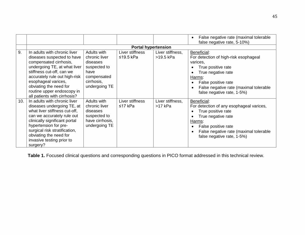
45
Table 1. Focused clinical questions and corresponding questions in PICO format addressed in this technical review.
False negative rate (maximal tolerable false negative rate, 5-10%)
Portal hypertension
9. In adults with chronic liver diseases suspected to have compensated cirrhosis, undergoing TE, at what liver stiffness cut-off, can we accurately rule out high-risk esophageal varices, obviating the need for routine upper endoscopy in all patients with cirrhosis?
Adults with chronic liver diseases suspected to have compensated cirrhosis, undergoing TE
Liver stiffness ≤19.5 kPa
Liver stiffness, >19.5 kPa
Beneficial: For detection of high-risk esophageal varices,
True positive rate
True negative rate Harms:
False positive rate
False negative rate (maximal tolerable false negative rate, 1-5%)
10. In adults with chronic liver diseases undergoing TE, at what liver stiffness cut-off, can we accurately rule out clinically significant portal hypertension for pre-surgical risk stratification, obviating the need for invasive testing prior to surgery?
Adults with chronic liver diseases suspected to have cirrhosis, undergoing TE
Liver stiffness ≤17 kPa
Liver stiffness, >17 kPa
Beneficial: For detection of any esophageal varices,
True positive rate
True negative rate Harms:
False positive rate
False negative rate (maximal tolerable false negative rate, 1-5%)

46
PICO Condition/ Outcome
Liver stiffness cut-off
Sensitivity Specificity
Positive predictive value Negative predictive value
Low prevalence§
High prevalence¶
Low prevalence§
High prevalence¶
2 Hepatitis C; Cirrhosis
12.5 (±1) kPa
86% 91% 33% (25-42) 80% (76-85) 99% (98-100) 94% (92-96)
3 Hepatitis C; Advanced fibrosis or cirrhosis
9.5 (±1) kPa
78% 86% 23% (17-30) 70% (65-75) 99% (98-99) 90% (88-92)
5 Hepatitis B; Cirrhosis
11 (±1) kPa
81% 83% 20% (15-27) 67% (62-72) 99% (98-99) 91% (89-93)
8 Alcoholic liver disease; Cirrhosis
12.5 (±1) kPa
95% 71% 15% (11-19) 100% (99-100) 58% (54-63) 97% (95-98)
9 Chronic liver disease; High-risk EVs§
19.5 (±2) kPa
89% 56% 10% (7-13) 34% (30-38) 99% (98-100) 95% (93-97)
10 Suspected liver disease; Any EVs
17.0 (±2) kPa
83% 52% Very low risk: 1% (0-2) Low risk: 8% (6-11)
54% (50-58) Very low risk: 100% (99-100)
Low risk: 98% (97-99)
82% (78-86)
§Low prevalence corresponds to an illustrative low-risk population with prevalence of outcome, 5% ¶High prevalence corresponds to an illustrative high-risk population with a prevalence of cirrhosis/advanced fibrosis (PICOs# 2, 3, 5, 8) of 30%, prevalence of high-risk EVs (PICO# 9) of 20% and prevalence of any EVs (PICO# 10) of 40%
Table 2. Performance characteristics of suggested stiffness cut-offs in different disease conditions. Please note that while identifying cut-offs we placed more emphasis on high negative predictive value.
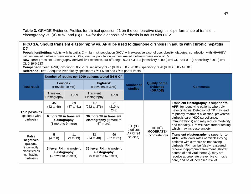
47
Table 3. GRADE Evidence Profiles for clinical question #1 on the comparative diagnostic performance of transient elastography vs. (A) APRI and (B) FIB-4 for the diagnosis of cirrhosis in adults with HCV
PICO 1A. Should transient elastography vs. APRI be used to diagnose cirrhosis in adults with chronic hepatitis C? Population/Setting: Adults with hepatitis C – high-risk population (HCV with excessive alcohol use, obesity, diabetes, co-infection with HIV/HBV) with estimated cirrhosis prevalence of 30%; low-risk population with estimated cirrhosis prevalence of 5% New Test: Transient Elastography-derived liver stiffness, cut-off range: 9.2-17.3 kPa [sensitivity: 0.89 (95% CI, 0.84-0.92); specificity: 0.91 (95% CI, 0.89-0.92)] Comparison Test: APRI, low cut-off: 0.75-1.0 [sensitivity: 0.77 (95% CI, 0.73-0.81); specificity: 0.78 (95% CI: 0.74-0.81)] Reference Test: Adequate liver biopsy specimen; >/= 1.5 cm and >/= 6 portal tracts
Test result
Number of results per 1000 patients tested (95% CI)
Number of studies
Quality of the Evidence (GRADE)
Comments Low-risk
(Prevalence 5%) High-risk
(Prevalence 30%)
Transient Elastography
APRI Transient
Elastography APRI
True positives (patients with
cirrhosis)
45 (42 to 46)
39 (37 to 41)
267 (252 to 276)
231 (219 to 243)
TE (36 studies); APRI (24 studies)
⨁⨁⨁◯ MODERATE1
(Inconsistency)
Transient elastography is superior to APRI for identifying patients who truly have cirrhosis. Detection of TP may lead to priority treatment allocation, preventive cirrhosis care (HCC surveillance, immunizations) and may reduce morbidity and mortality. TPs will have further testing which may increase anxiety.
6 more TP in transient elastography
(1 more to 9 more)
36 more TP in transient elastography (9 more to
57 more)
False negatives (patients
incorrectly classified as not having cirrhosis)
5 (4 to 8)
11 (9 to 13)
33 (24 to 48)
69 (57 to 81)
Transient elastography is superior to APRI, with lower rates of misclassifying patients with cirrhosis as not having cirrhosis. FN may be falsely reassured, receive inappropriate treatment (shorter course of anti-viral therapy), may not receive appropriate preventive cirrhosis care, and be at increased risk of
6 fewer FN in transient elastography
(1 fewer to 9 fewer)
36 fewer FN in transient elastography
(9 fewer to 57 fewer)

48
Test result
Number of results per 1000 patients tested (95% CI)
Number of studies
Quality of the Evidence (GRADE)
Comments Low-risk
(Prevalence 5%) High-risk
(Prevalence 30%)
Transient Elastography
APRI Transient
Elastography APRI
progression to hepatic decompensation, and potentially increased morbidity and mortality.
True negatives (patients without
cirrhosis)
864 (845 to 884)
741 (703 to 770)
637 (623 to 651)
546 (518 to 567)
⨁⨁⨁◯ MODERATE1
(Inconsistency)
Transient elastography is superior to APRI for identifying patients who truly do not have cirrhosis. TN may be reassured and obviate the need for invasive testing with liver biopsy, although they may need to undergo repeated assessment of liver stiffness at periodic intervals.
123 more TN in transient elastography
(75 more to 181 more)
91 more TN in transient elastography
(56 more to 133 more)
False positives (patients
incorrectly classified as
having cirrhosis)
86 (66 to 105)
209 (180 to 247)
63 (49 to 77)
154 (133 to 182)
Transient elastography is superior to APRI, with lower rates of misclassifying patients without cirrhosis as having cirrhosis. FP may receive unnecessary testing (HCC surveillance, immunization) and treatment (shorter course of anti-viral therapy) and have avoidable anxiety, potential testing- or treatment-related complications and excessive resource utilization.
123 fewer FP in transient elastography
(75 fewer to 181 fewer)
91 fewer FP in transient elastography
(56 fewer to 133 fewer)
1High heterogeneity, with wide range of liver stiffness cut-offs

49
PICO 1B. Should transient elastography vs. FIB-4 be used to diagnose cirrhosis in adults with chronic hepatitis C? Population/Setting: Adults with hepatitis C – high-risk population (HCV with excessive alcohol use, obesity, diabetes, co-infection with HIV/HBV) with estimated cirrhosis prevalence of 30%; low-risk population with estimated cirrhosis prevalence of 5% New Test: Transient Elastography-derived liver stiffness, cut-off range: 9.2-17.3 kPa [sensitivity: 0.89 (95% CI, 0.84-0.92); specificity: 0.91 (95% CI, 0.89-0.92)] Comparison Test: FIB-4, low cut-off: 1.45 [sensitivity: 0.87 (95% CI, 0.74-0.94); specificity: 0.61 (95% CI: 0.53-0.69)] Reference Test: Adequate liver biopsy specimen; >/= 1.5 cm and >/= 6 portal tracts
Test result
Number of results per 1000 patients tested (95% CI)
Number of studies
Quality of the Evidence (GRADE)
Comments Low-risk
(Prevalence 5%) High-risk
(Prevalence 30%)
Transient Elastography
FIB-4 Transient
Elastography FIB-4
True positives (patients with
cirrhosis)
45 (42 to 46)
44 (37 to 47)
267 (252 to 276)
261 (222 to 282)
TE (36 studies); FIB-4 (2 studies)
⨁⨁◯◯ LOW1,2
(Inconsistency, Imprecision)
Transient elastography may be superior to FIB-4 for identifying patients who truly have cirrhosis. Detection of TP may lead to priority treatment allocation, preventive cirrhosis care (HCC surveillance, immunizations) and may reduce morbidity and mortality. TPs will have further testing which may increase anxiety.
1 more TP in transient elastography
(5 fewer to 9 more)
6 more TP in transient elastography
(30 fewer to 54 more)
False negatives (patients
incorrectly classified as not having cirrhosis)
5 (4 to 8)
6 (3 to 13)
33 (24 to 48)
39 (18 to 78)
Transient elastography may be superior to FIB-4, with lower rates of misclassifying patients with cirrhosis as not having cirrhosis. FN may be falsely reassured, receive inappropriate treatment (shorter course of anti-viral therapy), may not receive appropriate preventive cirrhosis care, and be at increased risk of progression to hepatic decompensation, and potentially increased morbidity and mortality.
1 fewer FN in transient elastography
(5 more to 9 fewer)
6 fewer FN in transient elastography
(30 more to 54 fewer)

50
Test result
Number of results per 1000 patients tested (95% CI)
Number of studies
Quality of the Evidence (GRADE)
Comments Low-risk
(Prevalence 5%) High-risk
(Prevalence 30%)
Transient Elastography
FIB-4 Transient
Elastography FIB-4
True negatives (patients without
cirrhosis)
864 (845 to 874)
580 (503 to 656)
637 (623 to 644)
427 (371 to 483)
⨁⨁⨁◯ MODERATE1
(Inconsistency)
Transient elastography is superior to FIB-4 for identifying patients who truly do not have cirrhosis. TN may be reassured and obviate the need for invasive testing with liver biopsy, although they may need to undergo repeated assessment of liver stiffness at periodic intervals.
284 more TN in transient elastography
(189 more to 371 more)
210 more TN in transient elastography
(140 more to 273 more)
False positives (patients
incorrectly classified as
having cirrhosis)
86 (76 to 105)
370 (294 to 447)
63 (56 to 77)
273 (217 to 329)
Transient elastography is superior to FIB-4, with lower rates of misclassifying patients without cirrhosis as having cirrhosis. FP may receive unnecessary testing (HCC surveillance, immunization) and treatment (longer course of anti-viral therapy) and have avoidable anxiety, potential testing- or treatment-related complications and excessive resource utilization.
284 fewer FP in transient elastography
(189 fewer to 371 fewer)
210 fewer FP in transient elastography
(140 fewer to 273 fewer)
1High heterogeneity, with wide range of liver stiffness cut-offs 2Overlapping confidence intervals for rates of TP and FN

51
Table 4. GRADE Evidence Profiles for clinical question #2 on the performance of transient elastography threshold of 12.5(±1) for the diagnosis of cirrhosis and anticipated downstream consequences in patients with HCV.
PICO 2. In adults with HCV undergoing TE, at what liver stiffness cut-off, can we accurately diagnose cirrhosis, obviating the need for liver biopsy? Population/Setting: Adults with hepatitis C – high-risk population (HCV with excessive alcohol use, obesity, diabetes, co-infection with HIV/HBV) with estimated cirrhosis prevalence of 30%; low-risk population with estimated cirrhosis prevalence of 5% Pooled sensitivity TE with cut-off 12.5(±1) kPa: 0.86 (95% CI: 0.83-0.88) | Pooled specificity TE with cut-off 12.5(±1) kPa: 0.91 (95% CI: 0.89-0.92) Reference Test: Adequate liver biopsy specimen; >/= 1.5 cm and >/= 6 portal tracts
Test result
Number of results per 1000 patients tested (95% CI)
Number of studies,
participants
Quality of the Evidence (GRADE)
Comments
Low-risk (Prevalence
5%)
High-risk (Prevalence
30%)
True positives (patients with
cirrhosis)
43 (42 to 44)
258 (249 to 264)
17 studies, 5812 patients
⨁⨁◯◯ LOW1,2
(Inconsistency, Indirectness)
Detection of TP with TE may lead to priority treatment allocation, preventive cirrhosis care (HCC surveillance, immunizations) and may reduce morbidity and mortality. TPs will have further testing which may increase anxiety. By avoiding liver biopsy, these patients would avoid potential complications of liver biopsy, like pain, bleeding, etc.
False negatives (patients
incorrectly classified as not having cirrhosis)
7 (6 to 8)
42 (36 to 51)
FN may be falsely reassured, receive inappropriate treatment (shorter course of anti-viral therapy), may not receive appropriate preventive cirrhosis care, and be at increased risk of progression to hepatic decompensation, and potentially increased morbidity and mortality. This potential under-diagnosis may have been avoided if all patients had undergone liver biopsy instead

52
Test result
Number of results per 1000 patients tested (95% CI)
Number of studies,
participants
Quality of the Evidence (GRADE)
Comments
Low-risk (Prevalence
5%)
High-risk (Prevalence
30%)
of TE, but that would put patients at risk for liver biopsy-related complications.
True negatives (patients without
cirrhosis)
864 (845 to 874)
637 (623 to 644)
TN may be reassured and obviate the need for invasive testing with liver biopsy (and potential complications related to it), although they may need to undergo repeated assessment of liver stiffness at periodic intervals.
False positives (patients
incorrectly classified as
having cirrhosis)
86 (76 to 105)
63 (56 to 77)
FP may receive unnecessary testing (HCC surveillance, immunization) and treatment (longer course of anti-viral therapy) and have avoidable anxiety, potential testing- or treatment-related complications and excessive resource utilization. This potential over-diagnosis may have been avoided if all patients had undergone liver biopsy instead of TE, but that would put patients at risk for liver biopsy-related complications.
1High heterogeneity, selective inclusion of studies corresponding to cut-off of 12.5±1 kPa; 2Surrogate patient-important outcomes (FP, FN are surrogates for presumed downstream consequences on patient-important outcomes), no predetermined liver stiffness cut-off, which was determined post-hoc in individual studies, corresponding to AUROC

53
Table 5. GRADE Evidence Profiles for clinical question #3 on the performance of transient elastography threshold of 9.5(±1) for the diagnosis of advanced fibrosis and anticipated downstream consequences in patients with HCV who achieve sustained virologic response after treatment.
PICO 3. In adults with HCV, can TE-defined liver stiffness cut-off ≤9.5 kPa accurately rule out advanced fibrosis, so patient may be discharged from a dedicated liver clinic? Population/Setting: Adults with hepatitis C – high-risk population (HCV patients who have achieved sustained virologic response with anti-viral therapy, with ongoing risk factors for liver disease - excessive alcohol use, obesity, diabetes, co-infection with HIV/HBV) with estimated advanced fibrosis prevalence of 30%; low-risk population with estimated advanced fibrosis prevalence of 5% Pooled sensitivity TE with cut-off 9.5 (±1) kPa: 0.78 (95% CI: 0.75-0.81) | Pooled specificity TE with cut-off 9.5 (±1) kPa: 0.86 (95% CI: 0.84-0.88) Reference Test: Adequate liver biopsy specimen; >/= 1.5 cm and >/= 6 portal tracts
Test result
Number of results per 1000 patients tested (95% CI)
Number of studies,
participants
Quality of the Evidence (GRADE)
Comments
Low-risk (Prevalence
5%)
High-risk (Prevalence
30%)
True positives (patients with advanced fibrosis)
39 (38 to 41)
234 (225 to 243)
13 studies, 4106 patients
⨁◯◯◯ VERY LOW1,2
(Inconsistency, Very serious indirectness)
TP will have further follow-up in dedicated liver clinic. Detection of TP would lead to continuation of preventive cirrhosis care (HCC surveillance) and may reduce morbidity and mortality. TP may experience excessive resource utilization and anxiety.
False negatives (patients incorrectly classified as not having advanced fibrosis)
11 (9 to 12)
66 (57 to 75)
FN may be falsely reassured and discharged from dedicated liver clinic, and may not receive appropriate preventive cirrhosis care, and be at increased risk of progression to hepatic decompensation, and potentially increased morbidity and mortality.

54
Test result
Number of results per 1000 patients tested (95% CI)
Number of studies,
participants
Quality of the Evidence (GRADE)
Comments
Low-risk (Prevalence
5%)
High-risk (Prevalence
30%)
True negatives (patients without advanced fibrosis)
817 (798 to 836)
602 (588 to 616)
TN would be discharged from dedicated liver clinic, and may not need ongoing preventive cirrhosis care. TN may still be at low risk for liver-related events.
False positives (patients incorrectly classified as having advanced fibrosis)
133 (114 to 152)
98 (84 to 112)
FP will receive continued care in dedicated liver clinic, receiving unnecessary testing (HCC surveillance) and have avoidable anxiety, potential testing-related complications and excessive resource utilization.
1High heterogeneity, selective inclusion of studies corresponding to cut-off of 9.5±1 kPa; 2Diagnostic performance inferred from studies in the general population with HCV with or without therapy, rather than studies in patients who achieved SVR; surrogate patient-important outcomes (FP, FN are surrogates for presumed downstream consequences on patient-important outcomes), no predetermined liver stiffness cut-off which was determined post-hoc in individual studies, corresponding to AUROC
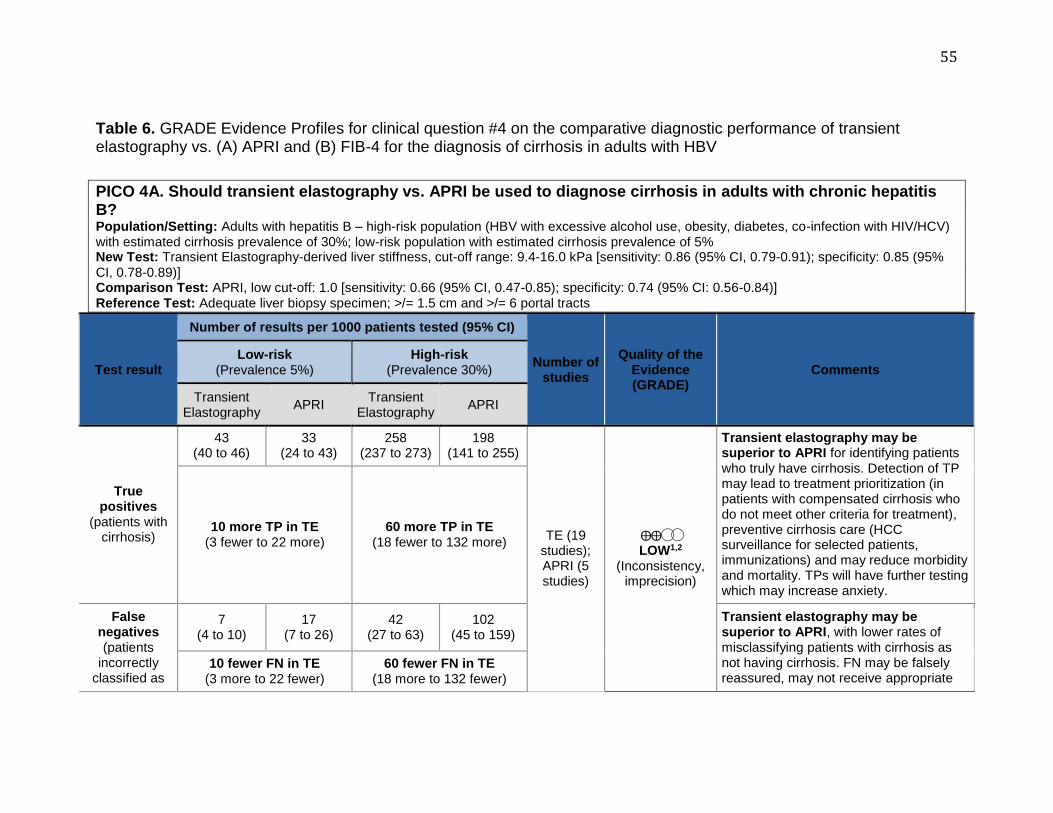
55
Table 6. GRADE Evidence Profiles for clinical question #4 on the comparative diagnostic performance of transient elastography vs. (A) APRI and (B) FIB-4 for the diagnosis of cirrhosis in adults with HBV
PICO 4A. Should transient elastography vs. APRI be used to diagnose cirrhosis in adults with chronic hepatitis B? Population/Setting: Adults with hepatitis B – high-risk population (HBV with excessive alcohol use, obesity, diabetes, co-infection with HIV/HCV) with estimated cirrhosis prevalence of 30%; low-risk population with estimated cirrhosis prevalence of 5% New Test: Transient Elastography-derived liver stiffness, cut-off range: 9.4-16.0 kPa [sensitivity: 0.86 (95% CI, 0.79-0.91); specificity: 0.85 (95% CI, 0.78-0.89)] Comparison Test: APRI, low cut-off: 1.0 [sensitivity: 0.66 (95% CI, 0.47-0.85); specificity: 0.74 (95% CI: 0.56-0.84)] Reference Test: Adequate liver biopsy specimen; >/= 1.5 cm and >/= 6 portal tracts
Test result
Number of results per 1000 patients tested (95% CI)
Number of studies
Quality of the Evidence (GRADE)
Comments Low-risk
(Prevalence 5%) High-risk
(Prevalence 30%)
Transient Elastography
APRI Transient
Elastography APRI
True positives
(patients with cirrhosis)
43 (40 to 46)
33 (24 to 43)
258 (237 to 273)
198 (141 to 255)
TE (19 studies); APRI (5 studies)
⨁⨁◯◯ LOW1,2
(Inconsistency, imprecision)
Transient elastography may be superior to APRI for identifying patients who truly have cirrhosis. Detection of TP may lead to treatment prioritization (in patients with compensated cirrhosis who do not meet other criteria for treatment), preventive cirrhosis care (HCC surveillance for selected patients, immunizations) and may reduce morbidity and mortality. TPs will have further testing which may increase anxiety.
10 more TP in TE (3 fewer to 22 more)
60 more TP in TE (18 fewer to 132 more)
False negatives (patients
incorrectly classified as
7 (4 to 10)
17 (7 to 26)
42 (27 to 63)
102 (45 to 159)
Transient elastography may be superior to APRI, with lower rates of misclassifying patients with cirrhosis as not having cirrhosis. FN may be falsely reassured, may not receive appropriate
10 fewer FN in TE (3 more to 22 fewer)
60 fewer FN in TE (18 more to 132 fewer)

56
Test result
Number of results per 1000 patients tested (95% CI)
Number of studies
Quality of the Evidence (GRADE)
Comments Low-risk
(Prevalence 5%) High-risk
(Prevalence 30%)
Transient Elastography
APRI Transient
Elastography APRI
not having cirrhosis)
preventive cirrhosis care, and be at increased risk of progression to hepatic decompensation, and potentially increased morbidity and mortality.
True negatives (patients without
cirrhosis)
808 (741 to 845)
703 (532 to 798)
595 (546 to 623)
518 (392 to 588)
⨁⨁◯◯ LOW1,2
(Inconsistency, imprecision)
Transient elastography may be superior to APRI for identifying patients who truly do not have cirrhosis. TN may be reassured and obviate the need for invasive testing with liver biopsy, although they may need to undergo repeated assessment of liver stiffness at periodic intervals.
105 more TN in TE (57 fewer to 313 more)
77 more TN in TE (42 fewer to 231 more)
False positives (patients
incorrectly classified as
having cirrhosis)
142 (105 to 209)
247 (152 to 418)
105 (77 to 154)
182 (112 to 308)
Transient elastography may be superior to APRI, with lower rates of misclassifying patients without cirrhosis as having cirrhosis. FP may receive unnecessary testing (HCC surveillance, immunization) and treatment (HBV therapy) and have avoidable anxiety, potential testing- or treatment-related complications and excessive resource utilization.
105 fewer FP in TE (57 more to 313 fewer)
77 fewer FP in TE (42 more to 231 fewer)
1High heterogeneity, with wide range of liver stiffness cut-offs 2Overlapping confidence intervals for rates of TP, FN, TN, FP
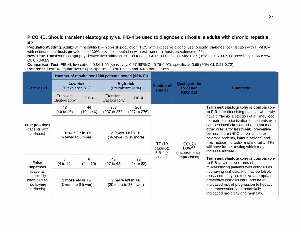
57
PICO 4B. Should transient elastography vs. FIB-4 be used to diagnose cirrhosis in adults with chronic hepatitis B? Population/Setting: Adults with hepatitis B – high-risk population (HBV with excessive alcohol use, obesity, diabetes, co-infection with HIV/HCV) with estimated cirrhosis prevalence of 30%; low-risk population with estimated cirrhosis prevalence of 5% New Test: Transient Elastography-derived liver stiffness, cut-off range: 9.4-16.0 kPa [sensitivity: 0.86 (95% CI, 0.79-0.91); specificity: 0.85 (95% CI, 0.78-0.89)] Comparison Test: FIB-4I, low cut-off: 0.84-1.05 [sensitivity: 0.87 (95% CI, 0.79-0.92); specificity: 0.65 (95% CI: 0.51-0.73)] Reference Test: Adequate liver biopsy specimen; >/= 1.5 cm and >/= 6 portal tracts
Test result
Number of results per 1000 patients tested (95% CI)
Number of studies
Quality of the Evidence (GRADE)
Comments Low-risk
(Prevalence 5%) High-risk
(Prevalence 30%)
Transient Elastography
FIB-4 Transient
Elastography FIB-4
True positives (patients with
cirrhosis)
43 (40 to 46)
44 (40 to 46)
258 (237 to 273)
261 (237 to 276)
TE (19 studies); FIB-4 (4 studies)
⨁⨁◯◯ LOW1,2
(Inconsistency, imprecision)
Transient elastography is comparable to FIB-4 for identifying patients who truly have cirrhosis. Detection of TP may lead to treatment prioritization (in patients with compensated cirrhosis who do not meet other criteria for treatment), preventive cirrhosis care (HCC surveillance for selected patients, immunizations) and may reduce morbidity and mortality. TPs will have further testing which may increase anxiety.
1 fewer TP in TE (6 fewer to 6 more)
3 fewer TP in TE (39 fewer to 36 more)
False negatives (patients
incorrectly classified as not having cirrhosis)
7 (4 to 10)
6 (4 to 10)
42 (27 to 63)
39 (24 to 63)
Transient elastography is comparable to FIB-4, with lower rates of misclassifying patients with cirrhosis as not having cirrhosis. FN may be falsely reassured, may not receive appropriate preventive cirrhosis care, and be at increased risk of progression to hepatic decompensation, and potentially increased morbidity and mortality.
1 more FN in TE (6 more to 6 fewer)
3 more FN in TE (39 more to 36 fewer)

58
Test result
Number of results per 1000 patients tested (95% CI)
Number of studies
Quality of the Evidence (GRADE)
Comments Low-risk
(Prevalence 5%) High-risk
(Prevalence 30%)
Transient Elastography
FIB-4 Transient
Elastography FIB-4
True negatives (patients without
cirrhosis)
808 (741 to 845)
617 (485 to 694)
595 (546 to 623)
455 (357 to 511)
⨁⨁⨁◯ MODERATE1
(Inconsistency)
Transient elastography is superior to FIB-4 for identifying patients who truly do not have cirrhosis. TN may be reassured and obviate the need for invasive testing with liver biopsy, although they may need to undergo repeated assessment of liver stiffness at periodic intervals.
191 more TN in TE (47 more to 360 more)
140 more TN in TE (35 more to 266 more)
False positives (patients
incorrectly classified as
having cirrhosis)
142 (105 to 209)
333 (256 to 465)
105 (77 to 154)
245 (189 to 343)
Transient elastography is superior to FIB-4, with lower rates of misclassifying patients without cirrhosis as having cirrhosis. FP may receive unnecessary testing (HCC surveillance, immunization) and treatment (HBV therapy) and have avoidable anxiety, potential testing- or treatment-related complications and excessive resource utilization.
191 fewer FP in TE (47 fewer to 360 fewer)
140 fewer FP in TE (35 fewer to 266 fewer)
1High heterogeneity, with wide range of liver stiffness cut-offs 2Overlapping confidence intervals for rates of TP and FN

59
Table 7. GRADE Evidence Profiles for clinical question #5 on the performance of transient elastography threshold of 11.0(±1) for the diagnosis of cirrhosis and anticipated downstream consequences in patients with HBV.
PICO 5. In adults with HBV undergoing TE, at what liver stiffness cut-off, can we accurately diagnose cirrhosis, obviating the need for liver biopsy? Population/Setting: Adults with hepatitis B – high-risk population (HBV with excessive alcohol use, obesity, diabetes, co-infection with HIV/HCV) with estimated cirrhosis prevalence of 30%; low-risk population with estimated cirrhosis prevalence of 5% Pooled sensitivity TE with cut-off 11.0(±1) kPa: 0.81 (95% CI: 0.79-0.0.84 | Pooled specificity TE with cut-off 11.0(±1) kPa: 0.83 (95% CI: 0.82-0.84 Reference Test: Adequate liver biopsy specimen; >/= 1.5 cm and >/= 6 portal tracts
Test result
Number of results per 1000 patients tested (95% CI)
Number of studies,
participants
Quality of the Evidence (GRADE)
Comments
Low-risk (Prevalence
5%)
High-risk (Prevalence
30%)
True positives (patients with
cirrhosis) 41 (40 to 42) 243 (237 to 252)
17 studies, 4864 patients
⨁⨁◯◯ LOW1,2
(inconsistency, indirectness)
Detection of TP may lead to treatment prioritization (in patients with compensated cirrhosis who do not meet other criteria for treatment), preventive cirrhosis care (HCC surveillance for selected patients, immunizations) and may reduce morbidity and mortality. TPs will have further testing which may increase anxiety. By avoiding liver biopsy, these patients would avoid potential complications of liver biopsy, like pain, bleeding, etc.
False negatives (patients
incorrectly classified as not having cirrhosis)
9 (8 to 10) 57 (48 to 63)
FN may be falsely reassured, may not receive appropriate preventive cirrhosis care, and be at increased risk of progression to hepatic decompensation, and potentially increased morbidity and mortality. This potential under-diagnosis may have been

60
Test result
Number of results per 1000 patients tested (95% CI)
Number of studies,
participants
Quality of the Evidence (GRADE)
Comments
Low-risk (Prevalence
5%)
High-risk (Prevalence
30%)
avoided if all patients had undergone liver biopsy instead of TE, but that would put patients at risk for liver biopsy-related complications.
True negatives (patients without
cirrhosis)
789 (779 to 798)
581 (574 to 588)
TN may be reassured and obviate the need for invasive testing with liver biopsy (and potential complications related to it), although they may need to undergo repeated assessment of liver stiffness at periodic intervals.
False positives (patients
incorrectly classified as
having cirrhosis)
161 (152 to 171)
119 (112 to 126)
FP may receive unnecessary testing (HCC surveillance, immunization) and treatment (HBV therapy) and have avoidable anxiety, potential testing- or treatment-related complications and excessive resource utilization. This potential over-diagnosis may have been avoided if all patients had undergone liver biopsy instead of TE, but that would put patients at risk for liver biopsy-related complications.
1High heterogeneity, selective inclusion of studies corresponding to cut-off of 11.0±1 kPa; 2Surrogate patient-important outcomes (FP, FN are surrogates for presumed downstream consequences on patient-important outcomes), no predetermined liver stiffness cut-off, which was determined post-hoc in individual studies, corresponding to AUROC

61
Table 8. GRADE Evidence Profiles for clinical question #6 on the comparative diagnostic performance of transient elastography vs. (A) APRI and (B) FIB-4 for the diagnosis of cirrhosis in adults with NAFLD
PICO 6A. Should transient elastography vs. APRI be used to diagnose cirrhosis in adults with NAFLD? Population/Setting: Adults with NAFLD – high-risk population (NAFLD with advanced age, obesity, particularly central adiposity, diabetes, alanine elevated >2x upper limit of normal) with estimated cirrhosis prevalence of 30%; low-risk population with estimated cirrhosis prevalence of 5% New Test: Transient Elastography-derived liver stiffness, cut-off range: 10.3-22.3 kPa [sensitivity: 0.90 (95% CI, 0.82-0.95); specificity: 0.87 (95% CI, 0.85-0.89)] Comparison Test: APRI, cut-off: 0.50 [sensitivity: 0.78 (95% CI, 0.71-0.99); specificity: 0.71 (95% CI: 0.30-0.93)] Reference Test: Adequate liver biopsy specimen; >/= 1.5 cm and >/= 6 portal tracts
Test result
Number of results per 1000 patients tested (95% CI)
Number of studies
Quality of the Evidence (GRADE)
Comments Low-risk
(Prevalence 5%) High-risk
(Prevalence 30%)
Transient Elastography
APRI Transient
Elastography APRI
True positives (patients with
cirrhosis)
45 (41 to 48)
39 (36 to 50)
270 (246 to 285)
234 (213 to 297)
TE (9 studies); APRI (2 studies)
⨁◯◯◯ VERY LOW1,2,3 (Risk of bias, inconsistency, imprecision)
Transient elastography may be superior to APRI for identifying patients who truly have cirrhosis. Detection of TP may lead to preventive cirrhosis care (HCC surveillance, immunizations) and may reduce morbidity and mortality. TPs will have further testing which may increase anxiety.
6 more TP in transient elastography
(9 fewer to 12 more)
36 more TP in transient elastography
(51 fewer to 72 more)
False negatives (patients
incorrectly classified as not having cirrhosis)
5 (2 to 9)
11 (0 to 14)
30 (15 to 54)
66 (3 to 87)
Transient elastography may be superior to APRI, with lower rates of misclassifying patients with cirrhosis as not having cirrhosis. FN may be falsely reassured, may not receive appropriate preventive cirrhosis care, and be at increased risk of progression to hepatic
6 fewer FN in transient elastography
(9 more to 12 fewer)
36 fewer FN in transient elastography
(51 more to 72 fewer)

62
Test result
Number of results per 1000 patients tested (95% CI)
Number of studies
Quality of the Evidence (GRADE)
Comments Low-risk
(Prevalence 5%) High-risk
(Prevalence 30%)
Transient Elastography
APRI Transient
Elastography APRI
decompensation, and potentially increased morbidity and mortality.
True negatives (patients without
cirrhosis)
827 (808 to 850)
675 (285 to 884)
609 (595 to 627)
497 (210 to 651)
⨁◯◯◯ VERY LOW1,2,3 (Risk of bias, inconsistency, imprecision)
Transient elastography may be superior to APRI for identifying patients who truly do not have cirrhosis. TN may be reassured and obviate the need for invasive testing with liver biopsy, although they may need to undergo repeated assessment of liver stiffness at periodic intervals.
152 more TN in transient elastography
(76 fewer to 365 more)
112 more TN in transient elastography
(56 fewer to 407 more)
False positives (patients
incorrectly classified as
having cirrhosis)
123 (100 to 142)
275 (66 to 665)
91 (73 to 105)
203 (49 to 490)
Transient elastography may be superior to APRI, with lower rates of misclassifying patients without cirrhosis as having cirrhosis. FP may receive unnecessary testing (HCC surveillance, immunization) and have avoidable anxiety, potential testing- or treatment-related complications and excessive resource utilization.
152 fewer FP in transient elastography
(76 more to 365 fewer)
112 fewer FP in transient elastography
(56 more to 407 fewer)
1Spectrum bias with diagnostic accuracy based on only patients with successful TE (recognizing high failure rate of TE due to high body mass index), rather than intention-to-diagnose analysis 2High heterogeneity, with wide range of liver stiffness cut-offs 3Overlapping confidence intervals for rates of TP, FN, TN, FP

63
PICO 6B. Should transient elastography vs. FIB-4 be used to diagnose cirrhosis in adults with NAFLD? Population/Setting: Adults with NAFLD – high-risk population (NAFLD with advanced age, obesity, particularly central adiposity, diabetes, alanine elevated >2x upper limit of normal) with estimated cirrhosis prevalence of 30%; low-risk population with estimated cirrhosis prevalence of 5% New Test: Transient Elastography-derived liver stiffness, cut-off range: 10.3-22.3 kPa [sensitivity: 0.90 (95% CI, 0.82-0.95); specificity: 0.87 (95% CI, 0.85-0.89)] Comparison Test: FIB-4, cut-off: 1.92 [sensitivity: 0.74 (95% CI, 0.54-0.87); specificity: 0.71 (95% CI: 0.64-0.76)] Reference Test: Adequate liver biopsy specimen; >/= 1.5 cm and >/= 6 portal tracts
Test result
Number of results per 1000 patients tested (95% CI)
Number of studies
Quality of the Evidence (GRADE)
Comments Low-risk
(Prevalence 5%) High-risk
(Prevalence 30%)
Transient Elastography
FIB-4 Transient
Elastography FIB-4
True positives (patients with
cirrhosis)
45 (41 to 48)
37 (27 to 44)
270 (246 to 285)
222 (162 to 261)
TE (9 studies); FIB-4 (1 study)
⨁◯◯◯ VERY LOW1,2,3 (Risk of bias, inconsistency, imprecision)
Transient elastography may be superior to FIB-4 for identifying patients who truly have cirrhosis. Detection of TP may lead to preventive cirrhosis care (HCC surveillance, immunizations) and may reduce morbidity and mortality. TPs will have further testing which may increase anxiety.
8 more TP in transient elastography
(3 fewer to 21 more)
48 more TP in transient elastography
(15 fewer to 123 more)
False negatives (patients
incorrectly classified as not having cirrhosis)
5 (2 to 9)
13 (6 to 23)
30 (15 to 54)
78 (39 to 138)
Transient elastography may be superior to FIB-4, with lower rates of misclassifying patients with cirrhosis as not having cirrhosis. FN may be falsely reassured, may not receive appropriate preventive cirrhosis care, and be at increased risk of progression to hepatic decompensation, and potentially increased morbidity and mortality.
8 fewer FN in transient elastography
(3 more to 21 fewer)
48 fewer FN in transient elastography
(15 more to 123 fewer)
827 (808 to 845)
675 (608 to 722)
609 (595 to 623)
497 (448 to 532)
⨁⨁◯◯ Transient elastography may be superior to FIB-4 for identifying patients

64
Test result
Number of results per 1000 patients tested (95% CI)
Number of studies
Quality of the Evidence (GRADE)
Comments Low-risk
(Prevalence 5%) High-risk
(Prevalence 30%)
Transient Elastography
FIB-4 Transient
Elastography FIB-4
True negatives (patients without
cirrhosis)
152 more TN in transient elastography
(86 more to 237 more)
112 more TN in transient elastography
(63 more to 175 more)
LOW1,2 (Risk of bias,
inconsistency)
who truly do not have cirrhosis. TN may be reassured and obviate the need for invasive testing with liver biopsy, although they may need to undergo repeated assessment of liver stiffness at periodic intervals.
False positives (patients
incorrectly classified as
having cirrhosis)
123 (105 to 142)
275 (228 to 342)
91 (77 to 105)
203 (168 to 252)
Transient elastography may be superior to FIB-4, with lower rates of misclassifying patients without cirrhosis as having cirrhosis. FP may receive unnecessary testing (HCC surveillance, immunization) and have avoidable anxiety, potential testing- or treatment-related complications and excessive resource utilization.
152 fewer FP in transient elastography
(86 fewer to 237 fewer)
112 fewer FP in transient elastography
(63 fewer to 175 fewer)
1Spectrum bias with diagnostic accuracy based on only patients with successful TE (recognizing high failure rate of TE due to high body mass index), rather than intention-to-diagnose analysis 2High heterogeneity, with wide range of liver stiffness cut-offs 3Overlapping confidence intervals for rates of TP and FN

65
Table 9. GRADE Evidence Profiles for clinical question #8 on the performance of transient elastography threshold of 12.5(±1) for the diagnosis of cirrhosis and anticipated downstream consequences in patients with chronic alcoholic liver disease.
PICO 8. In adults with chronic alcoholic liver disease undergoing TE, at what liver stiffness cut-off, can we accurately diagnose cirrhosis, obviating the need for liver biopsy? Population/Setting: Adults with alcoholic liver disease – high-risk population (chronic alcoholic liver disease with advanced age, obesity, diabetes, co-infection with HIV/HBV/HCV) with estimated cirrhosis prevalence of 30%; low-risk population with estimated cirrhosis prevalence of 5% Pooled sensitivity TE with cut-off 12.5 kPa: 0.95 (95% CI: 0.87-0.98) | Pooled specificity TE with cut-off 12.5 kPa: 0.71 (95% CI: 0.56-0.82) Reference Test: Adequate liver biopsy specimen; >/= 1.5 cm and >/= 6 portal tracts
Test result
Number of results per 1000 patients tested (95% CI)
Number of studies,
participants
Quality of the Evidence (GRADE)
Comments
Low-risk (Prevalence
5%)
High-risk (Prevalence
30%)
True positives (patients with
cirrhosis)
48 (44 to 49)
285 (261 to 294)
7 studies, 330 patients
⨁⨁◯◯ LOW1,2
(Inconsistency, Indirectness)
Detection of TP may lead to preventive cirrhosis care (HCC surveillance, immunizations), potentially enhance motivation to abstain from alcohol and may reduce morbidity and mortality. TPs will have further testing which may increase anxiety. By avoiding liver biopsy, these patients would avoid potential complications of liver biopsy, like pain, bleeding, etc.
False negatives (patients
incorrectly classified as not having cirrhosis)
2 (1 to 6)
15 (6 to 39)
FN may be falsely reassured and may not receive appropriate preventive cirrhosis care, and be at increased risk of progression to hepatic decompensation, and potentially increased morbidity and mortality. This potential under-diagnosis may have been avoided if all patients had undergone liver

66
Test result
Number of results per 1000 patients tested (95% CI)
Number of studies,
participants
Quality of the Evidence (GRADE)
Comments
Low-risk (Prevalence
5%)
High-risk (Prevalence
30%)
biopsy instead of TE, but that would put patients at risk for liver biopsy-related complications.
True negatives (patients without
cirrhosis)
675 (532 to 779)
497 (392 to 574)
TN may be reassured and obviate the need for invasive testing with liver biopsy (and potential complications related to it), although they may need to undergo repeated assessment of liver stiffness at periodic intervals.
False positives (patients
incorrectly classified as
having cirrhosis)
275 (171 to 418)
203 (126 to 308)
FP may receive unnecessary testing (HCC surveillance, immunization) and have avoidable anxiety, potential testing- or treatment-related complications and excessive resource utilization. This potential over-diagnosis may have been avoided if all patients had undergone liver biopsy instead of TE, but that would put patients at risk for liver biopsy-related complications.
1High heterogeneity, selective inclusion of studies corresponding to cut-off of 12.5±1 kPa; 2Surrogate patient-important outcomes (FP, FN are surrogates for presumed downstream consequences on patient-important outcomes), no predetermined liver stiffness cut-off, which was determined post-hoc in individual studies, corresponding to AUROC

67
Table 10. GRADE Evidence Profiles for clinical question #9 on the performance of transient elastography threshold of 19.5(±2) for triaging patients having high-risk esophageal varices and anticipated downstream consequences in patients with compensated cirrhosis.
PICO 9. In adults with suspected cirrhosis due to chronic liver diseases undergoing TE, at what liver stiffness cut-off, can we accurately rule out high-risk esophageal varices, obviating the need for routine endoscopic screening? Population/Setting: Adults with chronic liver diseases suspected to have cirrhosis, without prior esophageal variceal bleeding – high pre-test probability of having high-risk EVs (known compensated cirrhosis, platelet count <150,000/μL) with estimated prevalence of high-risk EVs of 20%; low pre-test probability of having high-risk EVs (newly diagnosed compensated cirrhosis either based on cross-sectional imaging, with platelet count >150,000/μL) with estimated prevalence of high-risk EVs of 5% Pooled sensitivity TE with cut-off 19.5(±2) kPa: 0.89 (95% CI: 0.84 to 0.92) | Pooled specificity TE with cut-off 19.5(±2) kPa: 0.56 (95% CI: 0.52 to 0.59) Reference Test: Upper endoscopy
Test result
Number of results per 1000 patients tested (95% CI)
Number of studies,
participants
Quality of the Evidence (GRADE)
Comments
Low-risk (Prevalence
5%)
High-risk (Prevalence
20%)
True positives (patients with high-
risk EVs)
44 (42 to 46)
178 (168 to 185)
8 studies, 964 patients
⨁⨁◯◯ LOW1,2
(Inconsistency, Indirectness)
TP may lead to preventive endoscopy for confirmation and initiation of therapy with beta-blockers, potentially reducing mortality. TP will have further testing (EGD) and/or intervention which may lead to side effects
False negatives (patients
incorrectly classified as not having high-risk
EVs)
6 (4 to 8)
22 (15 to 32)
FN may lead to increased risk of variceal bleeding with associated increased mortality due to delayed diagnosis

68
Test result
Number of results per 1000 patients tested (95% CI)
Number of studies,
participants
Quality of the Evidence (GRADE)
Comments
Low-risk (Prevalence
5%)
High-risk (Prevalence
20%)
True negatives (patients without high-risk EVs)
532 (494 to 565)
448 (416 to 476)
TN will likely be reassured, avoid an invasive test but may still be retested with TE periodically
False positives (patients
incorrectly classified as
having high-risk EVs)
418 (385 to 456)
352 (324 to 384)
FP will likely have further testing (which they anyways would have with current standard of care) and will increase anxiety, complications and resources use.
1High heterogeneity, selective inclusion of studies corresponding to cut-off of 19.5±2 kPa; 2Surrogate patient-important outcomes (FP, FN are surrogates for presumed downstream consequences on patient-important outcomes); no predetermined liver stiffness cut-off, which was determined post-hoc in individual studies, corresponding to AUROC; observed prevalence of high-risk EV in studies was much higher than that used in illustrative examples (median, 24.7%; range, 10.3-37)
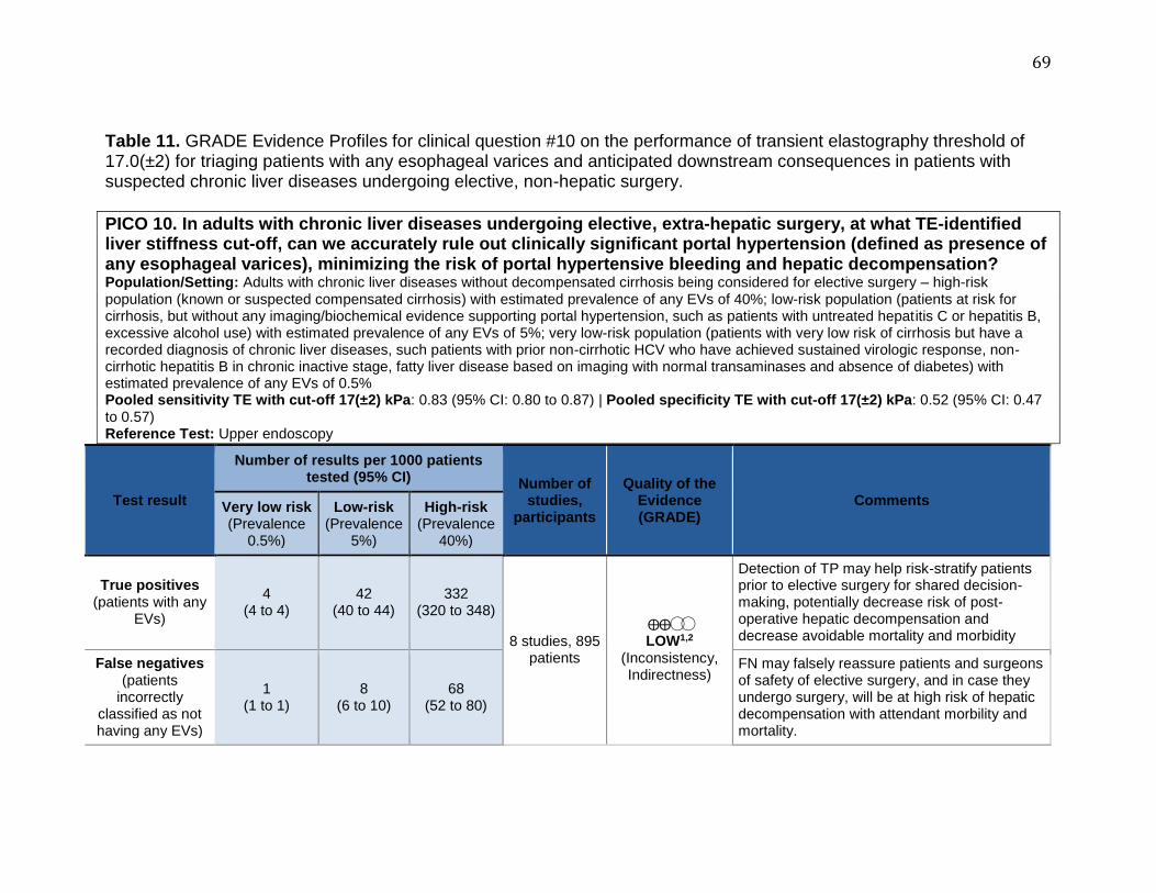
69
Table 11. GRADE Evidence Profiles for clinical question #10 on the performance of transient elastography threshold of 17.0(±2) for triaging patients with any esophageal varices and anticipated downstream consequences in patients with suspected chronic liver diseases undergoing elective, non-hepatic surgery.
PICO 10. In adults with chronic liver diseases undergoing elective, extra-hepatic surgery, at what TE-identified liver stiffness cut-off, can we accurately rule out clinically significant portal hypertension (defined as presence of any esophageal varices), minimizing the risk of portal hypertensive bleeding and hepatic decompensation? Population/Setting: Adults with chronic liver diseases without decompensated cirrhosis being considered for elective surgery – high-risk population (known or suspected compensated cirrhosis) with estimated prevalence of any EVs of 40%; low-risk population (patients at risk for cirrhosis, but without any imaging/biochemical evidence supporting portal hypertension, such as patients with untreated hepatitis C or hepatitis B, excessive alcohol use) with estimated prevalence of any EVs of 5%; very low-risk population (patients with very low risk of cirrhosis but have a recorded diagnosis of chronic liver diseases, such patients with prior non-cirrhotic HCV who have achieved sustained virologic response, non-cirrhotic hepatitis B in chronic inactive stage, fatty liver disease based on imaging with normal transaminases and absence of diabetes) with estimated prevalence of any EVs of 0.5% Pooled sensitivity TE with cut-off 17(±2) kPa: 0.83 (95% CI: 0.80 to 0.87) | Pooled specificity TE with cut-off 17(±2) kPa: 0.52 (95% CI: 0.47 to 0.57) Reference Test: Upper endoscopy
Test result
Number of results per 1000 patients tested (95% CI) Number of
studies, participants
Quality of the Evidence (GRADE)
Comments Very low risk (Prevalence
0.5%)
Low-risk (Prevalence
5%)
High-risk (Prevalence
40%)
True positives (patients with any
EVs)
4 (4 to 4)
42 (40 to 44)
332 (320 to 348)
8 studies, 895 patients
⨁⨁◯◯ LOW1,2
(Inconsistency, Indirectness)
Detection of TP may help risk-stratify patients prior to elective surgery for shared decision-making, potentially decrease risk of post-operative hepatic decompensation and decrease avoidable mortality and morbidity
False negatives (patients
incorrectly classified as not having any EVs)
1 (1 to 1)
8 (6 to 10)
68 (52 to 80)
FN may falsely reassure patients and surgeons of safety of elective surgery, and in case they undergo surgery, will be at high risk of hepatic decompensation with attendant morbility and mortality.

70
Test result
Number of results per 1000 patients tested (95% CI) Number of
studies, participants
Quality of the Evidence (GRADE)
Comments Very low risk (Prevalence
0.5%)
Low-risk (Prevalence
5%)
High-risk (Prevalence
40%)
True negatives (patients without
any EVs)
517 (468 to 567)
494 (446 to 542)
312 (282 to 342)
TN will be reassured, and safely undergo elective surgery with reasonable surgical risk
False positives (patients
incorrectly classified as
having any EVs)
478 (428 to 527)
456 (408 to 504)
288 (258 to 318)
FP may falsely be considered to have high risk of hepatic decompensation, and be denied elective surgery due to fear of decompensation, leading to anxiety and distress; this may be avoided if confirmatory tests are conducted.
1High heterogeneity, selective inclusion of studies corresponding to cut-off of 17±2 kPa; 2Surrogate patient-important outcomes (FP, FN are surrogates for presumed downstream consequences on patient-important outcomes); no predetermined liver stiffness cut-off, which was determined post-hoc in individual studies, corresponding to AUROC; observed prevalence of any EV in studies was much higher than that used in illustrative examples (median, 60%; range, 10-78.3)

71
ONLINE SUPPLEMENT
American Gastroenterological Association Technical Review on the Use of Transient Elastography in Chronic Liver Diseases
Siddharth Singh, MD, MS1; Andrew J. Muir, MD2; Douglas T. Dieterich, MD3;
Yngve T. Falck-Ytter, MD4
1Division of Gastroenterology, University of California San Diego, La Jolla, CA; 2Division of Gastroenterology and Hepatology, Duke University School of
Medicine, Durham, NC; 3Division of Liver Disease, Icahn School of Medicine, New York, NY; 4Division of Gastroenterology and Hepatology, Case and VA
Medical Center, Case Western Reserve University, Cleveland, Ohio

72
Table of Contents
1. eMethods …………………………………………… Pages 74 – 87
2. eTables ……………………………………………... Pages 88 – 94
3. eFigures …………………………………………….. Pages 95 – 101

73
eMethods Search Strategy
To inform evidence pertaining to these focused questions, a systematic
literature search of multiple electronic databases on the diagnostic performance of
transient elastography in CLDs, was conducted by an experienced medical
librarian using a combination of controlled vocabulary supplemented with
keywords, with input from the technical review authors. The search was conducted
from January 1, 2005 to January 24, 2016, and the databases included: Ovid
Medline In-Process & Other Non-Indexed Citations, Ovid MEDLINE, Ovid
EMBASE, Ovid Cochrane Database of Systematic Reviews, and PsychInfo. One
GRADE methodologist (SS) independently reviewed the title and abstract of
studies identified in the search, to exclude studies that did not address the focused
question, based on pre-specified inclusion and exclusion criteria. The full text of
the remaining articles was examined to determine whether it contained relevant
information, and relevant articles at end of this process, were reviewed by a
second author (YFY) for appropriateness. Conflicts in study selection at this stage
were resolved by consensus, referring back to the original article in consultation
with clinical content experts. This search was supplemented with a recursive
search of the bibliographies of recently published systematic reviews on this topic,
to identify any additional studies. We restricted our search to English language and
human studies. Filters were applied to exclude conference proceedings, editorials,
letters to the editor and case reports.
Study selection criteria
There were two broad themes for our focused questions. The first set of
questions for each population of interest (HCV, HBV, NAFLD and alcoholic liver
diseases) were centered around the overall diagnostic performance (across a
broad range of cut-offs) of TE in relation to commonly used, non-proprietary, non-
invasive serum biomarkers of fibrosis in these conditions (APRI, FIB-4) (PICO# 1,
4, 6). For these questions, we included randomized controlled trials or
observational studies of diagnostic accuracy which met the following inclusion

74
criteria: (a) performed in adults with CLD (corresponding to PICO question), (b)
provided adequate description of liver stiffness measurement using TE, as well as
(c) assessment of fibrosis stage based on liver biopsy as gold standard (for the
purposes of this technical review, all histological systems were converted to
METAVIR), and (d) provided sufficient data to allow estimation of diagnostic
accuracy of TE (sensitivity, specificity) for detection of cirrhosis. This diagnostic
performance of TE was compared with reported diagnostic performance of
corresponding fibrosis biomarkers for detection of cirrhosis, obtained from a recent
well-conducted systematic reviews. Since proprietary, serum-based fibrosis
markers are expensive and generally have performance comparable to non-
proprietary, inexpensive alternatives, we did not compare the performance of TE
to other serum-based fibrosis markers.
The second set of focused questions were focused on identifying reliable
TE-derived liver stiffness cut-offs to either diagnose cirrhosis (PICO# 2, 4, 6, 8), or
rule out (PICO# 3) advanced fibrosis or rule out high-risk esophageal varices (EV)
[defined as any medium/large EV, or small varices with high risk stigmata for
bleeding] (PICO# 9) or clinically significant portal hypertension (CSPH, defined as
presence of any EV) (PICO# 10). Studies included to answer this question were
randomized controlled trials or observational studies of diagnostic accuracy which
met the following inclusion criteria: (a) performed in adults with CLD
(corresponding to PICO question), (b) provided adequate description of liver
stiffness measurement using TE with cut-off corresponding to diagnosis of
cirrhosis, (c) with liver biopsy as gold standard, and (d) provided sufficient data to
allow estimation of diagnostic accuracy of TE (sensitivity, specificity) for detection
of cirrhosis. For these questions, we a priori sought to identify TE cut-off
maximizing sensitivity (to rule out advanced fibrosis, high-risk EV or CSPH), or
maximizing specificity (to diagnose cirrhosis).
Data Abstraction and Risk of Bias Assessment
The following data from each study was abstracted by a single author (SS)
using a standardized data abstraction form: (a) study characteristics: primary

75
author; time period of study/year of publication; country of study, inclusion and
exclusion criteria (to determine population in which the diagnostic performance of
TE has been studied); (b) patient characteristics: age, sex, body mass index,
underlying etiology of the chronic liver disease, liver fibrosis stage (stage 0-4, or
F0-F4); (c) liver stiffness assessment using TE: failure rate, or cut-off
corresponding to maximum sensitivity (to rule out cirrhosis, high-risk EV or CSPH),
maximum specificity (to diagnose cirrhosis) and cut-off corresponding to best
trade-off of sensitivity and specificity corresponding to area-under-the-receiver-
operator-curve (AUROC) or Youden index; (d) outcomes reported: presence or
absence of cirrhosis, high-risk EV, any EV (as a measure of CSPH) as well as
hepatic venous pressure gradient (HVPG)-defined CSPH (HVPG>10mm Hg); (e)
test performance of TE-derived liver stiffness measurement: sensitivity, specificity,
prevalence of outcome of interest in study (to impute numbers of true-positive,
true-negative, false-positive, and false-negative results).
The quality assessment of included studies was performed by a single
author (SS) using the quality assessment of diagnostic accuracy studies
(QUADAS) questionnaire, which was designed to assess the internal and external
validity of diagnostic accuracy studies included in systematic reviews. This tool is
a 14-item instrument that allows for the identification of important design elements
in diagnostic accuracy studies such as patient spectrum, the presence or absence
of observer blinding and verification bias, handling of indeterminate results, and
reporting of patient loss to follow-up evaluation.
Data Synthesis and Statistical Analysis
For the first set of PICOs, comparing overall diagnostic performance of TE
with other non-invasive markers, pooled summary statistics (and 95% confidence
intervals [CI]) for sensitivity and specificity were derived using random-effects
model of DerSimonian and Laird, regardless of cut-off in individual studies.
For the second set of PICOs related to identifying reliable cut-offs, as
mentioned previously, we a priori sought to identify TE cut-off maximizing
sensitivity (to rule out cirrhosis, high-risk EV or CSPH), or maximizing specificity

76
(to diagnose cirrhosis). However, during the data abstraction process, we
recognized that variable cut-offs were not consistently reported in included studies;
moreover, most studies did not prospectively study a particular cut-off, but rather
retrospectively applied the cut-off corresponding to AUROC. Hence, to identify
reliable cut-offs, we used the most commonly reported cut-off in studies, confirmed
their clinical use with content experts (and use in clinical trials which recruited
patients with cirrhosis based on TE cut-offs), and calculated sensitivity and
specificity corresponding to these, as described previously. Though ideally we
wanted a single cut-off for each PICO, this was not feasible due to variable cut-
offs, which were reported in individual studies, and hence, we used a narrow range
(±1-2kPa) of ideal cut-offs. These cut-offs were:
9.5(±1) kPa to rule out advanced fibrosis or cirrhosis (low FN rate) in
patients with HCV;
12.5(±1) kPa to diagnose cirrhosis (desired trade-off of FP and FN) cirrhosis
in patients with HCV;
11.0(±1) kPa to diagnose cirrhosis in patients with HBV
11.0(±1) kPa to diagnose cirrhosis in patients with NAFLD;
12.5(±1) kPa to diagnose cirrhosis in patients with alcoholic liver disease;
17.0(±2) kPa to rule out any EV (as a measure of clinically significant portal
hypertension) for pre-surgical risk stratification;
19.5(±2) kPa to rule out high-risk EV in patients with compensated cirrhosis.
Next, using the calculated sensitivity and specificity, we calculated rates of TP, FP,
TN and FN rates for different clinical scenarios. In addition, using pre-defined
illustrative prevalence of outcome, we estimated positive predictive value (PPV)
and negative predictive value (NPV) of a liver stiffness cut-off.
Between-study heterogeneity for all diagnostic test parameters was
evaluated initially by graphic examination of Forest plots for sensitivity and
specificity. Statistical assessment then was performed using the inconsistency
index (I2), which estimates what proportion of total variation across studies was
due to heterogeneity rather than chance; a value of >50% was suggestive of
considerable heterogeneity. We tested for the presence of publication bias by

77
using a regression of the diagnostic log odds ratio against 1/(Effective Sample
Size)1/2 and weighting according to the effective sample size, with p-value <0.10
indicating significant asymmetry, and suggestive of a significant publication bias.
All analyses were performed using the statistical software Meta-DiSc®
(Ramón y Cajal Hospital, Madrid, Spain) (version 1.1.1).
Determination of Maximal Tolerable False Negative Rate - Questionnaire
A pre-meeting questionnaire was administered to both the content experts
in the technical review team and the guideline panel to determine their a priori
maximal tolerable false negative rate for each PICO (i.e., what level of FN rate
would they be willing to accept for a particular test, for their patient). As the
maximally tolerable rates of false negative tests for any diagnostic strategy is
highly context sensitive, we devised different clinical scenarios with corresponding
downstream consequences for each PICO to arrive at fully contextualized
estimates of false negative thresholds. The following questionnaire was sent to all
participants in the clinical guideline panel and technical review team.
Questionnaire:
“Tests are never completely accurate. This questionnaire is about your willingness
to accept a certain level of being wrong (e.g., stating a patient does not have
cirrhosis based on a certain kPa cut-off), when in reality cirrhosis is present (which
could be detected through the reference standard, liver biopsy). Sometimes
undesirable consequences of being wrong are not severe, even trivial. In those
situations we are likely to accept a higher rate of being wrong. On the other hand,
if missing the condition in question (e.g., cirrhosis, varices) will have more severe
downstream consequences, we are likely to accept only lower rates of
misclassification. Typically, newer tests are less invasive than the reference test,
but also less accurate. Therefore there will always be a balance between
optimizing for a low false negative rate at the expense of an increasing false
positive rate, limiting our degree to optimize.
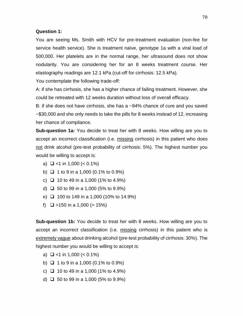
78
Question 1:
You are seeing Ms. Smith with HCV for pre-treatment evaluation (non-fee for
service health service). She is treatment naïve, genotype 1a with a viral load of
500,000. Her platelets are in the normal range, her ultrasound does not show
nodularity. You are considering her for an 8 weeks treatment course. Her
elastography readings are 12.1 kPa (cut-off for cirrhosis: 12.5 kPa).
You contemplate the following trade-off:
A: if she has cirrhosis, she has a higher chance of failing treatment. However, she
could be retreated with 12 weeks duration without loss of overall efficacy.
B: if she does not have cirrhosis, she has a ~94% chance of cure and you saved
~$30,000 and she only needs to take the pills for 8 weeks instead of 12, increasing
her chance of compliance.
Sub-question 1a: You decide to treat her with 8 weeks. How willing are you to
accept an incorrect classification (i.e. missing cirrhosis) in this patient who does
not drink alcohol (pre-test probability of cirrhosis: 5%). The highest number you
would be willing to accept is:
a) <1 in 1,000 (< 0.1%)
b) 1 to 9 in a 1,000 (0.1% to 0.9%)
c) 10 to 49 in a 1,000 (1% to 4.9%)
d) 50 to 99 in a 1,000 (5% to 9.9%)
e) 100 to 149 in a 1,000 (10% to 14.9%)
f) >150 in a 1,000 (> 15%)
Sub-question 1b: You decide to treat her with 8 weeks. How willing are you to
accept an incorrect classification (i.e. missing cirrhosis) in this patient who is
extremely vague about drinking alcohol (pre-test probability of cirrhosis: 30%). The
highest number you would be willing to accept is:
a) <1 in 1,000 (< 0.1%)
b) 1 to 9 in a 1,000 (0.1% to 0.9%)
c) 10 to 49 in a 1,000 (1% to 4.9%)
d) 50 to 99 in a 1,000 (5% to 9.9%)

79
e) 100 to 149 in a 1,000 (10% to 14.9%)
f) >150 in a 1,000 (> 15%)
Question 2:
You are seeing Mr. Miller who is transferring his care to your facility as he recently
moved. He finished hep c treatment 5 months ago. A pre-treatment TE result is
unavailable. The results of your test show that he has achieved SVR, has a normal
platelet count, and no nodularity on ultrasound.
TE result show a stiffness of 9.1 kPa (cut-off for ruling out advanced fibrotic liver
disease (F3 and F4): 9.5 kPA). You consider discharging the patient from your liver
clinic back to his PCP.
You contemplate the following trade-off:
A: if he has advanced fibrosis (F3 or early F4), he has a higher chance to develop
consequences of unrecognized advanced fibrotic liver disease. However, your
letter to the PCP states that if routine care (physical exam, standard labs, including
CBC and liver function tests) should become abnormal to refer back to you.
B: if he does not have advanced fibrotic liver disease, he can enjoy not needing to
go to his liver specialist and will have less worry associated with these visits.
Sub-question 2a: You decide to discharge the patient. How willing are you to
accept an incorrect classification (i.e. missing advanced fibrotic liver disease) in
this patient who does not drink alcohol (pre-test probability of cirrhosis: 5%). The
highest number you would be willing to accept is:
a) <1 in 1,000 (< 0.1%)
b) 1 to 9 in a 1,000 (0.1% to 0.9%)
c) 10 to 49 in a 1,000 (1% to 4.9%)
d) 50 to 99 in a 1,000 (5% to 9.9%)
e) 100 to 149 in a 1,000 (10% to 14.9%)
f) >150 in a 1,000 (> 15%)
Sub-question 2b: You decide to discharge the patient. How willing are you to
accept an incorrect classification (i.e. missing advanced fibrotic liver disease) in

80
this patient who is extremely vague about drinking alcohol (pre-test probability of
cirrhosis: 30%). The highest number you would be willing to accept is:
a) <1 in 1,000 (< 0.1%)
b) 1 to 9 in a 1,000 (0.1% to 0.9%)
c) 10 to 49 in a 1,000 (1% to 4.9%)
d) 50 to 99 in a 1,000 (5% to 9.9%)
e) 100 to 149 in a 1,000 (10% to 14.9%)
f) >150 in a 1,000 (> 15%)
Question 3:
You are seeing Ms. Jones with chronic hep B. Her transaminases are borderline
for treatment and do not fulfil criteria according to the guideline. She is not
interested in a liver biopsy.
Her TE reading is 10.5 kPa (cut-off for ruling out cirrhosis: 11.0 kPa). You
contemplate the following trade-off:
A: If she has cirrhosis, you failed to start her on treatment her and her liver disease
will likely progress. However, she could get a repeat TE in a year to see if her liver
stiffness gets worse, since you have been following her closely for years.
B: If she does not have cirrhosis, you are not exposing her to daily medications,
which may have some minor side effects, and she might seroconvert
spontaneously obviating the need to be treated.
Sub-question 3a: You decide to hold off treatment. How willing are you to accept
an incorrect classification (i.e. missing cirrhosis) in this patient who is not drinking
alcohol (pre-test probability of cirrhosis: 5%). The highest number you would be
willing to accept is:
a) <1 in 1,000 (< 0.1%)
b) 1 to 9 in a 1,000 (0.1% to 0.9%)
c) 10 to 49 in a 1,000 (1% to 4.9%)
d) 50 to 99 in a 1,000 (5% to 9.9%)
e) 100 to 149 in a 1,000 (10% to 14.9%)
f) >150 in a 1,000 (> 15%)

81
Sub-question 3b: You decide to hold off treatment. How willing are you to accept
an incorrect classification (i.e. missing cirrhosis) in this patient who is extremely
vague about drinking alcohol (pre-test probability of cirrhosis: 30%). The highest
number you would be willing to accept is:
a) <1 in 1,000 (< 0.1%)
b) 1 to 9 in a 1,000 (0.1% to 0.9%)
c) 10 to 49 in a 1,000 (1% to 4.9%)
d) 50 to 99 in a 1,000 (5% to 9.9%)
e) 100 to 149 in a 1,000 (10% to 14.9%)
f) >150 in a 1,000 (> 15%)
Question 4:
You are seeing Mr. Dickens with fatty liver on ultrasound and mild abnormal
transaminases. The patient requests a TE test.
His TE reading is 10.5 kPa (cut-off for ruling out cirrhosis: 11.0 kPa). You
contemplate the following trade-off:
A: If he has cirrhosis, you failed to counsel him adequately (including risk of HCC),
he fails to intensify his life style changes, and his liver disease may progress.
However, he could get a repeat TE in a year to see if his liver stiffness gets worse.
Also, you tell him that he needs to lose weight now.
B: If he does not have cirrhosis, you correctly counseled him that he is not in
immediate danger of having cirrhosis, but that he needs to lose weight now to
prevent worsening liver disease.
Sub-question 4a: You decide to tell him he does not yet have cirrhosis. How
willing are you to accept an incorrect classification (i.e. missing cirrhosis) in this
patient who does NOT have diabetes and is not of advanced age and is not majorly
obese (pre-test probability of cirrhosis: 5%). The highest number you would be
willing to accept is:
a) <1 in 1,000 (< 0.1%)
b) 1 to 9 in a 1,000 (0.1% to 0.9%)
c) 10 to 49 in a 1,000 (1% to 4.9%)

82
d) 50 to 99 in a 1,000 (5% to 9.9%)
e) 100 to 149 in a 1,000 (10% to 14.9%)
f) >150 in a 1,000 (> 15%)
Sub-question 4b: You decide to tell him he does not yet have cirrhosis. How
willing are you to accept an incorrect classification (i.e. missing cirrhosis) in this
patient who has diabetes, is older and majorly obese (pre-test probability of
cirrhosis: 30%). The highest number you would be willing to accept is:
a) <1 in 1,000 (< 0.1%)
b) 1 to 9 in a 1,000 (0.1% to 0.9%)
c) 10 to 49 in a 1,000 (1% to 4.9%)
d) 50 to 99 in a 1,000 (5% to 9.9%)
e) 100 to 149 in a 1,000 (10% to 14.9%)
f) >150 in a 1,000 (> 15%)
Question 5:
You are seeing Mr. Keeler with fatty liver on ultrasound, intermittent mild abnormal
transaminases (currently normal), and a history of heavy drinking in the past. You
decide to perform a TE test.
His TE reading is 11.5 kPa (cut-off for ruling out cirrhosis: 12.5 kPa). You
contemplate the following trade-off:
A: If he has cirrhosis, you failed to counsel him adequately (including risk of HCC),
and his liver disease may progress. However, he could get a repeat TE in a year
to see if his liver stiffness gets worse. Also, you tell him that he should never drink
alcohol again.
B: If he does not have cirrhosis, you correctly counseled him that he is not in
immediate danger of having cirrhosis, but that he should never drink again to
prevent worsening liver disease.
Sub-question 5a: You decide to tell him he does not yet have cirrhosis. How
willing are you to accept an incorrect classification (i.e. missing cirrhosis) in this
patient who does NOT have viral co-infections (e.g., HIV), is not of advanced age

83
and is not majorly obese (pre-test probability of cirrhosis: 5%). The highest number
you would be willing to accept is:
a) <1 in 1,000 (< 0.1%)
b) 1 to 9 in a 1,000 (0.1% to 0.9%)
c) 10 to 49 in a 1,000 (1% to 4.9%)
d) 50 to 99 in a 1,000 (5% to 9.9%)
e) 100 to 149 in a 1,000 (10% to 14.9%)
f) >150 in a 1,000 (> 15%)
Sub-question 5b: You decide to tell him he does not yet have cirrhosis. How
willing are you to accept an incorrect classification (i.e. missing cirrhosis) in this
patient who is older and majorly obese (pre-test probability of cirrhosis: 30%). The
highest number you would be willing to accept is:
a) <1 in 1,000 (< 0.1%)
b) 1 to 9 in a 1,000 (0.1% to 0.9%)
c) 10 to 49 in a 1,000 (1% to 4.9%)
d) 50 to 99 in a 1,000 (5% to 9.9%)
e) 100 to 149 in a 1,000 (10% to 14.9%)
f) >150 in a 1,000 (> 15%)
Question 6:
You are seeing Mr. Clark who had hep c 3 years ago (now cured). The outside
hepatologist told him at that time that he has cirrhosis (no liver biopsy was
performed), but his platelets are still in the normal range and a current ultrasound
does not show nodularity.
His TE reading is 18.5 kPa (cut-off for ruling out high-risk esophageal varices: 19.5
kPa). You contemplate the following trade-off:
A: If he has high risk varices, you failed to start beta-blockers since he did not get
an EGD. However, he could get a repeat TE in a year to see if his liver stiffness
gets worse as you are following him in your liver clinic anyway.
B: If he does not have high-risk varices, you avoided the small risk associated with
him undergoing EGD (you practice in a non-fee for service health care setting).

84
Sub-question 6a: You decide not to send him for EGD to screen for varices. How
willing are you to accept an incorrect classification (i.e. missing high risk varices)
in this patient who does NOT drink (pre-test probability of high risk varices: 5%).
The highest number you would be willing to accept is:
a) <1 in 1,000 (< 0.1%)
b) 1 to 9 in a 1,000 (0.1% to 0.9%)
c) 10 to 49 in a 1,000 (1% to 4.9%)
d) 50 to 99 in a 1,000 (5% to 9.9%)
e) 100 to 149 in a 1,000 (10% to 14.9%)
f) >150 in a 1,000 (> 15%)
Sub-question 6b: You decide not to send him for EGD to screen for varices. How
willing are you to accept an incorrect classification (i.e. missing high risk varices)
in this patient who is extremely vague about drinking alcohol (pre-test probability
of high risk varices: 20%). The highest number you would be willing to accept is:
a) <1 in 1,000 (< 0.1%)
b) 1 to 9 in a 1,000 (0.1% to 0.9%)
c) 10 to 49 in a 1,000 (1% to 4.9%)
d) 50 to 99 in a 1,000 (5% to 9.9%)
e) 100 to 149 in a 1,000 (10% to 14.9%)
f) >150 in a 1,000 (> 15%)
Question 7:
You are seeing Mrs. Templeton in pre-op who will undergo laparoscopic
cholecystectomy. She has had hep c for many years, but has achieved an SVR 3
years ago. Her surgeon is concerned about risks associated with intraabdominal
varices. You decide to perform a TE test.
Her TE reading is 16 kPa (cut-off for ruling out any varices: 17 kPa). You
contemplate the following trade-off:
A: If she has any varices, she might have a higher operative risk. However, the
surgeon could convert to an open cholecystectomy if needed.

85
B: If she does not have any varices, you avoided the small risk associated with her
undergoing additional testing such as EGD (you practice in a non-fee for service
health care setting).
Sub-question 7a: You decide to tell the surgeon that she is unlikely to have any
varices, even if she already has very early cirrhosis. How willing are you to accept
an incorrect classification (i.e. missing any varices) in this patient who does NOT
drink (pre-test probability of any varices: 5%). The highest number you would be
willing to accept is:
a) <1 in 1,000 (< 0.1%)
b) 1 to 9 in a 1,000 (0.1% to 0.9%)
c) 10 to 49 in a 1,000 (1% to 4.9%)
d) 50 to 99 in a 1,000 (5% to 9.9%)
e) 100 to 149 in a 1,000 (10% to 14.9%)
f) >150 in a 1,000 (> 15%)
Sub-question 7b: You decide to tell the surgeon that she is unlikely to have any
varices, even if she already has very early cirrhosis. How willing are you to accept
an incorrect classification (i.e. missing any varices) in this patient who is extremely
vague about drinking alcohol (pre-test probability of any varices: 40%). The highest
number you would be willing to accept is:
a) <1 in 1,000 (< 0.1%)
b) 1 to 9 in a 1,000 (0.1% to 0.9%)
c) 10 to 49 in a 1,000 (1% to 4.9%)
d) 50 to 99 in a 1,000 (5% to 9.9%)
e) 100 to 149 in a 1,000 (10% to 14.9%)
f) >150 in a 1,000 (> 15%)

86
eTable 1. Characteristics of studies reporting diagnostic performance of liver stiffness cut-off of 12.5 (±1) kPa for diagnosis
of cirrhosis in patients with hepatitis C
Study Prevalence of
cirrhosis TE failure rate TP FP FN TN
Bota 2013 105/512, 20.5% NR 97 101 8 306
Cardoso 2012 31/363; 8.5% 48/613; 7.8% 26 20 5 312
Castera 2014 13/116; 11.2% NR 10 14 3 89
Castera 2009 70/298; 23.5% 10/298; 3.4% 55 10 11 212
Carrion 2006 15/124; 12.1% 2/135; 1.5% 19 20 0 131
D'Ambrosio 2013 13/33, 39.4% 4/37; 10.8% 8 1 5 19
Degos 2010 126/913; 13.8% NR 130 115 50 1012
De Ledinghen 2006 17/72; 23.6% 0 17 4 0 51
Gaia 2011 13/77; 16.8% 21/290 (7.2%) 9 4 4 60
Gara 2013 13/109; 11.9% 11/120 (9.2%) 13 11 0 85
Liu 2011 14/284; 4.9% 7/304; 2.3% 5 0 9 270
Lupsor 2013 374/1202; 31.1% 27/1202 (2.2%) 355 55 23 773
Nitta 2009 24/165; 14.5% NR 22 31 2 110
Patel 2011 18/214; 8.4% NR 17 24 1 172
Sporea 2010 39/317; 12.3% NR 30 19 9 259
Sporea 2012 53/197; 26.9% 10/197; 5.1% 51 15 2 129
Verveer 2012 56/382; 14.7% 57/456; 12.5% 43 34 13 292 [Abbreviations: FN, false negative; FP, false positive; NR, not reported; TE, transient elastography; TN, true negative; TP, true positive]

87
eTable 2. Characteristics of studies reporting diagnostic performance of liver stiffness cut-off of 9.5 (±1) kPa for diagnosis
of advanced fibrosis or cirrhosis in patients with hepatitis C
Study Prevalence of
advanced fibrosis TP FP FN TN
Cassinotto 2014 41.3% 124 56 17 135
Cardoso 2012 87/363; 24% 58 22 29 254
Castera 2005 45.3% 61 9 22 91
Cho 2011 43.8% 28 7 3 48
Crissan 2012 122/446; 30.7% 95 47 42 262
Gaia 2011 17/77; 22% 13 6 4 54
Liu 2011 40/284; 14.1% 18 2 22 242
Lupsor 2013 500/1202; 46.6% 497 75 63 567
Nitta 2009 57/165; 34.5% 50 19 7 89
Obara 2008 16/51; 31.4% 15 11 1 24
Seo 2015 118/349; 33.8% 94 39 24 192
Sporea 2010 132/317; 41.6% 82 35 50 150
Ziol 2005 76/251; 30.3% 65 26 11 149 [Abbreviations: FN, false negative; FP, false positive; TN, true negative; TP, true positive]
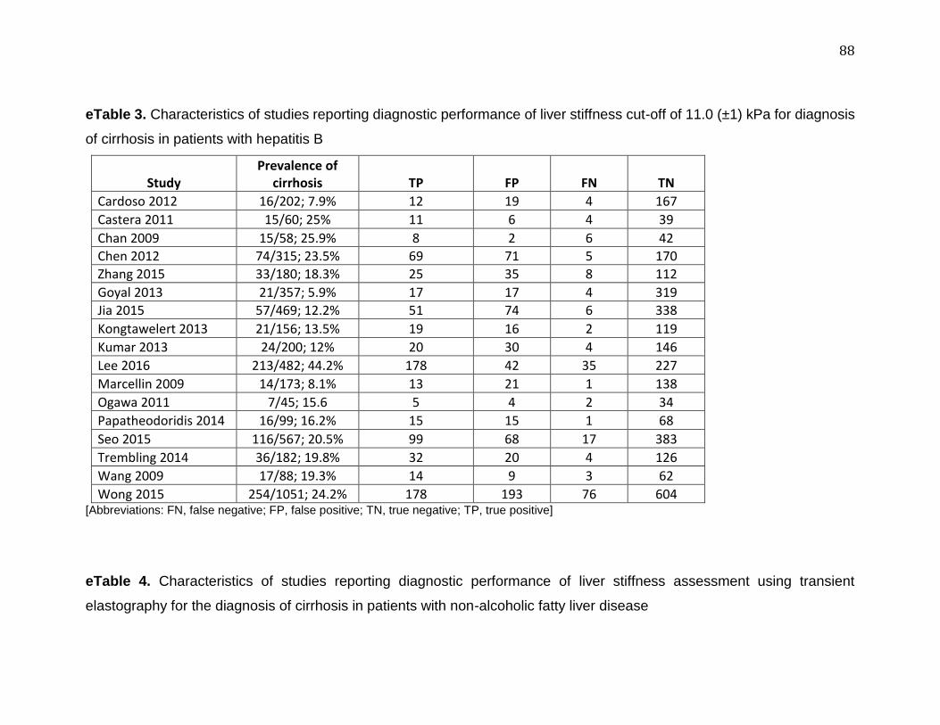
88
eTable 3. Characteristics of studies reporting diagnostic performance of liver stiffness cut-off of 11.0 (±1) kPa for diagnosis
of cirrhosis in patients with hepatitis B
Study Prevalence of
cirrhosis TP FP FN TN
Cardoso 2012 16/202; 7.9% 12 19 4 167
Castera 2011 15/60; 25% 11 6 4 39
Chan 2009 15/58; 25.9% 8 2 6 42
Chen 2012 74/315; 23.5% 69 71 5 170
Zhang 2015 33/180; 18.3% 25 35 8 112
Goyal 2013 21/357; 5.9% 17 17 4 319
Jia 2015 57/469; 12.2% 51 74 6 338
Kongtawelert 2013 21/156; 13.5% 19 16 2 119
Kumar 2013 24/200; 12% 20 30 4 146
Lee 2016 213/482; 44.2% 178 42 35 227
Marcellin 2009 14/173; 8.1% 13 21 1 138
Ogawa 2011 7/45; 15.6 5 4 2 34
Papatheodoridis 2014 16/99; 16.2% 15 15 1 68
Seo 2015 116/567; 20.5% 99 68 17 383
Trembling 2014 36/182; 19.8% 32 20 4 126
Wang 2009 17/88; 19.3% 14 9 3 62
Wong 2015 254/1051; 24.2% 178 193 76 604 [Abbreviations: FN, false negative; FP, false positive; TN, true negative; TP, true positive]
eTable 4. Characteristics of studies reporting diagnostic performance of liver stiffness assessment using transient
elastography for the diagnosis of cirrhosis in patients with non-alcoholic fatty liver disease

89
Study Cut-off (kPa)
Prevalence of cirrhosis (%) BMI (kg/m2)
TE Failure Rate TP FP FN TN
Chan 2015 17 2 29.3 ± 4.3;
83.7% obese 4.1% 3 9 0 135
Gaia 2011 10.5 12.5 27.5 (21.1-40.4);
26% obese 8.0% 7 3 2 60
Imjao 2015 14 7.7 28 ± 4.6; NR 10.6% 10 28 0 89
Kumar 2013 11.8 8.3 26.1 ± 4.1; NR 10.1% 9 13 1 97
Mahadeva 2013 11.3 6.1 NR; 32.8% obese 8.4% 6 12 1 101
Myers 2010 11.1 12 NR NR 6 8 0 36
Myers 2012 22.3 12 33 (30-36); NR 26.0% 7 6 2 60
Wong 2012 10.3 10.2 28.9 ± 4.8; 35% obese 19.0% 13 24 3 116
Wong 2010 10.3 10.2 28 ± 4.5;
28.5% obese 10.2% 23 27 2 194
Yoneda 2008 17.5 9.3 26.6 ± 4.2; NR 4.9% 9 3 0 85
Yoneda 2010 16 11.1 M: 28.2 ± 5.0; F: 26.2 ± 4.4 NR 6 1 0 47
[Abbreviations: BMI, body mass index; FN, false negative; FP, false positive; NR, not reported; TE, transient elastography; TN, true negative; TP, true positive]
eTable 5. Characteristics of studies reporting diagnostic performance of liver stiffness cut-off of 12.5 (±1) kPa for diagnosis
of cirrhosis in patients with chronic alcoholic liver disease

90
Study Prevalence of
cirrhosis TP FP FN TN
Bardou-Jacquet 2013 5/8; 63% 5 2 0 1
Boursier 2009 69/106; 65% 62 10 7 27
de Ledinghen 2013 27/34; 79% 25 2 2 5
Dolman 2013 3/20; 15% 3 1 0 16
Kim 2009 29/45 64%; 29 8 0 8
Lannerstedt 2013 8/16; 50% 8 4 0 4
Mueller 2010 26/101; 26% 25 15 1 60 [Abbreviations: FN, false negative; FP, false positive; TN, true negative; TP, true positive]

91
eTable 6. Characteristics of studies reporting diagnostic performance of liver stiffness cut-off of 19.5 (±2) kPa for ruling out
high-risk esophageal varices in patients with chronic liver diseases
Study Cut-off (kPa)
Prevalence of high-risk EVs (%) TP TN FP FN
Calvaruso 2013 19 27.1 19 39 31 7
Castera 2009 21.5 19 11 39 18 2
Kazemi 2006 19 28.5 42 70 48 5
Nguyen-Khac 2010 19.8 22.4 12 25 20 1
Pineda 2009 21 19 19 27 56 0
Pritchett 2011 19 37 72 71 61 7
Vermehren 2011 20.5 36 45 24 63 4
Wang 2012 21 10.3 10 98 15 3 [Abbreviations: EV, esophageal varices; FN, false negative; FP, false positive; TN, true negative; TP, true positive]

92
eTable 7. Characteristics of studies reporting diagnostic performance of liver stiffness cut-off of 17.0 (±2) kPa for ruling out
any esophageal varices in patients with chronic liver diseases
Study Cut-off (kPa)
Prevalence of any EVs (%) TP TN FP FN
Al-Dashan 2012 16.2 50 25 18 12 5
Bintintan 2015 15 78.3 45 13 0 2
Calvaruso 2013 17 56.3 38 24 18 16
Colecchia 2012 16.4 53 51 28 19 2
Fraquelli 2014 19 10 8 47 52 3
Pritchett 2011 17.6 68 112 42 26 31
Stefanescu 2011 19 68 132 24 50 25
Vizutti 2007 17.6 63.8 27 7 10 3 [Abbreviations: EV, esophageal varices; FN, false negative; FP, false positive; TN, true negative; TP, true positive]

93
eFigure 1. (A) Sensitivity and (B) specificity of liver stiffness cut-off of 12.5 (±1) kPa for diagnosing cirrhosis in patients with hepatitis C A. Sensitivity
B. Specificity
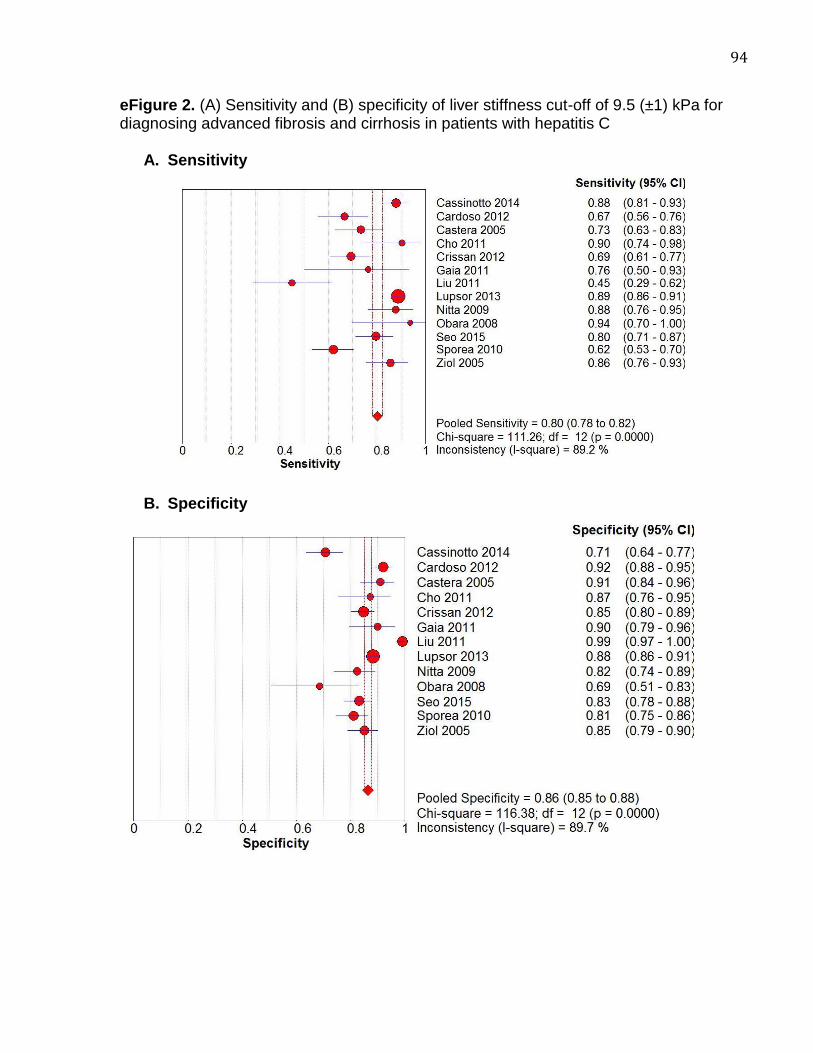
94
eFigure 2. (A) Sensitivity and (B) specificity of liver stiffness cut-off of 9.5 (±1) kPa for diagnosing advanced fibrosis and cirrhosis in patients with hepatitis C
A. Sensitivity
B. Specificity

95
eFigure 3. (A) Sensitivity and (B) specificity of liver stiffness cut-off of 11.0 (±1) kPa for diagnosing cirrhosis in patients with hepatitis B
A. Sensitivity
B. Specificity

96
eFigure 4. (A) Sensitivity and (B) specificity of transient elastography across a range of liver stiffness cut-offs (10.3-22.3 kPa) for diagnosing cirrhosis in patients with non-alcoholic fatty liver disease A. Sensitivity
B. Specificity
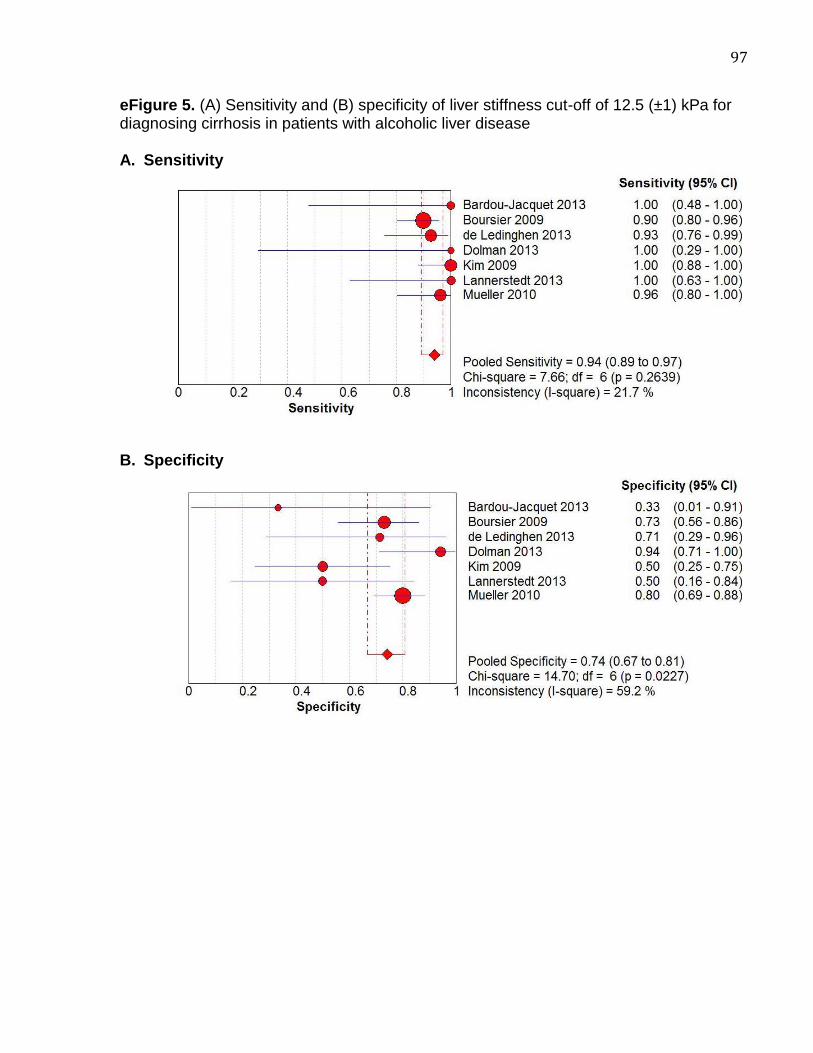
97
eFigure 5. (A) Sensitivity and (B) specificity of liver stiffness cut-off of 12.5 (±1) kPa for diagnosing cirrhosis in patients with alcoholic liver disease A. Sensitivity
B. Specificity

98
eFigure 6. (A) Sensitivity and (B) specificity of liver stiffness cut-off of 19.5 (±2) kPa for ruling out high-risk esophageal varices in patients with chronic liver diseases A. Sensitivity
B. Specificity

99
eFigure 7. (A) Sensitivity and (B) specificity of liver stiffness cut-off of 17.0 (±2) kPa for ruling out any esophageal varices in patients with suspected chronic liver diseases prior to elective, non-hepatic surgery
A. Sensitivity
B. Specificity

100