Embed Size (px)
Citation preview

Description of the disease: American foulbrood (AFB) affects the larval stage of the honey bee
Apis mellifera and other Apis spp., and occurs throughout the world. Paenibacillus larvae, the
causative organism, is a bacterium that can produce over one billion spores in each infected larva.
The spores are extremely resistant to heat and chemical agents, and can survive for many years in
scales (from diseased dead brood), hive products and equipment. Only the spores are capable of
inducing the infection.
Combs of infected colonies have a mottled appearance due to a mixture of healthy capped brood,
uncapped cells containing the remains of diseased larvae, and empty cells. This is not a
characteristic of AFB only. Cell cappings of a diseased larva appear moist and darkened, becoming
concave and possibly punctured as infection progresses. The larval or pupal colour changes to
creamy brown and then to a dark brown with a ropy appearance when drawn out. In some cases
the larval remains are rather watery. The diseased brood eventually dries out to form characteristic
brittle scales that adhere tightly to the lower sides of the cell. The formation of a pupal tongue is one
of the most characteristic but rarely seen signs of the disease and precedes the formation of the
scales. The clinical signs of AFB are very diverse and depend on the genotype involved, the stage
of the disease and the strength of the bee colony (and possibly its resistance to AFB). Genotypes
ERIC I–IV are all pathogenic for honey bees
Identification of the agent: Diagnosis of AFB is based on identification of the pathogenic agent
and the presence of clinical signs. The analyst can make use of a broad range of sample types.
However, in practice, the samples of choice will depend on whether it concerns a suspicious or
diseased honey bee colony/apiary, or analysis in the context of an AFB monitoring/prevention
programme. Some of the identification methods require a previous culturing step, while others can
be performed directly on collected samples. Five solid culture media are recommended: PLA
(Paenibacillus larvae agar), MYPGP agar, BHIT agar, J-agar and Columbia sheep blood agar. Two
polymerase chain reaction (PCR) protocols are described in this chapter that can be used for rapid
confirmation of clinical AFB and for identification of bacterial colonies after a cultivation step. The
biochemical profiling of P. larvae is based on the catalase test, the production of acid from
carbohydrates and the hydrolysis of casein depending on the genotype involved. Further, antibody-
based techniques and the microscopic identification of the pathogenic agent are described.
Serological tests: There are no serological tests available.
Requirements for vaccines: No vaccines are available.
American foulbrood (AFB) is an infectious disease of the larval stage of the honey bee Apis mellifera and other Apis spp., and occurs throughout the world where such bees are kept. Paenibacillus larvae, the causative organism, is a Gram-positive bacterium that can produce over one billion spores in each infected larva. The bacterium is a round-ended, straight or sometimes curved rod, which varies greatly in size (0.5–0.8 µm wide by 1.5 to 6 µm long), occurring singly and in chains and filaments; most strains are motile. The sporangia are often sparse in vitro, and the ellipsoidal, central to subterminal spores, which may swell the sporangia, are often found free, measuring 0.6 × 1.3 μm (Heyndrickx et al., 1996).
By using repetitive element polymerase chain reaction (rep-PCR) and primers ERIC1R-ERIC2, four different genotypes (ERIC I, II, III and IV) are differentiated (Genersch et al., 2006). Genotypes ERIC I and II correspond to the former subspecies P. l. larvae while genotypes ERIC III and IV corresponds to the former subspecies P. l. pulvifaciens (Genersch, 2010). All four genotypes differ in colony and spore morphology, in their metabolism of carbon sources and most importantly in virulence. Exposure bioassays revealed that members of ERIC II, III and IV are highly virulent against larvae in terms of the time course of mortality. All larvae infected with these genotypes are killed within approximately 7 days (Genersch et al., 2005, 2006). This means that only a minor
proportion of the larvae die after cell capping resulting in the described clinical signs of AFB (ropy stage, foulbrood scale). In contrast, genotype ERIC I needed around 12 days to kill all infected larvae and, hence, is considered less virulent than ERIC II, III and IV for the individual larva (Genersch et al., 2005, 2006; Genersch, 2010; Djukic et al., 2014). Epidemiological studies showed that only ERIC I and ERIC II are frequently isolated from AFB-disease colonies. Paenibacillus larvae genotype ERIC I is the most frequent genotype while genotype ERIC II seems to be more restricted although both have been reported worldwide. Genotypes ERIC III and IV have not been identified in the field for decades, but exist as few isolates in culture collections (Genersch, 2010; De Graaf et al., 2013). Because fingerprinting profiles generated via electrophoresis of rep-PCR amplified DNA are not consistently reproducible between laboratories, it is necessary to include reference P. larvae strains previously
typed. To enhance the discrimination of strains, the analysis using ERIC primers can be complemented with the use of other primers (De Graaf et al., 2013). A multilocus sequence typing scheme revealed the distribution and biogeography of 294 samples of P. larvae across six continents (Morrissey et al., 2015)
The spores are extremely heat stable and resistant to chemical agents. Only spores are capable of inducing the infection. The infection can be transmitted to larvae by nurse bees or by spores remaining at the base of a brood cell. Although the larval stages of worker bees, drones and queens are susceptible to infection, infected queens and drone larvae are rarely seen under natural conditions. The susceptibility of larvae to AFB disease decreases with increasing age (Woodrow, 1941); larvae cannot be infected later than 53 hours after the egg has hatched. The mean infective dose (LD50= spore dose at which 50% of the larvae are killed) needed to initiate infection,
though very variable, is 8.49 ± 1.49 spores in 24–28 hour-old bee larvae (Hansen & Brødsgaard, 1999). Exchanging combs containing the remains of diseased brood is the most common way of spreading the disease from colony to colony. In addition, feeding or robbing of spore-laden honey or bee bread, package bees and the introduction of queens from infected colonies can also spread the disease. Wax contaminated with the spores of P. larvae, used in the production of comb foundations, can also spread the disease if not properly treated. The
early detection of AFB helps to prevent further spread.
While there is generally a low risk of human infection with AFB organisms, it should be noted that fatal bacterial septicaemia has been reported in drug users injected with honey contaminated with P. larvae spores (Rieg et al., 2010). Biocontainment measures should be determined by risk analysis as described in Chapter 1.1.4 Biosafety and biosecurity: Standard for managing biological risk in the veterinary laboratory and animal facilities.
Spores of P. larvae can survive in bee products (honey, wax, dead larvae) and in the environment for 3 to 10 years and for 35 years in dry larval scales (Haseman, 1961). Purified spores can survive even more than 70 years (Rudenko, 1987).
The clinical signs of AFB are very diverse and depend on the genotype involved, the stage of the disease and the strength of the bee colony (and possibly its resistance to AFB) (Genersch et al., 2005). Larvae can be killed rapidly at an early age when they are curled at the base of uncapped brood cells. Adult worker bees will remove these dead larvae leaving only an empty cell (Brødsgaard et al., 2000). Other larvae will die later on in their development, when they are in an upright position, filling most of the brood cell. Usually the larvae or pupae will die after brood cell capping.
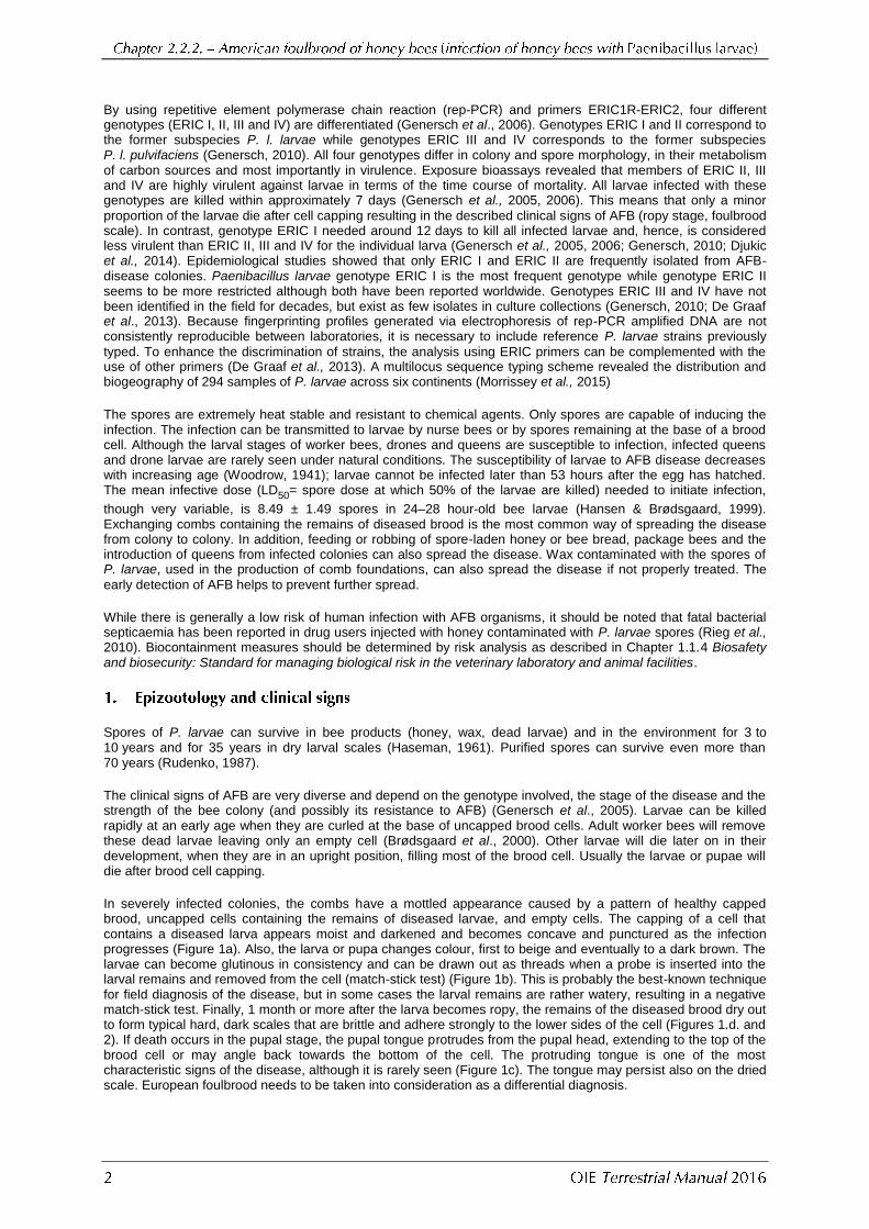
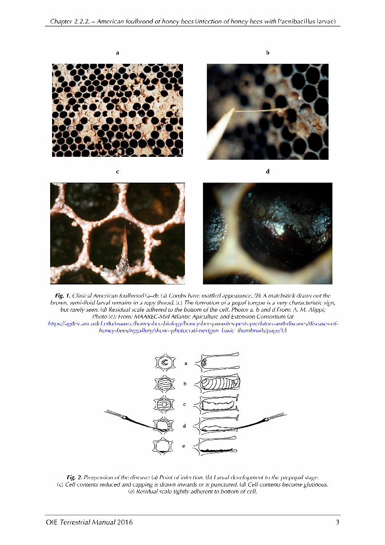
In severely infected colonies, the combs have a mottled appearance caused by a pattern of healthy capped brood, uncapped cells containing the remains of diseased larvae, and empty cells. The capping of a cell that contains a diseased larva appears moist and darkened and becomes concave and punctured as the infection progresses (Figure 1a). Also, the larva or pupa changes colour, first to beige and eventually to a dark brown. The larvae can become glutinous in consistency and can be drawn out as threads when a probe is inserted into the larval remains and removed from the cell (match-stick test) (Figure 1b). This is probably the best-known technique for field diagnosis of the disease, but in some cases the larval remains are rather watery, resulting in a negative match-stick test. Finally, 1 month or more after the larva becomes ropy, the remains of the diseased brood dry out to form typical hard, dark scales that are brittle and adhere strongly to the lower sides of the cell (Figures 1.d. and 2). If death occurs in the pupal stage, the pupal tongue protrudes from the pupal head, extending to the top of the brood cell or may angle back towards the bottom of the cell. The protruding tongue is one of the most characteristic signs of the disease, although it is rarely seen (Figure 1c). The tongue may persist also on the dried scale. European foulbrood needs to be taken into consideration as a differential diagnosis.
It has been demonstrated that different genotypes differ in virulence; ERIC I strains lead to 100% mortality of infected larvae within 12 days while ERIC II strains kill infected larvae in about 7 days (Djukic et al., 2014; Genersch, 2010; Genersch et al., 2005;).The faster P. larvae kills infected larvae, the more infected larvae will be removed as nurse bees seem to recognise dead larvae at lower rates after cells capping (Rauch et al., 2009). Therefore, the proportion of larvae developing into a ropy mass under the cell cappings is higher for infections with strains of genotype ERIC I. As veterinarians and beekeepers look for a ropy mass inside capped cells as the main sign of the disease, false negative diagnoses are likely to occur if AFB-diseased colonies are infected with strains of genotype ERIC II because only a few infected cells may be present (Genersch, 2007; Rauch et al., 2009).
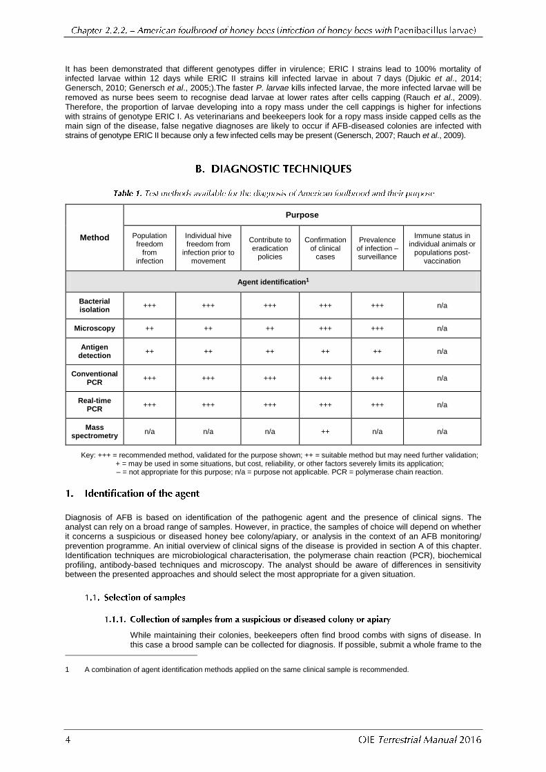
Method
Purpose
Population freedom
from infection
Individual hive freedom from
infection prior to movement
Contribute to eradication
policies
Confirmation of clinical
cases
Prevalence of infection – surveillance
Immune status in individual animals or
populations post-vaccination
Agent identification1
Bacterial isolation
+++ +++ +++ +++ +++ n/a
Microscopy ++ ++ ++ +++ +++ n/a
Antigen detection
++ ++ ++ ++ ++ n/a
Conventional PCR
+++ +++ +++ +++ +++ n/a
Real-time PCR
+++ +++ +++ +++ +++ n/a
Mass spectrometry
n/a n/a n/a ++ n/a n/a
Key: +++ = recommended method, validated for the purpose shown; ++ = suitable method but may need further validation; + = may be used in some situations, but cost, reliability, or other factors severely limits its application; – = not appropriate for this purpose; n/a = purpose not applicable. PCR = polymerase chain reaction.
Diagnosis of AFB is based on identification of the pathogenic agent and the presence of clinical signs. The analyst can rely on a broad range of samples. However, in practice, the samples of choice will depend on whether it concerns a suspicious or diseased honey bee colony/apiary, or analysis in the context of an AFB monitoring/ prevention programme. An initial overview of clinical signs of the disease is provided in section A of this chapter. Identification techniques are microbiological characterisation, the polymerase chain reaction (PCR), biochemical profiling, antibody-based techniques and microscopy. The analyst should be aware of differences in sensitivity between the presented approaches and should select the most appropriate for a given situation.
While maintaining their colonies, beekeepers often find brood combs with signs of disease. In this case a brood sample can be collected for diagnosis. If possible, submit a whole frame to the
1 A combination of agent identification methods applied on the same clinical sample is recommended.
laboratory, to avoid risk of distortion in transit. Alternatively the brood may be sampled by
cutting out a piece comb of about 20 cm2 in size, containing as much of the dead or discoloured brood as possible. An experienced person can collect infected larval or pupal remains directly from the cells with a sterile swab, significantly reducing the sample size and facilitating packaging and sample transportation to the laboratory (see below). When microscopic examination is the method of choice, smears of the remains of diseased larvae can also be made at the apiary (Hornitzky & Wilson, 1989). After air-drying the smears are packaged to be forwarded to the laboratory for microscopic examination and culture.
Every bee colony in the vicinity of such a clinical case of AFB should be considered as suspicious and a broad range of samples should be taken for confirmation. Apart from brood samples, food stores (honey [Ritter & Kiefer, 1995; von der Ohe & Dustmann, 1997], pollen [Gochnauer & Corner, 1987] and royal jelly), adult workers (Lindström & Fries, 2005) and wax debris (Bzdil, 2007; Titera & Haklova, 2003) can be used to detect the presence of P. larvae
spores. Honey samples can be collected from cells close to the brood with separate disposable spoons to prevent cross-contamination between samples; however, honey may have been sitting in the comb for months at the time of sampling. Adult bees can be shaken or brushed from the combs of the brood chamber or the honey supers into a plastic bag or container. For the most reliable picture of the actual situation, bees from the brood nest (and not the honey supers) should be analysed. Wax debris can be collected at the hive bottom all year round.
To prevent the propagation of diseased brood, honey, adult bee and debris samples can be used to detect AFB in colonies where no clinical signs are observed. Routine collection of samples from colonies or from harvested honey can be used as part of an operational or regional AFB detection programme.
Microscopic examination of smears from larvae with no clinical signs is far less sensitive at detecting spores in colonies compared with bacteriological or PCR-based methods. In fact, bacteriological and PCR-based methods will often detect spores in colonies that never develop clinical signs of AFB. High numbers of spores cultured from honey and bee samples using bacteriological methods, however, can often predict the presence of clinical AFB signs at colony, apiary and operational levels.
Brood comb should be wrapped in a paper bag, paper towel or newspaper and placed in a wooden or heavy cardboard box for transport; avoid any form of plastic wrapping to prevent fungal growth. Swabs with larval remains can be put into appropriate test tubes with a cap. Smears of dead larvae on microscope slides are placed into individual slide holders. These are commercially available. Adult bees can be kept frozen or submerged in 70% ethanol in leak-proof containers during transportation, although dried bees are adequate, a sample size should be at least 30 bees. Food supplies can be put into a test tube or a suitable pot, or wrapped in a plastic bag together with the spoon. Leaking and cross-contamination of the samples must be prevented. If possible, fresh material for laboratory tests should be sent refrigerated.
Hive debris and wax should be wrapped in a paper bag, plastic pots covered with a paper lid or in paper tubes covered with a plastic lid. Secondary packaging consists of a plastic bag as a protection against cross-contamination. In the case of bulk samples, samples may be stored even in tertiary packaging (large cardboard boxes) that protects samples from mechanical damage.
The minimum amount of honey for detection of viable spores of P. larvae is 50 g and should be placed in a leak-proof plastic container (one per sample with an identification label).
i) Samples for cultivation
In general, an aqueous solution containing P. larvae spores should be prepared for further analysis. This spore suspension is heat-shocked at 80°C for 10 minutes or 95–96°C for 3–5 minutes in order to kill vegetative forms of other microorganisms, including other spore formers. Different genotypes of P. larvae show variation in germination ability and their

response to heat treatment is variable (Forsgren et al., 2008). Direct plating of larval remains for cultivation on agar without heat treatment is possible.
Larval/pupal remains from brood comb are collected with a sterile swab and suspended in 5–10 ml of sterile water or physiological solution (0.01 M phosphate-buffered saline [PBS] or 0.9% NaCl) in a test tube. For larval or pupal remains submitted on a glass slide, add 2–3 drops of sterile water. Emulsify with an orange stick or sterile loop. Place a loopful of emulsified material onto a suitable agar plate and streak with a sterile loop to obtain isolated colonies. Plates are incubated in 5–7% CO2 and examined daily for up to seven
days. Colonies are visible from day 2 onwards.
Each spore-suspension sample should be divided and treated in triplicate:
a) without heat treatment;
b) with heat treatment at 80°C for 10 minutes;
and
c) with heat treatment at 95°C for 3 minutes.
Steps ii) and iii) are for killing vegetative forms of other microorganisms. The heat step will significantly reduce the risk that P. larvae colonies will become masked by these competitors. Nevertheless, bacteria of the genera Bacillus, Paenibacillus and Brevibacillus may continue to swarm over the plates which necessitates the use of semi-selective media through addition of the antibiotics nalidixic acid (Hornitzky & Clark, 1991) and pipemidic acid (Alippi, 1992; 1995). In step i) (no heat treatment), as well as both antibiotics, amphotericin B at a final concentration of 16.8 µg/ml of culture medium should be used to avoid fungal contamination in the isolation plates. Incubate the plates at 37+1°C for 2–4 days.
For smears prepared from dead larvae, add 1–2 drops of sterile water and mix on the slide. Use a wire loop to prepare a new smear for Gram stain and microscopic examination for spores. A second loop of reconstituted material is used to culture onto a suitable agar plate.
Honey samples to be examined for spores are heated to 45–50°C and shaken to distribute any spores that may be present, then each honey sample should be diluted (1/1) in 0.01 M PBS pH 7.2 or 0.9% NaCl, transferred to a centrifuge tube and centrifuged at 6000 g for
40 minutes. The supernatant is discarded leaving approximately 3 ml per tube that is then vortex-mixed for 1 minute to re-suspend the pellet, and treated as described for larval remains. The samples are high speed vortex-mixed again for 2 minutes and 100–200 µl of the sediment-fluid mixture poured on suitable culture media with the addition of antibiotics and incubate at 37°C for 7–8 days (de Graaf et al., 2013).
Direct plating of diluted honey (Ritter & Kiefer, 1995) is widely used, but its sensitivity is inferior to that of the centrifugation method as only a fraction of the total volume will be plated out. Whatever the method of choice is, when honey is analysed quantitatively and threshold values are set, the methodology that was used to establish these values should always be strictly followed.
An aqueous filtrate of pollen can be made by thoroughly dispersing 1 g of pollen in 10 ml final volume sterile distilled water or 0.01 M sodium PBS pH 7.2, and filtering it through Whatman No. 1 paper (Gochnauer & Corner, 1987).
When adult bees are dispatched in ethanol, the latter should be decanted and replaced by sterile water or physiological solution before crushing.
Hornitzky & Karlovskis (1989) developed a culture technique that provides a rapid means of detecting P. larvae spores in adult bees that could act as a source of AFB infection for
young larvae. Briefly, each sample of 30 nurse bees is homogenised in 20 ml sterile PBS for 30 seconds. The homogenate is filtered through Whatman No. 1 paper, centrifuged and the pellet is resuspended in PBS. The samples are heat-shocked (see spore-suspension sample treatments) and plated onto a suitable culture medium supplemented with nalidixic acid and pipemidic acid to inhibit the spread of P. alvei and other bacteria that can swarm over the plates.

Debris and bee wax (1.5 g) should be dissolved in an organic solvent (10 ml): toluene (Titera & Haklova, 2003), chloroform (Kostecki, 1969) or diethyl ether (Ritter, 2003). The liquid part (2 ml) is then diluted in physiological solution (6 ml). After shaking roughly, this suspension can immediately be plated out (no heat-shock) (Titera & Haklova, 2003). In another protocol, bee wax is first diluted in water (wax/water 1/10) and heated up to 90°C for 6 minutes. After cooling down, the organic solvent is added (organic solvent/water 1/9) and the mixture is shaken carefully. After 2 minutes standing time, a deposit of a watery solution containing P. larvae spores forms (Ritter, 2003).
ii) Cultivation by Tween 80 Method (Bzdil, 2007)
One gram of debris or 1 g of wax is put in a test tube with an airtight seal. Larger pieces of wax should be cut by sterile instruments into very small pieces (ideally up to 3 mm in size). Wax pieces contained in debris do not have to be further cut because they are usually very small. The smaller the pieces, the easier and faster is the process of homogenisation. Dry material prepared this way should be stirred thoroughly and diluted with 8.5 ml of sterile distilled water. The resulting suspension is then supplemented with 0.5 ml of Tween 80. Approximately 30 minutes before pipetting, required volume of Tween 80 should be withdrawn from the original container, put into another sterile container with airtight seal and immersed in a hot water bath (70±2°C) to reduce the viscosity of Tween 80 and facilitate its pipetting. The suspension of debris, water and Tween 80 is shaken thoroughly and the test tube is placed in a hot water bath (70±2°C) for 30 minutes. If the wax dissolves slowly or there are pieces of wax larger than 5 mm, the test tube can be left in the water bath for up to 1 hour. While warming the sealed tube in water bath, it should be thoroughly shaken in a longitudinal direction at least three times (preferably in several 5- to 30-second cycles 5–10 minutes apart). Thorough homogenisation results in development of homogenous greyish brown pulpy material, which can harden as it cools down. Afterwards, tubes are removed from the water bath and allowed to cool down to room temperature, by which time they should be stored for 2–4 hours until a sufficient amount of liquid is separated at the bottom of tubes. Then 2–5 ml of this liquid is withdrawn with a disposable balloon pipette and mixed with the same volume of distilled water in another sterile sealable tube. Again, the resulting mixture should be shaken thoroughly in a longitudinal direction for at least 5 minutes and put into a hot water bath (90±2°C). After 10 minutes, tubes are removed from the bath, allowed to cool down to room temperature and shaken again. Then the material is inoculated in 0.2 ml doses to 3–5 plates of MYPGP with nalidixic acid and at least one plate of blood agar serving as a control. Before culturing, the plates should be dried in a thermostat at 37±1°C. Drying time is selected according to the humidity of the culture medium surface (approximately 30 minutes). Petri dishes must be labelled accurately with the sample identity. The liquid is spread over the plates using a bent sterile plastic/glass stick or the tip of the pipette. The liquid is allowed to dry and the plates are inverted and incubated at 37±1°C for 5–8 days.
i) Nalidixic acid stock solution (Hornitzky & Clark, 1991)
Prepare by dissolving 0.1 g in 2 ml of 0.1 N NaOH and diluting to 100 ml with 0.01 M phosphate buffer (pH 7.2), (stock concentration 1000 µg/ml). Filter sterilise. Final concentration: 10 µg/ml for larval samples and 20 µg/ml for honey samples.
ii) Pipemidic acid stock (Alippi, 1995)
Prepare by dissolving 0.2 g in 2 ml of 0.1 N NaOH and then diluting to 100 ml with 0.01 M phosphate buffer (pH 7.2), (stock concentration 2000 µg/ml). Filter sterilise. Final concentration: 10 µg/ml for honey samples.
After autoclaving and cooling to 50°C add the antibiotics to the media at the final concentration required and pour into sterile Petri plates (20 ml per plate) (De Graaf et al,
2013).
Several media for cultivating P. larvae have been described but best results were obtained with PLA (Paenibacillus larvae agar) (Schuch et al., 2001), MYPGP agar (Dingmann & Stahly, 1983), BHIT agar (brain–heart infusion medium supplemented with thiamine) (Gochnauer, 1973), J-agar (Gordon et al., 1973) and CSA (Columbia sheep-blood agar) (Hornitzky & Karlovskis, 1989). The formulations of the media are as follows:

i) PLA (Paenibacillus larvae agar)
This selective medium combines three different media to comprise the base, to which antibiotics and egg yolk supplements are added (Schuch et al., 2001). Equal quantities (100 ml) of sterile, molten Bacillus cereus selective agar base, trypticase soy agar and supplemented nutrient agar (SNA) are combined and mixed. SNA is composed of (per litre): nutrient agar 23 g, yeast extract 6 g, meat extract 3 g, NaCl 10 g, Na
2HPO
4 2 g: final
pH is 7.4 ± 0.2. All solid media are sterilised at 121°C/15 minutes. After the three molten media are combined, 3 ml of stock nalidixic acid, 3 ml of stock pipemidic acid, and 30 ml of 50% egg-yolk suspension (Gordon et al., 1973) are added to form the PLA medium (final
concentration 9 µg/ml pipemidic acid and 18 µg/ml nalidixic acid). The PLA medium is poured (20 ml) into sterile Petri dishes and plates are dried before use (45–50°C for 15 minutes).
ii) MYPGP agar (The abbreviation refers to its constituents: Mueller-Hinton broth, yeast extract, potassium phosphate, glucose and pyruvate)
MYPGP agar is composed of (per litre): Mueller-Hinton broth 10 g, yeast extract 15 g, K
2PO
4 3 g, glucose 2 g, Na-pyruvate 1 g and agar 20 g (pH 7.1) (Dingmann & Stahly,
1983). Addition of nalidixic acid and pipemidic acid is as above.
iii) J-agar
J-agar is composed of (per litre): tryptone 5 g, yeast extract 15 g, K2PO
4 3 g, glucose 2 g,
agar 20 g (pH 7.3–7.5) (Gordon et al., 1973). Addition of nalidixic acid and pipemidic acid
is as above.
iv) CSA (Columbia sheep-blood agar)
CSA is composed of (per litre): 39 g Columbia blood agar base (pH 7.3). After autoclaving and cooling to 50°C, add 5% sterile defibrinated blood (Hornitzky & Karlovskis, 1989). Addition of nalidixic acid and pipemidic acid is as above. Cultivation on CSA slants induces sporulation and allows microscopic detection of the flagellar bundles.
v) BHIT (brain–heart infusion medium supplemented with thiamine) agar
BHIT agar is composed of (per litre): 47 g brain–heart infusion agar (adjusted to pH 6.6 with HCl). After autoclaving and cooling to 50°C, a sterile solution of thiamine hydrochloride is added to obtain a final concentration of 1 mg per litre (Gochnauer, 1973).
MYPGP agar is routinely used to cultivate P. larvae for AFB diagnosis and yielded the highest percentage of spore recovery, while J-agar, BHI and CSA proved to be less efficient in this respect. PLA medium also shows superior plating efficacy and inhibits the majority of micro-organisms normally present in hive and bee products (Schuch et al., 2001).
If cultivation of P. larvae is hampered by the occurrence of fungi, the addition of 16.8 µg/ml medium of amphotericin B works very well.
A sterile cotton swab is used to transfer a portion of the sample on to the surface of the solid medium. For a quantitative evaluation, it is recommended to spread a fixed volume of the suspension on the solid agar with a sterile scraper or pipette rather than using cotton swabs.
Inoculated plates are best incubated at 37+1°C for 2–4 days in an atmosphere of 5–10% CO2 in
air, although aerobic incubation can also be used. Honey samples should be incubated longer – for at least 6 days and up to 15 days – and checked for suspect colonies at 3 and 6 days.
i) Colony morphology
Samples from clinically diseased larvae will produce confluent growth on plates after 2–4 days, leading to a subculturing step in order to isolate single colonies.
On PLA, colonies of P. larvae are small, pale green to yellow (= the same colour as the
medium), with a slightly opaque and rough surface; sometimes the centre is raised.
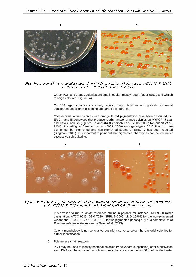
On MYPGP and J-agar, colonies are small, regular, mostly rough, flat or raised and whitish to beige coloured (Figure 3a)
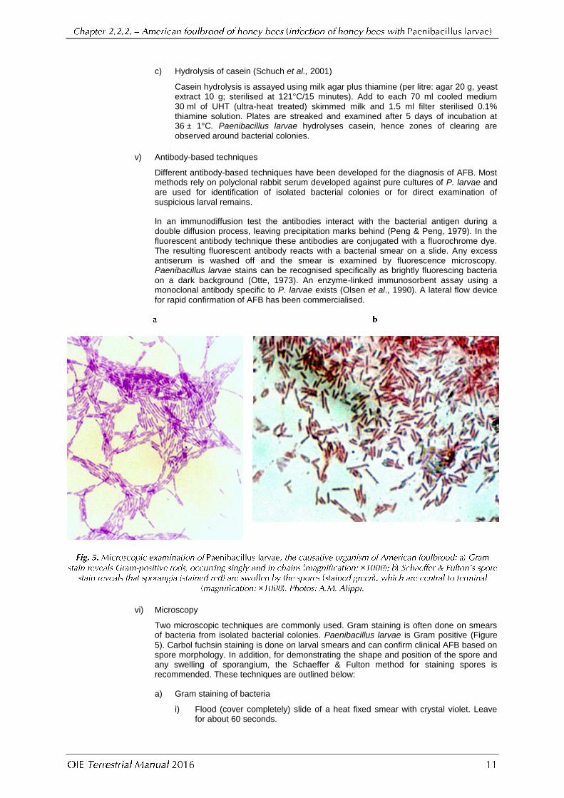
On CSA agar, colonies are small, regular, rough, butyrous and greyish, somewhat transparent and slightly glistening appearance (Figure 4a).
Paenibacillus larvae colonies with orange to red pigmentation have been described, i.e. ERIC II and III genotypes that produce reddish and/or orange colonies on MYPGP, J-agar and CSA (Table 2) (Figures 3b and 4b) (Genersch et al., 2005; 2006; Neuendorf et al., 2004). According to Genersch et al. (2005; 2006) only genotypes ERIC II and III are pigmented, but pigmented and non-pigmented strains of ERIC IV has been reported (Dingman, 2015). It is important to point out that pigmented phenotypes can be lost under successive sub-culturing.
It is advised to run P. larvae reference strains in parallel, for instance LMG 9820 (other designation: ATCC 9545, DSM 7030, NRRL B-2605, LMG 15969) for the non-pigmented variant and DSM 16115 or DSM 16116 for the pigmented genotype. (For a complete list of P. larvae reference strains see de Graaf et al., 2013).
Colony morphology is not conclusive but might serve to select the bacterial colonies for further identification.
ii) Polymerase chain reaction
PCR may be used to identify bacterial colonies (= cell/spore suspension) after a cultivation step. DNA can be extracted as follows: one colony is suspended in 50 µl of distilled water

and heated to 95°C for 15 minutes. Following centrifugation at 5000 g for 5 minutes, 1–5 µl of the supernatant is used as template DNA in a PCR 50 µl mixture (Dobbelaere et al., 2001b). Commercial DNA extraction kits can also be used, following the manufacturer’s instructions for Gram-positive bacteria. See section 1.4.2 below for the PCR method.
Characteristic ERIC I ERIC II ERIC III ERIC IV
Pigmented colonies – + + Variable
Spore surface (as seen by SEM) Smooth Convoluted With ridges With ridges
Growth in nutrient broth – + + +
Fermentation of mannitol – + + +
Fermentation of salicin + – – –
Alkaline phosphatase + – + +
Acid phosphatase + – + +
Catalase – – Weak, delayed + Weak, delayed +
iii) Mass spectrometry
Using a toothpick, a bacterial colony is smeared to two wells of the target plate and allowed to dry at room temperature before 1 µl of matrix (α-cyano-4-hydroxycinnamic acid) is added to each spot and prior to placing the target plate into the machine. MALDI-TOF MS measurement is then performed. The device detects specific mass spectra of bacterial ribosomal peptides. After comparison of detected spectra with the database of known spectra, the bacterial strain is identified (Schäfer et al., 2014).
iv) Biochemical tests
Paenibacillus larvae can also be identified by its biochemical profile. The bacteria are catalase negative or weak delayed positive, they have a typical carbohydrate fermentation profile with acid production from glucose and trehalose, but not from arabinose and xylose, and they can hydrolyse casein or milk. The results of some tests vary within the genotypes, e.g. fermentation of mannitol and salicin (Table 2).
a) Catalase test
A drop of 3% hydrogen peroxide is placed on an actively growing culture on solid medium. Most aerobic bacteria break down the peroxide to water and oxygen, producing a bubbly foam, but P. larvae is negative or weak delayed positive for this reaction, depending on genotype (Table 2) (Haynes, 1972). Organisms can lose their catalase activity with age, resulting in a false negative reaction. When Colombia sheep blood agar is used for cultivation, the test cannot be done on the solid medium, as the presence of sheep blood will cause a false-positive reaction. In this case, colonies should be transferred to a clean microscope slide for the execution of the test. Here the evaluation of the test occurs as above with the naked eye.
b) Production of acid from carbohydrates (Gordon et al., 1973)
Bacteria are grown in J-broth (per litre: yeast extract 15 g, tryptone 5 g and K2HPO
4
3 g) in which 0.5% of the test substrate, separately sterilised in aqueous solution, is substituted for the glucose. The carbohydrates used are L (+)-arabinose, D (+)-glucose, D (+)-xylose and D (+)-trehalose. The cultures are tested at 14 days by aseptically removing one ml or less to a spot plate, mixing the sample with a drop of 0.04% alcoholic bromocresol purple, and observing the colour of the indicator. Paenibacillus larvae produces acid aerobically from glucose and trehalose. No acid is produced from arabinose and xylose (Alippi, 1992) and variable results are obtained with mannitol and salicin according to the isolate tested (Genersch et al., 2005; 2006) (Table 2).
Commercial kits are also available for the biochemical characterisation of P. larvae (Carpana et al., 1995; Dobbelaere et al., 2001a; Neuendorf et al., 2004).
c) Hydrolysis of casein (Schuch et al., 2001)
Casein hydrolysis is assayed using milk agar plus thiamine (per litre: agar 20 g, yeast extract 10 g; sterilised at 121°C/15 minutes). Add to each 70 ml cooled medium 30 ml of UHT (ultra-heat treated) skimmed milk and 1.5 ml filter sterilised 0.1% thiamine solution. Plates are streaked and examined after 5 days of incubation at 36 ± 1°C. Paenibacillus larvae hydrolyses casein, hence zones of clearing are observed around bacterial colonies.
v) Antibody-based techniques
Different antibody-based techniques have been developed for the diagnosis of AFB. Most methods rely on polyclonal rabbit serum developed against pure cultures of P. larvae and are used for identification of isolated bacterial colonies or for direct examination of suspicious larval remains.
In an immunodiffusion test the antibodies interact with the bacterial antigen during a double diffusion process, leaving precipitation marks behind (Peng & Peng, 1979). In the fluorescent antibody technique these antibodies are conjugated with a fluorochrome dye. The resulting fluorescent antibody reacts with a bacterial smear on a slide. Any excess antiserum is washed off and the smear is examined by fluorescence microscopy. Paenibacillus larvae stains can be recognised specifically as brightly fluorescing bacteria
on a dark background (Otte, 1973). An enzyme-linked immunosorbent assay using a monoclonal antibody specific to P. larvae exists (Olsen et al., 1990). A lateral flow device for rapid confirmation of AFB has been commercialised.
×
×
vi) Microscopy
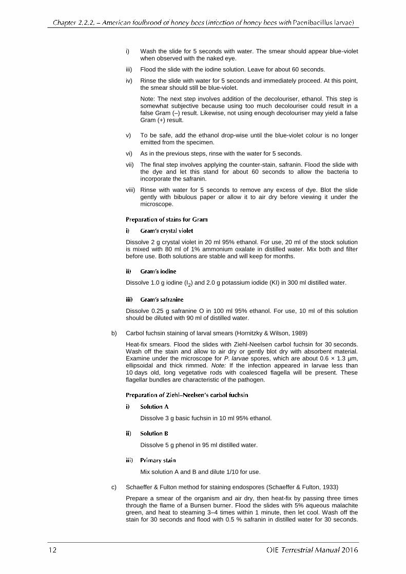
Two microscopic techniques are commonly used. Gram staining is often done on smears of bacteria from isolated bacterial colonies. Paenibacillus larvae is Gram positive (Figure
5). Carbol fuchsin staining is done on larval smears and can confirm clinical AFB based on spore morphology. In addition, for demonstrating the shape and position of the spore and any swelling of sporangium, the Schaeffer & Fulton method for staining spores is recommended. These techniques are outlined below:
a) Gram staining of bacteria
i) Flood (cover completely) slide of a heat fixed smear with crystal violet. Leave for about 60 seconds.
i) Wash the slide for 5 seconds with water. The smear should appear blue-violet when observed with the naked eye.
iii) Flood the slide with the iodine solution. Leave for about 60 seconds.
iv) Rinse the slide with water for 5 seconds and immediately proceed. At this point, the smear should still be blue-violet.
Note: The next step involves addition of the decolouriser, ethanol. This step is somewhat subjective because using too much decolouriser could result in a false Gram (–) result. Likewise, not using enough decolouriser may yield a false Gram (+) result.
v) To be safe, add the ethanol drop-wise until the blue-violet colour is no longer emitted from the specimen.
vi) As in the previous steps, rinse with the water for 5 seconds.
vii) The final step involves applying the counter-stain, safranin. Flood the slide with the dye and let this stand for about 60 seconds to allow the bacteria to incorporate the safranin.
viii) Rinse with water for 5 seconds to remove any excess of dye. Blot the slide gently with bibulous paper or allow it to air dry before viewing it under the microscope.
Dissolve 2 g crystal violet in 20 ml 95% ethanol. For use, 20 ml of the stock solution is mixed with 80 ml of 1% ammonium oxalate in distilled water. Mix both and filter before use. Both solutions are stable and will keep for months.
Dissolve 1.0 g iodine (I2) and 2.0 g potassium iodide (KI) in 300 ml distilled water.
Dissolve 0.25 g safranine O in 100 ml 95% ethanol. For use, 10 ml of this solution should be diluted with 90 ml of distilled water.
b) Carbol fuchsin staining of larval smears (Hornitzky & Wilson, 1989)
Heat-fix smears. Flood the slides with Ziehl-Neelsen carbol fuchsin for 30 seconds. Wash off the stain and allow to air dry or gently blot dry with absorbent material. Examine under the microscope for P. larvae spores, which are about 0.6 × 1.3 µm, ellipsoidal and thick rimmed. Note: If the infection appeared in larvae less than 10 days old, long vegetative rods with coalesced flagella will be present. These flagellar bundles are characteristic of the pathogen.
Dissolve 3 g basic fuchsin in 10 ml 95% ethanol.
Dissolve 5 g phenol in 95 ml distilled water.
Mix solution A and B and dilute 1/10 for use.
c) Schaeffer & Fulton method for staining endospores (Schaeffer & Fulton, 1933)
Prepare a smear of the organism and air dry, then heat-fix by passing three times through the flame of a Bunsen burner. Flood the slides with 5% aqueous malachite green, and heat to steaming 3–4 times within 1 minute, then let cool. Wash off the stain for 30 seconds and flood with 0.5 % safranin in distilled water for 30 seconds.

Wash off the stain very lightly, blot dry and examine under the microscope. The spores should be stained green and the vegetative cells red. P. larvae spores are ellipsoidal, central to terminal, swelling the sporangium and thick rimmed, measuring about 0.6 × 1.3 µm.
Dissolve 5 g of malachite green chloride with 100 ml of distilled water. This solution is very stable.
Dissolve 0.5 g of safranin O chloride with 100 ml of distilled water. This solution is very stable.
d) Nigrosin negative staining
Take a drop of the fluid phase of the CSA slant, mix it on the slide with 5% nigrosin solution, air dry then heat fix by passing three times through the flame of a Bunsen burner.
Cell/spore suspensions and suspensions containing only spores have to be differentiated, the latter requiring a more complex DNA extraction step.
For rapid confirmation of clinical AFB, the samples should be prepared as follows: the remains of two diseased honey bee larvae (= cell/spore suspension) are emulsified in 1 ml of sterile distilled water and mixed thoroughly. 100 µl of this suspension is diluted with 900 µl distilled water. This dilution is vortexed and 100 µl of it is used to extract DNA by heating and centrifugation (see above) (Dobbelaere et al., 2001b). Extraction of DNA can also be done using commercial kits according to the manufacturer´s instructions.
All aqueous solutions resulting from the sampling of adult bees, debris, bee wax, pollen and royal jelly should be considered as a spore suspension. Here, the extraction of DNA demands another approach. Indeed, spore suspensions are centrifuged at 6000 g and 4°C for 30 minutes. Next, the pellet is subjected to microwave treatment for 5 minutes at maximum power to break the spore coat, and the released DNA is suspended in 30 µl of 10 mM Tris/HCl, pH 8.0, containing 1 mM EDTA (Piccini et al., 2002).
When spores are to be detected from honey, DNA is serially diluted with sterile distilled water to eliminate PCR inhibition caused by honey (Piccini et al., 2002). Another DNA extraction method, based on lysozyme and proteinase K treatment, has been described (Bakonyi et al., 2003).
Good results can also be obtained by incubating a pelleted spore suspension in MYPGP broth at 37°C for 2–24 hours. Thereafter, the suspension is centrifuged at 14,500 g for 5 minutes,
washed with sterile distilled water and resuspended in 200 µl of sterile distilled water. This short incubation step causes spores to germinate, making them sensitive for DNA preparation by heat treatment again (see above) (Lauro et al., 2003).
Positive and negative controls should be run in parallel with the test samples.
Several PCR protocols have been described to identify P. larvae (reviewed by de Graaf et al., 2006 and De Graaf et al., 2013), but two of them, based on the 16S rRNA gene, have proven robustness and are described as follows:
Note: if using commercial PCR mixes the required ingredients may already be included. Check and follow the manufacturer’s instructions
PCR reactions (modified from Dobbelaere et al., 2001) are set up as 50 μl mixtures containing:
i) 1–5 μl template DNA (see sample preparation);
ii) 50 pmol forward (AFB-F) and reverse primer (AFB-R);
iii) 10 nmol of each dNTP;
iv) 2 mM MgCl2;
v) 1–2.5 U of Taq polymerase in the appropriate PCR buffer containing 2 mM MgCl2.
Reducing the volume of the PCR mixtures to 25 µl is possible.
Use the following PCR conditions: a 95°C (1–15 minutes) step; 30 cycles of 93°C (1 minute), 55°C (30 seconds), and 72°C (1 minute); and a final cycle of 72°C (5 minutes).
PCR reactions by using PL1 and PL2 primers (Govan et al., 1999) are set up as 50 µl mixtures containing:
i) 1–5 µl template DNA (see sample preparation);
ii) 2 mM MgCl2;
iii) 50 pmol of forward PL1) and reverse (PL2) primer;
iv) 25 mM concentration of each dNTP;
v) 1 U of Taq polymerase per ml.
Reducing the volume of the PCR mixtures to 25 µl is possible.
Use the following PCR conditions: a 95°C (1 minute) step; 30 cycles of 93°C (1 minute), 55°C (30 seconds), and 72°C (1 minute); and a final cycle of 72°C (5 minutes). The molecular weights of the PCR products are determined by electrophoresis in a 0.8% agarose gel and staining with a suitable DNA dye.
Ref. Name Sequence PCR-product size
Specificity level
(Dobbelaere et al., 2001b)
AFB-F AFB-R
5’-CTT-GTG-TTT-CTT-TCG-GGA-GAC-GCC-A-3’ 5’-TCT-TAG-AGT-GCC-CAC-CTC-TGC-G-3’
1106 bp species
(Govan et al., 1999)
PL1 PL 2
5’-AAG-TCG-AGC-GGA-CCT-TGT-GTT-TC-3’ 5-’TCT-ATC-TCA-AAA-CCG-GTC-AGA-GG-3’
973 bp species
No serological tests are available.
No vaccines are available.
Illustrations by Karl Weiss, extracted from Bienen-Pathologie, 1984, are reproduced with the kind permission of the author and Ehrenwirth-Verlag, Munich (Germany). Photographs are from the Animal and Plant Health Agency (APHA) York (UK) and the Informatiecentrum voor Bijenteelt, Ghent (Belgium) and published with kind permission of respectively Ruth Waite and Frans J. Jacobs. Figures 3, 4 and 5 are reproduced by kind permission of Dr A.M. Alippi, National University of La Plata (Argentina).
An FAO publication, Honey bee diseases and pests: a practical guide, W. Ritter & P. Akratanakul (eds). Agricultural and Food Engineering Technical Report No. 4. FAO, Rome, Italy, 42 pp. ISSN 1814-1137 TC/D/A0849/E, is available free of charge at: ftp://ftp.fao.org/docrep/fao/012/a0849e/a0849e00.pdf

ALIPPI A.M. (1992). Characterization of Bacillus larvae White, the causative agent of American foulbrood of honey-bees. First record of its occurrence in Argentina. Rev. Argent. Microbiol., 24, 67–72.
ALIPPI A.M. (1995). Detection of Bacillus larvae spores in Argentinian honeys by using a semi-selective medium. Microbiologia SEM, 11, 343–350.
BAKONYI T., DERAKHSHIFAR I, GRABENSTEINER E. & NOWOTNY N. (2003). Development and evaluation of PCR assays for the detection of Paenibacillus larvae in honey samples: comparison with isolation and biochemical characterization. Appl. Eviron. Microbiol., 69 (3), 1504–1510.
BRØDSGAARD C.J., HANSEN H. & RITTER W. (2000). Progress of Paenibacillus larvae larvae infection in individually inoculated honey bee larvae reared single in vitro, in micro colonies, or in full-size colonies. J. Apicult. Res., 39 (1–2), 19–27.
BZDIL J. (2007): Detection of Paenibacillus larvae spores in the debris and wax of honey bee by the Tween 80 method. Acta Vet. Brno, 76, 643–648.
CARPANA E., MAROCCHI L. & GELMINI L. (1995). Evaluation of the API 50CHB system for the identification and biochemical characterization of Bacillus larvae. Apidologie, 26, 11–16.
DE GRAAF D.C., ALIPPI A.M., ANTÚNEZ K., ARONSTEIN K.A., BUDGE G., DE KOKER D., DE SMET L., DINGMAN D.W., EVANS J.D., FOSTER L.J., FÜNFHAUS A., GARCIA-GONZALEZ E., GREGORC A., HUMAN H., MURRAY K.D., NGUYEN B.K., POPPINGA L., SPIVAK M., VAN ENGELSDORP D., WILKINS S. & GENERSCH E. (2013). Review Article: Standard methods for American foulbrood research. J. Apicult. Res., 52 (1), DOI 10.3896/IBRA.1.52.1.11.
DE GRAAF D.C., ALIPPI A.M., BROWN M., EVANS J.D., FELDLAUFER M., GREGORC A., HORNITZKY M., PERNAL S.F., SCHUCH D.M.T., TITĔRA D., TOMKIES V. & RITTER W. (2006). Under the microscope. Diagnosis of American foulbrood disease in honeybees: A synthesis and proposed analytical protocols. Lett. Appl. Microbiol., 43, 583–
590.
DINGMANN D.W. (2015). Comparative analysis of Paenibacillus larvae genotypes isolated in Connecticut. Arch. Microbiol., DOI 10.1007/s00203-015-1113-4.
DINGMANN D.W. & STAHLY D.P. (1983). Medium promoting sporulation of Bacillus larvae and metabolism of medium components. Appl. Environ. Microbiol., 46(4), 860–869.
DJUKIC M., BRZUSZKIEWICZ E., FÜNFHAUS A, VOSS J., GOLLNOW K., POPPINGA L., LIESEGANG H., GARCIA-GONZALEZ E., GENERSCH E. & ROLF D. (2014). How to kill the honey bee larva: Genomic potential and virulence mechanisms of Paenibacillus larvae. PLos One, 9 (3): e90914. Doi: 10.1371/journal.pone.0090914.
DOBBELAERE W., DE GRAAF D.C., PEETERS J.E & JACOBS F.J. (2001a). Comparison of two commercial kits for the biochemical characterization of Paenibacillus larvae larvae in the diagnosis of AFB. J. Apic. Res., 40, 37–40.
DOBBELAERE W., DE GRAAF D.C., PEETERS J.E & JACOBS F.J. (2001b). Development of a fast and reliable diagnostic method for American foulbrood disease (Paenibacillus larvae subsp. larvae) using a 16S rRNA gene based PCR. Apidologie, 32, 363–370.
FORSGREN E., STEVANOVIC J. & FRIES I. (2008). Variability in germination and in temperature and storage resistance among Paenibacillus larvae genotypes. Vet. Microbiol., 129, 342–349.
GENERSCH E. (2010) American Foulbrood in honeybees and its causative agent, Paenibacillus larvae. J. Invert. Pathol., 103 (Suppl. 1), 10–19.
GENERSCH E., ASHIRALIEVA A. & FRIES I. (2005). Strain- and genotype-specific differences in virulence of Paenibacillus larvae subsp. larvae, a bacterial pathogen causing American foulbrood disease in honey bees. Appl. Environ. Microbiol., 71, (11), 7551–7555.

GENERSCH E., FORSGREN E., PENTIKÅINEN J., ASHIRALIEVA A., RAUCH S., KILWINSKI J. & FRIES I. (2006). Reclassification of Paenibacillus larvae subsp. pulvifaciens and Paenibacillus larvae subsp. larvae as Paenibacillus larvae without subspecies differentiation. Int. J. Syst. Evol. Microbiol. 56, 501–511.
GOCHNAUER T.A. (1973). Growth, protease formation, and sporulation of Bacillus larvae in aerated broth culture. J. Invertebr. Pathol., 22, 251–257.
GOCHNAUER T.A. & CORNER J. (1987). Detection and identification of Bacillus larvae in a commercial pollen sample. J. Apic. Res., 13, 264–267.
GORDON R.E., HAYNES W.C. & PANG C.H.N. (1973). The genus Bacillus. Agriculture Handbook N° 427, USDA, Washington D.C.
GOVAN V.A., ALLSOPP M.H. & DAVIDSON S. (1999). A PCR detection method for rapid identification of Paenibacillus larvae. Appl. Environm. Microbiol., 65, 2243–2245.
HANSEN H. & BRØDSGAARD C.J. (1999). American foulbrood: a review of its biology, diagnosis and control. Bee World, 80 (1), 5–23.
HASEMAN L. (1961). How long can spores of American Foulbrood live? Amer. Bee J., 101, 298–299.
HAYNES W.C. (1972). The catalase test. An aid in the identification of Bacillus larvae. Am. Bee J., 112, 130–131.
HEYNDRICKX M., VANDEMEULEBROECKE K., HOSTE B., JANSSEN P., KERSTERS K., DE VOS P., LOGAN N.A., ALI N. &
BERKELEY R.C. (1996). Reclassification of Paenibacillus (formerly Bacillus) pulvifaciens (Nakamura 1984) Ash et al. 1994, a later subjective synonym of Paenibacillus (formerly Bacillus) larvae (White 1906) Ash et al. 1994, as a subspecies of P. larvae, with emended descriptions of P. larvae as P. larvae subsp. larvae and P. larvae subsp. pulvifaciens. Int. J. Syst. Bacteriol., 46, 270–279.
HORNITZKY M.A.Z. & CLARK S. (1991). Culture of Bacillus larvae from bulk honey samples for the detection of American foulbrood. J. Apicult. Res., 30 (1), 13–16.
HORNITZKY M.A.Z. & KARLOVSKIS S. (1989). A culture technique for the detection of Bacillus larvae in honeybees. J. Apicult. Res. 28, 118–120.
HORNITZKY M.A.Z. & WILSON S.C. (1989). A system for the diagnosis of the major bacterial brood diseases of honeybees. J. Apicult. Res., 28, 191–195.
KOSTECKI R. (1969). Studies on improvement of control of American foulbrood of the honey bee (in Polish). Pszczelnicze Zeszyty Naukowe, 13, 97–135.
LAURO F.M., FAVARETTO M., COVOLO L., RASSU M. & BERTOLONI G. (2003). Rapid detection of Paenibacillus larvae from honey and hive samples with a novel nested PCR protocol. Int. J. Food Microbiol., 81, 195–201.
LINDSTRÖM A. & FRIES I. (2005). Sampling of adult bees for detection of American foulbrood (Paenibacillus larvae subsp. larvae) spores in honey bee (Apis mellifera) colonies. J. Apicult. Res., 44 (2), 82–86.
MORRISSEY B.J., HELGASON T., POPPINGA L., FÜNFHAUS A., GENERSCH E. & BUDGE G.E. (2015). Biogeography of Paenibacillus larvae, the causative agent of American foulbrood using a new multilocus sequence typing scheme. Environ. Microbiol., 17, 1414–1424.
NEUENDORF S., HEDTKE K., TANGEN G. & GENERSCH E. (2004) Biochemical characterization of different genotypes of Paenibacillus larvae subsp. larvae, a honey bee bacterial pathogen. Microbiology SGM, 150, 2381–2390.
OLSEN P.E., GRANT G.A., NELSON D.L. & RICE W.A. (1990). Detection of American foulbrood disease of the honeybee, using a monoclonal antibody specific to Bacillus larvae in an enzyme-linked immunosorbent assay. Can. J. Microbiol., 36, 732–735.
OTTE E. (1973). Contribution to the laboratory diagnosis of American foulbrood of the honey bee with particular reference to the fluorescent antibody technique. Apidologie, 4 (4), 331–339.
PENG Y.S. & PENG K.Y. (1979). A study on the possible utilization of immunodiffusion and immunofluorescence techniques as diagnostic methods for American foulbrood of honeybees (Apis mellifera), J. Invertebr. Pathol., 33, 284–289.
PICCINI C., D’ALESSANDRO B., ANTUNEZ K. & ZUNINO P. (2002). Detection of Paenibacillus larvae subsp. larvae spores in naturally infected bee larvae and artificially contaminated honey by PCR. World J. Microbiol. Biotechnol., 18, 761–765.
RAUCH S., ASHIRALIEVA A., HEDTKE K.& GENERSCH E. (2009). Negative correlation between individual-insect-level virulence and colony-level virulence of Paenibacillus larvae, the etiological agent of American Foulbrood of honey bees. Appl. Environm. Microbiol., 75, 3344–3347.
RIEG S., BAUER T.M., PEYERL-HOFFMANN G., HELD J., RITTER W., WAGNER D., KERN W.V. & SERR A. (2010). Paenibacillus larvae bacteremia in injection drug users. Emerg. Infect. Dis. 16 (3), 487–489.
RITTER W. (2003). Early detection of American foulbrood by honey and wax analysis. Apiacta, 38, 125–130.
RITTER W. & KIEFER M.B. (1995). A method for diagnosing Bacillus larvae in honey samples. Animal Res. Dev., 42, 7–13.
RUDENKO E.V. (1987). Manuscript. Dissertation for Doctorate of Veterinary Science American foulbrood of honey bees and its vaccine prophylaxis (in Russia), Minsk, Belarus.
SCHÄFER M.O., GENERSCH E., FÜNFHAUS A., POPPINGA L., FORMELLA N., BETTIN B. & KARGER A. (2014). Rapid identification of differentially virulent genotypes of Paenibacillus larvae, the causative organism of American foulbrood of honey bees, by whole cell MALDI-TOF mass spectrometry. Vet. Microbiol., 170, 291–297.
SCHAEFFER A.B. & FULTON M. (1933). A simplified method of staining endospores. Science, 77, 194, New York.
SCHUCH D.M.T., MADDEN R.H. & SATTLER A. (2001). An improved method for the detection and presumptive identification of Paenibacillus larvae subsp. larvae spores in honey. J. Apicult. Res., 40 (2), 59–64.
TITERA D & HAKLOVA M. (2003). Detection method of Paenibacillus larvae larvae from beehive winter debris. Apiacta, 38, 131–133.
VON DER OHE W. & DUSTMANN J.H. (1997). Efficient prophylactic measures against American foulbrood by bacteriological analysis of honey for spore contamination. Am. Bee J., 137 (8), 603–606.
WOODROW A.W. (1941). Susceptibility of honey bee larvae to American foulbrood. Gleanings Bee Cult., 69, 148–151.
*
* *
NB: There is an OIE Reference Laboratory for American foulbrood of honey bees (see Table in Part 4 of this Terrestrial Manual or consult the OIE Web site for the most up-to-date list:
http://www.oie.int/en/our-scientific-expertise/reference-laboratories/list-of-laboratories/ http://www.oie.int/). Please contact the OIE Reference Laboratories for any further information on
diagnostic tests and reagents for bee diseases


![kumpulanpublikasi.files.wordpress.com...negara dan . pwrn yang itu Sudah ... Ma] ell s 1071, 1937 tahm 1992 yang kaidah tesis maka taža rakyat. itu Identifikasi Ma—lah di . 2) yang](https://img.dokumen.tips/doc/110x75/5e30e40a62837d474472fe34/-negara-dan-pwrn-yang-itu-sudah-ma-ell-s-1071-1937-tahm-1992-yang-kaidah.jpg)


![СНБ 2.02.02-01 [+] Эвакуация людей из ЗиС при пожаре · 2019-11-11 · ТКП 45-3.02-108–2008 Высотные здания. Строительные](https://img.dokumen.tips/doc/110x75/5e659878f95fac48cb13b4da/-20202-01-f-.jpg)


