Embed Size (px)
Citation preview

1
Ambulance Emergency Assistants (AEA) CPD Activity for 2015
Activity: E1 (15)
General Theme: Clinical
Topics:
Do not underscore the value of a trauma assessment
Consider All Differentials for Respiratory Distress
Learn the signs and symptoms of acute angle closure glaucoma
Syncopal episodes can warn of cardiac conditions
Timely Prehospital Management of Stroke Victims Crucial for Patient
Be Wary During ECG Analysis in the Geriatric Population
To the Hospital or Not? Debating How to Handle Out-of-Hospital Cardiac Arrest
Non-invasive Positive Pressure Ventilation an Alternative to Intubation?
Approved for (4) Clinical Continuing Educational Units (CEU’s)

2
Don’t Underscore the Value of a Trauma Assessment
Fran Hildwine, BS, NREMT-P, CCEMT-P | From the October 2014 Issue | Wednesday, October 1, 2014
At 4:12 p.m. on a sunny summer day, Ambulance 5 is dispatched for a “sick person.”
Dispatch notes advise the patient is a 25-year-old male who’s had abdominal pain for two days. While responding to the initial dispatch, the police radio crackles with an officer requesting paramedics to expedite to a motor vehicle crash involving a motorcycle. The location of the crash is three blocks ahead, directly in your path to the initial dispatch.
As you approach the scene you notice police have the road blocked and are frantically waving you down to treat the seriously injured motorcyclist. You notify the 9-1-1 centre you’re on location with police and request the next due BLS ambulance be dispatched to your initial call.
Police direct you to an area of high grass behind the guardrail, where you find the 34-year-old helmeted male, whose motorcycle struck the driver’s side of a small, two-door car, ejecting him from the bike and over the car, where he struck the guardrail with his left side and landed in the grass.
As you approach the supine patient you notice a severely angulated left arm and left leg and immediately think, “This isn’t going to be good.”
Expecting the worst, you’re surprised to find an adequate respiratory effort at a rate of 24 as you kneel by the patient’s head in order to quickly stabilize the C-spine before calling out to the patient to check responsiveness.
“Hello! Can you hear me?” you ask.
“Yeah,” he groans. You’re again amazed; he’s awake, alert and fully oriented to person, place, time and events. He tells you he was driving the posted 40 mph speed limit when the car pulled out in front of him. Police confirm this with a statement from the driver of the car.
At the risk of sounding stupid, you ask him what hurts. “Everything!” he replies. “Is my leg still there?”
Your rapid trauma assessment reveals cervical point tenderness in the area of C-5. His pupils are equal, round and reactive, and he has equal chest expansion with full, clear breath sounds. His abdomen is soft, non-tender and his pelvis is stable. His right arm is intact with a radial pulse of 124 and you notice his right thigh is swollen and tender with crepitus indicative of a femur fracture, but you do note distal pulse, motor and sensation.
The extremities on the left side are much worse. The left arm has multiple open fractures with profuse bleeding and you can’t feel a distal pulse. His left leg has a deep laceration from the knee to the hip with the fascia of the muscle clearly visible. His left foot is severely

3
angulated, positioned roughly adjacent to the left knee, with multiple open fractures of the tibia and fibula. You can’t feel distal pulses and are worried that any movement may cause the few threads of tissue connecting the foot to tear and the foot to fall off.
At this time ALS arrives on scene, quickly eyeballs the patient and begins to set up the ambulance while your crew finishes packaging the patient.
Prehospital Treatment Your partner and police assist in cutting away the patient’s clothes while you secure a C-collar. Another member of your crew quickly places trauma dressings over the gaping wounds and positions the backboard. The log roll allows you to examine the spine and you’re surprised to find no further injuries.
The patient is secured to a backboard, a cervical immobilization device is applied and the patient is transferred to the ambulance. You place a non-rebreather mask on him with O2 at 15 Lpm while your partner obtains a blood pressure of 96/60. The paramedic supervisor says she’ll notify the trauma centre. During the four-minute transport to the Level 2 trauma centre ALS initiates a 14-gauge large bore IV in the right antecubital fossa and infuses a 500 cc fluid bolus. You continue to dress the wounds to the patient’s left extremities and actually manage to obtain a SAMPLE (signs and symptoms, allergies, medications, past history, last meal, events leading to injury) history.
Hospital Treatment The trauma team is awaiting your arrival and quickly transfers the patient to a trauma resuscitation bed. After quickly confirming your SAMPLE history, the anaesthesiologist uses rapid sequence intubation to paralyze and sedate the patient in order to pass an 8.0 endotracheal tube and secure his airway.
The trauma team completes its primary assessment, places a large bore femoral line and begins rapid transfusion of three units of blood. This causes a rise in blood pressure that reveals three small lacerations to the left brachial artery, which is now clearly visible.
A Combat Application Tourniquet is placed high on the left humerus and the bleeding is controlled. The left arm and left leg are quickly bandaged and splinted before taking the patient to get a CT scan then directly to the operating room.
The CT scan confirms the C-spine X-ray that shows a compression fracture of C-5 without neurological impairment. Initial surgeries included a below-knee amputation of the left leg, vascular repair of the left arm, external fixation of the left arm and placement of a halo brace for the C-spine fracture.
The hospital and rehabilitation stays were lengthy, but uneventful. Six months later the patient walks into your station to thank you and the crew for saving his life.
Discussion There’s a very good reason trauma assessment is taught as a skill that needs to be completed the same way every time. Deviation from standard trauma assessment during Advanced Trauma Life Support (ATLS), which is mirrored in Prehospital Trauma Life Support (PHTLS), provides an opportunity for important findings to be missed.

4
The primary assessment of airway, breathing, circulation, disability and exposure addresses the most life-threatening issues in order of importance and also provides an easy-to-remember standard that serves to minimize mistakes. This patient had a potentially life-threatening cervical spine fracture. Had the first arriving ambulance crew not taken the precautions to protect the C-spine while they progressed through the primary assessment this patient may not have survived.
EMTs and paramedics also need to acknowledge the impact of distracting injuries. It’s easy to internalize the pain upon seeing a grossly deformed arm and leg, to get tunnel vision and hyper-focus on these injuries. The reality is that extremity trauma is rarely life-threatening. Most haemorrhage from extremity fractures can be treated with direct pressure and, in extreme cases such as this, a tourniquet.
An organized, thorough and rapid secondary assessment assures that severe, possibly life-threatening injuries are found and treated. Although there’s often some variation among EMS and medical professional with the assessment of a medical patient, trauma assessments are generally done head to toe in order to provide consistency of results.
Field treatment for the multisystem trauma patient needs to be focused on addressing life threats, stabilizing the C-spine and rapidly transporting to the appropriate facility. Due to the short scene and transport times, pain management wasn’t a factor in this case.
Lastly, does your department have a policy on how to address the need to triage calls when presented with a life-threatening situation while responding to a different initial call? Some departments require you to proceed to the initial dispatch, others allow you to stop if you make sure a back-up unit is dispatched to the initial call for which you were responding.
Conclusion This case illustrates how trauma care is a team effort. Every person who touched this patient contributed to the successful outcome of this case. From the first police officer on scene, to the EMTs, paramedics, trauma physicians, nurses, radiology technicians, operating room staff, shock-trauma ICU, rehabilitation personnel and many others, there were many opportunities for a poor outcome, but when we do what we do well, every time, we can rest assured that these results are the norm. jems
Resource Dickinson E, Politis J. Painful distractions: The importance of splinting and managing the pain associated with fractures. JEMS. 2011;36(1):50–57.

5
Consider All Differentials for Respiratory Distress
Wes Ogilvie, MPA, JD, NRP, LP | From the November 2014 Issue | Friday, November 7, 2014
At 11:52 p.m., the county sheriff’s dispatcher tones out Medic 41 to an address in a rural
subdivision about 15 miles from their small town’s EMS station. Dispatch advises an out-of-breath male is in the back bedroom and may not be able to unlock the front door. Since the fire department doesn’t have any trained EMS providers, dispatch also assigns a sheriff’s deputy to the call in the event the EMS crew needs to force entry to the house.
The crew arrives at the address almost the same time as the deputy. There appear to be two houses on the property, both with the same address. The medics and deputy knock on the door of the larger house, whose resident states she didn’t call 9-1-1. The medics and deputy knock on the locked door of the second house but receive no response. The deputy forces it open.
The EMS crew makes it to the back bedroom of the home, where they find an approximately 70-year-old male in a tripod position at the foot of his bed. He’s breathing with the aid of a nasal cannula on a home oxygen generator at a rate of two litres per minute. The patient gasps for breath between each word as he tells the crew, “Can’t … breathe!” The patient weighs approximately 330 pounds.
Prehospital Treatment The paramedic immediately auscultates the lungs and hears little air movement. His EMT partner hooks the patient to the capnography and pulse oximetry functions on the cardiac monitor and then transfers the patient’s oxygen to their oxygen cylinder. Due to the confined quarters of the residence, the crew was unable to bring the stretcher inside.
The cardiac monitor shows a heart rate of approximately 94 in a sinus rhythm, a blood pressure of 104/48, and a pulse oximetry of 86% while on the oxygen at four litres per minute. The capnography waveform is normal.
The patient’s wife is able to provide some of the patient’s prior history, including several previous heart attacks, chronic obstructive pulmonary disease, hypertension and congestive heart failure (CHF). She also provides a list of his medications, which include lisinopril with HCTZ (hydrochlorothiazide), nitroglycerin, albuterol, Advair (fluticasone) and simvastatin. She says her husband has been ill for several days and hasn’t been compliant with his prescription medications for the past three days. He went to the back room to see if he

6
could sleep sitting up and told her not to disturb him. In fact, she didn’t know he called for EMS assistance.
In part because of the critical nature of the call, the size of the patient and the difficulty in accessing the back bedroom, the crew contacts dispatch by radio and requests additional personnel. Shortly after the tones go out, a volunteer first responder and an off-duty paramedic who lives in town are en route.
Meanwhile, the paramedic asks his partner and the deputy to get the continuous positive airway pressure (CPAP) kit and a stair chair. While this is occurring, the paramedic turns the oxygen flow up to 15 litres per minute by nasal cannula for passive oxygenation and prepares for a possible emergent intubation. As the team begins assembling the CPAP device, the two additional responders arrive and the team manoeuvres the patient onto the stair chair to move the patient to the ambulance.
The providers move the patient to the stretcher. At this point, the patient’s head is beginning to bob and he seems to be confused and lethargic. Both paramedics agree that the patient’s level of consciousness is insufficient to maintain compliance with CPAP therapy and agree that a crash airway situation exists.
They ask the EMT to get the video laryngoscope, connect the powered suction, obtain a rescue airway from the shelf and pre-oxygenate the patient. As one of the paramedics prepares the laryngoscope, the other draws up the appropriate rapid sequence intubation medications per protocol: 450 mg of ketamine (3 mg/kg at 150 kg) and 150 mg of rocuronium (1 mg/kg at 150 kg). He also prepares 5 mg of midazolam per protocol.
After both medics confirm the medications and dosages, the second establishes IV access via an 18-gauge catheter to the left forearm. After pre-oxygenating the patient, the other paramedic administers the ketamine, then the rocuronium. He uses the video laryngoscope to pass a 7.5 mm endotracheal (ET) tube and captures the image of the tube passing through the vocal cords onto the digital memory card. They then confirm placement via waveform end-tidal carbon dioxide capnography and secure the tube with a commercial device. Next, the patient is sedated with midazolam. The two paramedics agree they should both attend to the patient and ask the EMT to initiate emergency transport to a large community hospital on the outskirts of the nearest large city, about 35 miles away.
En route, the patient’s condition remains relatively unchanged, especially since he’s been sedated. The crew also obtains a 12-lead ECG, which doesn’t reveal any acute ST segment changes. The relative hypotension, especially in consideration of previous patient encounters where he’s routinely noncompliant with his anti-hypertensives, cause both providers to rapidly eliminate the possibility of a CHF exacerbation and thus rule out nitrate or diuretic therapy. The primary paramedic also obtains an axillary temperature of 103.5 degrees F. Based on this, combined with the patient’s relative hypotension, the paramedics suspect the possibility of sepsis and begin administering a fluid bolus. They contact the hospital via radio and advise the hospital of their treatment and the possible concern of sepsis, requesting that the hospital have a respiratory therapist in the ED on their arrival.
Hospital Treatment The patient is brought into the primary resuscitation room where the emergency physician on duty evaluates the patient. A chest X-ray is ordered for confirmation of ET tube placement. Upon viewing the film, the physician determines the patient has pneumonia. A

7
dosage of Levaquin (levofloxacin) is ordered via IV pump along with several fluid boluses. The patient remains on the ventilator and is admitted to the intensive care unit. After a course of antibiotic therapy, the patient is weaned from the ventilator and returns home several weeks later after discharge from the hospital to a skilled nursing facility for rehabilitation.
Discussion & Clinical Pearls Pneumonia is a relatively common but overlooked cause of respiratory distress. Pneumonia is a lung infection that causes fluid to collect in the alveoli of the lungs. It causes inflammation, which may cause dyspnoea, fever, chills, chest pain, chest wall pain or coughing.1
Several key clinical points bear mentioning. First, the entire EMS team practiced crew resource management throughout the course of the call. The initial crew recognized the need for additional personnel for both patient movement and patient treatment. The assisting first responder recognized the patient’s condition had deteriorated to the point where CPAP was contraindicated. Both paramedics recognized the patient’s condition might require both of them to attend to the patient during transport and they additionally worked together as a team, particularly during the rapid sequence intubation.
Also, while treating the patient, the providers used passive oxygenation with the nasal cannula to boost the patient’s oxygen saturation prior to intubation. The nasal cannula can even remain on the patient during the intubation attempt to maintain oxygen saturation. High-flow oxygen through the cannula has been shown to maintain saturations in excess of 95% during intubation.2
Finally, the EMS team identified the possibility of sepsis when they obtained the patient’s temperature and noted the relatively low blood pressure (particularly for a patient with a history of hypertension).
Conclusion This case presented challenges in assessment and diagnosis, especially considering the patient’s history of respiratory diseases and possible other comorbidities. Even with obtaining the patient’s history from the patient, family, and his medications, several likely differential diagnoses exist. The use of waveform capnography can help rule in or out the possibility of a reactive airway disease process. This case also illustrates that, even in a patient with a history of several respiratory disease processes, a new cause of respiratory failure can occur. Again, like any form of medicine, clinically competent and progressive emergency medical care is dependent on a through physical assessment and history-taking.
References 1. Abrahamson L, Mosesso V Jr., editors: Advanced medical life support: An assessment based approach. Elsevier Mosby: St. Louis, Mo.,
pp. 124–126, 2011.
2. Levitan R. (Dec. 9, 2010.) No desat! Emergency Physicians Monthly. Retrieved July 11, 2014,
fromwww.epmonthly.com/archives/features/no-desat-/.

8
Learn the Signs & Symptoms of Acute Angle Closure
Glaucoma
Judy Torres, BFA, JD, NREMT-P | From the October 2013 Issue | Tuesday, October 8, 2013
Patients with trauma to the eye should have their eye bandaged as appropriate to protect from additional damage. Also, whenever bandaging one eye, cover/bandage the other eye to reduce tandem eye movement.
Key Terms
Aqueous humor: The watery fluid that bathes and nourishes the anterior part of the eye. Ciliary body: The part of the eye above the lens that produces aqueous humor. Cornea: The outer transparent structure that covers and protects the iris and pupil. Choroid: A layer of the eye behind the retina that contains the blood vessels that supply the retina. Iris: The colored tissue behind the cornea that regulates the light entering your eye by changing the size of the pupil. Lens: The clear part of the eye which is suspended behind the iris. Optic nerve: A bundle of nerves located in the posterior eye that carries messages from the retina to the brain. Trabecular meshwork: The spongy tissue near the cornea. This is where the aqueous humor flows out of your eye.
Decision-making by paramedics in the field has the potential to become a challenging
situation. The decision of whether to transport has to be made quickly and efficiently. It often becomes a collaborative effort among the responding unit, the patient and their family members or friends. Insufficient information related to the actual complaint and limited access to the patient’s past medical history can also contribute to on-scene confusion. Balancing the patient’s care, safety and patient choice can sometimes be overwhelming.

9
When confronted with a patient who has one or more complaints that range from blurred vision and seeing halos around lights to severe eye pain and vomiting, time is of the essence.
This article discusses the classic signs and symptoms of acute angle closure glaucoma and will help you make critical transport decisions and understand available treatment options when faced with making a choice that could save someone’s vision.
What Is It?
Acute angle closure glaucoma (AACG), sometimes called acute narrow angle glaucoma or acute closed angle glaucoma, is a true medical emergency. Rapid and immediate treatment is necessary to prevent optic nerve damage and permanent loss of vision, which can occur within hours without prompt and appropriate treatment.
AACG is caused by a rapid or sudden increase in pressure inside the eye or intraocular pressure (IOP). This typically happens when the iris is pushed against the trabecular meshwork (the spongy tissue located near the cornea where the aqueous humor flows out of the eye) or drainage channels, and the fluid or aqueous humor (the watery fluid that nourishes the interior of your eye) that normally drains out of the eye is blocked. This creates the increase in IOP. (See Figure 2, p. 44.)
10
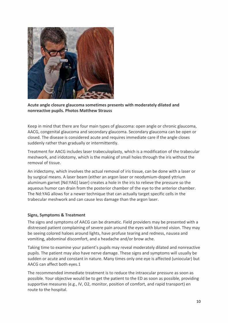
Acute angle closure glaucoma sometimes presents with moderately dilated and nonreactive pupils. Photos Matthew Strauss
Keep in mind that there are four main types of glaucoma: open angle or chronic glaucoma, AACG, congenital glaucoma and secondary glaucoma. Secondary glaucoma can be open or closed. The disease is considered acute and requires immediate care if the angle closes suddenly rather than gradually or intermittently.
Treatment for AACG includes laser trabeculoplasty, which is a modification of the trabecular meshwork, and iridotomy, which is the making of small holes through the iris without the removal of tissue.
An iridectomy, which involves the actual removal of iris tissue, can be done with a laser or by surgical means. A laser beam (either an argon laser or neodymium-doped yttrium aluminum garnet [Nd:YAG] laser) creates a hole in the iris to relieve the pressure so the aqueous humor can drain from the posterior chamber of the eye to the anterior chamber. The Nd:YAG allows for a newer technique that can actually target specific cells in the trabecular meshwork and can cause less damage than the argon laser.
Signs, Symptoms & Treatment
The signs and symptoms of AACG can be dramatic. Field providers may be presented with a distressed patient complaining of severe pain around the eyes with blurred vision. They may be seeing colored haloes around lights, have profuse tearing and redness, nausea and vomiting, abdominal discomfort, and a headache and/or brow ache.
Taking time to examine your patient’s pupils may reveal moderately dilated and nonreactive pupils. The patient may also have nerve damage. These signs and symptoms will usually be sudden or acute and constant in nature. Many times only one eye is affected (uniocular) but AACG can affect both eyes.1
The recommended immediate treatment is to reduce the intraocular pressure as soon as possible. Your objective would be to get the patient to the ED as soon as possible, providing supportive measures (e.g., IV, O2, monitor, position of comfort, and rapid transport) en route to the hospital.

11
These patients should be transported to the nearest appropriate ED. It’s also important to know which hospital in your area has an ophthalmologist on call or on staff since the vision loss can occur within a day of its onset.
Risk Factors
Knowing the risk factors can help you if you suspect if a patient with the above signs and symptoms might have AACG. The following risk factors can increase the incidence of AACG:1–4
Dim lighting, eye drops administered by the eye practitioner during the course of a routine eye exam, antihistamines/decongestant drops or cold medications. Anything that causes dilation of the pupil and anything that can block the drainage chamber of the eye.
Hyperopia (farsightedness). Farsighted people are at an increased risk because their anterior chambers are shallow and their angles are narrow.
Race. Some races are more prone to narrow angles and are more likely to develop AACG. Notable are: Asians, people of African descent, and Eskimos or Inuits.
Gender. In Caucasians, AACG is three times more likely in females than in males.
Family history. In general, glaucoma tends to run in families. Be sure to inquire about a patient's family history when performing your assessment.
Diabetes, hypertension and heart disease. These three conditions can be contributing factors.
Tumors behind the iris and physical injuries to the eye. Both cause internal damage that can contribute to an increased risk towards AACG. Ask the patient if they have experienced any recent trauma to their head, eye or eyes.
Inflammation. Eye infections causing inflammation inside the eye increase the risk of developing AACG.
Age. As we age the lens of our eye tends to enlarge and push our iris forward also increasing the risk for AACG.
Pupillary block. If the back of the iris (the colored part of the eye) adheres to the lens inside the eye the channel becomes blocked and fluid builds up behind the iris pushing it forward causing it to close the drainage of fluid in the anterior chamber of the eye.
Studies have shown occurrences of the disease rise with the intentional dilation of the eye during a routine eye examination.5,6 One study indicated that three in 10,000 patients were likely to develop AACG after diagnostic mydriasis (intentional prolonged dilatation of the pupil of the eye) even though the exams were followed by miotic drops (which constrict the pupil and increase the flow of aqueous humor).5
Conclusion
Decision-making in the field when a patient has signs and symptoms consistent with AACG can be aided by being familiar with the condition. Early recognition along with correct transport and treatment decisions can help give these patients the best chance for a positive outcome. Always being sensitive to your patient’s complaints and concerns is key to providing the best possible care. Your knowledge of this condition and its signs and symptoms can be the deciding factor in someone living with future blindness or not.

12
References
1. American Optometric Assocation. (n.d.) Glaucoma. Retrieved on Aug. 26, 2013, fromwww.aoa.org/patients-and-
public/eye-and-vision-problems/glossary-of-eye-....
2. Glaucoma Research Foundation. (Jan. 11, 2011.) Are you at risk for glaucoma? Retrieved on Aug. 26, 2013,
from www.glaucoma.org/glaucoma/are-you-at-risk-for-glaucoma.php.
3. Heiting G. (April 2010.) Narrow-angle glaucoma. All About Vision. Retrieved on Aug. 26, 2013,
fromwww.allaboutvision.com/conditions/narrow-angle-glaucoma.htm.
4. BrightFocus Foundation. (Apr. 28, 2013.) Glaucoma facts & statistics. Retrieved on Aug. 26, 2013,
fromwww.brightfocus.org/glaucoma/about/understanding/facts.html.
5. Liew G, Mitchell P, Wang JJ, et al. Fundoscopy: To dilate or not to dilate? BMJ. 2006;332(7532):3.
6. Dahl AA. Acute angle-closure glaucoma. eMedicineHealth. Retrieved on Aug. 26, 2013,
fromwww.emedicinehealth.com/script/main/hp.asp.

13
Syncopal Episodes Can Warn of Cardiac Conditions
Dennis Edgerly, EMT-P | From the January 2015 Issue | Thursday, January 8, 2015
You’ve been dispatched to care for a person who’s unconscious. As you walk in, you’re
directed to a young male lying on a couch. He appears to be in his late 20s or early 30s. The patient, Mark, is awake and acknowledges you as you approach. Your initial assessment identifies no respiratory distress or signs of trauma or hemorrhage. His pulse is tachycardic but strong, and his skin is warm and dry.
Mark tells you he and his wife had just arrived at his parents’ home. Soon after, he became light-headed and felt like his heart was racing. He laid himself down on the couch but doesn’t remember much after. His wife tells you he was unresponsive for about a minute until he began to move and answer to his name.
Nothing like this has happened to him before. Your assessment reveals no neurological deficits, a pulse of 120, blood pressure of 132/78 and a breathing rate at about 12 times a minute without difficulty.
Mark denies any previous medical history, takes no medications and says he hasn’t had anything to drink this evening. He says he’s feeling better and is able to sit up without feeling dizzy. He doesn’t want to go to the hospital, though his mother is encouraging him to go. She’s concerned because she had an older brother who died when he was about 20 years old—he had no known medical history but passed out a couple of times prior to his death, when he was found dead in his bed one morning. Mark agrees to go be evaluated and is transported without incident.
Discussion
Why did a healthy, young adult male pass out, and is there any correlation between Mark’s syncopal episode and the untimely death of his uncle? When EMS is evaluating a patient who has experienced a syncopal episode, they should attempt to identify a cause. Commonly, syncope is caused by a transient loss of blood supply to the brain, which can be due to dehydration, blood loss, cardiac dysrhythmias or neurologic events such as transient ischemic attacks. The fact the patient is young and healthy doesn’t negate the fact something caused him to lose consciousness.
Another potential cause of syncope and sudden cardiac death in young persons are a group of heart conditions known as channelopathies. Channelopathies are congenital (born with) problems on cardiac cells. The ion channels on the wall of cardiac cells fail to properly regulate the exchange of electrolytes, such as sodium. Two common channelopathies are long QT syndrome (LQTS) and Brugada syndrome (BrS). Both of these conditions are difficult to diagnosis because there are no structural changes seen in the heart. The first symptom can be sudden cardiac death. In some cases there are changes in the ECG suggestive of the conditions.
There are currently 13 different types of LQTS and it’s estimated LQTS is seen in one out of every 2,500 people. 1 The condition can be congenital or acquired. Acquired LQTS is most commonly associated with medications. The condition can result in sudden cardiac

14
arrhythmias, causing syncope or cardiac arrest. Faster heart rates can be associated with the arrhythmias so it’s not uncommon to see patients with LQTS show symptoms during physical activity, such as collapsing on a soccer field.
Once identified, LQTS can be treated with beta blockers, which are medications that work to keep heart rates from becoming tachycardic. These patients may also be placed with an internal cardiac defibrillator (ICD).
BrS exists in an estimated four out of every 1,000 persons2 with prevalence in men over women 8:1.3 Similar to LQTS, the first presentation of BrS can be sudden syncope or sudden cardiac death. Many medications can be harmful to patients with BrS, and the only treatment is an ICD.
Conclusion
Both channelopathies briefly mentioned here are congenital and can be seen in multiple family members. In this case, the fact Mark’s uncle died unexplainably at a young age is very relevant to Mark’s syncopal episode. If one member of a family is thought to have a channelopathy, other family members are commonly tested.
EMS providers, although perhaps not versed in all potential channelopathies, should keep in mind the possibility of a cardiac condition as a cause of syncope, especially if there’s no other explainable cause. It’s easy to consider a heart problem when the patient who passes out is older, but even young, healthy people can have cardiac conditions causing syncope or sudden cardiac death. In many instances, your interaction with them may be the first indication they have a condition requiring evaluation.
References 1. Vincent GM. The molecular genetics of the long QT syndrome: Genes causing fainting and sudden death. Annu Rev
Med. 1998;49:263–274.
2. Patel SS, Anees S, Ferrick KJ. Prevalence of a Brugada pattern electrocardiogram in an urban population in the United
States. Pacing Clin Electrophysiol. 2009;32(6):704.
3. Benito B, Sarkozy A, Mont L, et al. Gender differences in clinical manifestations of Brugada syndrome. J Am Coll Cardiol.
2008;52(19):1567.

15
Timely Prehospital Management of Stroke Victims Crucial
for Patient
David French, MD | Edward Jauch, MD, MS | Todd McGeorge, BA, EMT-P | From the January 2015 Issue |
Tuesday, January 20, 2015
Photo Kevin Link
Although prevention and early treatment have markedly reduced the morbidity and
mortality of stroke, it remains a significant health and social burden. This past decade saw stroke drop from the third to the fourth leading cause of death in the United States, but it remains a leading cause of adult disability.1
The cost of stroke is crippling, both in direct costs and lost opportunity. The global circumstances of stroke are even more dire—in many countries, it’s the second leading cause of death, and the rates of stroke are projected to double by 2030.2 To make an impact on this growing societal epidemic, the healthcare community must continue to improve our prevention and overall management of stroke.3
Fortunately, professionals involved in stroke care can learn from other healthcare successes. For medical conditions where timely identification, transport and intervention may mean the difference between life or death, integrated systems of care, including prehospital care coordinated with regional hospital services, saves lives.4
Outcomes data clearly show the benefits of such systems of care for trauma and ST elevation myocardial infarction (STEMI). More recently, data from similar systems of care for stroke suggest improved outcomes and less morbidity.5,6
Biology of Stroke
The term “stroke” collectively refers to all acute cerebrovascular disease, including ischemic and haemorrhagic types. Both forms share many risk factors, including diabetes, hypertension, smoking and age, and typically produce focal neurologic symptoms that are important to recognize. Prevention strategies and early management principles are similar between the two forms of stroke, but definitive interventions are specific to each type. Regardless of cause, targeted therapies that improve patient outcomes must be delivered within minutes to hours from symptom onset in order to be maximally effective.
Ischemic strokes occur due to an arterial occlusion, which causes brain tissue to infarct from a lack of oxygen and glucose. (See Figure 1 download below).

16
Occlusions can occur from either local vessel narrowing, causing thrombotic strokes, or due to clots originating elsewhere like the heart, which embolize to the vessel and cause embolic strokes. Regardless of cause, brain tissue is extremely sensitive to ischemia and begins to suffer irreversible damage within minutes of onset. To minimize permanent disability, blood flow must be restored quickly and overall brain physiology carefully managed, including in the prehospital setting.
In the U.S., haemorrhagic strokes occur much less frequently than ischemic strokes, accounting for about 20% of acute infarcts. However, in other countries such as China, over half of all strokes are haemorrhagic. Intracerebral haemorrhage (ICH) is a form of stroke that causes bleeding into the brain tissue itself.8
In older patients, ICH typically occurs due to amyloid deposition in smaller arteries, producing typically smaller, less disabling strokes. In younger patients, ICH is associated with hypertension, producing vascular damage in small blood vessels deep within the brain. Due to the haemorrhage size and location, it’s more often disabling and lethal.
Subarachnoid haemorrhage (SAH) typically occurs in younger patients due to a structural weakness in an artery producing an aneurysm in the vessel wall, which suddenly ruptures. Both forms of haemorrhagic stroke tend to be more severe than ischemic stroke.
Goals of Treatment
The goals of emergent stroke treatment are twofold: First, prevent or reverse the source of injury (limit haemorrhage growth in ICH or restore blood flow in ischemic stroke), and second, optimize the patient’s physiology as soon as possible to maximize chances for injured, but not infarcted, tissue to recover. In ischemic stroke, the area of brain tissue that’s irreversibly injured is referred to as the “infarct” or “core.” The area of brain with decreased blood flow that hasn’t experienced irreversible damage is called the “penumbra.”
The penumbra is the target of directed interventions. Opening obstructed blood vessels with either thrombolytics, such as recombinant tissue plasminogen activator (rt-PA), or

17
direct intra-arterial clot removal, may restore blood flow to the penumbra, limiting damage and reversing some if not all neurologic deficits.
The key to these interventions is time. If blood flow isn’t restored within the first several hours from symptom onset, the tissue will infarct, leaving little chance of improving outcomes.
Physiologic management in ischemic stroke parallels many strategies employed for traumatic brain injury and post-cardiac arrest. Maintaining cerebral blood flow, ensuring appropriate oxygenation and maintaining optimal serum glucose levels are key to maximizing brain tissue function and limiting secondary injury.6
In the prehospital setting, physiologic management is largely based on optimizing the ABCs—airway, breathing and circulation. Currently, specific targeted stroke interventions are typically reserved for the hospital setting once the stroke type is established and the patient’s comorbidities considered. Yet, recognizing the importance of time, ongoing studies may demonstrate that similar strategies can and should be deployed in the prehospital setting to maximize patient outcomes.
Stroke Systems of Care
In 1996, the Food and Drug Administration (FDA) approved intravenous rt-PA as the first treatment of acute ischemic stroke. Despite the approval, the healthcare community took a decade to realize that hospitals needed to organize internal and external stroke systems of care to optimally utilize the drug and to maximize the chance for favorable outcomes in all stroke patients, not just those who received rt-PA.9
The development of primary stroke centres (PSCs) was the first and most important step in establishing stroke systems. From the very beginning, prehospital care was a critical stakeholder in the process and an enthusiastic promoter of emergency stroke care. Similar to lessons from the U.S. trauma model, stroke healthcare professionals soon realized that more comprehensive stroke centres (CSCs) were required to diagnose and treat more severe ischemic strokes and all forms of haemorrhagic strokes.10
Services provided by CSCs are highly specialized and require a significant commitment of personnel and resources. (See Table 1 download below.) Through various certifying organizations, roughly 1,600 of the 5,000 hospitals in the U.S. are certified as PSCs or CSCs. Recognizing that patients with suspected stroke may not be near a PSC or CSC, acute stroke-ready hospitals (ASRH) were established to provide the minimum service required to diagnose ischemic strokes and consider treatment with rt-PA.11,12

18
Most ASRHs don’t have onsite neurologic expertise, so telemedicine systems providing real-time video and voice communication and tele-radiology allow remote stroke experts to assess the patient and collaborate with onsite physicians to make treatment decisions. This organization makes a difference—certified stroke centres, especially those that treat large numbers of patients, are more likely to administer thrombolytics in a timely fashion to eligible patients.7,13,14 Although ED physicians can safely administer thrombolytics, dedicated stroke teams have better protocol compliance.15
Establishing individual stroke centres is necessary but not sufficient for optimizing stroke care. Again similar to trauma and STEMI care, regional resources must be integrated into a cohesive system that uniquely utilizes all local resources, including hospitals, prehospital care systems, dispatch centres and air medical services.5 (See Figure 2 download below.)

19
Engaging all stakeholders from the onset of stroke system development is essential to overcoming logistical and political barriers. Several states such as Florida, California, New York and South Carolina have used legislation to form task forces to establish stroke systems of care.16,17
Through these stroke systems of care, patient outcomes improve by providing optimal acute treatment and attention to overall stroke management and prevention strategies. Studies show that patients presenting to PSCs have better outcomes in general compared to non-stroke centres, and more severe strokes are better addressed at CSCs.18 As a result, CSCs are increasingly becoming integrated regional centres that provide immediate support to smaller, surrounding hospitals.5
Integrated stroke systems of care are a two-way street: not all patients can go to a CSC or PSC. Therefore, elevating the level of stroke care everywhere is key, and utilizing expertise at CSCs and PSCs can help provide critical feedback to healthcare providers who collaborate on public and healthcare provider education.
Regardless of stroke care capability, all hospitals within a region must have a stroke plan and a protocol for interfacing with other hospitals in the local stroke system.
Prehospital Stroke Management
Prehospital management begins when patients or bystanders recognize stroke signs and symptoms and call 9-1-1, but rapid assessment and transport are of little utility if the patients don’t arrive at the hospital within treatment windows.
The American Heart Association has developed the stroke chain of survival (detection, dispatch, delivery, door, data, decision, drug), where the initial links focus on stroke recognition and EMS engagement. Community education on stroke symptoms and early EMS access are critical.
Most recently, the pneumonic “think FAST”—facial droop, arm weakness, speech slurred and timeliness—is being used to educate the public on strokes. However, although some

20
educational programs have successfully increased awareness of stroke symptoms, the majority of patients still miss the treatment window.19
9-1-1 Activation
Once 9-1-1 is activated, stroke recognition is essential. With the advent of emergency medical dispatch tools and the use of dispatch protocols, patients with stroke-like symptoms are more easily recognized by 9-1-1 operators. However, there’s still variability, with correct identification varying between 30% and 83%.19
Any patient presenting within a 6–8 hour window of symptom onset should still be considered a candidate for acute endovascular intervention and appropriate response configurations utilized. Use of protocols clearly helps determine dispatch prioritization, which is critical to early intervention.
There are many barriers to treatment, primarily due to delays in hospital arrival after symptom onset.5 Patients who don’t call 9-1-1, have a stroke history or mild symptoms, or who are ethnic minorities or live in rural communities, all have lower rates of treatment.7
Most importantly for prehospital providers, the early EMS notification of the receiving hospital will make timely treatment more likely. Therefore, barriers should be identified to ensure optimal care.
Prehospital Assessment
As with all initial assessments, the airway should be assessed in standard fashion, but note that stroke patients may have difficulty managing their secretions and could be prone to vomiting.
If possible, the head of the stretcher should be elevated to 30 degrees. If the patient’s symptoms worsen with the head of the bed elevated, place the patient’s head back to flat since the patient may require the higher blood pressure to perfuse the area of stroke.
Typically for most stroke patients, breathing isn’t substantially altered, but if it is, ventilator assistance is warranted. Hyperventilation should be avoided unless the patient’s presentation suggests impending herniation (i.e., hypertension, bradycardia, irregular respiratory pattern) and is approved by medical control. Circulatory status is assessed with vital signs and ECG monitoring, as stroke patients are at risk for dysrhythmias. Reassessment is required as the patient’s condition may dramatically change en route.
After the primary survey and baseline vitals, performance of a validated stroke assessment tool aids in the recognition of possible stroke. There are a variety of prehospital scales and screens that are widely used, but the most common are the Cincinnati Prehospital Stroke Scale and the Los Angeles Motor Scale.20,21 (See Table 2 download below.)

21

22
These screening tools attempt to balance ease of use with accuracy in order to help identify the presence of neurologic impairment, but they have some limitations.
First, the gross motor exams utilized can miss subtle strokes. Conversely, most prehospital stroke scales fail to grade the severity of the stroke, which may have implications in selecting an appropriate destination facility. Second, these scales may suggest a stroke when another cause of the patient’s symptoms exist, conditions termed “mimics.” (See Table 3 download below.)
More comprehensive, graded exams may help to identify and quantify specific stroke characteristics that assist in determining triage and treatment options.22 These scales are also available online and as smartphone applications. Unfortunately, these scales are more time consuming and may be more difficult to remember than the earlier stroke assessment tools, but in conjunction with a good patient history, the newer scales can provide a clearer picture of the patient’s condition.
Stroke mimics may account for more than 20% of patients with neurologic symptoms being considered as an acute stroke in the prehospital setting.23
Although it’s impossible to exclude all stroke mimics in the prehospital setting, a basic understanding of mimics should prompt providers to ask pertinent questions of the patient or family, which may lead to more effective patient care. Regardless of the stroke tool used, providers should always consider stroke mimics in their differential but should err on the side of treating the patient as a stroke.
The patient’s medical history is critically important. Particular attention should be paid to potential stroke risk factors, such as atrial fibrillation, hypertension, diabetes, previous strokes, transient ischemic attacks, recent surgeries and smoking.6
One of the most important elements of the patient’s history is the time of symptom onset, which will dictate many treatment options. The time of onset is based on the last time the patient was known to be “normal” or at their baseline, as opposed to when the patient was found with the neurologic deficits.

23
Current guidelines support the use of rt-PA within 0–4.5 hours in carefully selected patients and endovascular therapies up to 8 hours from symptom onset in patients with more severe strokes.
It’s also important to document the patient’s baseline physical and mental state, especially for patients with previous neurologic, physical or cognitive deficits. To determine last “normal” time, ask the patient, family members, caregivers or bystanders.
If they’re unsure about a specific time, inquire about other time clues such as daily routines, TV shows or recent phone conversations.6
The process of stroke identification in the prehospital setting is constantly evolving as treatments become more advanced. Consider stroke severity and time from symptom onset when triaging a patient; this also helps to provide important prearrival information to the stroke team.
Rapid Initiation of Treatment & Transport
Once the assessment and history are complete, prehospital focus should be on rapid initiation of treatment and transport. On-scene time should be less than 15 minutes whenever possible and the patient should be treated with the same urgency as major trauma or STEMI.6
The management plan should include frequent reassessment and management of the ABCs, as well as vital signs and cardiac and pulse oximetry monitoring. (See Table 4 download below.) If necessary, oxygen therapy should be applied to maintain an SpO2 above 94%, though supplemental oxygen isn’t recommended in non-hypoxic patients with acute ischemic stroke.6

24
Finger-stick blood glucose testing should be performed in all patients with stroke-like symptoms, and hyper- or hypoglycaemia should be corrected accordingly.
Other interventions are rarely required in the prehospital setting, unless the patient begins to decompensate with airway or ventilatory compromise, cardiac dysrhythmias or hemodynamic instability. Currently, there’s no evidence to support prehospital lowering of blood pressure in hypertensive patients with possible stroke, and in some cases lowering blood pressures to normal levels could exacerbate the patient’s symptoms. Prehospital administration of aspirin or other antithrombotic agents to these patients is also not supported by published studies at this time.
Transport and destination facilities are critical decisions in effective stroke treatment. As previously mentioned, patient outcomes are better if they’re treated at a stroke centre, though in most cases bypassing the closest facility for a higher level of care shouldn’t extend transport time more than 20 minutes.5
Use of helicopter EMS (HEMS) increases access to thrombolytics for patients residing in communities that lack specialty facilities and should be utilized when necessary.24–26
If the patient is too unstable for a prolonged transport and HEMS isn’t possible, transport to the closest facility for rapid assessment, stabilization and preparation for transfer to a stroke centre.
Early EMS notification of the receiving hospital is critical and has been clearly shown to reduce ED times to definitive treatment.19 Reports should include last known normal time, stroke screen results, vital signs, blood glucose, current medications and any acute interventions.
ED Management
With prehospital notification, all the necessary components of the hospital-based stroke team can be at bedside prior to patient arrival. A rapid assessment of the ABCs on arrival allows for most patients to be taken directly to the CT scanner by EMS, significantly reducing imaging delays. Concurrent physical evaluation, diagnostic testing and medical history review substantially reduce door-to-needle time to less than the currently recommended 60 minutes.
A recent study of 58,353 patients treated with IV rt-PA clearly demonstrated the importance of time to treatment, finding that “among 1,000 treated patients, every 15-minute-faster acceleration of treatment was associated with 18 more patients having improved ambulation at discharge … and 13 more patients being discharged to a more independent environment.”27
Inter-hospital Transfers
The “drip and ship” practice—assessing a patient and initiating thrombolytics before transfer to a higher level of care—may be an appropriate treatment choice for patients presenting within the therapeutic window.5
EMS providers involved in such patient transfers should carefully monitor vital signs and neurologic exams. Maintaining blood pressure at least below 180/105 mmHg is required after thrombolytics. Clinical deterioration may indicate an intracranial haemorrhage.

25
Air medical transport has been shown to be safe and effective, including for those patients who’ve received thrombolytics. As with field transport of stroke patients, early hospital notification is critical. Preplanning of the transfer process is key to minimizing delays once a stroke patient requires transfer to a higher level of care.
The Future of Prehospital Stroke Care
Recognizing the need for timely interventions, more and more hospital-based strategies are being deployed in the prehospital setting, including diagnostic tools, directed therapies and physiologic management.
To further minimize delays to treatment, some centers are equipping ambulances with mobile CT scanners, video telemetry and, in some cases, neurologists. This model employed in a study by Audebert and colleagues in Berlin led to a reduction in call-to-needle time of 36 minutes.28
Similar models are being studied in Houston and Cleveland. This high-tech, high-resource approach may not be broadly applicable in more rural areas, but demonstrates the growing appreciation for incorporating the prehospital setting in acute treatment paradigms.
No drug or therapy administered in the prehospital setting has been shown to improve patient outcomes, but recent studies show early treatment is feasible.29
Summary
Stroke is a time-dependent emergency and prehospital involvement is crucial for maximizing patient outcomes. System development of regional resources into a cohesive system is the cornerstone of stroke care.
Early EMS activation, identification, management, and rapid transport and triage to the most appropriate stroke centre will give the patient the best chance to make a full recovery.
References 1. Krumholz HM, Normand SL, Wang Y. Trends in hospitalizations and outcomes for acute cardiovascular disease and stroke, 1999–2011.
Circulation. 2014;130(12):966–975.
2. Lozano R, Naghavi M, Foreman K, et al. Global and regional mortality from 235 causes of death for 20 age groups in 1990 and 2010: A
systematic analysis for the global burden of disease study 2010. Lancet. 2012;380(9859):2095–2128.
3. Sacco RL, Frieden TR, Blakeman DE, et al. What the million hearts initiative means for stroke: A presidential advisory from the American
Heart Association/American Stroke Association. Stroke. 2012;43(3):924–928.
4. Acker JE 3rd, Pancioli AM, Crocco TJ, et al. Implementation strategies for emergency medical services within stroke systems of care: A
policy statement from the American Heart Association/American Stroke Association expert panel on emergency medical services systems
and the stroke council. Stroke. 2007;38(11):3097–3115.
5. Higashida R, Alberts MJ, Alexander DN, et al. Interactions within stroke systems of care: A policy statement from the American Heart
Association/American Stroke Association. Stroke. 2013;44(10):2961–2984.
6. Jaunch EC, Saver JL, Adams HP Jr., et al. Guidelines for the early management of patients with acute ischemic stroke: A guideline for
healthcare professionals from the American Heart Association/American Stroke Association. Stroke. 2013;44(3):870–947.
7. Ekundayo OJ, Saver JL, Fonarow GC, et al. Patterns of emergency medical services use and its association with timely stroke treatment:
Findings from get with the guidelines-stroke. Circ Cardivasc Qual Outcomes. 2013;6(3):262–269.
8. Tsai CF, Thomas B, Sudlow CL. Epidemiology of stroke and its subtypes in Chinese vs white populations: A systematic review. Neurology.
2013;81(3):264–272.
9. Alberts MJ, Latchaw RE, Selman WR, et al. Recommendations for comprehensive stroke centers: A consensus statement from the brain
attack coalition. Stroke. 2005;36(7):1597–1616.

26
10. Leifer D, Bravata DM, Connors JJ 3rd, et al. Metrics for measuring quality of care in comprehensive stroke centers: Detailed follow-up
to brain attack coalition comprehensive stroke center recommendations: A statement for healthcare professionals from the American
Heart Association/American Stroke Association. Stroke. 2011;42(3):849–877.
11. Kazley AS, Wilkerson RC, Jaunch E, et al. Access to expert stroke care with telemedicine: Reach musc. Front Neurol. 2012;3:44.
12. Schwamm LH, Holloway RG, Amarenco P, et al. A review of the evidence for the use of telemedicine within stroke systems of care: A
scientific statement from the American Heart Association/American Stroke Association. Stroke. 2009;40(7):2616–2634.
13. Fonarow GC, Smith EE, Saver JL, et al. Timeliness of tissue-type plasminogen activator therapy in acute ischemic stroke: Patient
characteristics, hospital factors, and outcomes associated with door-to-needle times within 60 minutes. Circulation. 2011;123(7):750–758.
14. Fonarow GC, Smith EE, Saver JL, et al. Improving door-to-needle times in acute ischemic stroke: The design and rationale for the
American Heart Association/American Stroke Association’s target: stroke initiative. Stroke. 2011;42(10):2983–2989.
15. Scott PA, Frederiksen SM, Kalbfleisch JD, et al. Safety of intravenous thrombolytic use in four emergency departments without acute
stroke teams. Acad Emerg Med. 2010;17(10):1062–1071.
16. Centers for Disease Control and Prevention. (October 2011.) A summary of primary stroke center policy in the United States.
Retrieved Nov. 18, 2014, fromwww.cdc.gov/dhdsp/pubs/docs/primary_stroke_center_report.pdf.
17. Schuberg S, Song S, Saver JL, et al. Impact of emergency medical services stroke routing protocols on primary stroke center
certification in California. Stroke. 2013;44(12):3584–3586.
18. Ali SF, Singhal AB, Viswanathan A, et al. Characteristics and outcomes among patients transferred to a regional comprehensive stroke
center for tertiary care. Stroke. 2013;44(11):3148–3153.
19. Fassbender K, Balucani C, Walter S, et al. Streamlining of prehospital stroke management: the golden hour. Lancet Neurol.
2013;12(6):585–596.
20. Kothari RU, Pancioli A, Liu T, et al. Cincinnati prehospital stroke scale: Reproducibility and validity. Ann Emerg Med. 1999;33(4):373–
378.
21. Kidwell CS, Saver JL, Schubert GB, et al. Design and retrospective analysis of the Los Angeles Prehospital Stroke Screen (ALPSS).
Prehosp Emerg Care. 1998;2(4):267–273.
22. Llanes JN, Kidwell CS, Starkman S, et al. The Los Angeles Motor Scale (LAMS): A new measure to characterize stroke severity in the
field. Prehosp Emerg Care. 2004;8(1):46–50.
23. Bray JE, Martin J, Cooper G, et al. Paramedic identification of stroke: Community validation of the melbourne ambulance stroke
screen. Cerebrovasc Dis. 2005;20(1):28–33.
24. Silliman SL, Quinn B, Huggett V, et al. Use of a field-to-stroke center helicopter transport program to extend thrombolytic therapy to
rural residents. Stroke. 2003;34(3):729–733.
25. Floccare DJ, Stuhlmiller DF, Braithwaite SA, et al. Appropriate and safe utilization of helicopter emergency medical services: A joint
position statement with resource document. Prehosp Emerg Care. 2013;17(4):521–525.
26. Hutton K, Sand C. Appropriateness of medical transport and access to care in acute stroke syndromes: Position statement of the Air
Medical Physician Association. Air Med J. 2005;24(5):220–221.
27. Saver JL, Fonarow GC, Smith EE, et al. Time to treatment with intravenous tissue plasminogen activator and outcome from acute
ischemic stroke. JAMA. 2013;309(23):2480–2488.
28. Weber JE, Ebinger M, Rozanski M, et al. Prehospital thrombolysis in acute stroke: Results of the phantom-s pilot study. Neurology.
2013;80(2):163–168.
29. Saver JL, Starkman S, Eckstein M, et al. Methodology of the field administration of stroke therapy-magnesium (fast-mag) phase 3 trial:
Part 2—Prehospital study methods. Int J Stroke. 2014;9(2):220–225.
David French, MD
David French, MD, is an assistant professor in the Division of Emergency Medicine at Medical University of
South Carolina (MUSC), the prehospital director for MUSC and the associate medical director for Charleston
County EMS. He’s been involved in EMS for over 20 years, as an EMT, paramedic and medical director and is a
board certified emergency and EMS physician. He can be reached at [email protected].
BROWSE FULL BIO & ARTICLES >
Edward Jauch, MD, MS

27
Edward Jauch, MD, MS, is the division director for emergency medicine at MUSC. He has over 20 years’
experience in stroke care, research and systems development. He’s the immediate past chair of the AHA
Stroke Council and senior author on the current AHA Acute Ischemic Stroke guidelines. He can be reached at
BROWSE FULL BIO & ARTICLES >
Todd McGeorge, BA, EMT-P
Todd McGeorge, BA, EMT-P, is the training and operations officer for Charleston County EMS. He has over 20
years of experience in EMS. He’s the executive chair for the South Carolina EMS Educators Association and a
paramedic instructor. He can be reached at [email protected].

28
Be Wary During ECG Analysis in the Geriatric Population
John Gabrick, MS, NREMT-P | From the January 2015 Issue | Wednesday, January 7, 2015
It’s around 4 a.m. and staff members hear the tell-tale “thud” of someone falling onto the
floor. It’s not unusual in the dementia unit of this extended care facility, and they quickly find the 80-year-old female patient on the floor alert and oriented. She says she’d been sleeping and rolled out of bed and onto her nightstand. She complains of minor eye pain, but appears unhurt. At her own insistence, she’s helped back into bed.
Several hours later, the staff members discover her eye and the bridge of her nose are swollen and red. The pain has increased, so they call 9-1-1.
Assessment
Upon arrival, the patient is alert and oriented, but lethargic and somewhat somnolent. Staff members report no mental status changes. She states the reason she rolled out of bed earlier was she hadn’t slept well the previous night.
She has no specific complaints other than pain around her eye and nose. Although she’s able to answer all of the crew’s questions appropriately, she still seems a bit too lethargic. A Cincinnati stroke assessment was negative.
Initial vital signs are strong/slightly irregular pulse of 90, blood pressure of 112/90, oxygen saturation of 88% on room air and a respiratory rate of 24. Blood glucose is 120 mg/dL and oral temperature is 98.5 degrees F. Her lungs are clear in all fields and air movement is good. The patient is placed on oxygen at 2 L/min via nasal cannula and her saturation quickly rises to 98%.
She most likely could’ve been taken to the hospital as a BLS patient with no issue, but her lethargy causes the crew to suspect other possibilities, particularly since her symptoms started the night before. An IV is established and a 12-lead captured. However, the patient is jittery, and a stable 12-lead is difficult to obtain.
The 12-lead shows widespread depression with hyperacute T waves throughout. (See Figure 1 download below.)

29
The patient has no chest pain, but since she’s elderly, some of her subtle symptoms are reason for concern. The crew is also concerned with the 12-lead as it raises the possibility of cardiac ischemia and a possible occlusion of the left anterior descending artery. The crew is less than three minutes from a percutaneous coronary intervention-capable hospital, and on arrival, shows the 12-lead to the attending physician.
Although the patient didn’t present with classic cardiac symptoms, her lethargy and alarming 12-lead caused the patient to be evaluated for cardiac issues. Troponin was zero and a cardiology consult revealed that the patient’s 12-lead was an indication of cardiac memory and was a result of a recent pacemaker implantation.
Cardiac Memory
Just as the name implies, cardiac memory is the heart’s tendency to remember the electrical pathways it previously traveled. Consider this similar phenomenon: city workers are replacing the sidewalk on your street, and in order to walk to your house, you have to cut through the grass. Since it takes a while to complete the replacement sidewalk, you eventually wear a pathway on the lawn down to bare dirt. After a few weeks, the sidewalk is replaced, but the dirt pathway remains.
On your way home, every once in a while, you inadvertently follow this path instead of the sidewalk. You have a habit (or memory) of the old pathway and you occasionally use it. The dirt path is still clear in your mind and once the grass grows back, you’ll probably never use it again, preferring the sidewalk instead. In the case of this patient, her pacemaker had caused her heart to beat with hyper acute T waves—a fairly normal occurrence. This was a new pathway her heart learned. After the pacing stopped, her heart continued to remember this electrical pathway.
If a paced rhythm had been shown, the electrical morphology in the 12-lead would’ve looked fairly normal. Instead, when the pacing stopped, her heart continued to follow the same electrical pathway. Since she’d only just received her pacemaker several months earlier, her heart hadn’t returned to its normal electrical pathway for non-paced beats due to scar tissue at the site of the pacemaker. The scar tissue and necrotic tissue had caused a more permanent change to the electrical morphology, and because she’d been paced for so long (at least initially), this unusual electrical pathway persisted.
Understanding Pacemakers
Pacemakers are fairly widespread, and are becoming more commonplace as their size, durability and functionality improves. They’re usually titanium coated and about the size of a pack of matches. They have a battery that can last up to 10 years, and several wires that both detect as well as initiate cardiac beats in the upper and lower chambers. They’re classified according to the North American Society of Pacing and Electrophysiology (NASPE)1 table download available below.

30
Based on this classification method, up to five properties of the pacemaker can be easily represented, with each placeholder indicating a certain characteristic:
1. The first column represents which area of the heart will be paced. This is an indication the pacemaker is able to generate an electrical impulse to the specific chamber(s). Either the atria, ventricles or both (dual) can receive the pacemaker’s electrical signal.
2. The second column is what area of the heart the pacer is looking for a spontaneously generated electrical signal in. The pacer can look for a generated signal from either the atria, ventricles or both (dual).
3. The third column indicates the type of response that will occur when a signal by the heart is generated, and is specifically related to column 2. Inhibited indicates that the pacemaker will not (it will be inhibited) generate a signal if it senses an intrinsically generated electrical signal from the heart. So, if the pacemaker senses an atrial/ventricle beat, it will not generate an atrial/ventricle beat. The inhibited mode is usually used for either pacing atrial or ventricle beats—not both. Dual is used for the coordination of atrial and ventricle beats. If the column specifies dual, then when the pacer senses an atrial signal from the heart, it won’t generate an atrial beat. Instead, a timer starts that will cause the pacemaker to generate a ventricular beat after a certain interval. If the patient has a ventricular beat during this interval, the pacemaker won’t generate a ventricular beat. So, if column 3 has a mode of response of inhibited, it usually means the patient’s heart is reliably generating either atrial or ventricle beats, and that the unreliable chamber must be watched and paced. Column 3 is set to trigger during testing and diagnostics.
4. This column indicates how the pacemaker responds to an increase in patient activity. Rate-modulated pacemakers are responsive to changes in patient activity. This means the pacemaker can be programmed to respond to changes in rate, sensing, refractory periods, mode, hysteresis or others.

31
5. The fifth column indicates whether the pacemaker generates electrical signals in one or more locations in the different chambers. Ventricles indicate several stimulation sites in the ventricles, whereas atrium refers to a pacemaker that has several stimulation sites in the atria. Dual indicates that both chambers have multiple sites.
For example, a pacemaker labelled as “DDDRO” would indicate that the pacemaker will both sense and generate an electrical signal in both chambers if it doesn’t detect the heart’s own electrical beat. After the atrial beat (whether generated by the pacemaker or by the patient’s own heart), the pacemaker will start a timer, and generate another electrical signal if and only if the ventricle doesn’t beat on its own. The pacemaker will respond to changes in the patient’s activity, and it contains only one stimulation area in the atria and ventricles.
Discussion
In the case of this patient, she had several concerning issues during presentation. Namely, she had weakness and mild hypoxia along with a possible syncopal episode. Considering her age (> 80), she was less likely to experience the hallmark chest pain and dyspnoea of a younger patient.2 She was a candidate for possible ST segment elevated myocardial infarction (STEMI) and so a 12-lead was clearly in order. (See Figure 2 download below.)
Unfortunately, when she was discussing her medical history, the paramedics were less concerned about when she had received a pacemaker than the fact she had one. The crew was familiar with what paced rhythms looked like, and how they can be diagnosed to reveal a STEMI (Modified Sgarbossa Rule), but her 12-lead didn’t seem to meet that criteria.3
Although cardiac memory was documented several decades ago, its physiology and significance is still being researched because it can be caused by many etiologies. The important ramification for the prehospital environment is that it can mimic an acute myocardial infarction. Cardiac memory is characterized by persistent but reversible T-wave changes on the ECG caused by abnormal electrical activation patterns.

32
In this case, it was brought on by ventricular pacing, but cardiac memory has also been recorded in intermittent left bundle branch block, pre-excitation observed in Wolff-Parkinson-White syndrome, and episodes of tachycardia.4–7
The duration and direction/magnitude of the T-wave deflection depends on the type of abnormal stimulus and the duration. It can persist for several days or weeks.
References
1. Bernstein AD, Daubert JC, Fletcher RD, et al. NASPE position statement: The revised NASPE/BPEG generic code for antibradycardia, adaptive-rate, and multisite pacing. Pacing Clin Electrophysiol. 2002;25(2):260–264. 2. Glickman SW, Shofer FS, Wu MC, et al. Development and validation of a prioritization rule for obtaining an immediate 12-lead electrocardiogram in the emergency department to identify ST-elevation myocardial infarction. Am Heart J. 2012;163(3):372–382. 3. Smith SW, Dodd KW, Henry TD, et al. Diagnosis of ST-elevation myocardial infarction in the presence of left bundle branch block with the ST-elevation to S-wave ratio in a modified sgarbossa rule. Ann Emerg Med. 2012;60(6):766–776. 4. Chatterjee K, Harris A, Davies G, et al. Electrocardiographic changes subsequent to artificial ventricular depolarization. Br Heart J. 1969;31(6):770–779. 5. Rosenbaum MB, Elizari MV, Lazzari JO, et al. The mechanism of intermittent bundle branch block: Relationship to prolonged recovery, hypopolarization and spontaneous diastolic depolarization. Chest. 1973;63(5):666–677. 6. Kalbfleisch SJ, Sousa J, el-Atassi R, et al. Repolarization abnormalities after catheter ablation of accessory atrioventricular connections with radiofrequency current. J Am Coll Cardiol. 1991;18(7):1761–1766. 7. Kernohan RJ. Post-paroxysmal tachycardia syndrome. Br Heart J. 1969;31(6):803–806.

33
To the Hospital or Not? Debating How to Handle Out-of-
Hospital Cardiac Arrest
Michael O'Riordan
September 23, 2014
SAN ANTONIO, TX and BRISTOL, UK — Should patients who experience an out-of-hospital
cardiac arrest be taken to the hospital?
It might seem intuitive, says one expert, since "these patients are critically ill and the
hospital seems the obvious place to go." But for Dr Jonathan Benger (University of the West
of England, Bristol, UK), the strategy of taking these sick patients to the hospital made sense
when the hospital was the only place that had defibrillators.
"However, this is no longer the case, and hospitals have nothing to offer almost all such
patients beyond the care that is provided by a well-trained and equipped ambulance
service," writes Benger. "Preparing patients for transport, moving them, and driving them to
the hospital leads to pauses in CPR and suboptimal chest compressions, even with the most
skilled and committed staff."
Dr Bruce Adams (University of Texas Health Sciences Center, San Antonio), who advocates
for taking arrested patients to the emergency room, argues that clinical-decision rules over
the past 10 years have sought to clarify when further CPR and hospital transport is futile.
"The fear that CPR creates long-term neurovegetative survivors is not borne out by the data.
For the most part, patients that are destined to die do so fairly soon," he states.
For those who do die, 5% of all organs harvested are from legally brain-dead patients who
receive CPR. Reducing ambulance transport could have the unintended consequence of
diminishing organ donation, he suggests.
Benger and Adams, both experts in emergency-care medicine, argue their positions in a
"Head-to-Head" article published September 23, 2014 in the BMJ [1].
Clinical-Decision Rules, Such As BLS-TOR
The American Heart Association 2013 update on heart disease and stroke statistics notes
that fewer than 10% of individuals who have a cardiac arrest outside the hospital survive.
Adams says that those who see the glass as 90% empty are typically physicians who see one
arresting patient after another, patients who "come in dead and stay dead." The optimists
are looking to return more survivors to productive lives.

34
For Benger, the ambulance paramedics are the best option in managing out-of-hospital
cardiac arrest and provide the best chance at achieving spontaneous circulation. Given that
time is critical, the ambulance staff should use these early minutes to provide CPR, achieve
early defibrillation, and give the best possible care at the scene. Transportation should take
place only once spontaneous circulation has returned or the patient has no chance of
survival.
For the survivors, they can be transported to a specialized cardiac-arrest center capable of
performing coronary angiography, computed tomography, and critical care with
temperature control. "If spontaneous circulation does not return, then the patient's death
should be accepted and made as dignified as possible," writes Benger.
While clinical-decision rules can help guide resuscitation and hospital transport, the most
studied of which is the Basic Life Support Termination of Resuscitation (BLS-TOR) rule,
Adams says that validation studies have "almost always found a handful of survivors who
would have been declared dead."
Moreover, these clinical-decision rules have been shown to be fallible, especially when
applied to different patient populations, even across different US cities. Some paramedics
who have participated in studying these clinical-decision rules have expressed discomfort in
applying them, notes Adams.
If money is the issue, Adams states that hospital transport for patients with out-of-hospital
cardiac arrests has an almost negligible impact on costs to the healthcare system. Had
transport been avoided for every patient who died in a US emergency room, the total
savings to the Medicare program would have been just $58 million. If the goal is reducing
injury and accidents, 2178 ambulance transports would need to be stopped to prevent a
single injury. Adams says that with a "false-positive rate of just 1%, that effort would result
in more than 20 unnecessary deaths."
The authors report no conflicts of interest.
References
1. Adams BD, Benger J. Should we take patients to hospital in cardiac arrest. BMJ 2014; 349:g5659. Available at:http://www.thebmj.com.

35
Non-invasive Positive Pressure Ventilation an Alternative to
Intubation?
Gary A. Smith, MD, MMM, FAAFP | Randy Budd, RRT, CEP | From the July 2014 Issue | Monday, July 7, 2014
Noninvasive positive pressure ventilation can help patients get relief from respiratory distress and
impending respiratory failure. Photo courtesy Mesa Fire and Medical Department
At 3:30 p.m., Mesa Fire and Medical Department (MFMD) Engine 218 was dispatched for a
54-year-old male with difficulty breathing. Upon arrival, we encountered a small singlewide trailer with limited access. Our patient was in the back bedroom complaining of difficulty breathing while on his home bi-level positive airway pressure (BiPAP) machine.
He was in obvious distress, utilizing his accessory muscles to breathe and speaking in two- to three-word sentences. His distress worsened when speaking. He had diminished lung sounds in all fields and prolonged expiratory wheezing and rhonchi.
Because of his excessive weight—over 500 lbs.—we requested a bariatric ambulance—code three—through alarm, and were given a 30-minute estimated time of arrival. However, alarm then reported the availability of a bariatric-capable transitional response vehicle (TRV) less than 10 minutes away.
Due to the patient’s deteriorating condition, we chose the TRV to be dispatched to our location to transport the patient to the closest ED. This particular unit would also provide us with two additional fire personnel to assist with extricating him from his small mobile home.
Patient Assessment & Treatment During assessment, we determined we had two ways to stabilize the patient prior to transport: intubate using rapid sequence intubation (RSI) or utilize non-invasive positive pressure ventilation (NPPV).
Intubation wasn’t an option because of the patient’s previous malignant hyperthermia reaction to succinylcholine, but, in addition to his inability to tolerate our paralytic drug, his morbid obesity also posed a real challenge to intubate, even in a controlled environment.

36
Our crew initiated additional ALS treatment even before attempting patient extrication. We started a peripheral IV and placed the patient on a non-rebreather (NRB) mask until NPPV could be administered.
The patient was then rapidly moved out of the trailer utilizing his bariatric wheelchair and placed in the TRV for his emergent transport. This extrication required the assistance of the additional crew members from the TRV.
His level of respiratory distress was exacerbated during the extrication process. Once in the ambulance, we replaced the NRB mask with bi-level NPPV, using the CareFusion ReVel ventilator. The patient tolerated the ventilation well, and within minutes his condition improved in conjunction with additional ALS treatments.
The patient was given 125 mg slow IV push methylprednisolone sodium succinate and magnesium sulphate drip 2 grams in 50cc normal saline over five minutes. The patient tolerated transport well and appeared to be comfortable with a noticeable decrease in his level of respiratory distress.
The patient was then placed on the ventilator and two puffs of albuterol were given in line with a metered-dose inhaler.
NPPV was administered with pressure support of 10 cmH2O titrated up to 12 cmH2O. Fraction of inspired oxygen (FiO2) was 100% and weaned to 40% upon arrival at the ED. Inspiratory peak airway pressure (IPAP) of 18 cmH2O and expiratory positive airway pressure (EPAP) of 6 cmH2O.
NPPV was a modality that prevented a prehospital intubation on an extremely difficult patient to manage. Upon arrival at the ED, the patient was acutely ill but stable. A detailed report was given at the bedside to the ED staff.
Our patient was kept on our ventilator for approximately another 15 minutes due to a hospital equipment shortage. After an NPPV was located, the respiratory therapist placed the patient on similar settings and he appeared to tolerate the transition well.
Teaching Points NPPV has been utilized in the prehospital setting for many years, mainly continuous positive airway pressure (CPAP) for congestive heart failure with pulmonary edema. Depending on the literature referenced, CPAP isn’t always considered non-invasive ventilation. Yes, it does utilize a mask (interface), but it doesn’t ventilate; it just creates continuous positive airway pressure in the airway.
Bi-level NPPV actually assists the patient’s spontaneous efforts and decreases the work of breathing by giving a positive pressure breath when it senses a spontaneous effort or is able to deliver a machine-initiated breath if indicated. This increases the tidal volume the patient is able to take and helps decrease the work of breathing and increases alveolar ventilation.
Typically the IPAP is increased in 2 cmH2O increments titrated to meet the patient’s needs. Standard initial settings are different based on the doctor’s orders and the patient’s disease process.
NPPV Implementation One of the most challenging obstacles with MFMD’s implementation of NPPV in the prehospital setting was the absence of any provider protocols. We had to start from scratch,

37
utilizing current best practices from hospital-based medicine since no research has been published on NPPV use and application in the prehospital setting. We also had to teach concepts that hadn’t been taught to paramedics in the past. Initially it was like driving with the headlights off down a dark winding road.
The MFMD was faced with the challenge to provide our paramedics an offline algorithm that would encompass the majority of respiratory ailments/sequela that may present in the field. A continual assessment of chest rise, breath sounds, oxygen saturation (SpO2) and quantitative capnography is used to help titrate the appropriate settings to the patient’s needs.
The prehospital utilization of bi-level NPPV is in its infancy. There are several reasons for this. Affordable portable prehospital equipment that provides bi-level NPPV was previously unavailable, and research supporting its benefits and use in the prehospital setting hasn’t been well-studied.
Additionally, this is a treatment modality that isn’t traditionally taught in the paramedic curriculum. Therefore, the medical terminology, pathophysiology and concepts of invasive and non-invasive ventilation needed to be taught prior to rolling out this new program.
With rapidly expanding technology, bi-level NPPV devices are now more readily available. These portable devices are capable of providing various modes of invasive and non-invasive ventilation. The numerous risks of prehospital intubation versus the minimal risks associated with NPPV should support the justification of its trial use. In the past, if the patient failed to improve on an NRB mask the only other treatment modality the paramedic had was intubation.
Conclusion MFMD has been collecting data on NPPV since starting this new treatment modality in August 2012. Our EMS department performs quality assurance on 100% of our calls when a provider utilizes NPPV. We’ve had many positive reports from our members and hospital staff after its application.
In addition, our elective out-of-hospital intubation attempts have decreased significantly and we’re tracking this trend. Since we initiated the program, the most common statement we hear from the provider is, “We would’ve had to intubate that patient.” We also hear, “Using the vent prevented an out-of-hospital RSI.” The challenge we all face is maintaining our advanced airway management skills when NPPV has provided such a large decrease in intubation numbers.
Keep in mind this is just another option in our toolbox to help patients get relief from respiratory distress and impending respiratory failure. Most evidence has demonstrated the biggest complication associated with the application of NPPV is the healthcare provider’s inability to recognize when it’s not working and that the patient's condition is worsening. The message has to be clear when teaching NPPV: “If it’s not working, don’t force it; take it off and go to plan B.”
Medical Director’s Support Statement NPPV has been shown to decrease intubation rates, mortality and morbidity rates in patients presenting with chronic obstructive pulmonary disease, cardiogenic pulmonary edema, asthma and immunocompromised patients in respiratory failure. The benefits of

38
this respiratory adjunct is that it’s technically easier to use than intubation; avoids the administration of paralytics, sedation, and the complications that may come from intubation, such as nosocomial infections; it decreases morbidity/mortality and the cost associated with patient care. The patient selection is one who’s unable to speak in full sentences, has an SpO2 less than 90%, using accessory muscles, with a respiratory rate greater than 24 and altered mental status.
The use of NPPV is a tool that should be considered for routine use for out-of-hospital emergency medical care provided by well-trained paramedics with appropriate EMS medical director oversight.
Resources
Aehlert B: Division 3: Airway management. In Paramedic practice today, above and beyond, volume 1. Jones & Bartlett Learning: Burlington, Mass., pp. 548-549, 2011.
Branson RD, Johannigman JA. Pre-hospital oxygen therapy. Respir Care. 2013;58(1):86–97.
Daily JC, Wang HE. Noninvasive positive pressure ventilation: Resource document for the National Association of EMS Physicians position statement. Prehosp Emerg Care. 2011;15(3):432–438.
Hess DR. Noninvasive ventilation for acute respiratory failure. Respir Care. 2013;58(6):950–972.
Gary A. Smith, MD, MMM, FAAFP
Gary A. Smith, MD, MMM, FAAFP, is medical director for the Mesa Fire and Medical Department and medical
director and assistant professor at A.T. Still University Physician Assistant Program.
BROWSE FULL BIO & ARTICLES >
Randy Budd, RRT, CEP
Randy Budd, RRT, CEP, is EMS captain for the Mesa (Ariz.) Fire and Medical Department.
BROWSE FULL BIO & ARTICLES >

1
QUESTIONNAIRE E1 (15)
Question 1: Which of the following did the rapid trauma assessment reveal? A: Cervical point tenderness in the area of C-5 B: Pupils which were equal, round and reactive C: A hard and tender abdomen D: Inadequate respiratory effort E: All of the above Question 2: Is it TRUE that trauma assessment is a skill that is completed differently with each patient, and that this is exactly what ensures that no important findings are missed?
A: YES B: NO
Question 3: Which of the following statements are FALSE?
A: There is no need to acknowledge the impact of distracting injuries
B: Extremity trauma is often life-threatening C: Haemorrhage from extremity fractures can be treated with direct pressure and, in extreme cases, a tourniquet D: None of the above
CLINICAL
INSTRUCTIONS Read through the article and answer the multiple choice questions provided at the back of the article.
Please note that some questions may have more than one answer; in the case of the latter please “tick” every correct answer.
When done only fax through your answer sheet to the fax number given on the answer sheet.
DO NOT UNDERSCORE THE VALUE OF A TRAUMA ASSESSMENT
Question 6: En route, the crew does which of the following that have important diagnostic value?
A: Obtain a 12-lead ECG which does not reveal any
acute ST segment change B: Eliminate the possibility of a CHF exacerbation
and thus rule out nitrate therapy C: Obtain an axillary temperature of 103.5 degrees F D: All of the above
Question 7: Is it TRUE that pneumonia is a relatively common, but overlooked cause of respiratory distress?
A: YES B: NO
Question 8: Which of the following key points bear mentioning?
A: The entire EMS team practiced crew resource management throughout the course of the call
B: Both paramedics recognized that the patient’s condition might require both of them to attend to the patient during transport
C: Passive oxygenation with the nasal cannula was used to boost the patient’s oxygen saturation prior to intubation
D: The possibility of sepsis was identified E: All of the above
Question 9: Is it TRUE or FALSE that the use of waveform capnography can help rule in or out the possibility of a reactive airway disease process?
A: TRUE
B: FALSE
CONSIDER ALL DIFFERENTIALS FOR RESPIRATORY DISTRESS
Question 4: Is it TRUE that as the patient becomes confused and lethargic, compliance with CPAP therapy becomes an immediate problem and that a crash airway situation exists?
A: YES B: NO
Question 5: Is it TRUE that as the rapid sequence intubation medications are drawn up by protocol, confirmation of medication and dosage by the second paramedic is of little importance and should only be done if there is sufficient time?
A: YES
B: NO

2
LEARN THE SIGNS AND SYMPTOMS OF ACUTE ANGLE CLOSURE GLAUCOMA (AACG)
Question 10: Is it TRUE that AACG is a medical emergency and that rapid, immediate treatment is necessary to prevent optic nerve damage and permanent loss of vision?
A: YES
B: NO
Question 11: Is the following statement TRUE or FALSE with reference to the rapid increase in pressure inside the eye? “This typically happens when the iris is pushed against the trabecular meshwork or drainage channels, and the fluid or aqueous humor that normally drains out of the eye is blocked”.
A: TRUE
B: FALSE Question 12: Emergency care personnel may be confronted with which of the following signs and symptoms? A: Severe pain around the eyes B: Blurred vision C: Black and white haloes around lights D: Nausea and vomiting E: Abdominal discomfort
Question 13: Is it TRUE that in most cases AACG occurs in both eyes?
A: YES B: NO
Question 14: Which of the following are known risk factors for AACG?
A: Hyperopia (farsightedness) B: Gender – it is three times more likely in males C: Family history D: Inflammation E: All of the above
Question 15: Studies have shown occurrences of the disease rise with the intentional dilation of the eye during routine eye examination. Is this statement TRUE or FALSE? A: TRUE B: FALSE
SYNCOPAL EPISODES CAN WARN OF CARDIAC CONDITIONS
Question 16: The initial assessment identifies which of the following?
A: Respiratory distress B: Tachycardic, but strong pulse C: No neurological deficits D: A breathing rate of 19 times per minute E: All of the above
Question 17: Syncope is commonly caused by which of the following?
A: A transient loss of blood supply to the brain B: A group of heart conditions known as
channelopathies C: Both of the above
Question 18: Is it TRUE that the two common channelopathies, long QT syndrome and Brugada syndrome, are easy to diagnose because they cause structural changes to the heart?
A: YES B: NO
Question 20: Is following statement TRUE or FALSE? “Prevention strategies and early management principles are similar between the two forms of stroke, but definitive interventions are specific to each type”.
A: TRUE B: FALSE
Question 19: Which of the following statements are FALSE?
A: It is estimated that LQTS is seen in one out of every 2 500 people
B: Acquired LQTS is most commonly associated with medications
C: LQTS cannot be treated with beta blockers D: BrS exists in an estimated four out of every 1 000
people E: All of the above
TIMELY PREHOSPITAL MANAGEMENT OF STROKE VICTIMS CRUCIAL FOR PATIENT BIOLOGY OF STROKE

3
Question 27: Which of the following are stroke mimics? A: Bell’s palsy B: Bird flu symptoms C: Hypoglycaemia D: Complicated migraine E: Hypertensive encephalopathy Question 28: Which of the following is one of the most important elements of the patient’s history?
A: Identifying risk factors for stroke B: Establishing the time of symptom onset C: Establishing if stroke mimics have occurred in the
past
Question 29: Which of the following are recommended by the American Heart Association (AHA) for prehospital management of stroke?
A: Treat hypertension
B: Delay transport if necessary but establish 12-lead ECG
C: Routinely administrate oxygen D: Treat with oral medication E: None of the above
Question 30: “A recent study of 58 353 patients treated with IV rt-PA demonstrated the importance of time to treatment, finding that among 1 000 treated patients, every 15-minute faster acceleration of treatment was associated with 18 more patients having improved ambulation at discharge”. Is this statement TRUE?
A: YES
B: NO
Question 21: To minimize permanent disability, blood flow must be restored quickly and brain physiology managed carefully in ischemic stroke. Is this statement TRUE? A: YES B: NO Question 22: Is it TRUE that ischemic strokes occur more frequently than hemorrhagic strokes in all population groups?
A: YES B: NO
GOALS OF TREATMENT
Question 23: Goals of treatment include which of the following?
A: Prevent or reverse the source of injury B: Optimize the patient’s physiology C: Target the infarct for directed intervention
D: Ensure appropriate oxygenation and maintain optimal serum glucose levels
E: All of the above
PREHOSPITAL STROKE MANAGEMENT AND ASSESSMENT Question 24: The pneumonic “think FAST” is being used to educate the public on strokes. Which of the following fits the pneumonic?
A: Facial droop B: Arm weakness C: Slurred speech D: Timeliness
Question 25: Which of the following are TRUE with reference to the
prehospital assessment?
A: Stroke victims may be prone to vomiting B: Breathing is typically not substantially altered C: Hyperventilation should be avoided D: Stoke victims are at risk for dysrhythmias
Question 26: Is it TRUE that a score bigger than 4 on the Los Angeles Motor Scale (LAMS) carries an over seven-fold increase for large vessel occlusion?
A: YES B: NO
BE WARY DURING ECG ANALYSIS IN THE GERIATRIC POPULATION BIOLOGY OF STROKE
Question 31: Which of the following was found during the initial assessment?
A: An alert and oriented, but lethargic and somewhat somnolent patient B: A negative stroke assessment C: An irregular pulse of 110 and oxygen saturation of 68% D: Blood pressure of 112/90 E: All of the above

4
Question 32: What causes the crew to suspect other possibilities? A: Elevated oral temperature B: Low blood pressure C: Lethargy D: Irregular pulse E: B and D Question 33: Is it TRUE that because the patient only received her pacemaker several months earlier, her heart had not returned to its normal electrical pathway for non-paced beats due to scar tissue at the site of the pacemaker? A: YES B: NO
Question 34: Which of the following are the chief complaints of possible ST segment elevated myocardial infarction (STEMI)?
A: Dyspnea B: Syncope C: Weakness D: Headache E: All of the above
Question 35: Is the following statement TRUE or FALSE? “The important ramification of cardiac memory for the prehospital environment is that it can mimic an acute myocardial infarction”. A: TRUE B: FALSE
Question 37: Which of the following are arguments used in the debate?
A: Reducing ambulance transport could have the unintended consequence of diminishing organ donation B: Preparing patients for transport, moving them, and driving them to hospital leads to pauses in CPR and suboptimal chest compressions C: The fear that CPR creates long-term neurovegetative survivors is borne out by data D: Transportation should only take place once spontaneous circulation has returned E: All of the above
Question 36: Is it TRUE that according to the AHA 2013 update on heart disease and stroke statistics, fewer than 10% of individuals who have a cardiac arrest outside the hospital survive? A: YES B: NO
TO THE HOSPITAL OR NOT? DEBATING HOW TO HANDLE OUT-OF-HOSPITAL CARDIAC ARREST
NON-INVASIVE POSITIVE PRESSURE VENTILATION AN ALTERNATIVE TO INTUBATION?
Question 38: Is it TRUE that bi-level NPPV assists the patient’s spontaneous efforts and decreases the work of breathing by giving a positive pressure breath when it senses a spontaneous effort? A: YES B: NO Question 39: Most evidence has demonstrated that the biggest complication associated with the application of NPPV is the healthcare provider’s inability to recognize when it is not working. Is this statement TRUE or FALSE? A: TRUE B: FALSE Question 40: Patient selection is based on which following? A: Altered mental status B: Unable to speak at all C: Uses accessory muscles with a respiratory rate greater than 24 D: Has a SpO2 less than 90% E: All of the above

5
This article is accredited for FOUR Clinical (4 CEU’s)
ANSWER FORM (If your personal details have not changed, only complete the sections marked with an asterisk *)
E1 (15)
Do not underscore the value of a trauma assessment. Consider All Differentials for Respiratory Distress. Learn the signs and symptoms of acute angle closure glaucoma.
Syncopal episodes can warn of cardiac conditions . Timely Prehospital Management of Stroke Victims Crucial for Patient . Be Wary During ECG Analysis in the Geriatric
Population. To the Hospital or Not? Debating How to Handle Out-of-Hospital Cardiac Arrest. Non-invasive Positive Pressure Ventilation an Alternative to Intubation?
I hereby declare that the completion of this document is my own effort without any assistance. Signed:_________________________________ Date:_____________________
Please rate the article:
FAX TO 0866144200 OR 012 653 2073 AFTER COMPLETION
*HPCSA No Postal address
*Initials and Surname
*Employer
ID Number
E-mail Address Fax Number
FOH Number Contact Number
*How would you like to receive your results for this activity (IAR? FAX EMAIL POST
*Time spent on activity _____Hour _____Min Is this for an audit? YES
NO When did you receive this activity?
A B C D E A B C D E A B C D E
1 15 29
2 16 30
3 17 31
4 18 32
5 19 33
6 20 34
7 21 35
8 22 36
9 23 37
10 24 38
11 25 39
12 26 40
13 27
14 28
POOR 1
FAIR 2
AVERAGE 3
GOOD 4
EXCELLENT 5
Mark /30 PERCENTAGE % (PASS RATE 70%)
PASSED FAILED
MODERATED BY: DATE:
PO Box 71 Wierda Park 0149 400 Theuns van Niekerk Street, Wierda Park, 0149
www.fohcpd.co.za Cell: 074 230 3874 Tel: 012 653 0133/ 2373/2873 Mon-Fri: 07:30-16:30