Embed Size (px)
Citation preview
Cosynthesis of Cargo-Loaded Hydroxyapatite/Alginate Core−ShellNanoparticles (HAP@Alg) as pH-Responsive Nanovehicles by a Pre-gel MethodYung-He Liang,† Chia-Hung Liu,‡ Shih-Hsiang Liao,† Yuan-Yun Lin,† Hao-Wei Tang,† Shin-Yun Liu,§
I-Rue Lai,§ and Kevin C.-W. Wu*,†,⊥
†Department of Chemical Engineering, National Taiwan University, No. 1, Sec. 4, Roosevelt Road, Taipei 10617, Taiwan‡Department of Urology, Taipei Medical University-Shuang Ho Hospital, No. 291, Jhongjheng Road, Jhonghe District, New TaipeiCity 23561, Taiwan§Graduate Institute of Anatomy and Cell Biology, College of Medicine, National Taiwan University, Taipei 10764, Taiwan⊥Division of Medical Engineering Research, National Health Research Institutes, 35 Keyan Road, Zhunan, Miaoli County 350,Taiwan
*S Supporting Information
ABSTRACT: A new core−shell nanostructure consisting of inorganic hydrox-yapatite (HAP) nanoparticles as the core and organic alginate as the shell (denotedas HAP@Alg) was successfully synthesized by a pre-gel method and applied to pH-responsive drug delivery systems (DDS). HAP@Alg nanoparticles have theadvantages of hydroxyapatite and alginate, where hydroxyapatite provides pH-responsive degradability, and alginate provides excellent biocompatibility andCOOH functionality. Through the subsequent addition of CaCl2 and phosphatesolutions to the alginate solution, HAP@Alg nanoparticles with controllable particle sizes (ranging from 160 to 650 nm) wereobtained, and their core−shell structure was confirmed through transmission electron microscopy (TEM) observation.Rhodamine 6G (R6G), a positively charged dye, was selected as a model drug for pH-sensitive DDS. R6G was encapsulated inthe HAP/Alg nanoparticles upon synthesis, and its loading efficiency could reach up to approximately 63.0%. The in vitro releasebehavior of the loaded R6G at different pH values was systematically studied, and the results indicated that more R6G moleculeswere released at lower pH conditions. For example, after releasing for 8 h, the release amount of R6G at pH 2.0 was 2.53-fold theamount at pH 7.4. We attributed this pH-sensitive release behavior to the dissolution of the HAP core in acidic conditions. Theresults of the MTT assay and confocal laser scanning microscopy indicated that the HAP@Alg were successfully uptaken by livercancer cells (HepG2) without apparent cytotoxicity. The synthesized HAP@Alg nanoparticles show great potential as drugnanovehicles with high biocompatibility, enhanced drug loading, and pH-responsive features for future intracellular DDS.
KEYWORDS: hydroxyapatite, alginate, nanoparticles, pH-responsive release, DDS, HepG2
1. INTRODUCTION
The direct administration of conventional anticancer drugsresults in limited efficacy and exerts serious adverse side effectson the patient. A drug delivery system, in general, can refer toany one of numerous strategies to overcome these problems.1−3
Particles with nanometric sizes have proven useful for loadingtherapeutic drugs, protecting them from enzymatic degradation,alternating their release profiles, and transporting them totarget sites.4−6 An ideal nanocarrier should be biocompatibleand allow a high loading capacity for the drug.Alginate, a natural polysaccharide, is recognized as the most
abundant marine biopolymer in the world, and it is known forits high biocompatibility and biodegradability.7−9 For decades,alginate has been used as a thickener, an emulsifier, and astabilizer in the food industry because it is generally regarded assafe (GRAS) by the U.S. Food and Drug Administration (GRNNo. 328). Alginate is composed of the epimers D-mannuronicacid (M) and L-guluronic acid (G) residues, and these
monomers are (1→4) linked and arranged in homogeneousblocks (MM, GG), as well as heterogeneous blocks (MG).When multiple G-blocks are aligned side by side, they form athree-dimensional network, thereby producing a junction that issuitable for cooperative binding with divalent and multivalentcations (except Mg2+). The conformation of guluronic acidallows a high degree of coordination of the COO‑ and OH‑
groups with Ca2+. This characteristic structure is often called an“egg-box” model.9 After the reaction with Ca2+, alginate forms ahigh-viscosity gel. In contrast to conventional drug loadingprocesses wherein drugs are adsorbed onto the pre-synthesizedcarriers, the unique cation-induced gelation properties ofalginate enable the encapsulation of therapeutic agents withan enhanced loading. In terms of drug delivery, microparticles
Received: September 5, 2012Accepted: November 15, 2012Published: November 15, 2012
Research Article
www.acsami.org
© 2012 American Chemical Society 6720 dx.doi.org/10.1021/am301895u | ACS Appl. Mater. Interfaces 2012, 4, 6720−6727

or microcapsules made of alginate have been used success-fully,10−12 but the fabrication of alginate nanoparticles isrelatively limited because of the persistence of alginate, whichmakes the polymer chain stiff and hard to operate on ananometric scale.Drug carriers have been administrated widely by intravenous
injection, which guarantees the highest bioavailability (100% bydefinition).13 In such cases, particle size is the most importantfactor in terms of biodistribution and cellular uptake.14 Sizes ofapproximately 100 nm are preferred because particles of thissize are hardly recognized by the immune system. Furthermore,they show a high colloidal stability and long circulating half-lifetime. Therefore, they are more likely to accumulate in tumortissues through the enhanced permeability and retention (EPR)effect.15,16 In addition, particles with pH responsibility areuseful for controlled release inside cancer cells, because cellularendosome is an acidic environment with a pH of approximately4.5.17−19 Therefore, the development of alginate-based nano-particles with pH-sensitivity and a particle size of approximately100 nm is highly desired.Rajaonarivony et al. reported the first poly-L-lysine/alginate
nanoparticles by using a pre-gel method.20 The term “pre-gel”refers to a stage in the crosslinking process that extends up to,but not beyond, the gel point.21 By controlling the amount ofcalcium and the rate of calcium addition, a pre-gel state ofalginate (i.e., a microdomain with a high concentration of
alginate) can be obtained.9 De et al. reported a similarchitosan/alginate system produced by a modified pre-gelmethod using positively charged chitosan.22 Yu et al. reporteda modified pre-gel method used to synthesize CaCO3/alginatenanoparticles by sequent additions of calcium cations andphosphate anions. In addition, a low-molecular-weightanticancer drug, 5-fluorouracil (5-FU), was loaded in thesynthesized CaCO3/alginate nanoparticles for the DDS.23
In this study, we adopted the concept of the pre-gel methodto synthesize a new core-shell nanoparticle consisting of aninorganic hydroxyapatite as the core and an organic alginatebiopolymer as the shell (Scheme 1). This method was used forseveral reasons: (1) alginate provides excellent biocompatibilityand surface functionality (COOH group), (2) hydroxyapatitealso exhibits high biocompatibility and, more significantly, pH-responsible biodegradability, and (3) drugs can be loaded andcore−shell nanostructures can be synthesized simultaneously,thereby enhancing the drug packing capability. We thencharacterized the synthesized HAP@Alg nanoparticles withX-ray diffraction (XRD), transmission electron microscopy(TEM), Fourier transform infrared spectroscopy (FTIR), andthermo-gravimetric analysis (TGA). In addition, a model drug,rhodamine 6G (R6G), and an anticancer drug, doxorubicin(DOX), were encapsulated during the synthesis process withenhanced efficiency. The R6G-loaded HAP@Alg nanoparticlesshowed pH-dependent in vitro release behaviors, and for
Scheme 1. Illustration for Cosynthesis of Drug-Loaded Hydroxyapatite/Alginate Core-Shell Nanoparticles; Drugs Can Be Insitu Loaded into the Nanoparticles through (a) Route 1 and (b) Route 2
Table 1. Summary of the Effects of Various Synthetic Conditions on the Particle Sizes and Zeta-Potentials; Particles wereCollected by Dialysis or Centrifugation
synthetic conditions separation Ca (mmol) P/Ca (mol/mol) size (nm) PDI zeta-potential (mV)
1-1 dialysis 0.1 1.00 760.6 0.747 −69.91-2 0.1 2.25 527.6 0.899 −61.81-3 0.1 4.50 497.9 0.364 −71.01-4 0.1 7.50 1027 0.543 −76.71-5 0.2 1.00 1226 0.672 −49.41-6 0.2 2.25 654.2 0.427 −34.61-7 0.2 4.50 482.1 0.302 −77.61-8 0.2 7.50 1053 0.658 −56.32-1 centrifugation 0.1 1.00 590.0 0.554 −27.32-2 0.1 2.25 232.5 0.369 −22.12-3 0.1 4.50 190.3 0.132 −28.82-4 0.1 7.50 227.3 0.211 −54.32-5 0.2 1.00 254.3 0.210 −27.32-6 0.2 2.25 160.3 0.184 −27.82-7 0.2 4.50 173.9 0.153 −38.42-8 0.2 7.50 223.3 0.151 −59.2
ACS Applied Materials & Interfaces Research Article
dx.doi.org/10.1021/am301895u | ACS Appl. Mater. Interfaces 2012, 4, 6720−67276721
intracellular drug delivery, the DOX-loaded HAP@Alg nano-particles increased the drug’s cell-killing efficiency, compared tofree drugs.
2. MATERIALS AND METHODS2.1. Chemicals. Alginate (cat# A2158), sodium phosphate dibasic
(Na2HPO4), calcium chloride dihydrate (CaCl2·2H2O), rhodamine6G (R6G), sodium acetate, acetic acid, dimethyl sulfoxide (DMSO),4′,6-diamidino-2-phenylindole dihydrochloride (DAPI), phosphatebuffered saline (PBS), and thiazolyl blue tetrazolium bromide(MTT) were purchased from Sigma-Aldrich. 5(6)-Carboxyfluorescein(CF) was provided by Acros. MEM Eagle, RPMI, fetal bovine serum(FBS), sodium bicarbonate (7.5 wt %), L-glutamine (200 mM insaline), and Pen-Strep Sol. (penicillin 10 000 units/mL andstreptomycin 10 mg/mL) were provided by Biological Industries.2.2. Synthesis of HAP@Alg-Based Nanoparticles. For the
synthesis of HAP@Alg nanoparticles without cargo, alginate (0.1 g)was dissolved in DI water (25 mL) by stirring in a pre-equilibrated oilbath (55 °C) for 30 min. To this system was added a solutioncontaining CaCl2 (0.02M or 0.04M, 5 mL) at a rate of 30 mL/h. After1 h, an Na2HPO4 solution (3 mL) was added at the same rate. Themixture was stirred for an additional 20 h, and then cooled to roomtemperature in a water bath. The solids were collected by eitherdialysis or centrifugation/re-dispersion. For dialysis, samples weretransferred to a dialysis bag (Cellu•Sep T4; Nominal MWCO: 12000−14 000, Orange Scientific, Belgium) and dialyzed against 500 mLDI water for 1 day. The DI water was refreshed every 8 h. Forcentrifugation, samples were placed in a centrifuge (Mikro 120;Hettich ZENTRIFUGEN) at 13 000 rpm for 30 min to remove thesupernatant and washed with DI water 3 times to collect the pellets.The detailed synthetic conditions are shown in Table 1.For the synthesis of HAP@Alg nanoparticles loaded with cargo
molecules (i.e., R6G or CF), the synthetic condition 2-7 was used (i.e.,CaCl2 (0.04M, 5 mL), Na2HPO4 (0.3M, 3 mL), and centrifugation).During the synthesis, the R6G and CF molecules (1 mg) were firstdissolved in the CaCl2 solution and then added to the alginatesolution.2.3. Characterization. The hydrodynamic particle size and zeta
potential of the synthesized HAP@Alg nanoparticles were measuredby the Zetasizer Nano ZS system (Malvern Instruments Ltd., UK) at25 °C with DI H2O as the solvent. Samples were sonicated for 1 hbefore measuring. The average particle size was evaluated on the basisof dynamic light scattering (DLS). The morphology and core−shellstructure of the samples were observed using an SEM (Nova NanoSEM) and a TEM (JEOL JEM-1200EX II). Sample-containingsolutions were sonicated for 1 h, dropped onto copper grids (200mesh, carbon-coated) and dried under vacuum. Before SEMobservation, the samples were subjected to Pt coating. Spectraobtained from Fourier transform infrared spectroscopy (FTIR) weremeasured on a Perkin Elmer Spectrum 100 at a resolution of 4 cm‑1.Samples used for FTIR measurements were prepared by mixing thevacuum-dried samples with KBr (KBr : sample = 100:1). The mixturewas then ground extensively and pressed into a translucent disc. Thestructural properties of the samples were analyzed by X-ray diffraction(XRD) on a Rigaku ultima IV system with Cu Kα radiation (λ =1.5418 Å, 40 kV, 40 mA). The proportions of P and Ca were analyzedby inductively coupled plasma mass spectrometry (ICP-MS, Agilent7500ce). Thermogravimetric analysis (TGA) curves were recorded ona Perkin Elmer PYRIS 1 DSC. Samples (5 mg) were maintained at 50°C for 10 min, then heated to 800 °C at 10 °C/min and maintained at800 °C for 1 min. Nitrogen flow was fixed at 20.0 mL/min throughoutthe measurement process.2.4. Drug Loading and In vitro Release. For the loading of R6G
molecules, the R6G-loaded samples were separated from the solutionby centrifugation, and the supernatant was collected. The fluorescenceof the supernatant was measured by PL (Hitachi F-7000 FluorescenceSpectrophotometer) to quantify the amount of R6G in the supernatant(λex, 526 nm; λem, 551 nm). The loading efficiency and capacity werecalculated according to the following equations.23
=−
×
encapsulation efficiency(%)drug input (mg) drug in supernatant (mg)
drug input (mg)100%
=−
−
loading capacity (mg/g)drug input (mg) drug in supernatant (mg)
drug loaded material (g)
For the in vitro release of R6G molecules, the R6G-loaded particlesrecovered by centrifugation (2.8 mg) were mixed with 10 mM acetatebuffer at varying pH values (1.6 mL) at 37 °C. At predetermined timeintervals, the mixture was centrifuged (13000 rpm, 30 min), and 0.4mL of the supernatant was extracted and replaced with fresh buffers.R6G in the supernatant was quantified by measuring its fluorescence.
2.5. Cell Culture and MTT Assay. BT-20 human breast-cancercells (ATCC no. HTB-19TM) and HepG2 cells were purchased fromthe National Health Research Institutes (NHRI), Taiwan. BT-20 cellswere incubated in flasks with MEM medium at 37 °C, 5% CO2, and95% humidified atmosphere and were sub-cultured every 3 days. Each100 mL of MEM was supplemented with 10 mL of FBS, 2 mL ofNaHCO3, 1 mL of L-glutamine, and 1 mL of Pen-Strep Sol. (penicillin10 000 units/mL and streptomycin 10 mg/mL). HepG2 cells wereincubated in flasks with RPMI medium at 37 °C, 5% CO2, and 95%humidified atmosphere and were sub-cultured every 3 days. Each 100mL of RPMI was supplemented with 10 mL of FBS and 1 mL of Pen-Strep Sol. (penicillin 10 000 units/mL and streptomycin 10 mg/mL).Cell viability was measure by MTT assay. BT-20 and HepG2 cellswere seeded onto 24-well plates at a density of 1 × 10⁵ cells/well andallowed to attach overnight. The medium was then removed and eachwell was washed twice with 1 mL PBS. Media containing samples withvarious concentrations were added to each well, and the cells wereincubated at 37 °C for 24 h. The medium was then removed, and thewells were washed twice with 1 mL PBS. To each well 0.5 mL of MTTsolution (0.5 mg/mL in the medium) was added, and the cells wereincubated for an additional 4 h. The medium was then replaced with0.5 mL DMSO. The plates were left stationary for 4 h to dissolve theblue crystals, and the absorbance was recorded by a microplate readerat a wavelength of 570 nm. Cell viability was expressed as the averageabsorbance of treated samples, relative to untreated ones.
2.6. Confocal Laser Scanning Microscopy (CLSM). HepG2cells were seeded onto 4-well Lab-Tek slides at a density of 2.5 × 10⁴
cells per well. After incubation at 37 °C with 5% CO2 overnight, themedium was removed, and the slides were washed twice with PBS. Toeach well 0.5 mL PBS (10% FBS) containing CF-loaded HAP@Alg(100 μg/mL) was added. After incubation for an additional 2 h, thesupernatant was removed, and the slides were washed extensively withPBS. To each well was added 0.5 mL of PBS containing 28 μM DAPIto stain the nuclei for 20 min. The slides were then washed 3 timeswith PBS and covered with coverslips. CLSM was performed on aLeica TCS SP5 II equipped with a 63x oil immersion objective lens.Blue fluorescence was produced in the DAPI-stained nuclei by excitingthe cells with a UV laser (λex: 405 nm). The green fluorescenceemitted from the CF-loaded HAP@Alg was visualized by excitation at488 nm.
3. RESULTS AND DISCUSSION3.1. Synthesis and Characterization of HAP@Alg
Nanoparticles. As shown in Scheme 1, a CaCl2 solutionwith a desired concentration was added slowly to an alginatesolution to induce the pre-gel state of alginate. The amount ofCa2+ solution has a significant effect on the gelation of alginate.An excessive amount of Ca2+ ions caused aggregation, and themaximum amount of Ca2+ for obtaining a homogeneoussolution was 20 mmol (0.04M, 5 mL). Because alginate consistsof D-mannuronic acid (M) and L-guluronic acid (G) with highlynegative charges that can interact strongly with divalent cations
ACS Applied Materials & Interfaces Research Article
dx.doi.org/10.1021/am301895u | ACS Appl. Mater. Interfaces 2012, 4, 6720−67276722

such as Ca2+, the pre-gel state of alginate represents the egg-boxstructure of alginate−Ca2+,9 resulting in the formation ofnanoparticles.The addition of phosphate ions was essential for stabilizing
the nanoparticles. Without phosphate ions, the products werehighly unstable for the DLS and TEM measurements. Whenphosphate ions were added to the mixture, they interacted withCa2+, thereby resulting in the formation of calcium phosphatenanoparticles. The nanoparticles were collected by eitherdialysis or centrifugation. The particle sizes of the samplessynthesized at varying reaction conditions were analyzed byDLS, and they are summarized in Table 1. The samplescollected by dialysis (samples 1-1 to 1-8) exhibited largerparticle sizes (480−1200 nm) than those (samples 2-1 to 2-8)collected by centrifuge (160−250 nm, except for sample 2-1)under the same synthetic conditions. Furthermore, the dialysis-collected samples showed a higher negative surface charge thanthat of the centrifuge-collected samples. These results indicatethat more negatively charged phosphate anions and alginatemolecules were located on the external surface of the dialysis-collected samples. We suggest that this was because centrifugewould only cause precipitation of matters with “higher density”,matters with “lighter density” such as free phosphate ions andalginate oligomers would stay in the supernatant and would notbe collected with HAP@Alg NPs, resulting in less negativelycharged surface.The P/Ca molar ratio also affected the final particle size. We
found that the minimum particle size could be obtained whenthe P/Ca value was 4.5, regardless of the type of collectionmethod (i.e., samples 1-7 and 2-7). We analyzed the real P/Camolar ratio of sample 2-7 by ICP-MS, and the result of 0.64 wassignificantly close to the ratio of conventional hydroxyapatite(i.e., P/Ca = 0.6). This result suggests that a consolidated HAPcore can be formed under this condition. Further evidence wassupported by the lowest poly-dispersity indices (PDI) ofsample 2-7, among other samples. Therefore, we propose thatphosphate ions play a role in strengthening the structure of theHAP@Alg nanoparticles. If phosphate ions were too few, itwould be difficult to form HAP cores in the nanoparticles, andthus the synthesized nanoparticles would consist mainly oforganic alginate polymers that were loose and unstable despitetheir large sizes. Furthermore, previous studies have found thatthe dissolution/erosion of calcium-alginate can be acceleratedby the presence of a chelating reagent, such as phosphate orcitrate, in the system.24 An excessive amount of phosphate ionswould disturb the coordination between alginate and calcium,thereby resulting in larger particle sizes. The effect of phosphateis therefore delicate, and the amount of phosphate must bechosen carefully. We use sample 2-7 for further characterizationhereafter unless extra declaration.The morphology and the core-shell structure of Sample 2-7
were observed by SEM and TEM, respectively. As shown inFigure 1a, a particle exhibiting a nearly spherical shape and asize of approximately 100 nm in diameter was observed. Thisparticle size is significantly smaller than the one measured byDLS, because alginate interacts strongly with water andincreases significantly in volume when placed in aqueousenvironments. Even with the shrinkage of the HAP@Algnanoparticles after sample 2-7 was dried for observation underTEM, a clear difference between the inorganic HAP core andorganic alginate polymer shell could be observed in the TEMimage (Figure 1b). Although the shell was thin (approximately10 nm) compared to the inorganic core (80 nm), it could be
expected to swell when the sample is dispersed in an aqueoussolution because of the bibulous property of alginate. Wetherefore propose that the alginate shell can act as a barrier tocontrol molecule diffusion, whereas the inner HAP core canserve as a pH-sensitive element.To investigate the crystalline phase of the inner calcium
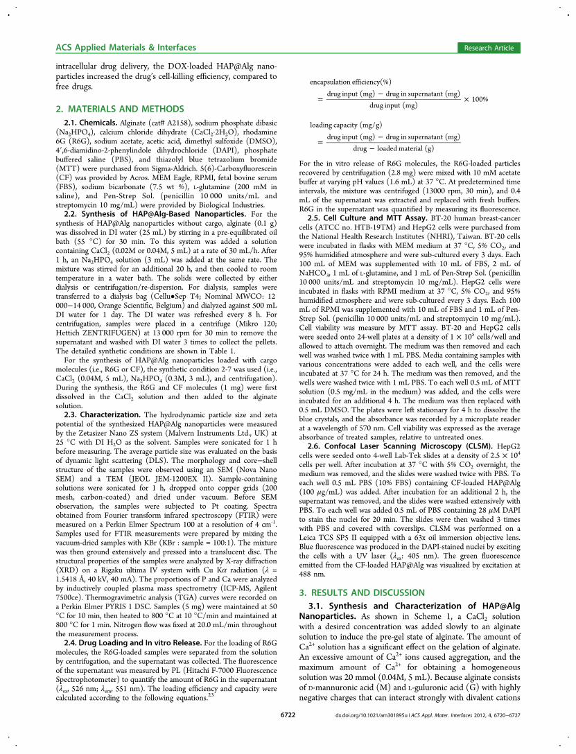
phosphate, we examined sample 2-7 by XRD and ICP-MS. Incontrast to the XRD pattern of pure amorphous alginateexhibiting no peak, (see Figure S1 in the SupportingInformation), the XRD peaks of the Sample 2-7 shown inFigure 2a correspond with those of hexagonal hydroxyapatite(HAP) (JAPCS card # 09-0432). HAP is a mineral form fromthe apatite family, with a formula of Ca10(PO4)6(OH)2. ThepKsp (Ksp = solubility product) of HAP is 117.3 at 37 °C, whichis significantly higher than other calcium phosphate phases(e.g., 6.6 for dicalcium phosphate dihydrate (CaHPO4·2H2O)and 29.5 for β-tricalcium phosphate (Ca3(PO4)2)). Therefore,HAP is regarded as the most stable phase of calcium phosphatewithin the pH range of 4.2-12.2.25 Based on the result of ICP-MS, the proportion of P and Ca in the dried sample 2-7 was13.04 wt % and 26.20 wt %, respectively, and thus the molarratio P/Ca was calculated at 0.642, which is close to that ofregular hydroxyapatite (i.e., P/Ca = 0.6). The ICP-MS resultalso evidenced the formation of hydroxyapatite.Chemical bonding in the synthesized HAP@Alg nano-
particles was analyzed by FTIR spectra. As shown in Figure 2band Figure S2 (Supporting Information), the bands at 1614 and1416 cm−1 were the result of asymmetric (ν8) and symmetric(ν3) stretching vibrations of the C−O bond from thecarboxylate group, respectively. The band at 2932 cm−1 isattributed to C−H stretching. In addition to the peakscorresponding to the organic alginate shell, the bands at 1096and 1030 cm−1 can be attributed to the triply degenerateasymmetric stretching mode vibration (ν3) of phosphategroups. The peak at 961 cm−1 is the non-degenerate symmetricstretching (ν1) of phosphate groups. The peaks at 602 and 562cm−1 result from the doubly degenerate bending mode (ν4) ofthe P−O bond, and the peak at 472 nm−1 corresponds to thedoubly degenerate bending mode (ν2) of the same group. TheFTIR spectra show the existence of both the inorganic HAPand the organic alginate component in the sample, which isconsistent with the XRD and TEM results.The thermogram of the HAP@Alg nanoparticles contrasts
that of the pure alginate polymer. As shown in Figure 2c, threeregions can be defined in the thermogravimetric analysis(TGA) curve for alginate. Initially, the weight loss was slowfrom 50 °C to 220 °C, which results from the desorption ofweakly-bound water and lactonization.26 It has been reportedthat commercial alginate contains moisture of up to 10 wt %.27
In the region of 220−280 °C, the weight dropped dramatically,and this weight loss can be attributed to the decarboxylate of
Figure 1. (a) SEM and (b) TEM images for typical HAP@Algnanoparticles synthesized at the synthetic condition 2−7.
ACS Applied Materials & Interfaces Research Article
dx.doi.org/10.1021/am301895u | ACS Appl. Mater. Interfaces 2012, 4, 6720−67276723
the alginate, in the form of CO2, because COO− groups usually
decompose in this temperature range. In the third region of280−600 °C, the gradual weight loss can be attributed to theloss of abundant hydroxyl groups in alginate.28 In contrast tothat of alginate, the TGA curve of HAP@Alg was smooththroughout the temperature range, remaining at a residualweight of 77.6 wt % after 600 °C. The weight loss in this casecan be ascribed to the decomposition of the organic alginateshell. Therefore, estimating the residual weights of alginate andHAP@Alg, the proportion of alginate in the HAP@Algnanoparticles was calculated at approximately 37.4 wt %. Forcomparison, we also performed the TGA curve of pure HAPnanoparticles (see Figure S3 in the Supporting Information),and it is clearly seen that there was no apparent weight lossduring calcination.3.2. Biocompatibility of HAP@Alg Nanoparticles.
Safety is of utmost importance when nanoparticles are usedfor biomedical applications. Although both alginate andhydroxyapatite have been reported to exhibit high biocompat-ibility, the cytotoxicity of the newly synthesized materials in thisstudy must be specified. MTT assay was used to quantify theeffect of HAP@Alg nanoparticles on human breast cancer cellsBT-20 and liver cancer cells HepG2. As shown in Figure 2d,our HAP@Alg clearly exhibited excellent biocompatibility, andno adverse effects were observed up to a high concentration of1000 μg/mL. The excellent biocompatibility of HAP@Algensures its potential for use in biomedical applications such asdrug delivery systems.To visualize the intracellular uptake of our HAP@Alg
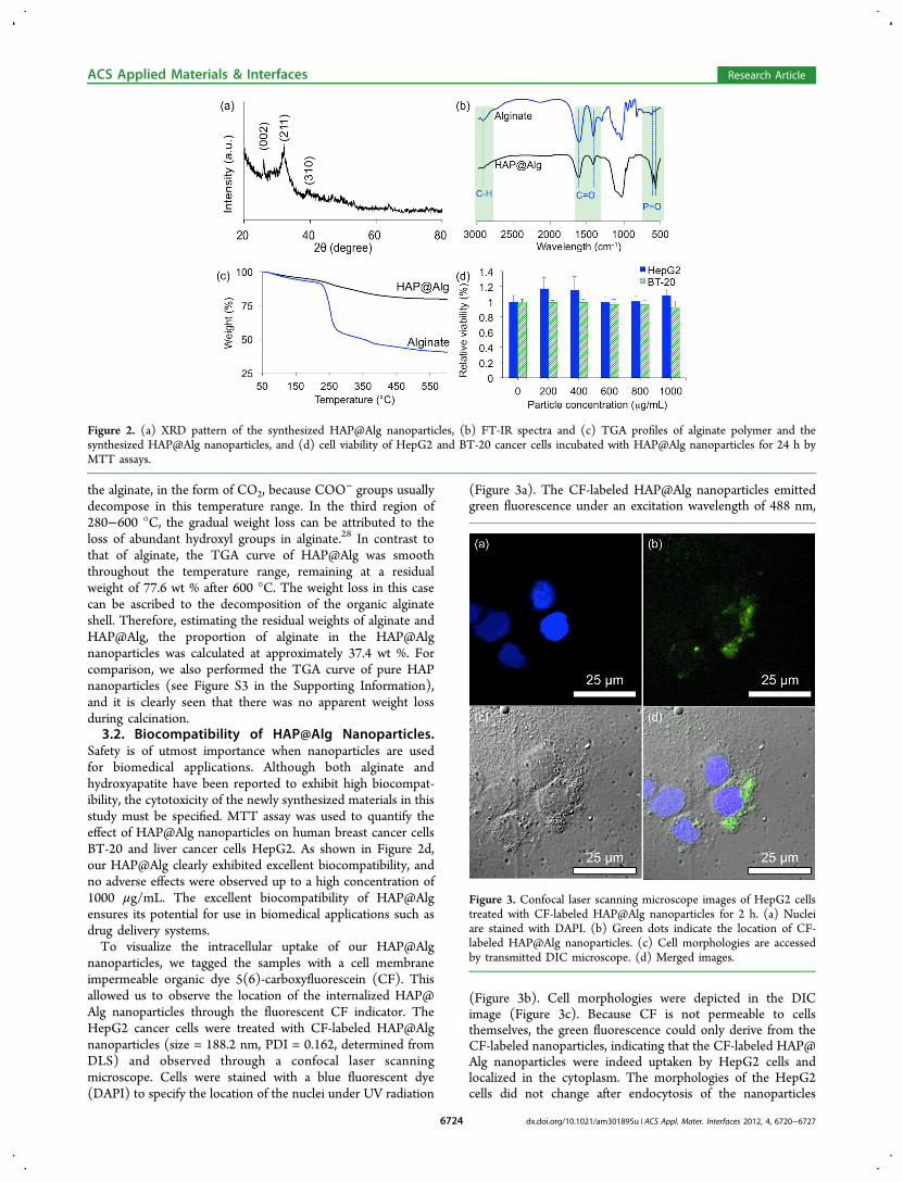
nanoparticles, we tagged the samples with a cell membraneimpermeable organic dye 5(6)-carboxyfluorescein (CF). Thisallowed us to observe the location of the internalized HAP@Alg nanoparticles through the fluorescent CF indicator. TheHepG2 cancer cells were treated with CF-labeled HAP@Algnanoparticles (size = 188.2 nm, PDI = 0.162, determined fromDLS) and observed through a confocal laser scanningmicroscope. Cells were stained with a blue fluorescent dye(DAPI) to specify the location of the nuclei under UV radiation
(Figure 3a). The CF-labeled HAP@Alg nanoparticles emittedgreen fluorescence under an excitation wavelength of 488 nm,
(Figure 3b). Cell morphologies were depicted in the DICimage (Figure 3c). Because CF is not permeable to cellsthemselves, the green fluorescence could only derive from theCF-labeled nanoparticles, indicating that the CF-labeled HAP@Alg nanoparticles were indeed uptaken by HepG2 cells andlocalized in the cytoplasm. The morphologies of the HepG2cells did not change after endocytosis of the nanoparticles
Figure 2. (a) XRD pattern of the synthesized HAP@Alg nanoparticles, (b) FT-IR spectra and (c) TGA profiles of alginate polymer and thesynthesized HAP@Alg nanoparticles, and (d) cell viability of HepG2 and BT-20 cancer cells incubated with HAP@Alg nanoparticles for 24 h byMTT assays.
Figure 3. Confocal laser scanning microscope images of HepG2 cellstreated with CF-labeled HAP@Alg nanoparticles for 2 h. (a) Nucleiare stained with DAPI. (b) Green dots indicate the location of CF-labeled HAP@Alg nanoparticles. (c) Cell morphologies are accessedby transmitted DIC microscope. (d) Merged images.
ACS Applied Materials & Interfaces Research Article
dx.doi.org/10.1021/am301895u | ACS Appl. Mater. Interfaces 2012, 4, 6720−67276724
(Figure 3d), thereby suggesting little or negligible cytotoxicityof our samples. This observation is in accordance with theresults of the MTT assays. It is interesting to note that althoughwe estimated the particle size to be around 100 nm by bothdirect SEM observation and DLS analysis, after HAP@Alg NPswere uptaken by cells, they aggregated inside the cells (in thecytoplasm) due to ionic interactions between charged HAP@Alg and cell medium. Therefore, only aggregated HAP@AlgNPs could be observed by confocal microscopy.3.3. In vitro Loading and pH-Responsible Release of
Drugs. To demonstrate the co-synthesis of drug-loadedHAP@Alg nanoparticles, we chose R6G, a positively charged,water-soluble dye, as the model drug because it can act as asurrogate for the most commonly used anti-cancer drugdoxorubicin, with which it shares a similar molecular weightand charge. We used two in situ loading routes: first, R6G wasdissolved in either alginate solution (route 1) or CaCl2 solution(route 2), as shown in Scheme 1. Route 1 has been introducedin several other studies;23,29 however, we expected route 2 toproduce a higher loading efficiency, based on the formationmechanism of the HAP@Alg core−shell structure. All loadingprocesses were conducted according to the synthetic conditionsof sample 2-7.For the loading of R6G through route 1, the loading amount
increased with the increase in input amount of R6G from 0.5 to2 mg and reached a plateau after 2 mg, as shown in Figure 4a.
The loading efficiency also increased as the input of R6Gincreased from 0.5 to 2 mg, but then decreased when moreR6G molecules were added (Figure 4b). We calculated the drugloading capacity to be approximately 65 mg for one gram ofHAP@Alg nanoparticles, with a maximum efficiency of 50%.The low loading capacity of route 1 can be explained by theshelter effect of R6G. Because positively charged R6Gmolecules were added to the alginate solution before theaddition of calcium, R6G molecules could interact withnegatively charged alginate electrostatically. Although sequen-tial additions of calcium and phosphate led to the formation ofHAP@Alg nanoparticles, we suggest that most of the R6Gmolecules were still encapsulated in the alginate shell, and notin the HAP core, resulting in a low loading capacity, as shownin Scheme 1a.For route 2, R6G molecules were mixed with Ca2+ solution.
As shown in Figure 4, both the loading capacity and efficiencyincreased as the R6G input increased. The trend is verydifferent from that of route 1, suggesting the presence of adifferent encapsulation mechanism. We propose that thepositively charged R6G molecules helped Ca2+ interact with
alginate; therefore they should be encapsulated within the HAPcore during the formation of nanoparticles, as shown in Scheme1b. Because of the limited solubility of R6G in the CaCl2solution (i.e., 7 mg of R6G in 5 mL of 0.04 M CaCl2), themaximum loading amount was limited to 275 mg/g, with a highloading efficiency of 67%. These in situ loading strategiesprovide varying loading capacities, efficiencies, and locations forguest molecules in HAP@Alg nanoparticles, which influencethe corresponding release behavior.The release behavior of the loaded R6G molecules at varying
pH values was also investigated. The release profiles of the R6Gmolecules encapsulated through the two routes are shown inFigure 5. For the R6G molecules encapsulated through route 1,
the release rates were significantly higher at acidic conditions(pH 2.0 and pH 4.5) than at the physiologic condition (pH7.4) (Figure 5a). For example, after 8 h, the release amounts ofR6G at pH 2.0 and pH 4.5 were found to be 1.87- and 1.68-fold, respectively, the amount at pH 7.4. A similar pH-responsive controlled release behavior was observed for route 2(Figure 5b). We propose that the pH-responsible releaseproperty is attributed to the higher solubility of HAP in acidicconditions. As shown in the Supporting Information, therelease amount of R6G was found to be proportional to therelease time for all cases, which indicates that the releasekinetics were closed to the first-order release, rather than to thepseudo-first-order release that has been widely reported whendrugs are loaded into porous nanocarriers.30,31 This first-orderrelease behavior suggests that the R6G was released upon thedissolution of the HAP core.This hypothesis was confirmed by FTIR spectra of HAP@
Alg nanoparticles after release at pH 2. As shown in Figure 6,for both route 1 and 2, the P−O−P bands disappeared afterrelease, indicating a complete dissolution of HAP. In additionto the dissolution of the HAP core, the protonation/de-protonation of alginate at different pH values is also critical tothe release behavior of guest molecules.8 At low pH values, thecarboxylate groups of alginate were protonated, so theelectrostatic interaction between negatively charged alginateand positively charged R6G was diminished, resulting in higherrelease amounts. Another evidence was the TEM image ofR6G-loaded HAP@Alg nanoparticles that were immersed atpH 2 for 12 h (see Figure S1 in the Supporting Information).From the TEM image, it is clearly seen that the HAP@Alg NPs
Figure 4. (a) Encapsulation capacity and (b) efficiencies of R6G intoHAP@Alg nanoparticles through two different routes.
Figure 5. Cumulative release of R6G from HAP@Alg nanoparticles(synthetic condition 2−7, input R6G: 7 mg) prepared by (a) route 1and (b) route 2. The release was conducted in 10 mM acetate buffersat different pH values and the temperature was kept at 37 °C.
ACS Applied Materials & Interfaces Research Article
dx.doi.org/10.1021/am301895u | ACS Appl. Mater. Interfaces 2012, 4, 6720−67276725

are uniform in size and the HAP cores were indeed dissolved inacidic conditions, which promote the release of R6G cargo.Although route 1 and 2 exhibited similar pH-sensitive release
behaviors, their release amount was different from each other.R6G molecules were loaded mostly in the alginate shell forroute 1 and in the HAP core for route 2. Therefore, when theR6G-loaded HAP@Alg nanoparticles prepared for route 1started to release, a burst release of 7% occurred at the initialstage. As the release time increased, HAP dissolved in thesolutions and thus forced the diffusion of R6G from the alginateshell to its surroundings. As shown in Figure 5a, because of thelower loading amount in this case, most of the R6G moleculescould be released (especially in acidic conditions) so the releaseefficiency was higher than the one in route 2. In contrast, whenthe R6G-loaded HAP@Alg nanoparticles prepared for route 2started to release, no burst release occurred in the initial stage,and the total release efficiency was low (only 35%). In this case,the positively charged R6G molecules needed to diffuse fromthe HAP core (Figure 5b). In this case, most of R6G moleculeswere entrapped in the HAP core. We here demonstrate thatdifferent loading methods could selectively place guestmolecules in the core or shell positions, which resulted indifferent release properties.3.4. Intracellular Drug Delivery with Drug-Loaded
HAP@Alg Nanoparticles. To investigate intracellular drugdelivery by HAP@Alg, we chose a chemotherapeutic drug (i.e.,doxorubicin (DOX)) and load it through route 2. The cellviability of BT-20 cells was evaluated by MTT assay after 16 hwith different dosages of DOX-loaded HAP@Alg nanoparticles.The amount of DOX released from HAP@Alg was fixed at 10and 20 M. For comparison, BT-20 cells were also treated withfree DOX molecules at the same concentrations. As shown inFigure 7, the free DOX molecules exhibited weak cytotoxicitytoward BT-20 cells (i.e., 73% viability for 10 μM). In contrast,the viability of cells treated with DOX-loaded HAP@Algdecreased to 58% under the same condition. This resultsuggests that the enhanced intracellular drug delivery wasattributed to DOX-loaded HAP@Alg-mediated endocytosis.The suitable particle size (around 100 nm) of the HAP@AlgNPs and the biocompatible alginate shell enhanced cellularinternalization, whereas the HAP core allowed drug loading. Inaddition, the acid-assisted biodegradability of the HAP corefacilitated drug release in acidic endosomes or lysosomes afterendocytosis. When HAP dissolves in acidic environments suchas endosomes or lysosomes, the release of calcium and
phosphate ions causes an osmotic pressure across theendosome membrane, which interrupts the endosome mem-brane and promotes the cargo to escape from the endosome tothe cytoplasm, resulting in cell death.
4. CONCLUSIONThis study reports the synthesis of hydroxyapatite/alginate(HAP@Alg) core−shell nanoparticles with controllable particlesizes through a pre-gel method. The synthesized HAP@Algnanoparticles exhibit excellent biocompatibility, superior drugloading capacity, and enhanced drug release efficacy. Severalguest molecules, such as organic dyes and therapeuticanticancer drugs, can be loaded efficiently into the core orshell position of the synthesized HAP@Alg, and releasedthrough acid-assisted dissolution controlled kinetics. TheHAP@Alg nanoparticles show significant potential as effectiveand visually observable transmembrane delivery carriers for theintracellular controlled release of cell-membrane-impermeabledrugs. Further development of this type of HAP@Alg materialcan lead to a new generation of nanodevices for biomedicalapplications.
■ ASSOCIATED CONTENT*S Supporting InformationXRD pattern for a pure alginate polymer, FT-IR spectrum andTGA curve of a pure HAP nanoparticles, TEM image of ahollow alginate nanoparticle, and release kinetics of R6G-loaded HAP@Alg NPs (route 2). This material is available freeof charge via the Internet at http://pubs.acs.org/.
■ AUTHOR INFORMATIONCorresponding Author*E-mail: [email protected] authors declare no competing financial interest.
■ ACKNOWLEDGMENTSThis research was supported by the National Science Councilof Taiwan (100-2113-M-002-017, 101-2623-E-002-005-ET, and101-2923-E-002-012-MY3) and by National Taiwan UniversityHospital (UN101-015).
Figure 6. FTIR spectra of R6G-loaded HAP@Alg nanoparticles(prepared by route 1 and route 2) before and after release at pH 2.
Figure 7. MTT assays of HepG2 cells treated with free DOXmolecules and DOX-loaded HAP@Alg nanoparticles at variousconcentrations.
ACS Applied Materials & Interfaces Research Article
dx.doi.org/10.1021/am301895u | ACS Appl. Mater. Interfaces 2012, 4, 6720−67276726

■ REFERENCES(1) Slowing, I. I.; Vivero-Escoto, J. L.; Wu, C.-W.; Lin, V. S. Y. Adv.Drug Delivery Rev. 2008, 60, 1278−1288.(2) Ariga, K.; Lvov, Y. M.; Kawakami, K.; Ji, Q.; Hill, J. P. Adv. DrugDelivery Rev. 2011, 63, 762−771.(3) Kawakami, K.; Ebara, M.; Izawa, H.; Sanchez-Ballester, N. M.;Hill, J. P.; Ariga, K. Curr. Med. Chem. 2012, 19, 2388−2398.(4) Petros, R. A.; DeSimone, J. M. Nat. Rev. Drug Discovery 2010, 9,615−627.(5) Rejman, J.; Oberle, V.; Zuhorn, I. S.; Hoekstra, D. Biochem. J.2004, 377, 159−169.(6) Fadeel, B.; Garcia-Bennett, A. E. Adv. Drug Delivery Rev. 2010, 62,362−374.(7) George, M.; Abraham, T. E. J. Controlled Release 2006, 114, 1−14.(8) Tonnesen, H. H.; Karlsen, J. Drug Dev. Ind. Pharm. 2002, 28,621−630.(9) Rajesh, P.; Khuller, G. Alginate as a Drug Delivery Carrier. InHandbook of Carbohydrate Engineering; CRC Press: Boca Raton, FL,2005; pp 799−816.(10) Ribeiro, C. C.; Barrias, C. C.; Barbosa, M. A. Biomaterials 2004,25, 4363−4373.(11) Zhu, H. G.; Srivastava, R.; Brown, J. Q.; McShane, M. J.Bioconjugate Chem. 2005, 16, 1451−1458.(12) Joshi, A.; Keerthiprasad, R.; Jayant, R. D.; Srivastava, R.Carbohydr. Polym. 2010, 81, 790−798.(13) de Lima, F. O.; Nonato, F. R.; Couto, R. D.; Barbosa Filho, J.M.; Nunes, X. P.; Ribeiro dos Santos, R.; Soares, M. B. P.; Villarreal, C.F. J. Nat. Prod. 2011, 74, 596−602.(14) Lu, F.; Wu, S. H.; Hung, Y.; Mou, C. Y. Small 2009, 5, 1408−1413.(15) Fernandez-Fernandez, A.; Manchanda, R.; McGoron, A. J. Appl.Biochem. Biotechnol. 2011, 165, 1628−1651.(16) Mackiewicz, N.; Gravel, E.; Garofalakis, A.; Ogier, J.; John, J.;Dupont, D. M.; Gombert, K.; Tavitian, B.; Doris, E.; Duconge, F.Small 2011, 7, 2786−2792.(17) Yu, H. J.; Zou, Y. L.; Wang, Y. G.; Huang, X. N.; Huang, G.;Sumer, B. D.; Boothman, D. A.; Gao, J. M. ACS Nano 2011, 5, 9246−9255.(18) Chang, Y. L.; Meng, X. L.; Zhao, Y. L.; Li, K.; Zhao, B.; Zhu, M.;Li, Y. P.; Chen, X. S.; Wang, J. Y. J. Colloid Interface Sci. 2011, 363,403−409.(19) Chen, D. Y.; Li, N. J.; Xia, X. W.; Xu, Q. F.; Ge, J. F.; Li, Y. G.;Lu, J. M.; Gu, H. W. J. Controlled Release 2011, 152, E67−E68.(20) Rajaonarivony, M.; Vauthier, C.; Couarraze, G.; Puisieux, F.;Couvreur, P. J. Pharm. Sci. 1993, 82, 912−917.(21) Aleman, J.; Chadwick, A. V.; He, J.; Hess, M.; Horie, K.; Jones,R. G.; Kratochvil, P.; Meisel, I.; Mita, I.; Moad, G.; Penczek, S.; Stepto,R. F. T. Pure Appl. Chem. 2007, 79, 1801−1827.(22) De, S. J.; Robinson, D. J. Controlled Release 2003, 89, 101−112.(23) Yu, C.-Y.; Jia, L.-H.; Yin, B.-C.; Zhang, X.-Z.; Cheng, S.-X.;Zhuo, R.-X. J. Phys. Chem. C 2008, 112, 16774−16778.(24) Murata, Y.; Nakada, K.; Miyamoto, E.; Kawashima, S.; Seo, S.-H.J. Controlled Release 1993, 23, 21−26.(25) Uskokovic, V.; Uskokovic, D. P. J. Biomed. Mater. Res., Part B2011, 96B, 152−191.(26) Achelhi, K.; Masse, S.; Laurent, G.; Saoiabi, A.; Laghzizil, A.;Coradin, T. Dalton Trans. 2010, 39, 10644−10651.(27) Zohuriaan, M. J.; Shokrolahi, F. Polym. Test. 2004, 23, 575−579.(28) Tripathy, T.; Singh, R. P. J. Appl. Polym. Sci. 2001, 81, 3296−3308.(29) Sarmento, B.; Ferreira, D.; Veiga, F.; Ribeiro, A. Carbohydr.Polym. 2006, 66, 1−7.(30) Lian, H.-Y.; Liang, Y.-H.; Yamauchi, Y.; Wu, K. C. W. J. Phys.Chem. C 2011, 115, 6581−6590.(31) Wu, K. C.; Yamauchi, Y.; Hong, C. Y.; Yang, Y. H.; Liang, Y. H.;Funatsu, T.; Tsunoda, M. Chem. Commun. 2011, 47, 5232−5234.
ACS Applied Materials & Interfaces Research Article
dx.doi.org/10.1021/am301895u | ACS Appl. Mater. Interfaces 2012, 4, 6720−67276727


