Embed Size (px)
Citation preview
ALTERNATING GROUP EXPLICIT METHOD FOR EDGE DETECTION ON
BRAIN AND BREAST TUMOUR IMAGES
ZAWANAH BINTI MD. ZUBAIDIN
UNIVERSITI TEKNOLOGI MALAYSIA

ALTERNATING GROUP EXPLICIT METHOD FOR EDGE DETECTION ON
BRAIN AND BREAST TUMOUR IMAGES
ZAWANAH BINTI MD. ZUBAIDIN
A dissertation submitted in partial fulfilment of the
requirements for the award of the degree of
Master of Science (Mathematics)
Faculty of Science
Universiti Teknologi Malaysia
JANUARY 2013

To my beloved family,
Md. Zubaidin Muhamad @ Mamat
Rohaya Ismail
Zawani Md. Zubaidin
Md. Zulkarami Md. Zubaidin
Zatil Syarafana Md. Zubaidin.
With love and much thanks.

ACKNOWLEDGEMENT
In the name of Allah, the most Gracious and the most Merciful. Firstly, I would
like to express my gratitude to Allah S.W.T. for His love and giving me strength and
patience so that I can completely finish this dissertation task.
In particular, I would like to thanks and wish a greatest appreciation to my
supervisors, Assoc. Prof. Dr. Norma Alias and En. Che Rahim Che Teh for their
guidances, encouragements, and knowledges. Their meaningful advices to me
throughout this period will never be forgotten.
Much love and many thanks I would like to express to my beloved mum, Mrs.
Rohaya Ismail, and dad, Mr. Md. Zubaidin for all their loves, cares, and support. For my
siblings, thank you for the great motivation. I am so blessed to have their loves in my
life.
Finally, I would like to express my sincere appreciation to my senior, Rosdiana
Shahril for her teaching and knowledge sharing. Greatest thanks to my fellow friends,
Nor Aziran, Maizatul Nadirah, Nor Hafizah, Wan Sri Nurul Huda, Nurul Alya, Hafizah
Farhah, Asnida and others for their helps throughout the way in completing this
dissertation.
ABSTRACT
In this research, we used Geodesic Active Contour (GAC) model to detect the
edges of brain and breast tumor on MRI images. An additive operator splitting (AOS)
method is employed in the two dimensional GAC model to maintain the numerical
consistency and makes the GAC model computationally efficient. The numerical
discretization scheme for GAC model is semi-implicit and unconditional stable lead to
sparse system matrix which is a block tridiagonal square matrix. The proposed AOS
scheme capable to decompose the sparse system matrix into a strictly diagonally
dominant tridiagonal matrix that can be solved very efficiently likes a one dimensional
problem. Gauss Seidel and AGE method is used to solve the linear system equations.
The AGE employs the fractional splitting strategy which is applied alternately at each
half (intermediate) time step on tridiagonal system of difference scheme and it is proved
to be stable. This advanced iterative method is extremely powerful, flexible and affords
users many advantages. MATLAB has been choosing as the development platform for
the implementations and the experiments since it is well suited for the kind of
computations required. In the implementation of GAC-AOS model for edges detection
of tumor, the experimental results demonstrate that the AGE method gives the best
performance compared to Gauss Seidel method in term of time execution, number of
iterations,.RMSE, accuracy and computational cost.

ABSTRAK
Model kontur aktif Geodesik digunakan dalam kajian ini untuk menjejak sisi-sisi
tumor bagi barah otak dan payudara pada imej MRI. Kaedah agihan separa tersirat
(AOS) diguna dalam model GAC dua dimensi untuk mengekalkan kekonsistenan
berangka dan membolehkan pengiraan dibuat secara berkesan bagi model GAC. Skema
pendiskritan berangka bagi GAC model ialah dalam skema separuh tersirat dan stabil
secara tidak mutlak. Hal ini menghasilkan sistem matrik yang jarang dan besar. Sistem
matrik adalah dalam bentuk segi empat yang mempunyai bilangan baris dan kolum yang
sama dan merupakan matrik blok yang mempunyai tiga unsur pada pepenjuru. Skema
AOS mampu menguraikan sistem matrik yang jarang kepada sistem matrik yang hanya
mempunyai tiga unsur pada pepenjuru. Sistem matrik ini boleh diselesaikan secara
berkesan sepertimana menyelesaikan masalah satu dimensi. Kaedah Gauss Seidel dan
AGE digunakan untuk menyelesaikan persamaan sistem linear. Kaedah AGE adalah
berorientasikan strategi belahan paras masa terkini secara berselang-seli bagi sistem
persamaan linear tiga pepenjuru dan terbukti stabil. Kaedah lelaran yang maju ini adalah
sangat berkuasa, fleksibel, dan memberi banyak kelebihan kepada para pengguna.
Perisian MATLAB dipilih sebagai platform pembangunan kerana ia sesuai untuk semua
pengiraan yang diperlukan. Dalam pelaksanakan model GAC-AOS untuk mengesan sisi-
sisi tumor, hasil kajian menunjukkan bahawa kaedah AGE memberi persembahan yang
bagus berbanding kaedah Gauss Seidel dalam aspek pelaksanaan masa, bilangan lelaran,
RMSE, ketepatan dan kompleksiti pengiraan.

TABLE OF CONTENTS
CHAPTER TITLE PAGE
DECLARATION ii
DEDICATION iii
ACKNOWLEDGEMENT iv
ABSTRACT v
ABSTRAK vi
TABLE OF CONTENTS vii
LIST OF TABLES x
LIST OF FIGURES xi
LIST OF ABBREVIATIONS xiii
LIST OF SYMBOLS xiv
LIST OF APPENDICES xv
1 INTRODUCTION 1
1.1 Introduction 1
1.2 Background of the Problem 5
1.3 Statement of the Problem 8
1.4 Objectives of the Study 9
1.5 Scope of the Study 9
1.6 Significance of the Study 10
1.7 The Organization of the Dissertation 10

2 LITERATURE REVIEW 12
2.1 Introduction 12
2.2 Geodesic Active Contour (GAC) Model 13
2.3 Semi-Implicit Additive Operator Splitting (AOS)
Scheme
15
2.4 Finite Difference Method 21
2.5 Linear System Equations 23
2.6 Iterative Method 24
2.6.1 Gauss-Seidel (GS) Method 26
2.6.2 Alternating Group Explicit (AGE) Method 29
2.7 Numerical Analysis of the Sequential Algorithm 35
2.8 Computational Platform System 37
2.8.1 Read an Image in MATLAB Programming 38
2.9 Summary of Literature Review 40
3 GEODESIC ACTIVE CONTOUR (GAC) MODEL
BASED ON AOS SCHEME
48
3.1 Introduction 48
3.2 The Use of Notations 49
3.3 The Discretization of GAC Model 50
3.3.1 The Discretization of Divergence ( div )
Operator
52
3.3.2 Semi Implicit Scheme 55
3.3.3 AOS Scheme 61
3.4 The Balloon Force 63
3.5 Linearization of the Discretized GAC-AOS Model 64
3.5.1 Evolution in x -Direction 65
3.5.2 Evolution in y -Direction 68
3.6 The Algorithm of Edges Detection on MRI Image 71

4 NUMERICAL IMPLEMENTATION 74
4.1 Introduction 74
4.2 Alternating Group Explicit (AGE) Method 75
4.2.1 Solving the System Matrix 1A 76
4.2.2 Solving the System Matrix 2A 85
4.3 Gauss-Seidel Method 93
5 ANALYSIS AND DISCUSSION 95
5.1 Introduction 95
5.2 Results of Edges Detection 96
5.2.1 AGE 96
5.2.2 Gauss Seidel 97
5.3 Analysis Results for Iterative Methods 99
5.4 Result of Accuracy 101
5.5 Computational Complexity 103
6 CONCLUSION AND RECOMMENDATIONS 105
6.1 Conclusion 105
6.2 Recommendations for The Future Research 106
REFERENCES 107
Appendices A-C 112

LIST OF TABLES
TABLE NO.
TITLE PAGE
2.1 Summary of Literature Review
35
5.1 Analysis of AGE and GS method for brain tumor MRI
image
100
5.2 Analysis of AGE and GS method for breast tumor MRI
image
100
5.3 Coordinate of the contour to the exact coordinate of the
object using AGE and GS method for brain tumor MRI
image. The pixels have size 1 in each direction.
102
5.4 Coordinate of the contour to the exact coordinate of the
object using AGE and GS method for breast tumor MRI
image. The pixels have size 1 in each direction.
102
5.5 Computational cost of one iteration for brain and breast
tumor MRI
103
5.6 Computational cost for brain tumor MRI image
104
5.7 Computational cost for breast tumor MRI image
104

LIST OF FIGURES
FIGURE NO. TITLE PAGE
1.1 Parametric curve in ),( yx plane
2
2.1 A uniform grid spacing system with hxy
21
2.2 The grid point of interest ( jiu , ) and its neighbouring
points for two dimensional problems
22
2.3 Initial partitioning of matrix A
25
2.4
The tic and toc function works together to measure
elapsed time in MATLAB
37
2.5
The desktop of MATLAB programming 38
2.6 MATLAB editor shows the used of imread command to
read the image of Brain in jpg type
39
2.7 The chart of research scope for edges detection on MRI
image
47
3.1 A Cartesian grid by Ralli (2011)
49
3.2 Pixels involved in the approximation of the divergence
term (3.3.14) for the difference schemes
54
3.3 Pixel of interest denoted by C (Central) and its
neighbourhood pixels denoted by N (North), W (West), E
(East), and S (South)
60
3.4 A table to represent the system matrix A of size
)33()33(
60

3.5 Column and row wise pixel ordering. (a) Column wise
ordering through x -direction; (b) row wise ordering
through y -direction
65
3.6 The algorithm of edges detection on MRI image 73
4.1 The computational molecule of AGE method (explicit) at
level )( 21k through x -direction
83
4.2 The computational molecule of AGE method (explicit) at
level )1( k through x -direction
85
4.3 The computational molecule of AGE method (explicit) at
level )( 21k through y -direction
90
4.4 The computational molecule of AGE method (explicit) at
level )1( k through y -direction
92
4.5 The computational molecule of GS method in x -direction 93
4.6 The computational molecule of GS method in y direction 94
5.1 Final contour of the brain tumor MRI image based on
AGE method
96
5.2 Final contour of the breast tumor MRI image based on
AGE method
97
5.3 Final contour of the brain tumor MRI image based on GS
method
98
5.4 Final contour of the breast tumor MRI image based on
GS method
98
5.5 To find the accuracy between (a) coordinate of the
original image, and (b) coordinate of the final contour
101
LIST OF ABBREVIATIONS
ACM - Active contour model
ADI - Alternating Direction explicit
AGE - Alternating Group Explicit
AOS - Additive Operator Splitting
C-N - Crank-Nicholson
GAC - Geodesic Active Contour
GE - Group explicit
GGAC - Generalized Geodesic Active Contour
GS - Gauss Seidel
IADE - Iterative Alternating Decomposition Explicit
JPEG - Join Photographic Experts Group
LSE - Linear system equations
MRI - Medical Resonance Image
PDE - Partial differential equation
RMSE - Root mean square error
SOR - Successive Over Relaxation
LIST OF SYMBOLS
u - Edge detection of tumor
yx , - The space at coordinate system
- Tolerance value
snakeE - Energy of snake
intE - Internal energy
extE - External energy
imageE - Image forces
conE - External constraint forces
lineE - Line functional
edgeE - Edge functional
termE - Termination functional
w,, - Weighted for energy of snake
- Gradient operator
- Image domain
I - Pixel of interest
J - Neighbourhood pixel of pixel of interest
)(IN - The set of four neighbourhood pixel
- Acceleration parameter
g - Stopping function
v - Positive constant
- Time
k - Number of iterations
LIST OF APPENDICES
APPENDIX NO.
TITLE PAGE
A
File main.m 112
B
File Gauss Seidel.m 113
C File AGE.m
114

CHAPTER 1
INTRODUCTION
1.1 Introduction
In the past several years, active contour models have been widely applied in
computer vision especially in image processing since they were first introduced by
Kass et al. (1988). It is an effective tool for image segmentation, object tracking,
shape recognition, edge detection and stereo matching.
According to representation and implementation, active contours can be
classified into two types which are parametric active contours Kass et al.(1988),
Cohen(1991), Eviatar and Samorjai(1996), Xu et al.(2000), Wang et al.(2009) and
geometric active contours Caselles et al.(1993), Caselles et al.(1997), Xu et al.(2000),
Goldenberg et al.(2001).
Parametric active contours are represented explicitly as parameterized curves
or splines. Geometric active contours are represented implicitly as level sets of two-
dimensional distance functions which its evolution does not depend on particular
parameterization. These models are based on the curve evolution theory and
geometric flows, Caselles et al.(1993)
2
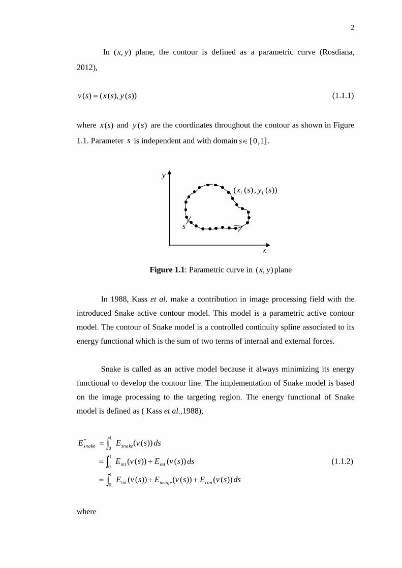
In ),( yx plane, the contour is defined as a parametric curve (Rosdiana,
2012),
))(),(()( sysxsv (1.1.1)
where )(sx and )(sy are the coordinates throughout the contour as shown in Figure
1.1. Parameter s is independent and with domain ]1,0[s .
Figure 1.1: Parametric curve in ),( yx plane
In 1988, Kass et al. make a contribution in image processing field with the
introduced Snake active contour model. This model is a parametric active contour
model. The contour of Snake model is a controlled continuity spline associated to its
energy functional which is the sum of two terms of internal and external forces.
Snake is called as an active model because it always minimizing its energy
functional to develop the contour line. The implementation of Snake model is based
on the image processing to the targeting region. The energy functional of Snake
model is defined as ( Kass et al.,1988),
1
0int
1
0int
1
0
*
))(())(())((
))(())((
))((
dssvEsvEsvE
dssvEsvE
dssvEE
conimage
ext
snakesnake
(1.1.2)
where
))(,)(( sysx ii
y
x
s

3
snakeE : Energy functional of Snake
intE : Internal energy of Snake to smooth the edge curve
extE : External Snake forces lead the curve to the edges of object in
the image.
imageE : Image forces pushing the Snake toward the desired object.
conE : External constraint forces
The internal energy can be expressed as ( Kass et al.,1988),
2
))()()()((22
int
svssvsE
sss (1.1.3)
The first-order term sv is controlled by )(s and the second-order term ssv is
controlled by )(s . The function of first-order term is to make the Snake act like a
membrane while the second-order term is to make Snake act like a thin-plate.
The relative importance of the membrane and thin-plate terms can be control
by adjusting the weighted )(s and )(s . By reviewing some previous researches, a
constant applied as a coefficient for the first-order term in (1.1.3) i.e., )(s ,
Wang et al.(2009). While the weight of )(s need to set as zero. This is to make
sure that Snake can be second-order discontinuous and extract a corner.
The total image energy is a weighted combination of the three energy
functionals. This energy can be represented as follows
termtermedgeedgelinelineimage EwEwEwE (1.1.4)
The three different energy functional ( timeedgeline EEE ,, ) can attract a the contour of
Snake to lines, edges, and terminations. A wide range of Snake can be created by
adjusting the weights ( linew , edgew , termw ).
4
A line functional lineE is the image intensity itself. It is defined as,
),( yxIEline (1.1.5)
where yxI , is the image function and it is viewed as a function of continuous
position variables yx, , Cheng et al. (2007). It is depending on the sign of linew so
that the contour or Snake will be attracted to either low level prediction contour or
high level of prediction contour.
The edges of the image can be found by the energy functional (1.1.6), Kass et
al.(1988). This allows Snake model to be attract to contours with large image
gradient.
2),( yxIEedge (1.1.6)
A different edge functional (1.1.7) issued by Kass et al.(1988) in order to
show the relationship of scale-space continuation to the theory of edge-detection by
Marr and Hildreth(1980).
22 ,, yxIyxGEedge (1.1.7)
where yxG , represent as a two dimensional Gaussian of standard deviation and
is the gradient operator. The functional lies on zero-crossing of IG 2 . The
location is minima which defined edges in the Marr-Hildreth theory. The Snake will
attract to zero-crossing if we add the edge functional term (1.1.7) to the existing
equation (1.1.4). Despite of adding this term to Snake model, it is still constrained by
its own smoothness.
Curvature of level lines in equation (1.1.8) is used to find the terminations of
line segments and corners in a slightly smoothed image.
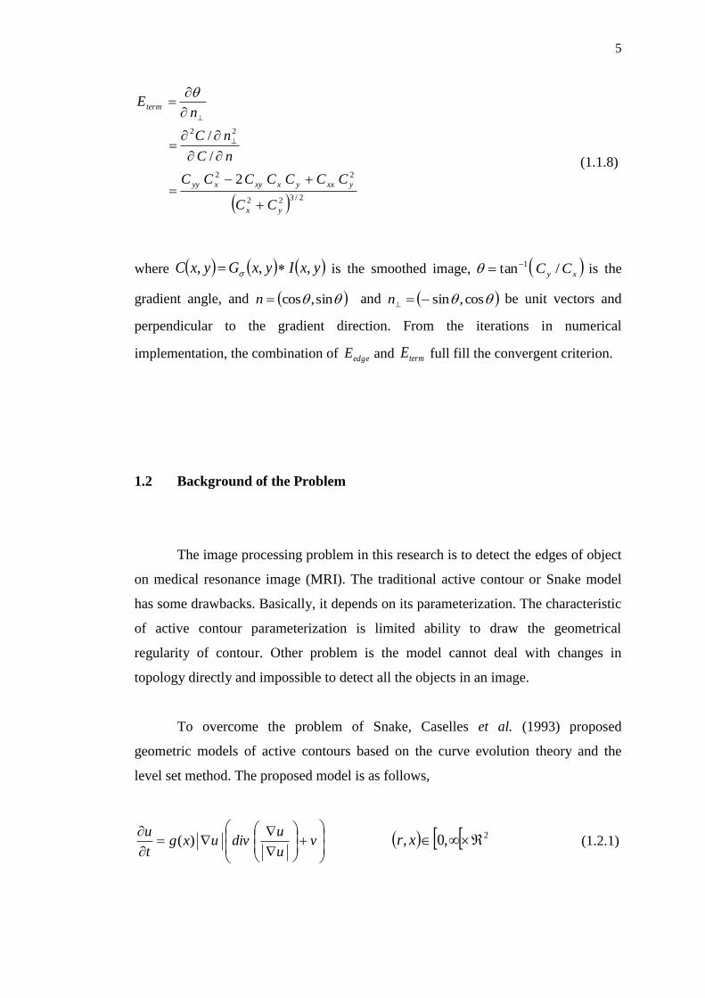
5
2/322
22
22
2
/
/
yx
yxxyxxyxyy
term
CC
CCCCCCC
nC
nC
nE
(1.1.8)
where yxIyxGyxC ,,, is the smoothed image, xy CC /tan 1 is the
gradient angle, and sin,cosn and cos,sinn be unit vectors and
perpendicular to the gradient direction. From the iterations in numerical
implementation, the combination of edgeE and termE full fill the convergent criterion.
1.2 Background of the Problem
The image processing problem in this research is to detect the edges of object
on medical resonance image (MRI). The traditional active contour or Snake model
has some drawbacks. Basically, it depends on its parameterization. The characteristic
of active contour parameterization is limited ability to draw the geometrical
regularity of contour. Other problem is the model cannot deal with changes in
topology directly and impossible to detect all the objects in an image.
To overcome the problem of Snake, Caselles et al. (1993) proposed
geometric models of active contours based on the curve evolution theory and the
level set method. The proposed model is as follows,
v
u
udivuxg
t
u)( 2,0, xr (1.2.1)
6
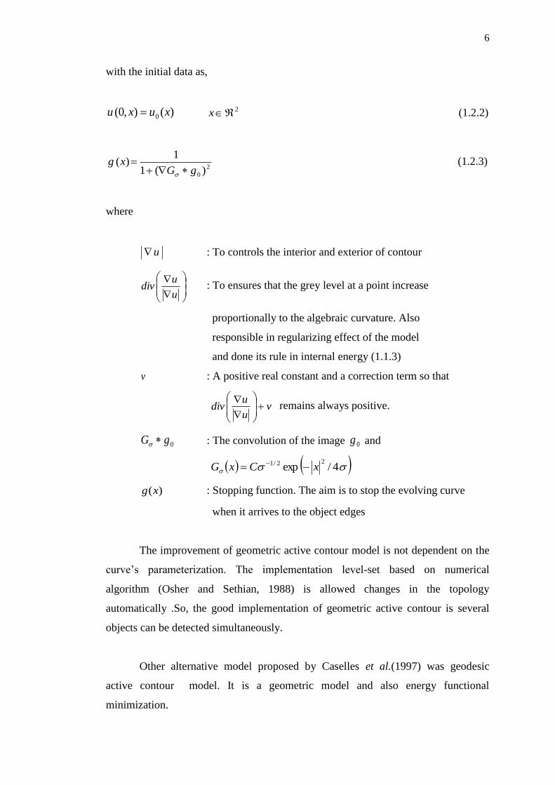
with the initial data as,
)(),0( 0 xuxu 2x (1.2.2)
2
0 )(1
1)(
gGxg
(1.2.3)
where
u : To controls the interior and exterior of contour
u
udiv : To ensures that the grey level at a point increase
proportionally to the algebraic curvature. Also
responsible in regularizing effect of the model
and done its rule in internal energy (1.1.3)
v : A positive real constant and a correction term so that
vu
udiv
remains always positive.
0gG : The convolution of the image 0g and
4/exp22/1 xCxG
)(xg : Stopping function. The aim is to stop the evolving curve
when it arrives to the object edges
The improvement of geometric active contour model is not dependent on the
curve’s parameterization. The implementation level-set based on numerical
algorithm (Osher and Sethian, 1988) is allowed changes in the topology
automatically .So, the good implementation of geometric active contour is several
objects can be detected simultaneously.
Other alternative model proposed by Caselles et al.(1997) was geodesic
active contour model. It is a geometric model and also energy functional
minimization.

7
Caselles et al. (1997) suggested the model of geodesic active contour as
follows
)()( xguvu
uxgdivu
t
u
2,0, xt (1.2.4)
The real fact is, geodesic active contour model yields the same result as that
of a simplified Snake model. It is up to arbitrary constant that depends on the initial
parameterization (Goldenberg et al. 2001).
However, geodesic active contour also has its drawbacks that we need to
consider in this research. The main disadvantage is its nonlinearity that will cause
bad implementation.
To linearize the geodesic active contour model, we apply the additive
operator splitting (AOS) scheme based on the Weickert et al. (1998). It can be
defined as follows,
km
l
k
l
k uuAmIm
u
1
1
1 1
(1.2.5)
where
k : Number of iteration
m : Dimension of the problem
l : Index running over the dimension
I : Unit matrix
: Time step
This numerical scheme is an unconditionally stable for nonlinear diffusion for
image processing problem. It is consistent, first order and semi-implicit scheme. In
this research, we are going to consider the two dimensional model of active contour.
So the AOS scheme for two dimensional cases is given by,
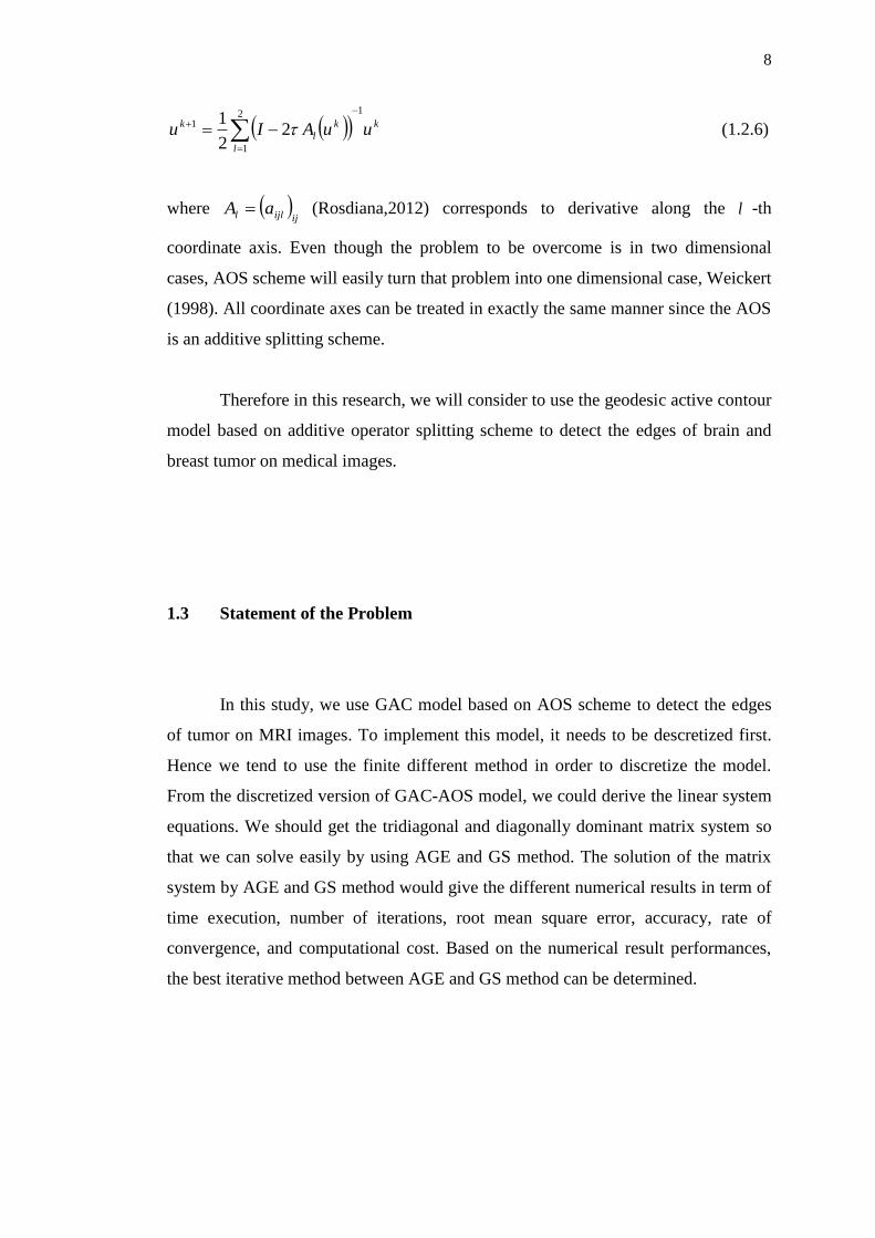
8
k
l
k
l
k uuAIu
12
1
1 22
1
(1.2.6)
where ijijll aA (Rosdiana,2012) corresponds to derivative along the l -th
coordinate axis. Even though the problem to be overcome is in two dimensional
cases, AOS scheme will easily turn that problem into one dimensional case, Weickert
(1998). All coordinate axes can be treated in exactly the same manner since the AOS
is an additive splitting scheme.
Therefore in this research, we will consider to use the geodesic active contour
model based on additive operator splitting scheme to detect the edges of brain and
breast tumor on medical images.
1.3 Statement of the Problem
In this study, we use GAC model based on AOS scheme to detect the edges
of tumor on MRI images. To implement this model, it needs to be descretized first.
Hence we tend to use the finite different method in order to discretize the model.
From the discretized version of GAC-AOS model, we could derive the linear system
equations. We should get the tridiagonal and diagonally dominant matrix system so
that we can solve easily by using AGE and GS method. The solution of the matrix
system by AGE and GS method would give the different numerical results in term of
time execution, number of iterations, root mean square error, accuracy, rate of
convergence, and computational cost. Based on the numerical result performances,
the best iterative method between AGE and GS method can be determined.
9
1.4 Objectives of the Study
The objectives of the study are:
i) To detect the edges of brain and breast tumor on MRI images.
ii) To apply some iterative methods (AGE and Gauss Seidel) to solve the linear
system equations.
iii) To compare the numerical analysis of the iterative methods (AGE, and Gauss
Seidel) in term of time execution, number of iterations, computational
complexity, root mean square error (RMSE), convergence rate and accuracy.
1.5 Scope of the Study
This study will focus on detecting the edges of tumor by using
Geodesic active contour (GAC) model based on additive operator splitting (AOS)
scheme. The solution for linear system of equation (LSE) can be done by using some
iterative methods. The iterative methods under consideration are Gauss-Seidel,
alternating group explicit (AGE). This experiment will be applied to brain and breast
tumor on MRI images. The MRI images are the real image of two patients from
Hospital Kubang Kerian, Kelantan and Hospital Kuala Batas, Pulau Pinang. The
algorithm will be run using MatlabR2011a.
10
1.6 Significance of the Study
From this study, it is hope that we can detect the edges of brain and breast
tumor on medical resonance image (MRI). Other than that, the numerical analysis
results can be the measurement in proving that AGE method is the best iterative
method with accuracy (2, 4) than Gauss Seidel method with accuracy (2, 2).
1.7 The Organization of the Dissertation
This dissertation consists of six chapters. Chapter 1 describes the introduction
of active contour models. In this chapter, we included the problem formulation,
active contour model under consideration, objectives, scope, and significance of the
research.
Chapter 2 focuses on the literature review. This chapter describes the use and
application of GAC model by different researchers year by year. There also have
descriptions about AOS scheme, finite difference method, Gauss Seidel method, and
AGE method. We also explain briefly about the numerical analysis of sequential
algorithm and the computational platform used in this research. At the end of the
chapter, we show the chart of our research scope.
Chapter 3 describes the discretization process for GAC-AOS model by using
finite differences method. From the discretized version of GAC-AOS, we derived the
linear system equations. Because of the AOS scheme, the linear system can be solves
for two directions separately. At the end of the chapter, we explain the flowchart of
the sequential algorithm for edge detection problem on MRI image.
In Chapter 4, we describe the solution of tridiagonal and diagonal matrix
system using AGE and GS methods. We show how the formulation of AGE and GS

11
method could solve the matrix system for two directions which are x -direction and
y -direction. We also included the computational molecule for each AGE and GS
method.
Chapter 5 presents the results of edge detection on MRI images. We analyse
the results based on number of iterations, time execution, root mean square error, rate
of convergence, accuracy and computational complexity. All the numerical results
are shown in the form of table while the visualization results of the captured edge of
tumor by contour line are shown by images.
The last chapter is the Chapter 6. In this chapter, we state the conclusions of
this research based on the results that we showed in Chapter 5 and relate them with
our objectives in Chapter 1. Then, there are some suggestions and recommendations
for the future researchers.
107
REFERENCES
Al Sharif, S. M. S., Deriche, M., and Maalej, N. (2010). A Fast Geodesic Active
Contour Model for Medical Images Segmentation Using Prior Analysis.
IEEE IPTA. 300-305.
Ananth, K. R., and Panniselvam, S. (2012). A Geodesic Active Contour Level Set
Method for Image Segmentation. International Journal of Image, Graphic,
and Signal Processing. 5, 31-37.
Burden, R. L., and Faires, J. D. (2005). Numerical Analysis. Eight edition. Canada.
Bob Pirtle.
Caselles, V., Catte, F., Coll, T., and Dibos, F. (1993). A Geometric Model for Active
Contours in Image Processing. Numerische Mathematik. 66, 1-31.
Caselles, V., Kimmel, R., and Sapiro, G. (1997). Geodesic Active Contour.
International Journal of Computer Vision. 22 (1), 61-79.
Cheng, J., Liu, Y., Jia, R., and Guo, W. (2007). A New Active Contour Model for
Medical Image Analysis-Wavelet Vector Flow. IAENG International Journal
of Applied Mathematics. 36 (2).
Che Rahim, C. T. (2009). Numerical Method: Algorithm and Matlab Programming.
Third Edition. Johor: Universiti Teknologi Malaysia

108
Chung, Y. L. (2004). Applied Numerical Methods For Partial Differential Equations.
First Edition. Singapore: Prentice Hall.
Cohen, L. D. (1991). On Active Contour Models and Balloons. CVGIP: Image
Process. 101(2).
Derraz, F., Beladgham, M., and Khelif, M. (2004). Application of Active Contour
Models in Medical Image Segmentation. Proceeding of The International
Conference on Information Technology.
Evans, D. J., and Abdullah, A. R. B. (1983). A New Explicit Method for The
Solution of Two Dimensional Parabolic Equation. International Journal
Computer Mathematic. 14, 325-353.
Evans, D. J. (1985). Group Explicit Iterative Methods for Solving Large Linear
Systems. International Journal Computer Mathematic.17, 81-108.
Evans, D. J., and Sahimi, M. S. (1988). The Alternating Group Explicit (AGE)
Iterative Method for Solving Parabolic Equations I. International Journal
Computer Mathematic. 24, 127-145.
Evans, D. J. (1997). Group Explicit Methods for The Numerical Solution of Partial
Differential Equations. First Edition. Netherlands. Gordon and Breach
Science Publisher.
Eviatar, H., and Samorjai, R. L. (1996). A Fast, Simple Active Contour Algorithm
for Medical Images. Elsevier Pattertn Recognition Letters. 17, 969-974.
Fairag, F., and Sahimi, M. S. (2005). The Alternating Group Explicit (AGE) Iterative
Method for Solving A Ladyzhenskaya Model For Stationary Incompressible
Viscous Flow.
Feng, Q. (2008). An Alternating Group Explicit Iterative Method for Solving Four-
order Parabolic Equations. Applied Mathematical Science. 2(52), 2591-2595.
109
Goldenberg, R., Kimmel, R., Rivlin, E., and Rudzsky, M. (2001). Fast Geodesic
Active Contours. IEEE Transaction on Image Processing. 10 (10), 1467-
1475.
Han, Z., and Liu, X. (2010). An Additive Operator Splitting Method for Microscopic
Image Segmentation. IEEE Transaction on Image Processing.
Hunt, B. R., Lipsman, R. L., and Rosenberg, J. M. (2001). A Guide To MATLAB for
Beginners and Experienced Users. First Edition. United States of America.
Cambridge University Press.
Kass, M., Witkin, A., and Terzopoulos, D. (1988). Snakes: Active Contour Models.
International Journal of Computer Vision. 1, 321-331.
Kuhne, G., Weickert, J., Beir, M., and Effelsberg, W. (2002). Fast Implicit Active
Contour Models. In Proceedings of the 24th
DAGM Symposium on Pattern
Recognition. Springer-Verlag, 133-140.
Liu, C., Ma, J., and Ye, G. Medical Image Segmentation by Geodesic Active
Contour Incorporating Region Statistical Information. (2007). Fourth
International Conference on Fuzzy Systems and Knowledge Discovery.IEEE.
Marr, D., and Hildreth, E. (1980). Theory of Edge Detection. Proceeding of the
Royal Society of London. Series B, Biological Sciences. 207, 187-217.
Norma, A. (2004). Development and Implementation of Parallel Algorithms in the
IADE and AGE Class of Methods to Solve Parabolic Equations on a
Distributed Parallel Computer Systems. Doctor of Philosophy, Universiti
Kebangsaan Malaysia, Bangi.
Osher, S., and Sethian, J. A. (1988). Fronts Propagating with Curvature Dependent
Speed: Algorithms Based on Hamiltonian-Jacobian Formulation. J. C. Ph. 79,
12-49.
110
Perona, P., and Malik, J. (1990). Scale-Space and Edge Detection Using Anisotropic
Diffusion. IEEE Transactions on Pattern Analysis and Machine Intelligence.
12(7), 629-639.
Ralli, J. (2011). Fusion and Regularisation of Image Information in Variational
Correspondence Methods. Doctor of Philosophy, University of Granada,
Granada.
Rosdiana, S. (2012). Two Dimensional Active Contour Model on Multigrids For
Edge Detection of Images. Master of Science, Universiti Teknologi Malaysia,
Skudai.
Saad, Y. (2000). Iterative Methods for Sparse Linear System. Second Edition with
Correction.
Wang, T., and Cheng, I. (2009). Fluid Vector Flow and Applications in Brain Tumor
Segmentation. IEEE Transactions on Biomedical Engineering. 56 (3).
Weickert, J., ter Haar Romeny, B. M., and Viergever, M. A. (1998). Efficient and
Reliable Schemes for Nonlinear Diffusion Filtering. IEEE Transactions on
Image Processing. 7 (3), 398-410.
Weickert, J. (2001). Efficient Image Segmentation Using Partial Differential
Equations and Morphology. Pattern Recognition Society. 34, 1813-1824.
Weickert, J. (2008). Anisotropic Diffusion in Image Processing. 3rd
Edition.
University of Copenhagen, Denmark. B. G. Teubner Stuttgart.
Xu, C., Jr, A. Y., and Prince, J. L. (2000). On the Relationship between Parametric
and Geometric Active Contours. In Proc. Of 34th
Asilomar Conference on
Signals,Systems, and Computers. October 2000. IEEE, 483-489.
111
Guo, Y., Jiang, J., Hao, S., and Zhan, S. (2011). Distribution-Based Active Contour
Model For Medical Image Segmentation. Sixth International Conference on
Image and Graphics.
Yun, J., Li, P., and Wen, Y. (2011). Contour Segmentation Using an Improved GAC
Model. IEEE Transaction on Image Processing.
Zaiton, M., Mohamed, O., and Chuah, C. Y. (1994). A Parallel AGE Method for
Parabolic Problems with Special Geometries. Pertanika Journal of Science
and Technology. 2(2), 149-158.


![THE EHRLICH-ABERTH METHOD FOR THEonal form [11], [14]. In the sparse case, the nonsymmetric Lanczos algorithm produces a nonsymmetric tridiagonal matrix. Other motivation for this](https://img.dokumen.tips/doc/110x75/60aa58269dcb0e7be2687539/the-ehrlich-aberth-method-for-onal-form-11-14-in-the-sparse-case-the-nonsymmetric.jpg)