Embed Size (px)
Citation preview

1
Alpha-1 Antitrypsin Enhances Islet Engraftment by Suppression of Instant Blood-
Mediated Inflammatory Reaction
Running head: AAT suppresses IBMIR
Jingjing Wang1, Zhen Sun
1, Wenyu Gou
1, David B Adams
1, Wanxing Cui
2, Katherine A
Morgan1, Charlie Strange
3, Hongjun Wang
1*
1 Department of Surgery and
3 Medicine, Medical University of South Carolina, Charleston, SC
29425.
2Medstar Georgetown University Hospital, Washington DC.
*To whom correspondence should be addressed: Hongjun Wang, Ph.D. Department of Surgery,
Medical University of South Carolina, BSB 641, 173 Ashley Avenue, Charleston, SC 29425.
Tel: 843-792-1800, Fax: 843-792-3315, Email: [email protected].
Page 1 of 36
For Peer Review Only
Diabetes
Diabetes Publish Ahead of Print, published online January 9, 2017

2
Abstract
Islet cell transplantation has limited effectiveness because of an instant blood-mediated
inflammatory reaction (IBMIR) that occurs immediately after cell infusion and leads to dramatic
β cell death. In intraportal islet transplantation models using mouse and human islets, we
demonstrated that alpha-1 antitrypsin (AAT, Prolastin-C), a serine protease inhibitor used for the
treatment of AAT deficiency, inhibits IBMIR and cytokine-induced inflammation in islets. In
mice, more diabetic recipients reached normoglycemia after intraportal islet transplantation when
they were treated with AAT compared to mice treated with saline. AAT suppressed blood-
mediated coagulation pathways by diminishing tissue factor production, reducing plasma
thrombin-antithrombin (TAT) complex levels and fibrinogen deposition on islet grafts, which
correlated with less graft damage and apoptosis. AAT-treated mice showed reduced serum TNF-
α levels, decreased lymphocytic infiltration, and decreased NF-κB activation compared with
controls. The potent anti-inflammatory effect of AAT is possibly mediated by suppression of
JNK phosphorylation. Blocking JNK activation failed to further reduce cytokine-induced
apoptosis in β cells. Taken together, AAT significantly improves islet graft survival after
intraportal islet transplantation by mitigation of coagulation in IBMIR and suppression of
cytokine-induced JNK and NF-κB activation. AAT-based therapy has the potential to improve
graft survival in human islet transplantation and other cellular therapies on the horizon.
Page 2 of 36
For Peer Review Only
Diabetes

3
Introduction
Allogeneic islet transplantation is a promising approach for treating patients with type 1
diabetes (T1D) (1). Islet autotransplantation is utilized to avoid pancreatogenic diabetes in
patients who undergo total pancreatectomy for chronic pancreatitis intractable to medical
management (2). In both situations, islet engraftment after transplantation is compromised and
ß cell death is problematic. Stresses induced during islet harvesting and post-transplantation may
lead to up to 60% islet death within 2-3 days after transplantation (3; 4). Many factors including
IBMIR, pro-inflammatory cytokines, hypoxia, and nutritional deprivation contribute to β cell
death. Strategies that maintain normal islet cell function and reduce cell death after
transplantation would improve patient outcomes and have ready clinical applicability.
Alpha-1 antitrypsin (AAT) is a serine protease inhibitor that belongs to the serpin
superfamily. It has a high concentration in serum, and is available for clinical use as a purified
human product (5). AAT inhibits various enzymes including neutrophil elastase, cathepsin G,
proteinase-3, thrombin, trypsin, and chymotrypsin (6). Patients with genetic deficiency of AAT
have higher risks for emphysema from alveolar destruction (7). Beside inhibition of elastase,
AAT exerts anti-inflammatory effects via suppressing cytokine production, complement
activation, and immune cell infiltration. AAT also functions as an anti-apoptotic factor for
endothelial cells and vascular smooth muscle cells (8; 9). AAT has beneficial effects in the
treatment of diabetes. AAT protects β cells from apoptosis induced by pro-inflammatory
cytokines and streptozotocin (STZ) (10). AAT injection to NOD mice reduces the intensity of
insulitis, preserves β cell mass and prevents the onset of diabetes via modulating T regulatory
cells (11; 12). AAT also protects islets from graft failure and immune rejection in mouse
transplantation models (13; 14). Adding AAT into islet digestion medium improves porcine islet
Page 3 of 36
For Peer Review Only
Diabetes

4
isolation by inhibiting trypsin activity during pancreas digestion (15). AAT monotherapy
prolongs allograft survival in mice by elevating VEGF expression and promotion of islet
revascularization (14). Short-term treatment with AAT in the peri-transplant period protects a
marginal mass of monkey islet autografts from long-term, non-immunological graft loss through
effects on expression of TGF-β, NF-κB and AKT (16). Thus, the beneficial effects of AAT in the
islet transplantation setting may be mediated by its anti-apoptotic and anti-inflammatory
properties and promotion of islet revascularization.
In many animal studies, islets are transplanted under the kidney capsule for easily
analysis. In clinical practice, islets are transplanted into the liver by portal vein infusion. The
major difference between these two methods is that in portal vein infusion, islets are directly
exposed to blood after transplantation, which leads to IBMIR, a thrombotic/inflammatory
reaction mediated by the innate immune system (17-19). IBMIR involves activation of the
coagulation cascade and the inflammatory pathway, and eventually leads to clot formation and
infiltration of leukocytes into the islets that cause islet destruction and failure of engraftment
(20). IBMIR is a significant factor in the damage to allogeneic, xenogeneic and autologous islet
transplants (21), which results in a prothrombotic state and secretion of proinflammatory factors
such as TNF-α IL-6, IL-8 and interferon-inducible protein-10 (18). IBMIR also happens after
hepatocyte and mesenchymal stem cell transplantation (22; 23). Therefore, IBMIR may represent
a major hurdle for all cell therapies, and is being targeted by NF-κB inhibitors or anticoagulants
such as low molecular dextran sulfate (24; 25). Furthermore, Herring et al. showed that a TNF-α
inhibitor, etanercept, in addition to prolonged heparin treatment, contributed to improved islet
engraftment in their clinical trial on single-donor, marginal dose islet transplants in T1D patients
Page 4 of 36
For Peer Review Only
Diabetes

5
(26). Therefore, suppression of IBMIR may contribute to a better outcome in islet transplantation
(26).
Although the protective effect of AAT in the kidney capsule islet transplantation model
has been reported (13; 14; 16; 17; 27), whether AAT protects intrahepatic islet grafts that mimic
the clinical islet transplantation setting remains unknown. The focus of this study is to evaluate
the effect of AAT on IBMIR-induced islet death and understand the molecular mechanisms of
the anti-inflammatory effects of AAT.
Methods
Mice:
Male C57BL/6 and NOD-SCID mice at 7-8 weeks of age were purchased from the
Jackson Laboratory (Bar Harbor, ME). The Animal Care and Use Committee at the Medical
University of South Carolina approved all mouse experiments.
Mouse islets isolation, diabetes induction and islet transplantation
Islets were isolated by collagenase digestion as described previously (28). Diabetes was
induced in mice by a single injection of STZ (225mg/kg, Sigma). Mice were considered diabetic
when non-fasting blood glucose exceeded 300 mg/dL for at least 2 consecutive days. For islet
transplantatin 200-250 mouse or 500 human islets were infused in a total volume of 200 µl of
HBSS and 0.5% BSA into the recipient liver as described (29). Mice with blood glucose levels <
200 mg/dl were considered normoglycmic.
In vitro miniature tube model for IBMIR
Miniaturized in vitro tube models were used as described (18). Briefly, mice blood was
collected in heparin-lithium coated 1.5mL Eppendorf tubes (Fisher Scientific). Islets were mixed
Page 5 of 36
For Peer Review Only
Diabetes

6
with blood (500 islets/500 µl blood) in the presence or absence of AAT (0.5 mg/ml) in an
incubator on a hematology mixer for 6 hours. Supernatant samples were collected at 0, 15 and 60
min and at 3 and 6 h.
AAT injection and its concentration in serum
C57BL/6 mice were injected with AAT (Prolastin-C, Grifols, 2mg/ mouse, i.p.) or saline
every 2 (Q2D) or 3 days (Q3D) for a total of 8 doses. Control mice (Ctrl) received saline at the
same schedule as Q2D group. Serum concentrations of human AAT were determined before the
last dose using a human AAT-specific ELISA kit (Abcam) according to the manufacturer’s
instructions. In the islet transplantation model, the first dose of AAT was given one day before
islet transplantation.
Immunohistochemical staining
Livers were harvested 6 h after transplantation. Sections of 5µm at 100-µm intervals were
incubated with anti-F4/80, anti-tissue factor (Abcam) and anti-insulin antibodies (Thermo
Scientific) overnight at 4°C. For miniature tube assays, islets in blood were span down, washed
on a cell strainer with cold PBS and subjected to frozen sections. Anti-p-JNK (Cell Signaling,
1:300) and anti-insulin antibodies were applied. A ZEISS AxioImager M2 microscope and Leica
SP5 confocal microscope were used. Biotinylated goat anti-mouse fibrinogen antibody (Accurate
Chemical & Scientific Corporation) was used to stain fibrin deposition (28).
Neutrophil infiltration
Naphthol AS-D chloroacetate (Specific Esterase) Kit (Sigma-Aldrich) was used to stain
for PMNs according to the manufacturer's instructions.
Page 6 of 36
For Peer Review Only
Diabetes

7
Preparation of nuclear extract and determination of NF-κB activation
Liver tissues were collected 24 h after transplantatin. A nuclear extraction kit followed by
a NF-κB (p65) transcription factor assay kit (Cayman) was used as described by the
manufacturer. Protein concentrations were determined by PierceTM
BCA protein assay (Thermo
Scientific).
Intravenous glucose tolerance test (IVGTT)
Recipient mice were fasted overnight and injected with glucose solution at 1g/kg via tail
vein. Blood glucose levels were measured at 0, 15, 30, 45, 60, 90, and 120 min after injection.
Measurements of coagulation factors and plasma cytokines
Serum thrombin-antithrombin (TAT), mouse tissue factor (Abcam ELISA), and mouse
TNF-α (RayBio ELISA) were measured according to the manufacturer’s instructions.
Cytokine treatment and Western blot
βTC3 cells were pre-incubated with AAT for 2 hours before the addition of 100 U/ml IL-
1β and 1000U/ml IFN-γ. Human islets were treated with 50U/ml TNFα + 50U/ml IL-1β + 1000
U/ml INF-γ until cells were collected at indicated time points. Proteins were analyzed by
immunoblotting with antibodies against phospho-JNK and total JNK. Histone H3 was used as a
loading control. This experiment was repeated five times. Western blots were further quantified
using standard densitometric analysis (NIH ImageJ software). Briefly, the bands on the films
were digitized on a flatbed photo scanner. Each band was manually selected and the intensities
were measured using the “gel analyzer” option in ImageJ software. The integrated band densities
were analyzed using GraphPad Prism 6 (GraphPad, San Diego, CA, USA).
Page 7 of 36
For Peer Review Only
Diabetes

8
Statistical analysis:
Graft survival was plotted using Statview Software and differences were compared by a
logrank test. Data are expressed as mean ± SD. Differences between groups were compared for
statistical significance by ANOVA or Student’s t test; p < 0.05 denoted significance.
Results
Effect of AAT on functional outcome of intrahepatic islet grafts
We first examined the protective effects of AAT in a syngeneic islet transplantation
model in which a marginal mass of islets (n=200-250) from C57BL/6 mice were infused into the
liver of syngeneic diabetic recipients. At 60 days post transplantation, only 2 in 18 (11%) of mice
in the control group reached normoglycemia, compared to 4 in 12 (33%) in the AAT Q3D group
(n=12, p=0.05 vs. Ctrl, logrank test, Fig 1A) and 11 in 19 (58%) in the AAT Q2D group (n=19,
p=0.001 vs. Ctrl). In mice with normoglycemia, average days needed to reach normoglycemia
was 54.5 ± 0.5 days in Ctrl, 21.8 ± 8.1 days in AAT Q3D and 5.0 ± 1.9 days in AAT Q2D
groups, respectively (Fig 1B). At 30 days post-transplantation, we performed IVGTT in AAT-
treated mice and controls that were normoglycemic. Mice that received AAT treatment (Q2D)
showed faster glucose clearance (Fig 1C) and a significantly smaller area under the curve (AUC)
after glucose challenge compared to controls (Fig 1C inset). This data provided strong evidence
that AAT treatment improved graft survival and function in the intrahepatic islet transplantation
model. We measured trough serum AAT levels before the last AAT injection for the two dose
regimens. Significantly higher plasma AAT level was observed in the Q2D group (3.52 ± 0.5153
mg/ml, n=8) compared with that in the Q3D group (2.03 ± 0.103 mg/ml, n=11) (Fig 1D).
Effects of AAT on plasma C-peptide and islet apoptosis
Page 8 of 36
For Peer Review Only
Diabetes
9
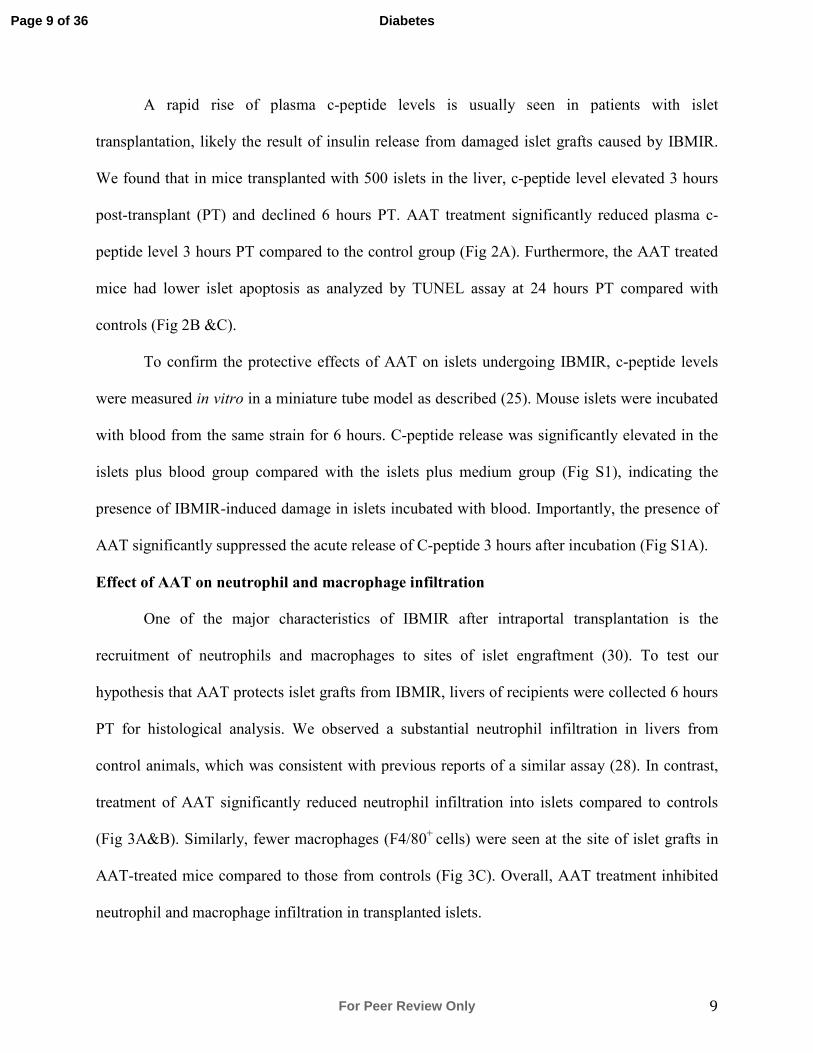
A rapid rise of plasma c-peptide levels is usually seen in patients with islet
transplantation, likely the result of insulin release from damaged islet grafts caused by IBMIR.
We found that in mice transplanted with 500 islets in the liver, c-peptide level elevated 3 hours
post-transplant (PT) and declined 6 hours PT. AAT treatment significantly reduced plasma c-
peptide level 3 hours PT compared to the control group (Fig 2A). Furthermore, the AAT treated
mice had lower islet apoptosis as analyzed by TUNEL assay at 24 hours PT compared with
controls (Fig 2B &C).
To confirm the protective effects of AAT on islets undergoing IBMIR, c-peptide levels
were measured in vitro in a miniature tube model as described (25). Mouse islets were incubated
with blood from the same strain for 6 hours. C-peptide release was significantly elevated in the
islets plus blood group compared with the islets plus medium group (Fig S1), indicating the
presence of IBMIR-induced damage in islets incubated with blood. Importantly, the presence of
AAT significantly suppressed the acute release of C-peptide 3 hours after incubation (Fig S1A).
Effect of AAT on neutrophil and macrophage infiltration
One of the major characteristics of IBMIR after intraportal transplantation is the
recruitment of neutrophils and macrophages to sites of islet engraftment (30). To test our
hypothesis that AAT protects islet grafts from IBMIR, livers of recipients were collected 6 hours
PT for histological analysis. We observed a substantial neutrophil infiltration in livers from
control animals, which was consistent with previous reports of a similar assay (28). In contrast,
treatment of AAT significantly reduced neutrophil infiltration into islets compared to controls
(Fig 3A&B). Similarly, fewer macrophages (F4/80+
cells) were seen at the site of islet grafts in
AAT-treated mice compared to those from controls (Fig 3C). Overall, AAT treatment inhibited
neutrophil and macrophage infiltration in transplanted islets.
Page 9 of 36
For Peer Review Only
Diabetes

10
We further measured serum TNF-α in mice treated with AAT or vehicle. The level of
serum TNF-α was not detected in mice before the transplantation, whereas a significant rise of
TNF-α was observed in the control animals 6 hours PT. Administration of AAT significantly
decreased the serum TNF-α level (Fig 3D).
AAT alters tissue factor (TF) expression, plasma TAT levels and fibrin deposition
TF expressed on islets is considered the trigger for IBMIR. To determine whether AAT
inhibits IBMIR through the suppression of TF, immunofluorescent staining was performed to
analyze the expression of TF by islet grafts after transplantation. At 6 hours PT, a dramatic
expression of TF was observed in control grafts. In contrast, AAT administration significantly
repressed expression of TF by islets, as indicated by immunohistochemistry and the relative
intensity calculated by dividing the intensity of insulin staining (Fig 4, A&B).
TAT is a sensitive indicator for the activation of blood coagulation. To test for the effect
of AAT on the thrombotic reaction in IBMIR, plasma TAT levels were monitored. AAT
treatment effectively suppressed the elevated plasma TAT level observed in the control group
within 6 hours PT (Fig 4C). In the in vitro miniature tube model, TAT was not detected in islets
incubated with medium only groups, but was markedly elevated in islets incubated with blood. In
contrast, AAT effectively blocked this elevation of TAT (Fig S1B). We also measured the
generation of fibrinogen deposition, which is normally caused by the activation of thrombin, and
is detrimental to islet grafts. As shown by immunohistochemistry, an apparent decrease of fibrin
staining was observed in islet grafts from the AAT group compared to grafts from the control
group 6 hours PT (Fig 4D). This data further confirmed the anti-thrombotic effect of AAT in
IBMIR.
Page 10 of 36
For Peer Review Only
Diabetes
11
Cytokine-induced JNK phosphorylation in vitro in βTC3 cells
IBMIR produces both systemic and local proinflamatory cytokine cascades.
Proinflammatory cytokines induce pancreatic β cell death via JNK phosphorylation and NF-κB
activation (20; 31). It is not currently known whether AAT has an effect on the JNK pathway.
Transcriptional profiles of islet grafts and surrounding liver tissues from control and AAT-
treated mice revealed significant down-regulation of the mRNA level of JUN, the substrate of
JNK, by AAT treatment. Western blot analysis revealed that AAT significantly suppressed JNK
phosphorylation in βTC3 cells 15 and 30 minutes after cytokine stimulation (Fig 5A&B). By
measuring cell death, we found that AAT treatment suppressed cytokine-induced β cell death in
a dose-dependent manner, an effect comparable to that of the JNK inhibitor SP600125 (Fig 5C).
Moreover, the addition of SP600125 failed in further reducing the AAT-suppressed cell death,
implying the possibility that the potent anti-inflammatory effect of AAT is mediated by
suppression of JNK phosphorylation.
AAT attenuates NF-κB activation in liver tissue and in βTC3 cells
Activation of NF-κB was studied in liver tissues harvested 24 hours PT. A significant
suppression of NF-κB activation (p65 nuclear translocation) was observed in livers of mice
treated with AAT compared with the control (Fig 5D), indicating that AAT attenuated the NF-κB
activation triggered by the IBMIR. Consistent with this, we observed that AAT treatment
prevented cytokine-induced inhibitor of B (IBα) degradation, indicating that NF-κB
activation was also suppressed by AAT treatment on βTC3 cells (Fig 5E). These experiments
show that AAT protects β cells from cytokine-induced apoptosis by suppression of JNK
phosphorylation and NF-κB activation.
AAT protects human islet grafts transplanted into diabetic NOD-SCID mice livers.
Page 11 of 36
For Peer Review Only
Diabetes

12
Since there are major differences between mouse and human islets (32; 33), it is
necessary to test the effects of AAT on human islets in IBMIR to confirm the potential clinical
relevance of this drug. To this end, a marginal mass of human islets were infused into diabetic
NOD-SCID mice livers in the presence or absence of one injection of AAT (2mg/ml, i.p.). We
found that human c-peptide level peaked at 1 hour PT in the control recipient serum samples, and
AAT treatment attenuated the rapid release of c-peptide from the islet grafts (Fig 6A). The
infusion of human islets triggered a substantial increase of plasma TAT, and in accordance with
our data in C57BL/6 mouse, AAT significantly lowered plasma TAT levels in recipients (Fig
6B). Furthermore, we found that neutrophilic infiltration to the human islet grafts was blocked
by AAT (Fig 6 C&D). Immunohistochemistry showed reduced macrophage accumulation in the
islet grafts in the AAT-treated livers (Fig 6E), suggesting that AAT treatment protected human
islet grafts from the damage of IBMIR.
AAT reduces tissue factor expression and inhibits neutrophil infiltration in a human islet in
vitro miniature tube model
To further confirm the protective effects of AAT on human islets in IBMIR, we
incubated human islets in vitro in NOD-SCID mouse blood for 15 min with constant disturbance.
We observed less tissue factor expression in islets pre-treated with AAT for 2 hours before the
exposure to blood. Compared with islets cultured in medium, addition of AAT significantly
decreased tissue factor (Fig 7, A&B), without other exogenous stimulation, indicating that AAT
suppresses tissue factor expression not only under pathological stress such as IBMIR, but also
under relatively normal conditions like an in vitro culture. The exposure to blood induced a
significant number of neutrophils infiltrating into human islets, with morphological damage. In
contrast, the islets obtained from the AAT-treated group exhibited fewer neutrophils with
Page 12 of 36
For Peer Review Only
Diabetes

13
preserved morphology (Fig 7, C&D). Thus, AAT could inhibit tissue factor expression and
neutrophilic damage to human islets in a miniature tube assay.
AAT suppresses JNK phosphorylation in human islets in vitro.
To determine whether AAT inhibits JNK phosphorylation in human islets, islets were
exposed to NOD-SCID mouse blood or a cytokine cocktail made up of TNF-α, IL-1β, and IFN.
By IHC, we observed that pre-treatment of AAT significantly reduced p-JNK positive staining
after a 15-minute incubation with blood (Fig 8A&B). In addition, cytokine-induced islet cell
apoptosis as detected by TUNEL at 72 hours after stimulation was significantly decreased by
AAT pre-treatment (Fig S2). Furthermore, AAT inhibited JNK phosphorylation in a dose-
dependent manner in these samples at 15, 30 and 60 minutes of cytokine exposure (Fig 8C).
These results confirmed that AAT could inhibit JNK phosphorylation in human islets.
Discussion
More than half of transplanted islets may be lost in the immediate post-transplant (PT)
period (34). Strong evidence shows that IBMIR is the major cause of early islet graft loss in both
autologous and allogeneic islet transplantation. In this study, we found that AAT protects islet
grafts from IBMIR-induced damage and thus, improves the outcome of syngeneic intraportal
islet transplantation in mice. We showed that AAT treatment significantly reduces the activation
of coagulation and inflammation pathways in IBMIR. This leads to less islet cell death 24 hours
PT, prolongs islet graft survival, and improves islet function 60 days after transplantation.
We demonstrate for the first time that AAT suppresses IBMIR in the intraportal islet
transplantation model with both mouse and human islets. Previous studies have shown that AAT
administration prolonged islet allograft survival when islets were transplanted under the kidney
Page 13 of 36
For Peer Review Only
Diabetes

14
capsule (17; 35-37). However, in the clinic, islets are infused to the patient’s liver through the
portal vein. The major difference between intraportal and subcapsular kidney islet transplantation
is that IBMIR is only present in the former where islets are directly exposed to the recipients’
blood circulation (19).
A short-term AAT treatment on diabetic cynomolgus monkeys underwent intraportal
autologous islet transplantation improved blood glucose levels and preserved graft morphology
at day 468 PT (16). Given the fact that IBMIR begins immediately after islet infusion and peaks
in less than 3 hours (18), our group focused on investigating the short-term effects of AAT with
regard to the inhibition of coagulation and inflammation. We found that AAT significantly
suppressed elevated plasma TAT levels, reduced TF expression and reduced fibrin deposition
within 6 hours PT in recipients that received either mouse or human islets. Also, AAT decreased
circulating TNF-α levels and leukocyte infiltration in the islet grafts compared with the control
group. All of these results translated into less islet cell damage and better blood glucose control
30 days PT in recipients treated with AAT. The evidence shows that AAT inhibits IBMIR, which
results in improved outcome of intraportal islet transplantation in mice.
TF is produced and secreted by the endocrine cells of the islets. Exposure of islet cell-
expressed TF to the recipient’s blood circulation is thought to initiate IBMIR (6). The interaction
of TF and factor VII results in the activation of the extrinsic pathway, leading to rapid elevation
of plasma TAT, regularly seen in patients undergoing islet transplantation (18). These thrombotic
reactions cause fibrin deposition on islet grafts as was observed in our experimental setting (28).
Therefore, methods that can suppress the thrombotic reaction become intriguing therapeutic
strategies in the prevention of IBMIR. Currently, most centers use systemic administration of
heparin to prevent blood clots in both autologous and allogeneic islet transplantations. However,
Page 14 of 36
For Peer Review Only
Diabetes

15
the potential therapeutic efficacy of heparin is largely limited by its short half-life and the risk of
systemic bleeding complications. Furthermore, heparin has failed to prevent infiltration of
CD11b+ cells (38), indicating that additional anti-inflammatory reagents are needed. Indeed, in
the Clinical Islet Transplantation 07 (CIT07) protocol, an extension of anticoagulation beyond
the islet infusion procedure was adopted in combination with peri-transplant administration of a
TNF- inhibitor, which contributed to improved islet engraftment in T1D patients (39).
Low molecular weight dextran sulfate (24) and a combination of Tirofiban and activated
protein C (40) have also been tested in tube loop models in vitro, and proved to be effective in
ameliorating the thrombotic reaction in IBMIR. However, to date, neither of these treatment
strategies has been reported. There is no current literature that describes the utilization of these
treatments in the in vivo islet transplant model. To address the inflammatory reactions in IBMIR,
Withaferin A, an NF-κB inhibitor, was applied in a miniature tube model and demonstrated
significant inhibitory effects on cytokine secretion and neutrophil infiltration, but no effect on
elevated TAT level in the tube model (25). In this study, we found that AAT markedly inhibited
tissue factor expression and suppressed elevated plasma TAT levels due to IBMIR in addition to
its inhibitory effects on inflammatory cell infiltration and cytokine release, both in mouse and
human islets. Since AAT has well-established anti-inflammatory effects in a large number of
other studies (17; 27; 37; 41; 42), we suspected that the effect was not specific to the coagulation
cascade.
Cytokines produced by infiltrated leukocytes and endocrine islet cells in IBMIR play a
crucial role in the destruction of islet grafts (17; 21; 30; 43). Cytokine-induced β cell failure and
death are key events that lead to diabetes (44). Cytokines rapidly activate the stress kinase
SAPK/JNK, as well as other proinflammatory kinases. Prolonged and pronounced activation of
Page 15 of 36
For Peer Review Only
Diabetes

16
JNK leads to ß cell death (45; 46). According to our expression profile-based analysis, the
mRNA level of JUN, which is the substrate of JNK, was significantly down-regulated by AAT
treatment. We hypothesized that inhibition of the JNK pathway served as one underlying
mechanism of the protective effect of AAT on cytokine-induced β cell death, seen both in our
experimental setting of IBMIR, and other models with type 1 diabetes (47) and cytokine
stimulation (37). The data that AAT significantly suppressed cytokine-induced JNK
phosphorylation and cell death, and that blocking JNK-phosphorylation showed similar
protective effects of AAT, indicated for the first time that AAT exerts its protective function via
suppression of JNK phosphorylation in the intrahepatic islet transplantation model. Feng et. al.
demonstrated that overexpression of AAT decreased hypoxia/reperfusion-induced p38
expression in human umbilical vein endothelial cells, supporting the hypothesis that normal AAT
is able to regulate the intracellular mitogen-activated protein kinase (MAPK) pathway, in
addition to its well-known protease inhibitor function (48; 49).
There are limitations to the study. By implicating multiple mechanisms by which AAT
protects islets from short- and long-term injury and apoptotic death, we run the risk these
mechanisms may be inter-dependent. More elaborate experiments could determine which of the
protective pathways has more impact than others. More work is also justified to determine
whether AAT directly suppresses JNK activation or the upstream factors in the JNK pathway in
islet β cells. It is fitting and proper that this work be done in a human islet transplantation setting,
since the demonstrated benefits of AAT are strong and the toxicities of this medication to
humans are minimal.
By careful examination of a number of cellular pathways, we have identified a critical
mechanism by which AAT exerts protective effects in a mouse intrahepatic islet transplantation
Page 16 of 36
For Peer Review Only
Diabetes

17
model. This data supports the feasibility of using AAT in clinical islet transplantation and other
cell therapies that involve IBMIR.
Acknowledgement
J.W. performed ex vivo experiments, analyzed data and wrote the manuscript. Z.S and
W.G performed in vivo experiments. C.W. participated in human islet study. D.A. and K.M
participated in manuscript writing. C.S participated in data discussion and manuscript writing.
H.W. designed the study, performed some in vivo experiments, analyzed the data and wrote the
manuscript. H.W take responsibility for the contents of the article. We thank Dr. Bashoo
Naziruddin for help setting up the in vitro miniature tube model for IBMIR. This study was
supported by NIH grant 1R01DK105183 and DK097544 to HW.
Disclosure of Conflicts of Interest:
The authors declare no conflict of interests.
Page 17 of 36
For Peer Review Only
Diabetes

18
References
1. Ichii H, Ricordi C: Current status of islet cell transplantation. J Hepatobiliary Pancreat Surg
2009;16:101-112
2. Wang H, Desai KD, Dong H, Owzarski S, Romagnuolo J, Morgan KA, Adams DB: Prior
surgery determines islet yield and insulin requirement in patients with chronic pancreatitis.
Transplantation 2013;95:1051-1057
3. Montana E, Bonner-Weir S, Weir GC: Beta cell mass and growth after syngeneic islet cell
transplantation in normal and streptozocin diabetic C57BL/6 mice. J Clin Invest 1993;91:780-
787
4. Davalli AM, Scaglia L, Zangen DH, Hollister J, Bonner-Weir S, Weir GC: Vulnerability of
islets in the immediate posttransplantation period. Dynamic changes in structure and function.
Diabetes 1996;45:1161-1167
5. Brantly ML, Wittes JT, Vogelmeier CF, Hubbard RC, Fells GA, Crystal RG: Use of a highly
purified alpha 1-antitrypsin standard to establish ranges for the common normal and deficient
alpha 1-antitrypsin phenotypes. Chest 1991;100:703-708
6. Breit SN, Wakefield D, Robinson JP, Luckhurst E, Clark P, Penny R: The role of alpha 1-
antitrypsin deficiency in the pathogenesis of immune disorders. Clin Immunol Immunopathol
1985;35:363-380
7. Mordwinkin NM, Louie SG: Aralast: an alpha 1-protease inhibitor for the treatment of alpha-
antitrypsin deficiency. Expert Opin Pharmacother 2007;8:2609-2614
8. Petrache I, Fijalkowska I, Medler TR, Skirball J, Cruz P, Zhen L, Petrache HI, Flotte TR,
Tuder RM: alpha-1 antitrypsin inhibits caspase-3 activity, preventing lung endothelial cell
apoptosis. Am J Pathol 2006;169:1155-1166
9. Tuder RM, Yoshida T, Fijalkowka I, Biswal S, Petrache I: Role of lung maintenance program
in the heterogeneity of lung destruction in emphysema. Proc Am Thorac Soc 2006;3:673-679
10. Zhang B, Lu Y, Campbell-Thompson M, Spencer T, Wasserfall C, Atkinson M, Song S:
Alpha1-antitrypsin protects beta-cells from apoptosis. Diabetes 2007;56:1316-1323
11. Bruning JC, Winnay J, Bonner-Weir S, Taylor SI, Accili D, Kahn CR: Development of a
novel polygenic model of NIDDM in mice heterozygous for IR and IRS-1 null alleles. Cell
1997;88:561-572
12. Koulmanda M, Bhasin M, Hoffman L, Fan Z, Qipo A, Shi H, Bonner-Weir S, Putheti P,
Degauque N, Libermann TA, Auchincloss H, Jr., Flier JS, Strom TB: Curative and beta cell
regenerative effects of alpha1-antitrypsin treatment in autoimmune diabetic NOD mice. Proc
Natl Acad Sci U S A 2008;105:16242-16247
13. Lewis EC, Shapiro L, Bowers OJ, Dinarello CA: Alpha1-antitrypsin monotherapy prolongs
islet allograft survival in mice. Proc Natl Acad Sci U S A 2005;102:12153-12158
14. Lewis EC, Mizrahi M, Toledano M, Defelice N, Wright JL, Churg A, Shapiro L, Dinarello
CA: alpha1-Antitrypsin monotherapy induces immune tolerance during islet allograft
transplantation in mice. Proc Natl Acad Sci U S A 2008;105:16236-16241
15. Shimoda M, Noguchi H, Fujita Y, Takita M, Ikemoto T, Chujo D, Naziruddin B, Levy MF,
Kobayashi N, Grayburn PA, Matsumoto S: Improvement of porcine islet isolation by inhibition
of trypsin activity during pancreas preservation and digestion using alpha1-antitrypsin. Cell
Transplant 2012;21:465-471
16. Koulmanda M, Sampathkumar RS, Bhasin M, Qipo A, Fan Z, Singh G, Movahedi B,
Duggan M, Chipashvili V, Strom TB: Prevention of nonimmunologic loss of transplanted islets
Page 18 of 36
For Peer Review Only
Diabetes

19
in monkeys. American journal of transplantation : official journal of the American Society of
Transplantation and the American Society of Transplant Surgeons 2014;14:1543-1551
17. Abecassis A, Schuster R, Shahaf G, Ozeri E, Green R, Ochayon DE, Rider P, Lewis EC:
alpha1-antitrypsin increases interleukin-1 receptor antagonist production during pancreatic islet
graft transplantation. Cellular & molecular immunology 2014;11:377-386
18. Naziruddin B, Iwahashi S, Kanak MA, Takita M, Itoh T, Levy MF: Evidence for instant
blood-mediated inflammatory reaction in clinical autologous islet transplantation. American
journal of transplantation : official journal of the American Society of Transplantation and the
American Society of Transplant Surgeons 2014;14:428-437
19. Sakata N, Tan A, Chan N, Obenaus A, Mace J, Peverini R, Sowers L, Chinnock R, Hathout
E: Efficacy comparison between intraportal and subcapsular islet transplants in a murine diabetic
model. Transplant Proc 2009;41:346-349
20. Nilsson B, Ekdahl KN, Korsgren O: Control of instant blood-mediated inflammatory reaction
to improve islets of Langerhans engraftment. Curr Opin Organ Transplant 2011;16:620-626
21. Bennet W, Groth CG, Larsson R, Nilsson B, Korsgren O: Isolated human islets trigger an
instant blood mediated inflammatory reaction: implications for intraportal islet transplantation as
a treatment for patients with type 1 diabetes. Upsala journal of medical sciences 2000;105:125-
133
22. Moll G, Rasmusson-Duprez I, von Bahr L, Connolly-Andersen AM, Elgue G, Funke L,
Hamad OA, Lonnies H, Magnusson PU, Sanchez J, Teramura Y, Nilsson-Ekdahl K, Ringden O,
Korsgren O, Nilsson B, Le Blanc K: Are therapeutic human mesenchymal stromal cells
compatible with human blood? Stem Cells 2012;30:1565-1574
23. Gustafson EK, Elgue G, Hughes RD, Mitry RR, Sanchez J, Haglund U, Meurling S, Dhawan
A, Korsgren O, Nilsson B: The instant blood-mediated inflammatory reaction characterized in
hepatocyte transplantation. Transplantation 2011;91:632-638
24. Johansson H, Goto M, Dufrane D, Siegbahn A, Elgue G, Gianello P, Korsgren O, Nilsson B:
Low molecular weight dextran sulfate: a strong candidate drug to block IBMIR in clinical islet
transplantation. American journal of transplantation : official journal of the American Society of
Transplantation and the American Society of Transplant Surgeons 2006;6:305-312
25. Kanak MA, Takita M, Itoh T, SoRelle JA, Murali S, Kunnathodi F, Shahbazov R, Lawrence
MC, Levy MF, Naziruddin B: Alleviation of instant blood-mediated inflammatory reaction in
autologous conditions through treatment of human islets with NF-kappaB inhibitors.
Transplantation 2014;98:578-584
26. Hering BJ, Kandaswamy R, Ansite JD, Eckman PM, Nakano M, Sawada T, Matsumoto I,
Ihm SH, Zhang HJ, Parkey J, Hunter DW, Sutherland DE: Single-donor, marginal-dose islet
transplantation in patients with type 1 diabetes. Jama 2005;293:830-835
27. Koulmanda M, Bhasin M, Fan Z, Hanidziar D, Goel N, Putheti P, Movahedi B, Libermann
TA, Strom TB: Alpha 1-antitrypsin reduces inflammation and enhances mouse pancreatic islet
transplant survival. Proc Natl Acad Sci U S A 2012;109:15443-15448
28. Cui W, Angsana J, Wen J, Chaikof EL: Liposomal formulations of thrombomodulin increase
engraftment after intraportal islet transplantation. Cell Transplant 2010;19:1359-1367
29. Kemp CB, Knight MJ, Scharp DW, Lacy PE, Ballinger WF: Transplantation of isolated
pancreatic islets into the portal vein of diabetic rats. Nature 1973;244:447
30. Martin BM, Samy KP, Lowe MC, Thompson PW, Cano J, Farris AB, Song M, Dove CR,
Leopardi FV, Strobert EA, Jenkins JB, Collins BH, Larsen CP, Kirk AD: Dual islet
transplantation modeling of the instant blood-mediated inflammatory reaction. American journal
Page 19 of 36
For Peer Review Only
Diabetes

20
of transplantation : official journal of the American Society of Transplantation and the American
Society of Transplant Surgeons 2015;15:1241-1252
31. Humphrey RK, Newcomb CJ, Yu SM, Hao E, Yu D, Krajewski S, Du K, Jhala US: Mixed
lineage kinase-3 stabilizes and functionally cooperates with TRIBBLES-3 to compromise
mitochondrial integrity in cytokine-induced death of pancreatic beta cells. The Journal of
biological chemistry 2010;285:22426-22436
32. Kulkarni RN, Mizrachi EB, Ocana AG, Stewart AF: Human beta-cell proliferation and
intracellular signaling: driving in the dark without a road map. Diabetes 2012;61:2205-2213
33. Bernal-Mizrachi E, Kulkarni RN, Scott DK, Mauvais-Jarvis F, Stewart AF, Garcia-Ocana A:
Human beta-cell proliferation and intracellular signaling part 2: still driving in the dark without a
road map. Diabetes 2014;63:819-831
34. Biarnes M, Montolio M, Nacher V, Raurell M, Soler J, Montanya E: Beta-cell death and
mass in syngeneically transplanted islets exposed to short- and long-term hyperglycemia.
Diabetes 2002;51:66-72
35. Rackham CL, Vargas AE, Hawkes RG, Amisten S, Persaud SJ, Austin AL, King AJ, Jones
PM: Annexin A1 Is a Key Modulator of Mesenchymal Stromal Cell-Mediated Improvements in
Islet Function. Diabetes 2016;65:129-139
36. Lewis EC, Mizrahi M, Toledano M, Defelice N, Wright JL, Churg A, Shapiro L, Dinarello
CA: alpha1-Antitrypsin monotherapy induces immune tolerance during islet allograft
transplantation in mice. Proceedings of the National Academy of Sciences of the United States of
America 2008;105:16236-16241
37. Lewis EC, Shapiro L, Bowers OJ, Dinarello CA: Alpha1-antitrypsin monotherapy prolongs
islet allograft survival in mice. Proceedings of the National Academy of Sciences of the United
States of America 2005;102:12153-12158
38. Bennet W, Sundberg B, Groth CG, Brendel MD, Brandhorst D, Brandhorst H, Bretzel RG,
Elgue G, Larsson R, Nilsson B, Korsgren O: Incompatibility between human blood and isolated
islets of Langerhans: a finding with implications for clinical intraportal islet transplantation?
Diabetes 1999;48:1907-1914
39. Rickels MR, Liu C, Shlansky-Goldberg RD, Soleimanpour SA, Vivek K, Kamoun M, Min Z,
Markmann E, Palangian M, Dalton-Bakes C, Fuller C, Chiou AJ, Barker CF, Luning Prak ET,
Naji A: Improvement in β-Cell Secretory Capacity After Human Islet Transplantation According
to the CIT07 Protocol. Diabetes 2013;62:2890-2897
40. Akima S, Hawthorne WJ, Favaloro E, Patel A, Blyth K, Mudaliar Y, Chapman JR, O'Connell
PJ: Tirofiban and activated protein C synergistically inhibit the Instant Blood Mediated
Inflammatory Reaction (IBMIR) from allogeneic islet cells exposure to human blood. American
journal of transplantation : official journal of the American Society of Transplantation and the
American Society of Transplant Surgeons 2009;9:1533-1540
41. Churg A, Dai J, Zay K, Karsan A, Hendricks R, Yee C, Martin R, MacKenzie R, Xie C,
Zhang L, Shapiro S, Wright JL: Alpha-1-antitrypsin and a broad spectrum metalloprotease
inhibitor, RS113456, have similar acute anti-inflammatory effects. Lab Invest 2001;81:1119-
1131
42. Pott GB, Chan ED, Dinarello CA, Shapiro L: α-1-Antitrypsin is an endogenous inhibitor of
proinflammatory cytokine production in whole blood. Journal of Leukocyte Biology
2009;85:886-895
Page 20 of 36
For Peer Review Only
Diabetes

21
43. SoRelle JA, Itoh T, Peng H, Kanak MA, Sugimoto K, Matsumoto S, Levy MF, Lawrence
MC, Naziruddin B: Withaferin A inhibits pro-inflammatory cytokine-induced damage to islets in
culture and following transplantation. Diabetologia 2013;56:814-824
44. Cnop M, Welsh N, Jonas JC, Jorns A, Lenzen S, Eizirik DL: Mechanisms of pancreatic beta-
cell death in type 1 and type 2 diabetes: many differences, few similarities. Diabetes 2005;54
Suppl 2:S97-107
45. Zhang S, Liu J, Dragunow M, Cooper GJ: Fibrillogenic amylin evokes islet beta-cell
apoptosis through linked activation of a caspase cascade and JNK1. The Journal of biological
chemistry 2003;278:52810-52819
46. Abdelli S, Ansite J, Roduit R, Borsello T, Matsumoto I, Sawada T, Allaman-Pillet N, Henry
H, Beckmann JS, Hering BJ, Bonny C: Intracellular stress signaling pathways activated during
human islet preparation and following acute cytokine exposure. Diabetes 2004;53:2815-2823
47. Koulmanda M, Bhasin M, Hoffman L, Fan Z, Qipo A, Shi H, Bonner-Weir S, Putheti P,
Degauque N, Libermann TA, Auchincloss H, Jr., Flier JS, Strom TB: Curative and beta cell
regenerative effects of alpha1-antitrypsin treatment in autoimmune diabetic NOD mice.
Proceedings of the National Academy of Sciences of the United States of America
2008;105:16242-16247
48. Feng Y, Hu L, Xu Q, Yuan H, Ba L, He Y, Che H: Cytoprotective Role of Alpha-1
Antitrypsin in Vascular Endothelial Cell Under Hypoxia/Reoxygenation Condition. Journal of
cardiovascular pharmacology 2015;66:96-107
49. Daigle JN, Femminella M, Shariat-Madar Z: Modeling the spontaneous reaction of
mammalian cells to external stimuli. In Ad Hoc Networks, Springer, 2013, p. 226-241
Page 21 of 36
For Peer Review Only
Diabetes

22
Figure legends:
Fig 1. AAT treatment improves islet survival and function in a syngeneic mouse intraportal
islet transplantation model. (A) Kaplan-Meier analysis shows significantly higher percentages
of post transplantation normoglycemia in diabetic mice that received AAT (2mg/kg) every two
days (Q2D) or every three days (Q3D) compared to control. (B) AAT treated mice reached
normoglycemia faster than control mice. (C) IVGTT shows that AAT treatment (Q2D) enhances
blood glucose disposal 30 days after transplantation. Inset, area under the curve (AUC) of
IVGTT. (D). Serum concentrations of human AAT before the last dose in each group, n=8-9 in
each group. Ctrl: mice received vehicle, AAT: mice received AAT every two days. Data are
expressed as mean ± SD, *P < 0.05, Student’s t test.
Fig 2. AAT reduces immediate release of C-peptide and inhibits islet cell death after
transplantation. (A) Serum c-peptide levels in AAT-treated (AAT) and control (Ctrl) recipients
3 and 6 hours post-transplant as measured by ELISA. (B) Islets in liver sections from AAT
treated mice showed fewer TUNEL+ dead cells compared to islets from control mice 24 h after
islet transplantation. Nuclei are stained with DAPI (blue), β cells with anti-insulin antibody (red),
and arrows point to TUNEL+ cells (green) within an islet. (C) Percentage of TUNEL-positive
cells per islet graft as determined after microscopic evaluation of intrahepatic islets randomly
selected from 3 different recipients in both Ctrl and AAT groups (Ctrl, 25 islets with sizes of
10,900.5 ± 3,700.7 µm2; AAT, 20 islets with mean sizes of 9,480.4 ± 4,840.2 µm
2). Results are
expressed as means ± SD, * P < 0.05, *** p <0.001. Student’ t test.
Fig 3. AAT suppresses neutrophil and macrophage infiltration, and serum TNF-α
production. (A) Accumulation of neutrophils identified by naphthol AS-D chloroacetate
esterase staining in AAT (n3) or control (n=3) grafts 6 h PT. Serial paraffin sections of an islet
Page 22 of 36
For Peer Review Only
Diabetes

23
graft were stained with anti-insulin (left, brown staining) and naphthol AS-D chloroacetate
esterase staining (right, purple staining). Arrows point to neutrophils. (B) Numbers of
neutrophils within the islet were lower in the AAT group compared to those from control group 6
h after islet transplantation. Twenty five Ctrl islets (average size: 8500.9 ± 6816.6 µm2) and 27
AAT islets (average size: 10,549.2 ± 6,596.9 µm2) were counted. (C) Representative
micrographs show fewer infiltrated macrophages surrounding transplanted islets identified by the
anti-F4/80 antibody in AAT compared to Ctrl mice 6 h after islet transplantation. Red staining
identifies macrophages, green staining identifies β cells and blue represents nuclear material
from all cells. (D) Serum TNF-α levels measured by ELISA are higher in serum of Ctrl (n=6)
than AAT-treated (n=4) recipients 6 h after islet transplantation. *p < 0.05 AAT versus Ctrl,
Student’s t test. Scale bars are 100 µm for figures A and C, and 20 µm for insets in panel A.
Fig 4. AAT inhibits activation of coagulation following intraportal transplantation. (A)
Reduced tissue factor expression in AAT treated compared to control grafts.
Immunohistochemistry staining of tissue factor (green) in transplanted islets (red for insulin)
from AAT or Ctrl mice. Arrows point to TF-positive cells. (B) Relative fluorescence intensity of
tissue factor divided by intensity of insulin graft from AAT or Ctrl mice. (C) Change of plasma
TAT levels in AAT and Ctrl mice at 3 and 6 h PT. (D) Fibrinogen deposition in transplanted
islets 6 h after intraportal transplantation in Ctrl and AAT mice. Serial paraffin sections of an
islet graft were stained with anti-insulin (left) and anti-fibrinogen (right). Dashed line outlines
islet grafts based on the insulin staining. Arrow indicates fibrin deposition on islet graft. Data are
expressed as mean ± SD. * p < 0.05 AAT vs. Ctrl, Student’s t test. Scale bar = 100 µm.
Fig 5. AAT protects islets from cytokine induced cell death by suppression of
phosphorylation of JNK and NF-κB activation. AAT treatment reduces p-JNK
Page 23 of 36
For Peer Review Only
Diabetes

24
phosphorylation after cytokine treatment as measured by Western blot. (A) Representative
immunoblot of phosphorylated (p-JNK) and total JNK and histone H3 by Western blot. (B) Ratio
of phosphorylated to total JNK from immunoblots quantified by densitometry. Data are
presented as mean ± SD for 5 individual experiments. (C) Cell death measured in βTC3 cells
pretreated with AAT at 0.1mg/ml or 0.5mg/ml and/or 20 nM SP600125, the JNK
phosphorylation blocker (JNK inh), for 1 h and stimulated with cytokines for 48 hours using the
Cell Death ELISA kit. Experiments were repeated 4 times. Data are presented as mean ± SD. *p
< 0.05, ** p<0.01 ***p<0.01 using one-way ANOVA followed by Tukey’s post hoc analysis.
(D) NF-κB activity (percentage to control) as measured by p65 level in nuclear fraction by
ELISA in livers bearing islet grafts. Experiments were repeated 3 times. (E) Representative
immunoblot of IκBα and β-actin in βTC3 cells pre-treated with vehicle or 0.5 mg/ml AAT, and
stimulated with cytokine for 0-30 mins as analyzed by Western blot. * p<0.05 AAT vs. Ctrl,
Student’s t test.
Fig 6. AAT reduces serum C-peptide and TAT levels and inhibits inflammatory cells
infiltration to human islet grafts. (A) Serum c-peptide levels in AAT-treated and control
recipients 1, 3 and 6 hours post-transplant as measured by ELISA. (B) Change of plasma TAT
levels in AAT and Ctrl mice. * P<0.05, ** P< 0.01 vs Ctrl. using two-way repeated-
measurement ANOVA followed by Bonferroni correction. (C) Less neutrophil infiltration was
identified in AAT grafts compared to or control grafts 6 h PT. Serial paraffin sections of an islet
graft were stained with anti-insulin (left) and naphthol AS-D chloroacetate esterase staining
(right). Dashed line illustrates islets with positive insulin staining and arrows point to
neutrophils. Scale bar = 100 µm. (D) Number of neutrophils per islets in AAT and control mice
(n =20 per group) 6 h after islet transplant. (E) Representative micrographs show more infiltrated
Page 24 of 36
For Peer Review Only
Diabetes
25
macrophages surrounding control vs. AAT islet grafts as identified by staining with the anti-
F4/80 antibody 6 h after islet transplantation. Green staining identifies macrophages, red staining
identifies insulin+ cells and blue represent nuclei. *p < 0.05 AAT versus Ctrl, Student’s t test.
Fig 7. AAT reduces tissue factor expression and inhibits neutrophils infiltration to human
islet in miniature tube assay. (A) Reduced tissue factor expression was observed in human
islets transplanted to AAT treated recipients compared to controls 15 min after exposure to
NOD-SCID blood. (B) Relative fluorescence intensity of tissue factor divided by intensity of
insulin from AAT or Ctrl group. In each group, 3-9 islets were measured using imagej. (C) More
PMNs infiltrations were observed in control islets compared to AAT-treated islets as determined
by naphthol AS-D chloroacetate esterase staining 15 min after exposure to NOD-SCID blood as
measured by immunohistochemistry staining (C) and cell counting (D). Scale bar = 50 µm. More
than 30 islets were counted in each group. *p < 0.05 AAT versus Ctrl, Student’s t test.
Fig 8. AAT suppressed JNK phosphorylation in human islets in vitro. (A)
Immunohistochemistry staining of tissue factor in human islets 15 min after exposure to NOD-
SCID blood. (B) Relative fluorescence intensity of pJNK divided by intensity of insulin from
AAT or Ctrl group. In each group, 10 islets were measured using NIH ImageJ software. (C)
Human islets were treated with cytokines with or without pre-incubation with AAT for the
indicated time periods. Representative immunoblot of JNK phosphorylation is shown. Ratio of
phosphorylated to total JNK from immunoblots quantified by densitometry.
Page 25 of 36
For Peer Review Only
Diabetes

Fig 1. AAT treatment improves islet survival and function in a syngeneic mouse intraportal islet transplantation model. (A) Kaplan-Meier analysis shows significantly higher percentages of post
transplantation normoglycemia in diabetic mice that received AAT (2mg/kg) every two days (Q2D) or every
three days (Q3D) compared to control. (B) AAT treated mice reached normoglycemia faster than control mice. (C) IVGTT shows that AAT treatment (Q2D) enhances blood glucose disposal 30 days after
transplantation. Inset, area under the curve (AUC) of IVGTT. (D). Serum concentrations of human AAT before the last dose in each group, n=8-11 in each group. Ctrl: mice received vehicle, AAT: mice received
AAT every two days. Data are expressed as mean ± SD, *P < 0.05, Student’s t test.
423x317mm (72 x 72 DPI)
Page 26 of 36
For Peer Review Only
Diabetes

Fig 2. AAT reduces immediate release of C-peptide and inhibits islet cell death after transplantation. (A) Serum c-peptide levels in AAT-treated (AAT) and control (Ctrl) recipients 3 and 6 hours post-transplant as
measured by ELISA. (B) Islets in liver sections from AAT treated mice showed fewer TUNEL+ dead cells
compared to islets from control mice 24 h after islet transplantation. Nuclei are stained with DAPI (blue), β cells with anti-insulin antibody (red), and arrows point to TUNEL+ cells (green) within an islet. (C)
Percentage of TUNEL-positive cells per islet graft as determined after microscopic evaluation of intrahepatic islets randomly selected from 3 different recipients in both Ctrl and AAT groups (Ctrl, 25 islets with sizes of 10,900.5 ± 3,700.7 µm2; AAT, 20 islets with mean sizes of 9,480.4 ± 4,840.2 µm2). Results are expressed
as means ± SD, * P < 0.05, *** p <0.001. Student’ t test.
423x317mm (72 x 72 DPI)
Page 27 of 36
For Peer Review Only
Diabetes

Fig 3. AAT suppresses neutrophil and macrophage infiltration, and serum TNF-α production. (A) Accumulation of neutrophils identified by naphthol AS-D chloroacetate esterase staining in AAT (n3) or control (n=3) grafts 6 h PT. Serial paraffin sections of an islet graft were stained with anti-insulin (left,
brown staining) and naphthol AS-D chloroacetate esterase staining (right, purple staining). Arrows point to neutrophils. (B) Numbers of neutrophils within the islet were lower in the AAT group compared to those from
control group 6 h after islet transplantation. Twenty five Ctrl islets (average size: 8500.9 ± 6816.6 µm2) and 27 AAT islets (average size: 10,549.2 ± 6,596.9 µm2) were counted. (C) Representative micrographs show fewer infiltrated macrophages surrounding transplanted islets identified by the anti-F4/80 antibody in
AAT compared to Ctrl mice 6 h after islet transplantation. Red staining identifies macrophages, green staining identifies β cells and blue represents nuclear material from all cells. (D) Serum TNF-α levels
measured by ELISA are higher in serum of Ctrl (n=6) than AAT-treated (n=4) recipients 6 h after islet transplantation. *p < 0.05 AAT versus Ctrl, Student’s t test. Scale bars are 100 µm for figures A and C, and
20 µm for insets in panel A.
423x317mm (72 x 72 DPI)
Page 28 of 36
For Peer Review Only
Diabetes

Fig 4. AAT inhibits activation of coagulation following intraportal transplantation. (A) Reduced tissue factor expression in AAT treated compared to control grafts. Immunohistochemistry staining of tissue factor
(green) in transplanted islets (red for insulin) from AAT or Ctrl mice. Arrows point to TF-positive cells. (B)
Relative fluorescence intensity of tissue factor divided by intensity of insulin graft from AAT or Ctrl mice. (C) Change of plasma TAT levels in AAT and Ctrl mice at 3 and 6 h PT. (D) Fibrinogen deposition in transplanted islets 6 h after intraportal transplantation in Ctrl and AAT mice. Serial paraffin sections of an islet graft were
stained with anti-insulin (left) and anti-fibrinogen (right). Dashed line outlines islet grafts based on the insulin staining. Arrow indicates fibrin deposition on islet graft. Data are expressed as mean ± SD. * p <
0.05 AAT vs. Ctrl, Student’s t test. Scale bar = 100 µm.
423x317mm (72 x 72 DPI)
Page 29 of 36
For Peer Review Only
Diabetes

Fig 5. AAT protects islets from cytokine induced cell death by suppression of phosphorylation of JNK and NF-κB activation. AAT treatment reduces p-JNK phosphorylation after cytokine treatment as measured by
Western blot. (A) Representative immunoblot of phosphorylated (p-JNK) and total JNK and histone H3 by
Western blot. (B) Ratio of phosphorylated to total JNK from immunoblots quantified by densitometry. Data are presented as mean ± SD for 5 individual experiments. (C) Cell death measured in βTC3 cells pretreated with AAT at 0.1mg/ml or 0.5mg/ml and/or 20 nM SP600125, the JNK phosphorylation blocker (JNK inh), for 1 h and stimulated with cytokines for 48 hours using the Cell Death ELISA kit. Experiments were repeated 4 times. Data are presented as mean ± SD. *p < 0.05, ** p<0.01 ***p<0.01 using one-way ANOVA followed by Tukey’s post hoc analysis. (D) NF-κB activity (percentage to control) as measured by p65 level in nuclear
fraction by ELISA in livers bearing islet grafts. Experiments were repeated 3 times. (E) Representative immunoblot of IκBα and β-actin in βTC3 cells pre-treated with vehicle or 0.5 mg/ml AAT, and stimulated
with cytokine for 0-30 mins as analyzed by Western blot. * p<0.05 AAT vs. Ctrl, Student’s t test.
423x317mm (72 x 72 DPI)
Page 30 of 36
For Peer Review Only
Diabetes

Fig 6. AAT reduces serum C-peptide and TAT levels and inhibits inflammatory cells infiltration to human islet grafts. (A) Serum c-peptide levels in AAT-treated and control recipients 1, 3 and 6 hours post-transplant as measured by ELISA. (B) Change of plasma TAT levels in AAT and Ctrl mice. * P<0.05, ** P< 0.01 vs Ctrl.
using two-way repeated-measurement ANOVA followed by Bonferroni correction. (C) Less neutrophil infiltration was identified in AAT grafts compared to or control grafts 6 h PT. Serial paraffin sections of an islet graft were stained with anti-insulin (left) and naphthol AS-D chloroacetate esterase staining (right). Dashed line illustrates islets with positive insulin staining and arrows point to neutrophils. Scale bar = 100
µm. (D) Number of neutrophils per islets in AAT and control mice (n =20 per group) 6 h after islet transplant. (E) Representative micrographs show more infiltrated macrophages surrounding control vs. AAT
islet grafts as identified by staining with the anti-F4/80 antibody 6 h after islet transplantation. Green staining identifies macrophages, red staining identifies insulin+ cells and blue represent nuclei. *p < 0.05
AAT versus Ctrl, Student’s t test.
423x317mm (72 x 72 DPI)
Page 31 of 36
For Peer Review Only
Diabetes

Fig 7. AAT reduces tissue factor expression and inhibits neutrophils infiltration to human islet in miniature tube assay. (A) Reduced tissue factor expression was observed in human islets transplanted to AAT treated
recipients compared to controls 15 min after exposure to NOD-SCID blood. (B) Relative fluorescence
intensity of tissue factor divided by intensity of insulin from AAT or Ctrl group. In each group, 3-9 islets were measured using imagej. (C) More PMNs infiltrations were observed in control islets compared to AAT-treated islets as determined by naphthol AS-D chloroacetate esterase staining 15 min after exposure to NOD-SCID blood as measured by immunohistochemistry staining (C) and cell counting (D). Scale bar = 50 µm. More
than 30 islets were counted in each group. *p < 0.05 AAT versus Ctrl, Student’s t test.
423x317mm (72 x 72 DPI)
Page 32 of 36
For Peer Review Only
Diabetes

Fig 8. AAT suppressed JNK phosphorylation in human islets in vitro. (A) Immunohistochemistry staining of tissue factor in human islets 15 min after exposure to NOD-SCID blood. (B) Relative fluorescence intensity of pJNK divided by intensity of insulin from AAT or Ctrl group. In each group, 10 islets were measured using
NIH ImageJ software. (C) Human islets were treated with cytokines with or without pre-incubation with AAT for the indicated time periods. Representative immunoblot of JNK phosphorylation is shown. Ratio of
phosphorylated to total JNK from immunoblots quantified by densitometry.
423x317mm (72 x 72 DPI)
Page 33 of 36
For Peer Review Only
Diabetes
1
Online supplemental Data
Fig S1. AAT decreases C-peptide and TAT levels in vitro. (A) C-peptide level in the in vitro
miniature tube model from islets treated with blood (Ctrl), islets treated with AAT and blood
(AAT) and islets cultured with complete medium (Medium). (B) TAT levels in the in vitro
miniature tube model from islets treated with blood, compared to islets treated with AAT and
blood. * P < 0.05, Student’ t test.
Fig S2. AAT reduces cytokine-induced human islet cell death as analyzed by TUNEL assay.
(A) TUNEL staining of human islets incubated with cytokine cocktail for 72 hours in the
presence or absence of 0.5 mg/ml AAT. (B) Fewer TUNEL+ cells per islets were observed in
AAT treated human islets when exposed to cytokine. More than 20 islets were counted in each
group. * p < 0.05 by one-way ANOVA with Turkey post test analysis.
Page 34 of 36
For Peer Review Only
Diabetes

423x317mm (72 x 72 DPI)
Page 35 of 36
For Peer Review Only
Diabetes

423x317mm (72 x 72 DPI)
Page 36 of 36
For Peer Review Only
Diabetes