Embed Size (px)
Citation preview

MOLECULAR PAINKurihara et al. Molecular Pain 2014, 10:17http://www.molecularpain.com/content/10/1/17
RESEARCH Open Access
Alleviation of behavioral hypersensitivity inmouse models of inflammatory pain with twostructurally different casein kinase 1 (CK1)inhibitorsTakashi Kurihara1,2*, Eri Sakurai2, Masayasu Toyomoto3, Isao Kii3, Daisuke Kawamoto1, Toshihide Asada1,Tsutomu Tanabe2, Megumu Yoshimura4, Masatoshi Hagiwara3 and Atsuro Miyata1
Abstract
Background: The phylogenetically highly conserved CK1 protein kinases consisting of at least seven isoforms forma distinct family within the eukaryotic protein kinases. CK1 family members play crucial roles in a wide range ofsignaling activities. However, the functional role of CK1 in somatosensory pain signaling has not yet been fullyunderstood. The aim of this study was to clarify the role of CK1 in the regulation of inflammatory pain in mousecarrageenan and complete Freund’s adjuvant (CFA) models.
Results: We have used two structurally different CK1 inhibitors, TG003 and IC261. TG003, which was originallyidentified as a cdc2-like kinase inhibitor, had potent inhibitory effects on CK1 isoforms in vitro and in cultured cells.Intrathecal injection of either TG003 (1-100 pmol) or IC261 (0.1-1 nmol) dose-dependently decreased mechanicalallodynia and thermal hyperalgesia induced by carrageenan or CFA. Bath-application of either TG003 (1 μM) orIC261 (1 μM) had only marginal effects on spontaneous excitatory postsynaptic currents (sEPSCs) recorded in thesubstantia gelatinosa neurons of control mice. However, both compounds decreased the frequency of sEPSCs inboth inflammatory pain models.
Conclusions: These results suggest that CK1 plays an important pathophysiological role in spinal inflammatory paintransmission, and that inhibition of the CK1 activity may provide a novel strategy for the treatment of inflammatorypain.
Keywords: Allodynia, Carrageenan, Complete Freund’s adjuvant, CFA, Hyperalgesia, Whole-cell patch-clamp
BackgroundIncreased sensitivity to both noxious and non-noxiousstimuli is a hallmark of persistent pain states followingtissue injury and inflammation. This hypersensitivity isassociated with both peripheral and spinal neuronalplasticities, leading to a reduction of activation thresholdin peripheral nociceptive sensory neurons in the dorsal
* Correspondence: [email protected] of Pharmacology, Graduate School of Medical and DentalSciences, Kagoshima University, 8-35-1 Sakuragaoka, Kagoshima City,Kagoshima 890-8544, Japan2Department of Pharmacology and Neurobiology, Graduate School ofMedicine, Tokyo Medical and Dental University, 1-5-45 Yushima, Bunkyo-ku,Tokyo 113-8519, JapanFull list of author information is available at the end of the article
© 2014 Kurihara et al.; licensee BioMed CentraCommons Attribution License (http://creativecreproduction in any medium, provided the orDedication waiver (http://creativecommons.orunless otherwise stated.
root ganglion (DRG) and trigeminal ganglion, as well asan increase in the synaptic activity between sensorynerve endings and second-order neurons in the spinaldorsal horn [1-3]. Inflammatory pain is typically treatedwith opioids and non-steroidal anti-inflammatory drugssuch as cyclooxygenase 2 inhibitors. However, thesetreatments are currently limited by well-known side ef-fects. Acute opioid treatment produces respiratory de-pression, sedation, nausea, constipation and vomiting,and long-term treatment with opioids and cyclooxygen-ase 2 inhibitors is associated with the development ofaddiction and cardiovascular defects, respectively. Thus,chronic pain associated with inflammation is still diffi-cult to treat, and development of new strategies leading
l Ltd. This is an Open Access article distributed under the terms of the Creativeommons.org/licenses/by/2.0), which permits unrestricted use, distribution, andiginal work is properly credited. The Creative Commons Public Domaing/publicdomain/zero/1.0/) applies to the data made available in this article,
Kurihara et al. Molecular Pain 2014, 10:17 Page 2 of 13http://www.molecularpain.com/content/10/1/17
to pharmacological treatment of inflammatory pain iseagerly awaited.Casein kinases (CK) were one of the first serine/threo-
nine protein kinases to be identified and characterized inthe 1970s [4-7]. Two distinct CK activities were recog-nized, leading to the identification of two different ki-nases, CK1 and CK2. Whereas CK2 belongs to theCMGC (cyclin-dependent kinase, mitogen-activated pro-tein kinase, glycogen synthase kinase, CDC-like kinase)group, CK1 forms one of the eight major groups of pro-tein kinases identified in the human and mouse genomes[8,9]. The CK1 family consists of several isoforms thatinclude CK1α, CK1γ1-CK1γ3, CK1δ, and CK1ε and theirvarious splice variants. CK1 is present in different celltypes and in subcellular compartments, including theplasma membrane, cytosol, and nucleus. The widespreaddistribution of CK1 suggests important regulatory rolesof this protein kinase. At present, CK1 has been impli-cated in diverse biological processes including circadianrhythms, membrane trafficking, cytoskeleton mainten-ance, DNA and RNA metabolism [4-6]. However, thefunction of CK1 in the somatosensory pathway has notyet been fully examined.IC261 is a commonly used and commercially available
CK1 inhibitor, which is reported to be relatively specificfor CK1δ and ε isoforms [10], although some of its ef-fects are likely to be independent from CK1 inhibition[11]. Previously we demonstrated that intrathecal admin-istration of IC261 effectively reversed neuropathic pain-like behavior in mice [12]. TG003 originally identified asa cdc2-like kinase (Clk) inhibitor [13,14], has recentlybeen shown to inhibit CK1δ and ε activities equally to,or more potently than IC261 in vitro [15,16]. In thisstudy, we examined the effects of these two structurallydifferent CK1 specific inhibitors on inflammatory paininduced by peripheral treatment of carrageenan or CFA.A preliminary report of this study has been presentedelsewhere [17].
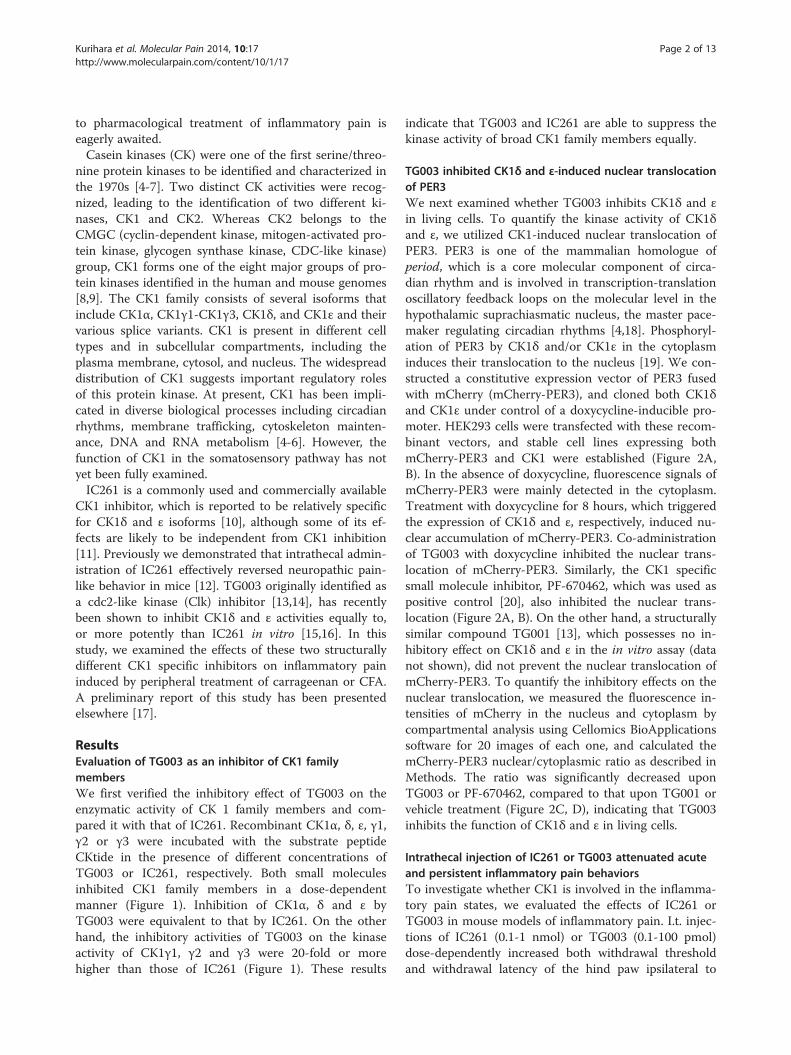
ResultsEvaluation of TG003 as an inhibitor of CK1 familymembersWe first verified the inhibitory effect of TG003 on theenzymatic activity of CK 1 family members and com-pared it with that of IC261. Recombinant CK1α, δ, ε, γ1,γ2 or γ3 were incubated with the substrate peptideCKtide in the presence of different concentrations ofTG003 or IC261, respectively. Both small moleculesinhibited CK1 family members in a dose-dependentmanner (Figure 1). Inhibition of CK1α, δ and ε byTG003 were equivalent to that by IC261. On the otherhand, the inhibitory activities of TG003 on the kinaseactivity of CK1γ1, γ2 and γ3 were 20-fold or morehigher than those of IC261 (Figure 1). These results
indicate that TG003 and IC261 are able to suppress thekinase activity of broad CK1 family members equally.
TG003 inhibited CK1δ and ε-induced nuclear translocationof PER3We next examined whether TG003 inhibits CK1δ and εin living cells. To quantify the kinase activity of CK1δand ε, we utilized CK1-induced nuclear translocation ofPER3. PER3 is one of the mammalian homologue ofperiod, which is a core molecular component of circa-dian rhythm and is involved in transcription-translationoscillatory feedback loops on the molecular level in thehypothalamic suprachiasmatic nucleus, the master pace-maker regulating circadian rhythms [4,18]. Phosphoryl-ation of PER3 by CK1δ and/or CK1ε in the cytoplasminduces their translocation to the nucleus [19]. We con-structed a constitutive expression vector of PER3 fusedwith mCherry (mCherry-PER3), and cloned both CK1δand CK1ε under control of a doxycycline-inducible pro-moter. HEK293 cells were transfected with these recom-binant vectors, and stable cell lines expressing bothmCherry-PER3 and CK1 were established (Figure 2A,B). In the absence of doxycycline, fluorescence signals ofmCherry-PER3 were mainly detected in the cytoplasm.Treatment with doxycycline for 8 hours, which triggeredthe expression of CK1δ and ε, respectively, induced nu-clear accumulation of mCherry-PER3. Co-administrationof TG003 with doxycycline inhibited the nuclear trans-location of mCherry-PER3. Similarly, the CK1 specificsmall molecule inhibitor, PF-670462, which was used aspositive control [20], also inhibited the nuclear trans-location (Figure 2A, B). On the other hand, a structurallysimilar compound TG001 [13], which possesses no in-hibitory effect on CK1δ and ε in the in vitro assay (datanot shown), did not prevent the nuclear translocation ofmCherry-PER3. To quantify the inhibitory effects on thenuclear translocation, we measured the fluorescence in-tensities of mCherry in the nucleus and cytoplasm bycompartmental analysis using Cellomics BioApplicationssoftware for 20 images of each one, and calculated themCherry-PER3 nuclear/cytoplasmic ratio as described inMethods. The ratio was significantly decreased uponTG003 or PF-670462, compared to that upon TG001 orvehicle treatment (Figure 2C, D), indicating that TG003inhibits the function of CK1δ and ε in living cells.
Intrathecal injection of IC261 or TG003 attenuated acuteand persistent inflammatory pain behaviorsTo investigate whether CK1 is involved in the inflamma-tory pain states, we evaluated the effects of IC261 orTG003 in mouse models of inflammatory pain. I.t. injec-tions of IC261 (0.1-1 nmol) or TG003 (0.1-100 pmol)dose-dependently increased both withdrawal thresholdand withdrawal latency of the hind paw ipsilateral to

Figure 1 TG003 suppresses kinase activity of CK1 family members in vitro. Recombinant CK1 family members were incubated with thesubstrate peptide CKtide in the presence of different concentrations of TG003 and IC261. TG003 (open circle) and IC261 (closed square) inhibitedCK1 family members in a dose-dependent manner. The IC50 values of TG003 on the kinase activity of CK1α, δ, ε, γ1, γ2 and γ3were 0.33, 0.34, 1.4,1.5, 0.93 and 0.88 μM, respectively. The IC50 values of IC261 were 0.19, 0.60, 0.86, > 30, > 30 and > 30 μM, respectively. Representative dose-responsecurves with Hill slope are shown. The results are presented as an average of duplicated experiments.
Kurihara et al. Molecular Pain 2014, 10:17 Page 3 of 13http://www.molecularpain.com/content/10/1/17
carrageenan or CFA-induced inflammation (Figures 3and 4). Spinal preemptive treatment of IC261 also dose-dependently attenuated the development of thermalhyperalgesia induced by carrageenan (Figure 3B). Thus,blocking the CK1 activity at the spinal level appeared tobe effective in reduction of inflammation-induced mech-anical allodynia and thermal hyperalgesia. The max-imum effects were observed 0.5-1 hour after theinjections of both inhibitors and significant analgesic ef-fects were still observed 3-4 hours after the injection ofthe highest doses used in this study (Figures 3 and 4).These inhibitors had no significant effects on the contra-lateral hind paw (Figures 3 and 4). I.t. injection of ve-hicle (1% DMSO in saline) used as a solvent for thedrugs did not show any effects (Figures 3A and 4B, datanot shown).
Carrageenan- and CFA-induced inflammation did notupregulate CK1α, δ and ε protein expressionWe next examined the protein expression levels ofCK1α, δ and ε protein in the spinal cord (L4-6) andDRGs (L4-6) by immunoblot analyses. Expression of thethree CK1 isoforms were not significantly altered in bothspinal cord (carrageenan model: CK1α, 95.6 ± 11.2%of control, n = 6; CK1δ, 143.4 ± 25.0%, n = 11; CK1ε,101.7 ± 9.25%, n = 6; CFA model : CK1α, 108.7 ± 18.3%,n = 6; CK1δ, 99.7 ± 13.7%, n = 11; CK1ε, 93.6 ± 10.8%,
n = 6) and DRGs (carrageenan model: CK1α, 111.4 ±23.2% of control, n = 6; CK1δ, 125.4 ± 33.4%, n = 7; CK1ε,109.8 ± 23.1%, n = 6; CFA model: CK1α, 92.4 ± 18.3% ofcontrol, n = 6; CK1δ, 102.2 ± 4.99%, n = 7; CK1ε, 96.8 ±11.0%, n = 6) after carrageenan (6 hours)- or CFA(3 days)-treatment, respectively (see also Additional file 1).
IC261 and TG003 decreased the frequency of sEPSCs ininflammatory pain model miceTo explore the mechanism of the antinociception in-duced by IC261 or TG003 at the spinal level, we pre-pared L5 spinal cord slice preparation from adult mice(7-10 weeks old) and performed patch-clamp recordingsin lamina II SG neurons ipsilateral to carrageenan, CFA,or vehicle injection [21-23]. The SG neurons of thespinal dorsal horn play an important role in the trans-mission and modulation of nociceptive information fromthe periphery to the CNS [24-26], and is one of the keysites generating synaptic plasticity (central sensitization)after tissue injury [3,26,27]. Such plasticity is exhibited inpart as changes in spontaneous excitatory and inhibitorypostsynaptic currents (sEPSCs and sIPSCs, respectively),which could point out both presynaptic mechanisms (fre-quency changes) and postsynaptic mechanisms (amplitudechanges) [28-32].We first examined the passive membrane properties
of SG neurons. All SG neurons examined had resting

Figure 2 TG003 inhibits CK1δ/ε-induced nuclear translocation of PER3. (A, B) Localization of mCherry-PER3 in HEK293 cells expressing CK1δ(A) or CK1ε (B). Prior to doxycycline (Dox)-induced expression of CK1δ/ε for 8 hours, the cells were treated with vehicle control, TG003, TG001, orPF-670462 for 1 hour. The treated cells were fixed and stained with Hoechst33342 to define nucleus. Representative images are shown. (C, D).Quantification of the nuclear/cytoplasmic fluorescence intensity ratio. The data are mean ± SEM (n = 20). #P < 0.0001 (Student’s t-test).
Kurihara et al. Molecular Pain 2014, 10:17 Page 4 of 13http://www.molecularpain.com/content/10/1/17
potentials more negative than -50 mV in control and twoinflammatory pain model mice. No differences were foundin the resting membrane potential and input membraneresistance among the groups (Additional file 2).Next we characterized sEPSCs, recorded under voltage-
clamp at a holding potential of -70 mV, from control andinflamed mice (Additional file 3A). The mean amplitude ofsEPSCs was not significantly different among the groups.The mean frequency of sEPSCs, on the other hand, was
significantly different. We found that the average frequencybut not the amplitude of sEPSCs was significantly in-creased in mice inflamed with CFA 3 days before, al-though carrageenan inflammation did not increase boththe average sEPSC frequency and amplitude 6 hoursafter the injection.We further examined sIPSCs from control and in-
flamed mice SG neurons at holding potentials of 0 mV(Additional file 3B). Although the mean amplitude of

0 2 4 6 80
5
10
15
20
25
Time after carrageenan injection (h)
Wit
hd
raw
al la
ten
cy (
s)
A
* *
********
***
IC261 (i.t.)
vehicle (n = 7)0.3 nmol (n = 11)1 nmol (n = 13)
Contra. Ipsi.
0 1 2 3 4 50
5
10
15
20
25
Time after carrageenan injection (h)
Wit
hd
raw
al la
ten
cy (
s)
B
******
**
IC261 (i.t.)(30 min before Car injection)
vehicle ( n = 8)0.1 nmol ( n = 8)1 nmol ( n = 8)
Contra. Ipsi.
Car
Car
0 2 4 6 8 100
1
2
3
4
5
Time after carrageenan injection (h)
Wit
hd
raw
al t
hre
sho
ld (
g)
C
Contra. Ipsi.0.1 pmol ( n = 8)
1 pmol ( n = 8)
10 pmol ( n = 8)
Car
TG003 (i.t.)
0 2 4 6 8 100
5
10
15
20
Time after carrageenan injection (h)
Wit
hd
raw
al la
ten
cy (
s)
D
TG003 (i.t.)
Car
** ******
******
***
*** *** ************
*********
*****
Contra. Ipsi.0.1 pmol (n = 8)
1 pmol (n = 7)
10 pmol (n = 8)
*** ** **
Figure 3 Effects of CK1 inhibitors on carrageenan-induced acute inflammatory pain behaviors. (A, B) Effects of intrathecal injection ofIC261 on carrageenan (Car)-induced thermal hyperalgesia. IC261 was injected 5 hour after (A) or 30 min before (B) carrageenan injection. (C, D)Effects of intrathecal injection of TG003 on carrageenan (Car)-induced mechanical allodynia (C) and thermal hyperalgesia (D). Paw withdrawalthreshold to mechanical stimulation and paw withdrawal latency to thermal stimuli are plotted against the time after carrageenan injection into ahindpaw. Data are mean ± SEM. *P < 0.05, **P < 0.01, ***P < 0.001, compared with pre-drug data (one-way ANOVA followed by Dunnett’s posthoc test).
Kurihara et al. Molecular Pain 2014, 10:17 Page 5 of 13http://www.molecularpain.com/content/10/1/17
sIPSCs was not different among the groups, the meanfrequency of sIPSCs was significantly reduced in CFAgroups.We then examined the possibility that the observed ef-
fects of the two CK1 inhibitors originate from the regula-tion of the excitatory and inhibitory synaptic transmissionin lamina II of inflamed mice. Superfusion of spinal cordslices from control mice either treated with IC261 (1 μM)or TG003 (1 μM) altered neither the frequency nor theamplitude of sEPSCs (Figures 5A and 6A). However, bothCK1 inhibitors significantly suppressed sEPSCs recordedfrom carrageenan (Figures 5B and 6B)- or CFA (Figures 5Cand 6C)-treated mice. It should be noted, however,that the inhibitory effects on sEPSC frequencies weremuch more dramatic than those on sEPSC amplitudes(Figures 5D and 6D), and furthermore, the magnitude
of inhibitory effects induced by TG003 on sEPSC frequen-cies was significantly higher than that induced by IC261(Figures 5D and 6D) in both carrageenan and CFA treatedanimals. On the other hand, IC261 (1 μM) did not affectsIPSCs from the inflamed mice, although slight but signifi-cant reduction of the mean amplitude of control sIPSCswas observed (Figure 7).
DiscussionThe present study showed for the first time that the twostructurally different CK1 inhibitors effectively reversedmechanical allodynia and thermal hyperalgesia inducedby acute or persistent hindpaw inflammation. Fromin vitro whole-cell patch-clamp studies, a part of the an-algesic mechanisms was suggested to be due to the in-hibitory effects of the CK1 inhibitors on excitatory

0 0.5 1 1.5 20
10
15
20
Time after IC261 injection (h)
Wit
hd
raw
al la
ten
cy (
s)
B
***
*********
******
vehicle (n = 9)0.1 nmol (n = 6)0.3 nmol (n = 6)1 nmol (n = 6)
Ipsi.Contra.
012340
1
2
3
4
5
Time after IC261 injection (h)
Wit
hd
raw
al t
hre
sho
ld (
g)
A
0.1 nmol (n = 6)0.3 nmol (n = 6)1 nmol (n = 6)
Contra. Ipsi.
** ******
**
*** ****** ***
00.511.522.530
1
2
3
4
5
Time after TG003 injection (h)
Wit
hd
raw
al t
hre
sho
ld (
g)
C
0.1 pmol (n = 7)1 pmol (n = 8)10 pmol (n = 7)
Contra. Ipsi.
****** **
*****
****
00.511.522.530
5
10
15
20
Time after TG003 injection (h)
Wit
hd
raw
al la
ten
cy (
s)
D
* *
**
*
0.1 pmol (n = 4)
10 pmol (n = 11)1 pmol (n = 8)
100 pmol (n = 7)
Contra. Ipsi.
Figure 4 Effects of CK1 inhibitors on CFA-induced persistent inflammatory pain behaviors. (A, B) Effects of intrathecal injection of IC261on CFA-induced mechanical allodynia (A) and thermal hyperalgesia (B). (C, D) Effects of intrathecal injection of TG003 on CFA-induced mechanicalallodynia (C) and thermal hyperalgesia (D). Paw withdrawal threshold to mechanical stimulation and paw withdrawal latency to thermal stimuli areplotted against the time after intrathecal injection into IC261 or TG003. CFA was injected 3 days before. Data are mean ± SEM. *P < 0.05, **P < 0.01,***P < 0.001, compared with pre-drug (at 0 hour) data (one-way ANOVA followed by Dunnett’s post hoc test).
Kurihara et al. Molecular Pain 2014, 10:17 Page 6 of 13http://www.molecularpain.com/content/10/1/17
synaptic transmission within SG neurons of the inflamedmice.
Pharmacological properties of IC261 and TG003In this study we clarified that both IC261 and TG003equally blocked CK1α, δ and ε activities. We also identi-fied that TG003 effectively blocked activities of CK1γisoforms. IC261 was initially reported as a selectiveCK1δ/ε inhibitor which blocked CK1δ and ε enzymaticactivities more potently than CK1α activity [10]. How-ever, our in vitro kinase assay and a recent report [16]indicated that IC261 exerted comparable inhibitory ef-fects against CK1α, δ and ε isoforms, but inhibitory ef-fects on three CK1γ isoforms were relatively weak. Incontrast, TG003 demonstrated almost equal inhibitoryeffects among CK1 isoforms. Results from our prelimin-ary screening experiments and reports from othergroups [15,16] suggested that common targets for IC261and TG003 are CK1α, δ and ε at this moment. Although
relative importance of each CK1 isoform in the allodyniaand hyperalgesia remains to be determined, CK1 mightplay an important role for the development and main-tenance of inflammatory pain.One important finding of this study is that TG003
produced antinociceptive effects on both carrageenan-and CFA-induced inflammatory pain models at lowerdoses than IC261. This difference may be due to the factthat TG003 also blocks CK1γ isoforms and Clks. In par-ticular, IC50 values of TG003 against Clk1 and 4 iso-forms (15-20 nM) [13] are smaller than those againstCK1 isoforms. However, it remains to be determinedwhether activation of CK1γ isoforms and/or Clks signifi-cantly contributes to the pathogenic mechanism of pain.In addition, we could not exclude the possibility thatother CK1-independent effects of TG003 and/or IC261might affect the antinociceptive effects.It would be noteworthy that TG003 preferentially alle-
viated mechanical allodynia than thermal hyperalgesia in

Figure 5 IC261 decreased the mean frequency and amplitude of sEPSCs in inflammatory pain model mice. Representative traces ofsEPSCs in SG neurons of the spinal cord slices from naïve control (A), carrageenan (B)- and CFA (C)-inflamed mice showing the effects of IC261(1 μM). Lower five traces represent sEPSCs at five given points in time presented above the upper trace, and are shown in an expanded timescale. (D) Summary of results, testing the effects of IC261 on the sEPSC frequencies and amplitudes The percentage compared to pre-drug re-sponse (as 100%) was shown as% control. *P < 0.05, **P < 0.01, compared with pretreatment control (Student’s t test).
Kurihara et al. Molecular Pain 2014, 10:17 Page 7 of 13http://www.molecularpain.com/content/10/1/17
both carrageenan and CFA models. Although 1 pmol ofTG003 did not affect CFA-induced thermal hyperalgesia,the same dosage of TG003 significantly reversed CFA-induced mechanical allodynia. IC261, on the other hand,was shown to be equally effective on both mechanicalallodynia and thermal hyperalgesia in the present inflam-matory pain models, as well as in our previously de-scribed spinal nerve injury model [12]. The reason ofthis difference is currently unknown and further rigor-ous studies would be necessary to evaluate the pharma-cological profiles of TG003 and IC261.
Inhibition of pain-related synaptic plasticity by the CK1inhibitorsSince intrathecal injection of these CK1 inhibitors re-versed both mechanical and thermal nociceptive behaviorsafter peripheral inflammation, we investigated whetherbath application of these CK1 inhibitors affects on sEPSCsand/or sIPSCs by using the whole-cell patch-clampmethod in SG neurons of adult spinal cord slices.
First, we characterized the effects of carrageenan- orCFA-induced peripheral inflammation on the sEPSCs andsIPSCs. In general accordance with previous reports[28,33,34], we found that CFA inflammation for 3 dayselicited significant increase in mean frequency of sEPSCs,and significant decrease of mean frequency, but not ampli-tude, of sIPSCs. In contrast, significant changes in frequen-cies and amplitudes of sEPSCs and sIPSCs were notobserved 6 hours after carrageenan injection, which may beconsistent with the previous report showing no alterationin frequencies and amplitudes of miniature EPSCs andIPSCs 1-2 days after carrageenan inflammation in imma-ture rats [35]. One apparent difference between our presentdata and the previous report using mice CFA model [28] isthat we could not detect significant increase in the meanamplitude of sEPSCs after CFA inflammation. The reasonfor this difference is at present unknown, but this mightbe due to the difference (this study vs. [28]) of strain(C57BL6/J vs. CD1), age (7-10 weeks old vs. 4-6 weeks old)or duration after CFA injection (3 days vs. 1 day).
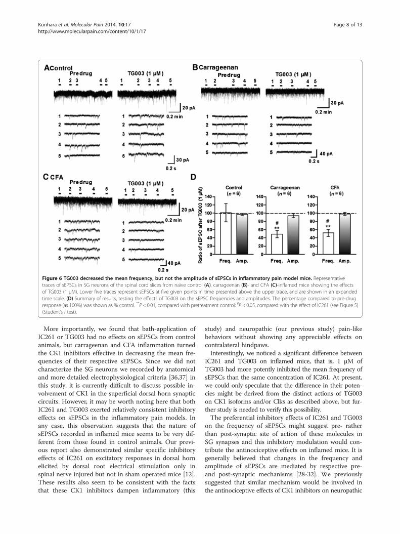
Figure 6 TG003 decreased the mean frequency, but not the amplitude of sEPSCs in inflammatory pain model mice. Representativetraces of sEPSCs in SG neurons of the spinal cord slices from naïve control (A), carrageenan (B)- and CFA (C)-inflamed mice showing the effectsof TG003 (1 μM). Lower five traces represent sEPSCs at five given points in time presented above the upper trace, and are shown in an expandedtime scale. (D) Summary of results, testing the effects of TG003 on the sEPSC frequencies and amplitudes. The percentage compared to pre-drugresponse (as 100%) was shown as % control. **P < 0.01, compared with pretreatment control; #P < 0.05, compared with the effect of IC261 (see Figure 5)(Student’s t test).
Kurihara et al. Molecular Pain 2014, 10:17 Page 8 of 13http://www.molecularpain.com/content/10/1/17
More importantly, we found that bath-application ofIC261 or TG003 had no effects on sEPSCs from controlanimals, but carrageenan and CFA inflammation turnedthe CK1 inhibitors effective in decreasing the mean fre-quencies of their respective sEPSCs. Since we did notcharacterize the SG neurons we recorded by anatomicaland more detailed electrophysiological criteria [36,37] inthis study, it is currently difficult to discuss possible in-volvement of CK1 in the superficial dorsal horn synapticcircuits. However, it may be worth noting here that bothIC261 and TG003 exerted relatively consistent inhibitoryeffects on sEPSCs in the inflammatory pain models. Inany case, this observation suggests that the nature ofsEPSCs recorded in inflamed mice seems to be very dif-ferent from those found in control animals. Our previ-ous report also demonstrated similar specific inhibitoryeffects of IC261 on excitatory responses in dorsal hornelicited by dorsal root electrical stimulation only inspinal nerve injured but not in sham operated mice [12].These results also seem to be consistent with the factsthat these CK1 inhibitors dampen inflammatory (this
study) and neuropathic (our previous study) pain-likebehaviors without showing any appreciable effects oncontralateral hindpaws.Interestingly, we noticed a significant difference between
IC261 and TG003 on inflamed mice, that is, 1 μM ofTG003 had more potently inhibited the mean frequency ofsEPSCs than the same concentration of IC261. At present,we could only speculate that the difference in their poten-cies might be derived from the distinct actions of TG003on CK1 isoforms and/or Clks as described above, but fur-ther study is needed to verify this possibility.The preferential inhibitory effects of IC261 and TG003
on the frequency of sEPSCs might suggest pre- ratherthan post-synaptic site of action of these molecules inSG synapses and this inhibitory modulation would con-tribute the antinociceptive effects on inflamed mice. It isgenerally believed that changes in the frequency andamplitude of sEPSCs are mediated by respective pre-and post-synaptic mechanisms [28-32]. We previouslysuggested that similar mechanism would be involved inthe antinociceptive effects of CK1 inhibitors on neuropathic

Figure 7 IC261 had no effects on the sIPSCs in inflammatory pain model mice. Representative traces of sIPSCs in SG neurons of the spinalcord slices from naïve control (A), carrageenan (B)- and CFA (C)-inflamed mice showing the effects of 1 μM IC261. Lower five traces representsIPSCs at five given points in time presented above the upper trace, and are shown in an expanded time scale. (D) Summary of results, testingthe effects of IC261 on the mean frequencies and amplitudes of sIPSCs. The percentage compared to pre-drug response (as 100%) was shown as% control. **P < 0.01, compared with pretreatment control (Student’s t test).
Kurihara et al. Molecular Pain 2014, 10:17 Page 9 of 13http://www.molecularpain.com/content/10/1/17
pain-like behaviors [12]. CK1 isoforms were shown tobe associated with cytosolic vesicles including small syn-aptic vesicles and to phosphorylate several small synapticvesicle-associated proteins in neuronal cells [38-40], sug-gesting a possible involvement of CK1 in the synapticvesicle exocytosis [5,40].At least CK1δ [41] and ε [12,42] proteins are shown to be
expressed in mouse spinal dorsal horn neurons and pri-mary sensory neurons at normal state. In contrast to ourprevious results that upregulation of CK1ε protein ex-pression was observed in the spinal dorsal horn (L5)and injured L5 DRG neurons ipsilateral to the nerve in-jury in the mouse L5/6 spinal nerve injury model [12],we could not detect significant increases of protein expres-sion levels of CK1α, δ, ε isoforms in the spinal cord (L4-6)and DRGs (L4-6) in the present immunoblot study asshown in Additional file 1. It would be, therefore, interest-ing to hypothesize that activity of CK1 in the primary sen-sory neurons and/or spinal dorsal horn neurons regulatedby the peripheral inflammation would contribute to the
spinal plasticity which has an important role in generatinginflammatory pain states. Several mechanisms, such as con-trol of subcellular localization by regulating membraneand/or nuclear trafficking, and modulation of the inhibitoryautophosphorylation sites located at C-terminal domains ofCK1, which have been identified to modulate CK1 activityin other experimental conditions [5,6], might also be rele-vant to our present observation. Targeting mechanisms thatcounter-regulate the spinal consequences of peripheral in-flammation by CK1 inhibitors or other methods may pro-vide an effective way to control chronic pain. Furtherelucidation of CK1 signaling mechanisms including spatialdistribution of CK1 isoforms before and after inflammationis considered to be critical in future clinical developmentfor directing the signaling pathways with small moleculeagents.
ConclusionsIn summary, the present study suggests an important roleof CK1 in inflammatory pain symptoms. Although the

Kurihara et al. Molecular Pain 2014, 10:17 Page 10 of 13http://www.molecularpain.com/content/10/1/17
specific role of each CK1 isoforms in inflammatory painremains elusive, CK1 inhibitors could be promising newtherapeutics for treating pain associated with inflamma-tion as well as neuropathic pain.
MethodsIn vitro kinase assayThe inhibitory effects of TG003 and IC261 against CK1isoforms were tested using the QuickScout screening as-sist mobility shift assay with the ATP concentration atthe Km (4.1 μM for CK1α, 6.3 μM for CK1γ1, 10 μM forCK1γ2, 3.2 μM for CK1γ3, 7.7 μM for CK1δ, and 16 μMfor CK1ε; Carna Biosciences, Inc., Kobe, Japan). Detailedinformation on the assay condition is available on thewebsite of Carna Biosciences (http://www.carnabio.com).Full-length human CK1α, CK1γ1, CK1γ2, CK1γ3 andcatalytic domain of human CK1ε were expressed as N-terminal GST-fusion protein using baculovirus system,and purified by using glutathione sepharose chromatog-raphy. Catalytic domain of CK1δ was expressed as N-terminal GST-fusion protein in E. coli, and purified byusing glutathione sepharose chromatography.
Vector constructionPCR-amplified fragments of mCherry (Clontech) andPER3 (Accession: NP_058515) were fused in-frame byoverlap-extension PCR method to generate mCherry-PER3, respectively, as described previously [43] withsome modifications. The combined fragment wasinserted into pCAGIPuro vector, an IRES-based bicis-tronic expression vector where the gene of interest anda puromycin resistant gene are expressed from a singlemRNA, which enables almost all of the cells selectedwith puromycin to express the gene product. PCR-amplified fragments of FLAG-tagged CK 1δ (Acces-sion: BC015775) and ε (Accession: BC006490) werefused in-frame to the amino-terminus of EGFP via F2Apeptide sequence by overlap-extension PCR method,which enables bicistronic expression of FLAG-taggedCK1 isoforms and EGFP. The combined fragmentswere inserted into pcDNA5/FRT/TO (Life Technolo-gies). The reconstituted vector sequences are availableupon request.
Cell culture and transfectionFlp-In/T-REx HEK293 cell (Life Technologies) wasmaintained in low glucose Dulbecco's modified Eagle'smedium (Nacalai Tesque, Kyoto, Japan) supplementedwith 10% fetal bovine serum (Nichirei Biosciences,Tokyo, Japan), 100 units/ml of penicillin and 100 μg/mlof streptomycin (Nacalai Tesque). Cells were transfectedwith plasmid DNAs using polyethylenimine MAX (Poly-sciences) as described previously [44], and then selectedwith hygromycin B (Life Technologies) for pcDNA5/
FRT/TO vectors and puromycin (Nacalai Tesque) forpCAGIPuro vectors to establish the stable cell lines.
PER3 nuclear translocation assayHEK293 cells seeded in a density of 1 × 10⁵ cells/dish inpolyethyleneimine-coated 35 mm glass bottom dishes(MatTek, Ashland, MA) were cultured for 2 days. Cellswere pre-incubated with 0.1% dimethyl sulfoxide(DMSO) containing 30 μM TG003, 30 μM TG001, or1 μM PF-670462 (Merck, Darmstadt, Germany) for1 hour at 37°C before expression of CK1δ or CK1ε wasinduced with 1 μg/ml of doxycycline. After 8 hour incu-bation with doxycycline at 37°C, cells were fixed with10% Formaldehyde Neutral Buffer Solution (Nacalai Tes-que) for 10 min at room temperature. Cells were washedtwice with PBS and then stained with 5 μg/ml ofHoechst33342 (Dojindo, Kumamoto, Japan) in PBS for30 min at room temperature. The Hoechst33342 solu-tion was removed and cells were washed with PBS, andstored in 1.5 ml PBS at 4°C in the dark until takingfluorescent images on the Confocal Laser Scanning Bio-logical Microscope FV10i (Olympus, Tokyo, Japan).The fluorescent images were analyzed by the compart-
mental analysis algorithm predefined in Cellomics BioAp-plications (Thermo Fisher Scientific, Waltham, MA). Thenuclear-cytoplasmic ratio of the mCherry-PER3 signal in-tensity was quantified by dividing the mean averagemCherry intensity in the nuclear area defined as “circ” bythe mean average mCherry intensity of a “ring” aroundthis area, which covered a cytoplasmic region. The dis-tance of the circ to the nuclear outline was 16 pixels. Thering had a width of 4 pixels and a distance of 1 pixel fromthe nuclear outline. The fluorescent image containing over15 objects (cells) counted by the compartmental analysisalgorithm was used for analysis. The objects that wereunder 650 of the mean average EGFP intensity in the nu-clear area were excluded. Analysis data was exported intoExcel file for statistical analysis.
AnimalsMale C57BL/6 J mice (5 weeks old) were purchasedfrom Clea Japan, Inc. (Tokyo, Japan) and housed undercontrolled temperature (24 ± 1°C) and humidity (55 ±10%) with a 12-hour light-dark cycle with food andwater freely available. The animal experiments were ap-proved by the Animal Care Committees of Tokyo Med-ical and Dental University (approval No. 0090173) andKagoshima University (approval No. MD10053), andwere conducted in accordance with the ethical guidelinesfor the study of experimental pain in conscious animalspublished by the International Association of the Study ofPain (1995) [45] and the European Communities CouncilDirective of 24 November, 1986 (86/609/EEC).

Kurihara et al. Molecular Pain 2014, 10:17 Page 11 of 13http://www.molecularpain.com/content/10/1/17
Animal models and behavioral studiesTo produce acute and persistent inflammatory pain, carra-geenan (2% lambda carrageenan in saline, 25 μl, Sigma, St.Louis, MO) and complete Freund’s adjuvant (CFA, 25 μl,Sigma) were injected into the plantar surface of the righthindpaw under light halothane anesthesia, respectively[46-49]. Control mice were treated with saline or incom-plete Freund’s adjuvant (IFA, Sigma), respectively. Mech-anical allodynia and thermal hyperalgesia were measuredusing the Dynamic Plantar Aesthesiometer (Ugo Basile,Comerio VA, Italy) and the Paw Thermal Stimulator(UCSD, San Diego, CA, USA), respectively as described[12]. In CFA model, these behavioral experiments wereconducted 3 days after the injection. Intrathecal (i.t.) injec-tion was given in a volume of 5 μl by percutaneous punc-ture through an intervertebral space at the level of the 5thor 6th lumbar vertebra, according to a previously reportedprocedure [12,50]. An investigator, who was unaware ofthe drug treatment, performed all of the behavioralexperiments.
Immunoblot analysisSix hours after carrageenan or saline injection, and 3 daysafter CFA or IFA injection, mice were anesthetized withsodium pentobarbital (50 mg/kg), and the lumbar spinalcord and DRGs (L4-L6) were quickly removed. Each sam-ple was homogenized in a lysis buffer [150 mM NaCl,1 mM EDTA, 1% NP-40, 0.5% sodium deoxycholate, 0.1%SDS, 1:100 diluted protease inhibitor cocktail (Sigma), and50 mM Tris-HCl, pH 8.0]. Protein concentrations weredetermined with a Bio-Rad protein assay kit (Bio-Rad,Hercules, CA). Proteins were separated by SDS-PAGE(7.5% gel) and then transferred to a polyvinylidene difluor-ide membrane (Millipore, Billerica, MA). Anti-CK1α (rabbitpolyclonal, raised against amino acids 281-337 at the C-terminus of human CK1α; 1: 2,000; no. sc-28886, SantaCruz Biotechnology, Santa Cruz, CA), anti-CK1ε (rabbitpolyclonal, raised against amino acids 301-360 near the C-terminus of human CK1ε; 1: 1,000; no. sc-25423, SantaCruz Biotechnology) and anti-CK1δ antibody (rabbit poly-clonal, NC10, 1:4,000; kindly donated by Prof. UweKnippschild, Univ. Ulm, Germany) were used. The spec-ificities of the three antibodies were characterized andreported previously in several studies including ours[12,41,42,51]. We have also conducted control stainingexperiments; omission of primary antibody or secondaryantibody, and substitution of primary antibody withnormal rabbit IgG. We did not obtain any signals fromthese control experiments (data not shown).Immunoreactivity was detected by using the ECL
system (GE Healthcare, Buckinghamshire, UK). An anti-glyceraldehyde-3-phosphate dehydrogenase (GAPDH) anti-body (mouse monoclonal, 1:20,000; no. MAB374, Che-micon, Temecula, CA) or β-actin (mouse monoclonal,
1:1,000; no. sc-47778, Santa Cruz Biotechnology) wereused to normalize protein loading. Relative intensities ofthe bands were quantified by using an image analysissystem with Image J software, version 1.40 g (NationalInstitutes of Health, Bethesda, MD). At least two inde-pendent immunoblot experiments of three independentspinal cord and DRG samples were analyzed.
Patch-clamp recordings from spinal dorsal horn neuronsAdult mouse spinal cord slices were prepared accordingto the method of Yoshimura & Jessell [21,22]. Briefly,6 hours after carrageenan or saline injection, and 3 daysafter CFA or IFA injection, transverse slices (thickness,800-900 μm) of the L5 spinal segments with the L5 dor-sal root attached were cut on a vibrating blade micro-tome. The slices were superfused with Krebs solution(10-15 ml/min) saturated with 95% O2 and 5% CO2 at36 ± 1°C. The composition of Krebs solution was as fol-lows (in mM): NaCl 117; KCl 3.6; NaHCO3 25;NaH2PO4 1.2; CaCl2 2.5; MgCl2 1.2, and glucose 11(pH 7.4 after gas saturation).Blind whole-cell patch-clamp recordings were made
from the lamina II (substantia gelatinosa: SG) neuronsipsilateral to carrageenan, CFA, or vehicle (saline or IFA)injection in voltage clamp mode. Patch pipettes werefabricated from thin-walled, borosilicate, glass-capillarytubing (1.5 mm o.d., World Precision Instruments).After establishing the whole-cell configuration, neuronswere held at the potential of -70 mV to record spontan-eous excitatory postsynaptic currents (sEPSCs) and atthe potential of 0 mV to record spontaneous inhibitorypostsynaptic currents (sIPSC). Under these conditions,GABA- and glycine-mediated IPSCs and glutamate-mediated EPSCs, respectively, were negligible, becausethese holding potential were close to the reversal poten-tials of IPSCs and EPSCs, respectively [52]. Recordingelectrodes were filled with either potassium gluconate-based solution (in mM: K-gluconate 135; KCl 5; CaCl20.5; MgCl2 2; EGTA 5; HEPES 5; ATP-Mg 5; adjustedwith KOH to pH 7.2) to investigate EPSCs, or Cs-basedsolution (in mM: Cs2SO4 110; tetraethylammonium 5;CaCl2 0.5; MgCl2 2; EGTA 5; HEPES 5; ATP-Mg 5; ad-justed with CsOH to pH 7.2) to examine IPSCs. The re-sistance of a typical patch pipette is 5-10 MΩ. Membranecurrents were amplified with an Axopatch 200B amplifier(Molecular Devices, Sunnyvale, CA, USA) in voltage-clamp mode. Signals were low-pass filtered at 5 kHz anddigitized at 333 kHz with an A/D converter (Digidata1322, Molecular Devices). Data were stored with a per-sonal computer using pCLAMP10 software and analyzedwith Mini Analysis software (Synaptosoft Inc., Decatur,GA, USA).The average values of both frequency and amplitude
of sEPSCs or sIPSCs during the control (1 min) and 5-

Kurihara et al. Molecular Pain 2014, 10:17 Page 12 of 13http://www.molecularpain.com/content/10/1/17
10 min after the drug application (1 min period after theattainment of steady effect of each drug) were calculatedand quantified as relative changes in frequency and amp-litude. Since the characteristics of sEPSCs and sIPSCsparameters (frequency and amplitude) were not signifi-cantly different among naïve-, saline- and IFA-control,data from each control were combined.
DrugsIC261 was from Calbiochem, LaJolla, CA, USA. PF-670462was obtained from Tocris bioscience, Bristol, UK. TG003and TG001 were synthesized according the proceduresdescribed previously [13]. These drugs were made up asconcentrated stock solution in DMSO, aliquoted andstored at –20°C. An aliquot was diluted to the desiredconcentration in saline or Krebs solution immediatelyprior to use. The dose ranges of IC261 and TG003 usedwere determined according to our previous report (forIC261) [12] and preliminary study (for TG003).
Statistical analysisExperimental data are expressed as mean ± SEM. Singlecomparisons were made using Student’s two-tailedpaired or unpaired t-test. One-way ANOVA followed bythe Dunnett’s or Tukey’s test was used for multiple com-parisons. P < 0.05 was considered statistically significant.
Additional files
Additional file 1: Carrageenan- and CFA-induced inflammation didnot upregulate CK1α, δ and ε expression. Immunoblot analyses ofCK1α (A), δ (B) and ε (C) expression levels in the spinal cord and DRGs.L4-6 spinal segments and DRGs ipsilateral to the inflammation weredissected 6 hours after carrageenan (Car) and 3 days after CFA injection.As a control, saline (Sal) and incomplete Freund’s adjuvant (IFA) wereinjected instead of Car and CFA, respectively.
Additional file 2: Comparison of passive membrane propertiesamong L5 SG neurons obtained from control and inflamed mice.
Additional file 3: Effects of inflammation on spontaneous EPSCs(sEPSCs, A) and IPSCs (sIPSCs, B). Hindpaw injection of CFA but notcarrageenan (Car) increased mean frequency of sEPSCs and decreasedmean frequency of sIPSCs. Neither CFA nor carrageenan changed meanamplitudes of sEPSCs and sIPSCs. Three days (CFA 3d) or 6 hours (Car6 h) after injection, spinal cord slices were prepared and blind whole-cellpatch-clamp recordings were made from the SG neurons ipsilateral toCar, CFA, or vehicle injection. *P < 0.05, **P < 0.01; one-way ANOVAfollowed by Tukey’s post hoc test.
AbbreviationsCFA: Complete Freund’s adjuvant; CK: Casein kinase; Clk: cdc2-like kinase;DRG: Dorsal root ganglion; GAPDH: Glyceraldehyde-3-phosphatedehydrogenase; IFA: Incomplete Freund’s adjuvant; i.t.: Intrathecal;sEPSC: Excitatory and inhibitory postsynaptic currents; sIPSC: Inhibitorypostsynaptic currents; SG: Substantia gelatinosa.
Competing interestsThe authors declare that they have no competing interests.
Authors’ contributionsTK participated in the design of the study, performed behavioral andelectrophysiological studies and wrote the manuscript. ES, TA and DK carriedout behavioral and immunoblot analysis. MT and IK performed in vitro kinaseassay and molecular biological study and wrote the manuscript. TT, MY, MHand AM participated in the design of the study and reviewed themanuscript. All authors read and approved the final manuscript.
AcknowledgementsThe authors are grateful to Professor Uwe Knippschild (University of Ulm,Germany) for helpful comments on the manuscript and for kind donation ofthe CK1δ antibody NC10. The authors also thank Drs. Kazuhiko Inoue andYuki Kambe for helpful discussions. This work was supported by a Grant-in-Aid for Young Scientists (A), JSPS (14704022) and a Grant-in-Aid for ScientificResearch (C), JSPS (22600001) to T.K. E.S. was supported by a grant from theMD/PhD Program of Tokyo Medical and Dental University, the 21st CenturyCOE Program on Brain Integration and its Disorders to Tokyo Medical andDental University, and Shouichi Kohashi Foundation.
Author details1Department of Pharmacology, Graduate School of Medical and DentalSciences, Kagoshima University, 8-35-1 Sakuragaoka, Kagoshima City,Kagoshima 890-8544, Japan. 2Department of Pharmacology andNeurobiology, Graduate School of Medicine, Tokyo Medical and DentalUniversity, 1-5-45 Yushima, Bunkyo-ku, Tokyo 113-8519, Japan. 3Departmentof Anatomy and Developmental Biology, Graduate School of Medicine, KyotoUniversity, Yoshida-Konoe-cho, Sakyo-ku, Kyoto 606-8501, Japan. 4GraduateSchool of Health Sciences, Kumamoto Health Science University, 325Izumi-machi, Kumamoto 861-5598, Japan.
Received: 2 December 2013 Accepted: 2 March 2014Published: 10 March 2014
References1. Kuner R: Central mechanisms of pathological pain. Nat Med 2010, 16:258–1266.2. Dawes JM, Anderson DA, Bennett DLH, Bevan S, McMahon SB:
Inflammatory mediators and modulators of pain. In Wall and Melzack’sTextbook of Pain. 6th edition. Edited by McMahon SB, Koltzenburg M, TracyI, Turk DC. Philadelphia, PA: Elsevier; 2013:48–67.
3. Sandkühler J: Spinal cord plasticity and pain. In Wall and Melzack’sTextbook of Pain. 6th edition. Edited by McMahon SB, Koltzenburg M, TracyI, Turk DC. Philadelphia, PA: Elsevier; 2013:94–110.
4. Gross SD, Anderson RA: Casein kinase I: spatial organization andpositioning of a multifunctional protein kinase family. Cell Signal 1998,10:699–711.
5. Knippschild U, Gocht A, Wolff S, Huber N, Löhler J, Stöter M: The caseinkinase 1 family: participation in multiple cellular processes in eukaryotes.Cell Signal 2005, 17:675–689.
6. Cheong JK, Virshup DM: Casein kinase 1: Complexity in the family. Int JBiochem Cell Biol 2011, 43:465–469.
7. Perez DI, Gil C, Martinez A: Protein kinases CK1 and CK2 as new targetsfor neurodegenerative diseases. Med Res Rev 2011, 31:924–954.
8. Manning G, Whyte DB, Martinez R, Hunter T, Sudarsanam S: The proteinkinase complement of the human genome. Science 2002, 298:1912–1934.
9. Caenepeel S, Charydczak G, Sudarsanam S, Hunter T, Manning G: Themouse kinome: discovery and comparative genomics of all mouseprotein kinases. Proc Natl Acad Sci U S A 2004, 101:11707–11712.
10. Mashhoon N, DeMaggio A, Tereshko V, Bergmeier SC, Egli M, Hoekstra MF,Kuret J: Crystal structure of a conformation-selective casein kinase-1 in-hibitor. J Biol Chem 2000, 275:20052–20060.
11. Cheong JK, Nguyen TH, Wang H, Tan P, Voorhoeve PM, Lee SH, Virshup DM:IC261 induces cell cycle arrest and apoptosis of human cancer cells viaCK1δ/ε and Wnt/β-catenin independent inhibition of mitotic spindleformation. Oncogene 2011, 30:2558–2569.
12. Sakurai E, Kurihara T, Kouchi K, Saegusa H, Zong S, Tanabe T: Upregulationof casein kinase 1ε in dorsal root ganglia and spinal cord after mousespinal nerve injury contributes to neuropathic pain. Mol Pain 2009, 5:74.
13. Muraki M, Ohkawara B, Hosoya T, Onogi H, Koizumi J, Koizumi T, Sumi K,Yomoda J, Murray MV, Kimura H, Furuichi K, Shibuya H, Krainer AR, SuzukiM, Hagiwara M: Manipulation of alternative splicing by a newlydeveloped inhibitor of Clks. J Biol Chem 2004, 279:24246–24254.

Kurihara et al. Molecular Pain 2014, 10:17 Page 13 of 13http://www.molecularpain.com/content/10/1/17
14. Nishida A, Kataoka N, Takeshima Y, Yagi M, Awano H, Ota M, Itoh K,Hagiwara M, Matsuo M: Chemical treatment enhances skipping of amutated exon in the dystrophin gene. Nat Commun 2011, 2:308.
15. Isojima Y, Nakajima M, Ukai H, Fujishima H, Yamada RG, Masumoto KH,Kiuchi R, Ishida M, Ukai-Tadenuma M, Minami Y, Kito R, Nakao K, KishimotoW, Yoo SH, Shimomura K, Takao T, Takano A, Kojima T, Nagai K, Sakaki Y,Takahashi JS, Ueda HR: CKI ε/δ-dependent phosphorylation is atemperature-insensitive, period-determining process in the mammaliancircadian clock. Proc Natl Acad Sci U S A 2009, 106:15744–15749.
16. Anastassiadis T, Deacon SW, Devarajan K, Ma H, Peterson J: Comprehensiveassay of kinase catalytic activity reveals features of kinase inhibitorselectivity. Nat Biotech 2011, 29:1039–1046.
17. Kurihara T, Sakurai E, Asada T, Miyata A, Tanabe T: Activation of caseinkinase 1δ/ε in dorsal root ganglia and spinal cord contributes tobehavioral hypersensitivity induced by inflammation in mice [abstract].J Pharmacol Sci 2011, 115(Suppl. 1):254P.
18. Ebisawa T: Circadian rhythm in the CNS and peripheral clock disorders:Human sleep disorders and clock genes. J Pharmacol Sci 2007, 103:150–154.
19. Akashi M, Tsuchiya Y, Yoshino T, Nishida E: Control of intracellulardynamics of mammalian period proteins by casein kinase 1ε (CK1ε) andCK1δ in culture cells. Mol Cell Biol 2002, 22:1693–1703.
20. Badura L, Swanson T, Adamowicz W, Adams J, Cianfrogna J, Fisher K,Holland J, Kleiman R, Nelson F, Reynolds L, St Germain K, Schaeffer E, Tate B,Sprouse J: An inhibitor of casein kinase 1ε induces phase delays incircadian rhythms under free-running and entrained conditions.J Pharmacol Exp Ther 2007, 322:730–738.
21. Yoshimura M, Jessell TM: Primary afferent-evoked synaptic responses andslow potential generation in rat substantia gelatinosa neurons in vitro.J Neurophysiol 1989, 62:96–108.
22. Yoshimura M, Jessell TM: Amino-acid mediated EPSPs at primary afferentsynapses with substantia gelatinosa neurons in the rat spinal cord.J Physiol (Lond) 1990, 430:315–335.
23. Yanagisawa Y, Furue H, Kawamata T, Uta D, Yamamoto J, Furuse S, KatafuchiT, Imoto K, Iwamoto Y, Yoshimura M: Bone cancer induces a uniquecentral sensitization through synaptic changes in a wide area of thespinal cord. Mol Pain 2010, 6:38.
24. Willis WD Jr, Coggeshall RE: Sensory mechanisms of the spinal cord. Volume 1.3rd edition. New York: Kluwer Academic/Plenum Publishers; 2004.
25. Todd AJ, Koerber HR: Neuroanatomical substrates of spinal nociception.In Wall and Melzack’s Textbook of Pain. 6th edition. Edited by McMahon SB,Koltzenburg M, Tracy I, Turk DC. Philadelphia, PA: Elsevier; 2013:77–93.
26. Latremoliere A, Woolf CJ: Central sensitization: A generator of painhypersensitivity by central neural plasticity. J Pain 2009, 10:895–926.
27. Ji R-R, Kohno T, Moore KA, Woolf CJ: Central sensitization and LTP: do painand memory share similar mechanisms? Trends Neurosci 2003, 26:696–705.
28. Park C-K, Xu Z-Z, Liu T, Lü N, Serhan CN, Ji R-R: Resolvin D2 is a potent en-dogenous inhibitor for transient receptor potential subtype V1/A1, in-flammatory pain, and spinal cord synaptic plasticity in mice: distinct roleof Resolvin D1, D2, and E1. J Neurosci 2011, 31:18433–18438.
29. Yang K, Kumamoto E, Furue H, Yoshimura M: Capsaicin facilitatesexcitatory but not inhibitory synaptic transmission in substantiagelatinosa of the rat spinal cord. Neurosci Lett 1998, 255:135–138.
30. Engelman HS, MacDermott AB: Presynaptic ionotropic receptors andcontrol of transmitter release. Nat Rev Neurosci 2004, 5:135–145.
31. Kawasaki Y, Zhang L, Cheng J-K, Ji R-R: Cytokine mechanisms of centralsensitization: Distinct and overlapping role of interleukin-1β, interleukin-6, and tumor necrosis factor-α, in regulating synaptic and neuronal activ-ity in the superficial spinal cord. J Neurosci 2008, 28:5189–5194.
32. Xu Z-Z, Zhang L, Liu T, Park JY, Berta T, Yang R, Serhan CN, Ji R-R: ResolvinsRvE1 and RvD1 attenuate inflammatory pain via central and peripheralactions. Nat Med 2010, 16:592–597.
33. Müller F, Heinke B, Sandkühler J: Reduction of glycine receptor-mediatedminiature inhibitory postsynaptic currents in rat spinal lamina I neuronsafter peripheral inflammation. Neuroscience 2003, 122:799–805.
34. Yang K, Takeuchi K, Wei F, Dubner R, Ren K: Activation of group I mGlureceptors contributes to facilitation of NMDA receptor membranecurrent in spinal dorsal horn neurons after hind paw inflammation inrats. Eur J Pharmacol 2011, 670:509–518.
35. Li J, Baccei ML: Excitatory synapses in the rat superficial dorsal horn arestrengthened following peripheral inflammation during early postnataldevelopment. Pain 2009, 143:56–64.
36. Yasaka T, Kato G, Furue H, Rashid MH, Sonohata M, Tamae A, Murata Y,Masuko S, Yoshimura M: Cell-type-specific excitatory and inhibitorycircuits involving primary afferents in the substantia gelatinosa of the ratspinal dorsal horn in vitro. J Physiol 2007, 581:603–618.
37. Yasaka T, Tiong SYX, Hughes DI, Riddell JS, Todd AJ: Populations ofinhibitory and excitatory interneurons in lamina II of the adult rat spinaldorsal horn revealed by a combined electrophysiological and anatomicalapproach. Pain 2010, 151:475–488.
38. Pyle RA, Schivell AE, Hidaka H, Bajjalieh SM: Phosphorylation of synapticvesicle protein 2 modulates binding to synaptotagmin. J Biol Chem 2000,275:17195–17200.
39. Takamori S, Holt M, Stenius K, Lemke E, Grønborg M, Riedel D, Urlaub H,Schenck S, Brügger B, Ringler P, Müller S, Rammner B, Gräter F, Hub JS,De Groot BL, Mieskes G, Moriyama Y, Klingauf J, Grubmüller H, Heuser J,Wieland F, Jahn R: Molecular anatomy of a trafficking organelle. Cell 2006,127:831–846.
40. Wolff S, Stöter M, Giamas G, Piesche M, Henne-Bruns D, Banting G, KnippschildU: Casein kinase 1 delta (CK1δ) interacts with the SNARE associated proteinsnapin. FEBS Lett 2006, 580:6477–6484.
41. Löhler J, Hirner H, Schmidt B, Kramer K, Fischer D, Thal DR, Leithäuser F,Knippschild U: Immunohistochemical characterisation of cell-type specificexpression of CK1δ in various tissues of young adult BALB/c mice. PLoSONE 2009, 4:e4174.
42. Utz CA, Hirner H, Blatz A, Hillenbrand A, Schmidt B, Deppert W, Henne-Bruns D, Fischer D, Thal DR, Leithӓuser F, Knippschild U: Analysis of celltype-specific expression of CK1ε in various tissues of young adult BALB/c mice and in mammary tumors of SV40 T-Ag-transgenic mice. J Histo-chem Cytochem 2010, 58:1–15.
43. Horton RM, Hunt HD, Ho SN, Pullen JK, Pease LR: Engineering hybridgenes without the use of restriction enzymes: gene splicing by overlapextension. Gene 1989, 77:61–68.
44. Thomas M, Lu JJ, Ge Q, Zhang C, Chen J, Klibanov AM: Full deacylation ofpolyethylenimine dramatically boosts its gene delivery efficiency andspecificity to mouse lung. Proc Natl Acad Sci U S A 2005, 102:5679–5684.
45. International Association for the Study of Pain: Animal models of pain andethics of animal experimentation. In Core Curriculum for ProfessionalEducation in Pain. Edited by Fields HL. Seattle: IASP Press; 1995:111–112.
46. Kayser V, Guilbaud G: Local and remote modifications of nociceptivesensitivity during carrageenan-induced inflammation in the rat. Pain1987, 28:99–107.
47. Iadarola MJ, Brady LS, Draisci G, Dubner R: Enhancement of dynorphingene expression in spinal cord following experimental inflammation:stimulus specificity, behavioral parameters and opioid receptor binding.Pain 1988, 45:313–326.
48. Stein C, Millan MJ, Herz A: Unilateral inflammation of the hind paw in ratsas a model of prolonged noxious stimulation: alterations in behaviorand nociceptive thresholds. Pharmacol Biochem Behav 1988, 31:445–451.
49. Sammons MJ, Raval P, Davey PT, Rogers D, Parsons AA, Bingham S:Carrageenan-induced thermal hyperalgesia in the mouse: role of nervegrowth factor and the mitogen-activated protein kinase pathway. BrainRes 2000, 876:48–54.
50. Hylden JLK, Wilcox GL: Intrathecal morphine in mice: a new technique.Eur J Pharmacol 1980, 67:313–316.
51. Chergui K, Svenningsson P, Greengard P: Physiological role for casein kinase1 in glutamatergic synaptic transmission. J Neurosci 2005, 25:6601–6609.
52. Yoshimura M, Nishi S: Blind patch-clamp recordings from substantia gelatinosaneurons in adult rat spinal cord slices: pharmacological properties of synapticcurrents. Neuroscience 1993, 53:519–526.
doi:10.1186/1744-8069-10-17Cite this article as: Kurihara et al.: Alleviation of behavioralhypersensitivity in mouse models of inflammatory pain with twostructurally different casein kinase 1 (CK1) inhibitors. Molecular Pain2014 10:17.