Embed Size (px)
Citation preview

Allergy & AsthmaProceedings
www.allergyandasthmaproceedings.com
The Official Journal of Regional, State, and Local Allergy, Asthma, and Immunology Societies (RSLAAIS)
and American Association of Certified Allergists (AACA)
Volume 33, Number 3May-June 2012
Supplement No. 1
Innate Adaptive Tissue damaging
The Immune System in Health The Immune System in Disease
Th2
Th0
Th17 Th3
Th1 TR1
Tc
B cell
Treg
Tolerance
Type I (IgE-mediated)Type II (cytotoxic)Type III (Ag/Ab complement complex)Type IV (delayed hypersensitivity)
Immune dysfunction,deviation and chronicinflammationAllergyChronic infectionsAutoimmunityMalignancy
Beneficial(eg,pregnancy)
Harmful(eg,malignancy)
Self response
Complement
Northwestern University Allergy-Immunology Syllabus 2012:
Residents and StudentsEdited By
Paul A Greenberger, MD and Leslie C Grammer, MD

Northwestern University Allergy-Immunology Syllabus 2012:Residents and Students
S1 OverviewP. A. Greenberger, and L. C. Grammer
S2 Chapter 1: An overview of allergensR. Shah, and L. C. Grammer
S6 Chapter 2: Skin testing in allergyT. F. Carr, and C. A. Saltoun
S9 Chapter 3: Allergen immunotherapy: Definition, indication, and reactionsM. S. Georgy, and C. A. Saltoun
S12 Chapter 4: Stinging insect allergy and venom immunotherapyA. P. Koterba, and P. A. Greenberger
S15 Chapter 5: Allergic rhinitisA. Uzzaman, and R. Story
S19 Chapter 6: Nonallergic rhinitisR. Shah, and K. G. McGrath
S22 Chapter 7: Nasal polypsM. S. Georgy, and A. T. Peters
S24 Chapter 8: RhinosinusitisM. S. Georgy, and A. T. Peters
S28 Chapter 9: Asthma classificationA. Koterba, and C. A. Saltoun
S32 Chapter 10: Pediatric asthma: Principles and treatmentR. G. Robison, and R. Kumar
S36 Chapter 11: The infant and toddler with wheezingR. G. Robison, and A. M. Singh
S39 Chapter 12: Asthma: Principles of treatmentT. F. Carr, and A. T. Peters
S44 Chapter 13: Potentially (near) fatal asthmaB. R. Sabin, and P. A. Greenberger
S47 Chapter 14: Acute severe asthma (status asthmaticus)R. Shah, and C. A. Saltoun
S51 Chapter 15: Lessons learned from clinical trials of asthmaB. R. Sabin, P. C. Avila, L. C. Grammer, and P. A. Greenberger
S55 Chapter 16: Asthma in pregnancyS. Bealert, and P. A. Greenberger
S58 Chapter 17: Occupational immunologic lung diseaseB. R. Sabin, and L. C. Grammer
S61 Chapter 18: Allergic bronchopulmonary aspergillosisP. A. Greenberger
S64 Chapter 19: Hypersensitivity pneumonitisK. H. Blatman, and L. C. Grammer
S67 Chapter 20: Atopic dermatitisB. R. Sabin, N. Peters, and A. T. Peters
S70 Chapter 21: Urticaria and angioedemaT. F. Carr, and C. A. Saltoun
TABLE OF CONTENTS Vol. 33, No. 3May–June 2012Supplement No. 1

S73 Chapter 22: Hereditary and acquired angioedemaM. S. Georgy, and J. A. Pongracic
S77 Chapter 23: Food allergyR. G. Robison, and J. A. Pongracic
S80 Chapter 24: AnaphylaxisP. A. Greenberger, and A. M. Ditto
S84 Chapter 25: Idiopathic anaphylaxisK. H. Blatman, and A. M. Ditto
S88 Chapter 26: Eosinophilic esophagitisK. H. Blatman, and A. M. Ditto
S91 Chapter 27: Approach to primary immunodeficiencyA. Uzzaman, and R. L. Fuleihan
S96 Chapter 28: Classification of hypersensitivity reactionsA. Uzzaman, and S. H. Cho
S100 Chapter 29: Unproved and controversial methods and theories in allergy–immunologyR. Shah, and P. A. Greenberger
S103 Chapter 30: Drug allergyP. A. Greenberger
S108 Chapter 31: Common in vitro tests for allergy and immunologyM. Makhija, and M. R. G. O’Gorman
About the cover: The cover image is reproduced with permission from Bellanti, JA (Ed). Immunology IV:Clinical Applications in Health and Disease, I Care Press, Bethesda, MD, 2012, www.immunologycenter.org.It is a schematic representation of the total immune capability of the host based upon the efficiency ofelimination of foreign matter and symbolizes the capacity of the immune system to maintain a balancebetween the internal and external environments. Nowhere are the expressions of an imbalance of thehost’s internal immune response to the external environment better illustrated than in the field ofallergy-immunology.
Copyright Disclosure
Allergy & Asthma Proceedings (ISSN 1088-5412 print, ISSN 1539-6304 online) is owned and publishedbi-monthly by OceanSide Publications, Inc., 95 Pitman Street, Providence, RI 02906.Copyright © 2012, OceanSide Publications, Inc. (401-331-2510; Fax 401-331-0223). Printed in the USA.All rights reserved. No part of this publication may be reproduced, stored in a retrieval system, ortransmitted, in any form or by any means, electronic, mechanical photocopying, recording, orotherwise, without prior written permission from the publisher. Statements and opinions expressedin articles and communications herein are those of the author(s) and not necessarily those of theEditor(s), publisher, the American Association of Certified Allergists (AACA), the affiliated regional,state and local allergy societies or the Northwestern University. The Editor(s), publisher, theAmerican Association of Certified Allergists (AACA), the affiliated regional, state and local allergysocieties and the Northwestern University disclaim any responsibility or liability for such materialand do not guarantee, warrant, or endorse any product or service in this publication, nor do theyguarantee any claim made by the manufacturer of such product or service. Allergy and AsthmaProceedings is registered with Copyright Clearance Center, Inc., 222 Rosewood Drive, Danvers, MA01923. To get permission to use copyrighted material from this publication, go towww.copyright.com.
TABLE OF CONTENTS Vol. 33, No. 3May–June 2012Supplement No. 1

Preface
Overview
Paul A. Greenberger, M.D., and Leslie C. Grammer, M.D.
Allergic and immunologic diseases continue to resultin frequent visits to physicians’ offices. Allergic
rhinitis, rhinosinusitis, asthma, and urticaria form thebulk of these visits, and although the number of fatalitiesfrom asthma in the United States has declined, the num-ber of hospitalizations remains unacceptably high. Epi-sodes of anaphylaxis may be encountered in the emer-gency department, primary care physician’s office, orhospital and for some physicians or health care profes-sionals, it is the very first time that they have been askedto diagnose and treat a patient with this potentially life-
threatening condition. Patients who experience recurrentinfections may have a treatable deficiency of their adap-tive immune system. In this syllabus we hope to aid allphysicians and health care professionals in diagnosingand treating commonly encountered allergic–immuno-logic diseases. We hope you enjoy this journal and willuse it as a constant resource.
Also, we would like to extend a special thank you tothe fellows and faculty at the Northwestern UniversityFeinberg School of Medicine, Division of Allergy–Immu-nology who have made this syllabus possible.
Allergy and Asthma Proceedings S1

Chapter 1
An overview of allergens
Rachna Shah, M.D., and Leslie C. Grammer, M.D.
ABSTRACT
Most allergens are proteins or glycoproteins that range in molecular weight from 5000 to 100,000 Da, although polysaccharidesand low molecular weight substances also may be allergenic. Common allergens include pollens, fungal spores, house-dust mites, andanimal epithelial materials but can also include drugs, biological products, and insect venoms. The allergic response is dependent onthe route of exposure. If exposure is to an inhaled aeroallergen, the allergic response will be a respiratory reaction in nature. Ingestedor injected exposure gives rise to gastrointestinal, cutaneous, or anaphylactic reactions. Size of pollen determines clinical manifes-tation of allergy. For example, particles between 20 and 60 �m in diameter can be carried in the wind and cause nasal and ocularsymptoms (allergic rhinoconjunctivitis). Particles �7 �m can deposit in the airways and cause symptoms of asthma. Animalsproduce allergens in forms unique to each species. Cat allergen, most importantly Fel d 1, is found mainly in cat saliva, sebaceousglands in the skin, and in urine of male cats. It is buoyant and “sticky,” which means it easily remains airborne and may last in ahome for up to 6–9 months after the source is removed. Cat allergen adheres to clothes and can be found in public places such asschools. Dog allergen, particularly Can f 1, is present in dander, saliva, urine, and serum. There are allergens specific to dog breeds,but all breeds produce allergenic proteins (even poodles and “hairless” dogs).
(Allergy Asthma Proc 33:S2–S5, 2012; doi: 10.2500/aap.2012.33.3531)
An allergen is any antigenic substance that can me-diate an immediate hypersensitivity reaction
with an associated clinical reaction in an individual.Common allergens include pollens, fungal spores,house-dust mites, and animal epithelial materials butcan also include drugs, biological products, and insectvenoms. Most allergens are proteins or glycoproteinsthat range in molecular weight from 5000 to 100,000Da, although polysaccharides and low molecularweight substances also may be allergenic.1 Little isknown, but much research is dedicated to determiningthe distinguishing facts that make an antigen capableof inducing IgE production (an allergen) in contrast toantigens that induce other immunologic responses(IgG and IgA). Factors that have already been shown toincrease immunogenicity of an antigen include molec-ular size, solubility, stability, conformational fold, andduration of exposure.
Allergens enter the body via inhalation, ingestion, ormay be injected. Genetic predisposition and environ-
mental factors determine if an individual will be sen-sitized to an allergen, and then subsequent allergenexposure of sufficient concentration triggers a physio-logical response by interacting with specific IgE boundto mast cells and basophils. The ensuing inflammatorycascade elicits a variety of signs and symptoms in theallergic spectrum. The allergic response is dependent onthe route of exposure. If exposure is to an inhaled aeroal-lergen, the allergic response will be a respiratory reactionin nature. Ingested or injected exposure gives rise togastrointestinal, cutaneous, or systemic reactions.
An allergen is recognized by the International Union ofImmunologic Societies as a protein that has allergenicityin at least five individuals.2 Individual allergens are fur-ther divided into major and minor allergens when itcomes from the same source, e.g., giant ragweed pollen orcat dander. Major allergens result in an IgE response in�50% of allergic individuals allergic to the specificsource, whereas minor allergens cause an allergic re-sponse in �50%. Although “minor” is in the name, theystill cause a significant allergic response in an individual.
Nomenclature for allergen proteins has been estab-lished by the International Union of Immunologic So-cieties. The standard nomenclature uses the first threeletters of the genus, followed by the first letter of thespecies, and then an Arabic numeral; they are notitalicized. For example, cat is Felis domesticus and theallergen protein nomenclature for the primary allergenis Fel d 1.3
From the Division of Allergy–Immunology, Department of Medicine, NorthwesternUniversity Feinberg School of Medicine, Chicago, IllinoisFunded by the Ernest S. Bazley Grant to Northwestern Memorial Hospital andNorthwestern UniversityThe authors have no conflicts of interest to declare pertaining to this articleAddress correspondence and reprint requests to Leslie C. Grammer, M.D. No. 14022,676 North St. Clair St., Chicago, IL 60611E-mail address: [email protected] © 2012, OceanSide Publications, Inc., U.S.A.
S2 May–June 2012, Vol. 33, No. 3

POLLENSFor pollen to be clinically significant as an aeroallergen,
it must be buoyant, present in significant numbers, andbe allergenic. Most pollens that cause clinical disease are20–60 �m in diameter.2 This small size allows exposurethrough wind carriage and contact with the respiratorymucosa and conjunctiva. Particles �7 �m tend to depositin the airways, and those �3 �m may enter the distalairways. Pollen immunogenicity, plant abundance, prox-imity to living environments, and regional geographydetermine specific pollens that are responsible for localallergic sensitization.2
Grass pollen is the most common cause of allergicrhinitis and asthma worldwide because of the wide dis-tribution of wind-pollinating grasses. Most are 20–25 �min diameter and, therefore, tend to cause symptoms ofrhinitis rather than asthma. Most grasses belong to thesame family (Poaceae) and have significant cross-reactiv-ity, with the exceptions of Bermuda and Bahia grass,which are subtropical grasses. Ryegrass (major allergenLol p 1) and Timothy grass (major allergen Phl p1) areamong the most important allergenic grasses. Grass pol-len is typically released in the afternoon, and in the Mid-west, is prevalent in the months of May through July.Many southern areas, such as Florida and southern Cal-ifornia, have grass seasons lasting as long as 10–11months.4
Ragweed pollen is the most important cause of aller-gic rhinitis and pollen asthma in North America. Am-brosia artemisiifolia (short ragweed; major allergen Amba 1) and Ambrosia trifida (giant ragweed; Amb t 5) arethe most important ragweed pollen allergens. Pollengrains are �16–20 �m in diameter and are notoriousfor triggering allergic symptoms in the central andeastern United States; Ontario, Canada; and increasinglocations in Europe.5,6 Weed pollen release depends onseasonal daylight variation and is released typically inthe morning during the autumn season in the UnitedStates. A single ragweed plant may expel 1 millionpollen grains in a single day.3 It possesses the ability totravel hundreds of miles from its source. In the Chi-cago area, ragweed pollen is prevalent from August 15to October 1.
Tree pollen allergens range from 20 to 60 �m indiameter and mostly come from the Angiosperm class.Geography determines which tree families are preva-lent in a community, and in contrast to grass pollen,cross-reactivity is uncommon. Tree pollen is a signifi-cant cause of allergic disease, and in most of the UnitedStates, it is typically released during a short springseason. In Chicago, tree pollination is from late Marchto late May. However, in Florida and the Californialowlands, there are trees with early and late pollensseasons spanning 6 months.4
FUNGIFungi produce airborne spores and mycelial elements
that are believed to contribute significantly to allergicdisease throughout the world. These allergens are typi-cally 3–30 �m in diameter. With few exceptions, such asAlternaria in asthma, Aspergillus in allergic bronchopul-monary aspergillosis, and various fungi in allergic fungalsinusitis, the clinical importance of common fungi hasbeen difficult to assess. Alternaria alternata (major allergenAlt a 1) species are common outdoor molds that havebeen associated with triggering respiratory arrest in pa-tients with asthma.7 Cladosporium (major allergens Cla h1, 2) is also a common outdoor mold species, and likeAlternaria, it has a seasonal prevalence in the warmermonths between spring and autumn. The first hard frostof late autumn decreases spore counts significantly untilwarm weather returns. In contrast, Aspergillus fumigatus(major allergen Asp f 1) and Penicillium citrinum (Pen c13,18) species are common indoor molds and may pro-vide allergenic triggers throughout the year. High sporecounts in homes are associated with warm, humid envi-ronments and may be reduced by air conditioning in thesummer, removal of mold in homes with contamination,preventing water damage, and dehumidification ifneeded.
ENVIRONMENTAL CHANGESClimate changes due to global warming are expected to
increase temperatures by 1–2°C in this century. This willaffect vegetation and will likely result in a higher allergicdisease burden. The 2006 U.S. Department of Agriculturehardiness zone map showed a shift northward of floristiczones, which influence the type of native vegetationfound in a region.8 This shift exemplifies the effect ofglobal warming on the type of trees and other plants thatcan survive in a given latitude. In addition, studies haveshown increased size and pollen production of ragweedwith increased CO2.9 This was especially seen in urbanareas where CO2 levels and temperatures were higherthan in rural areas.
DUST MITESDust mites, particularly Dermatophagoides pteronyssi-
nus (major allergen Der p 1) and Dermatophagoides fari-nae, ingest human epithelial scales and obtain waterfrom the ambient water in the air. They produce fecesthat provide a perennial allergen source within homes.Dust mites are small (0.33 mm long), eight-legged an-imals that are present in pillows, mattresses on boxsprings, sofas, and carpets (shag much more than low-nap carpets). They thrive in warm, humid conditionsand, therefore, peak in the summer months in theUnited States. The typical allergen size is 1–10 �m indiameter, and its ability to cause allergic respiratorydisease is enhanced by intrinsic enzymic activity that
Allergy and Asthma Proceedings S3

penetrates the respiratory mucosal barrier and pro-motes inflammation.
ANIMAL AEROALLERGENSAnimals produce allergens in forms unique to each
species. Dander (desquamated epithelium), saliva,urine, hair, and feathers are the major allergen sources.Cat allergen, most importantly Fel d 1, is found mainlyin cat saliva but also in sebaceous glands in the skinand in urine of male cats. Allergen size can be �5 �m,allowing cat allergen to reach the small bronchioles,causing symptoms of asthma. It is buoyant and“sticky,” which means it easily remains airborne andmay last in a home for up to 6–9 months after thesource is removed. Dog allergen, particularly Can f 1, ispresent in dander, saliva, urine, and serum. There areallergens specific to dog breeds, but all breeds produceallergenic proteins (even poodles and “hairless”dogs).10 Rodent dander sensitivity occurs in occupa-tional exposure of laboratory workers, but allergenicprotein in rodent urine may also contribute to allergicdisease in infested homes.
COCKROACHBlatella germanica (German cockroach) and Periplaneta
americana (American cockroach) are the two most com-mon species of cockroach infesting domestic homes andpublic buildings. The German cockroach is most preva-lent in the United States and has an affinity for warm,humid environments. Increased cockroach infestationshave also been noted in the inner cities. Sensitization tocockroach extract, including the best-studied allergensBla g 1 and Bla g 2, are more common in urban settings.11
Cockroach protein, like dust-mite allergen, becomes air-borne when disturbed and falls quickly.
HYMENOPTERAHymenoptera venoms are not aeroallergens and are
covered in a later chapter12 and are also discussed indetail in a practice parameter.13 In brief, the venoms areintroduced parenterally by an insect sting from eithervespids (yellow jackets, hornets, and wasps) or apids(honeybees). Vespid allergens are largely cross-reactive,but people sensitive to bee venom usually are not sensi-tive to vespid venom. Fire ants, located in the southeast-ern United States, also belong to the Hymenoptera order.
DRUGSMedicines often are implicated in triggering unde-
sired immunologic reactions. True drug allergy repre-sents �6–10% of all adverse drug reactions and isoverreported by individuals. Most drugs are too small(�1000 Da) to incite allergic sensitization alone. Tocause an allergic reaction, a reactive metabolite of thedrug must bind to a macromolecular carrier for antigen
processing. The drug metabolite in the carrier moleculecomplex is known as a hapten. This makes skin testingdifficult for diagnostic accuracy. Large molecularweight drugs (heterologous antisera, insulin, strepto-kinase, and l-asparaginase), which are �4000 Da, andmedicines that have enough distance between determi-nants to be bivalent (quaternary ammonium musclerelaxants and aminoglycosides), may provoke an aller-gic response without forming a hapten–protein com-plex.
LATEXNatural rubber latex is a product of the rubber tree
Hevea brasilensis. Sensitization is present in 75% of pa-tients with spina bifida and 6.5% of the general popula-tion. Health care providers and patients with urologicalproblems requiring catherization are also at increasedrisk. Clinical manifestations of IgE-mediated disease in-clude allergic rhinitis, asthma, contact urticaria, and ana-phylaxis. The incidence of IgE-mediated reactions to latexhas declined mostly because of the development of pow-der-free, low-protein gloves.
INGESTED ALLERGENSFood allergy is common and appears to be increasing;14
it can be divided into class 1 and class 2. Class 1 foodallergy is considered “traditional” and occurs in the gas-trointestinal tract. Class 2 food allergy is caused by aller-gic sensitization to inhalant allergens that cross-react withfood allergens. Class 1 allergens are 10–70 kDa and areheat, acid, and protease stable. Common class 1 allergensare cow’s milk, chicken egg, peanut, soybean, fish, andshrimp. Thirty-five percent of children with moderate tosevere atopic dermatitis have an associated food al-lergy,15 and 6% of asthmatic children have associatedfood allergy.16 Peanut is the most common food allergy inindividuals �4 years old. There has been a dramaticincrease in the number of children with peanut allergywith one study noting peanut allergy prevalence of 1.4%in 2008 versus 0.8% in 2002 in the United States.17 Studiesare currently looking at environmental factors that mayresult in this increased prevalence.
Cross-reactivity between members of a food allergengroup varies. Cross-reactivity between peanuts and otherlegumes is 5%, between tree nuts 35%, between differentfish 50%, and 75% between members of the shellfishfamily.16 Cross-reactivity also occurs between aeroaller-gens and certain food allergy resulting in class 2 foodallergies. This is known as pollen–food syndrome (previ-ously oral allergy syndrome) and manifests as prurituswith or without angioedema of the lips, tongue, palate,and posterior oropharynx. Shared allergen sensitivitieshave been reported between ragweed and the gourdfamily (watermelon, cantaloupe, zucchini, and cucum-bers) and bananas.18 Birch pollen shares allergen sensi-
S4 May–June 2012, Vol. 33, No. 3

tivities with apples, carrots, parsnips, celery, hazelnuts,potatoes, celery, and kiwi. Tree and grass pollen shareallergens with apples, tree nuts, peaches, oranges, pears,cherries, fennel, tomatoes, and carrots.16 Often, cookingor peeling these foods reduces symptoms of pollen–foodsyndrome.
Reviewing the properties of common allergens rein-forces the foundation of controlling clinical allergysymptoms, which is avoidance of allergen exposure.Their properties also form the basis for developingdiagnostic and therapeutic opportunities.
IMMUNOLOGY
• An allergen is typically a protein or glycoproteinthat can induce an IgE-mediated immune responsewith an associated clinical reaction.
• Size of pollen determines clinical manifestation ofallergy. Particles between 20 and 60 �m in diametercan be carried in the wind and cause nasal andocular symptoms. Particles �7 �m can deposit in theairways and cause symptoms of asthma.
• True drug allergy represents �6–10% of all adversedrug reactions. Skin testing is difficult because mostdrug allergens are small metabolites of the impli-cated drug and they must haptenize a carrier proteinto induce an immune response.
CLINICAL PEARLS
• Most tree pollens do not have significant cross-reac-tivity and are released in the spring in the UnitedStates. In the upper midwestern United States, treepollination occurs in mid to late March until May.
• Grass pollen comes from the Poaceae family, hassignificant cross-reactivity, and is typically releasedin the late spring and early summer in the UnitedStates. In the upper midwestern United States, thistime is from mid-May to the end of July.
• Weed pollens are released mostly in the autumn inthe United States with ragweed being the majorallergen from August 15 to October 1 in the uppermidwestern United States.
• Global warming has shifted the floristic zones inNorth America to higher latitudes. Ragweed hasincreased in size and pollen production because ofincreased CO2 and temperature.
• Fungal spores and mycelial elements are releasedpreferentially in warm, humid environments. Thefirst hard frost of late autumn decreases outdoormold spores.
• Dust mites thrive in warm, humid conditions andprovide allergen through their fecal particles, en-hanced by intrinsic enzymic activity.
• Cat allergen may last in a home for up to 6 monthsafter the source is removed. It can be isolated fromsaliva, urine, dander, and from sebaceous glands.
• The most common class I food allergens are cow’smilk, chicken egg, peanut, soybean, fish, andshrimp. There has been an increase in the prevalenceof peanut allergy in children.
• Pollen–food syndrome occurs because of cross-reac-tivity between aeroallergens and certain food pro-teins.
REFERENCES1. Stewart G, Zhang J, and Robinson C. The structure and function
of allergens. In Middleton’s Allergy Principles & Practice, 7thed. Adkinson NF Jr, Bochner BS, Busse WW, et al. (Eds). Phil-adelphia, PA: Mosby, 569–607, 2008.
2. Brooks GD, and Bush RK. Pathogenic and enviornmental as-pects in allergy and asthma. In Patterson’s Allergic Diseases, 7thed. Grammer LC, and Greenberger PA (Eds). Philadelphia, PA:Lippincott, Williams & Wilkins, 73–103, 2009.
3. Chapman MD. Allergen Nomenclature. In Allergens and Aller-gen Immunotherapy, 4th ed. Lockey RF, and Ledford DK (Eds).New York, NY: Informa Healthcare USA, 47–58, 2008.
4. Phillips JF, Jelks ML, and Lockey RF. Important Florida botan-ical aeroallergens. Allergy Asthma Proc 31:337–340, 2010.
5. Asero R. Birch and ragweed pollinosis north of Milan: A modelto investigate the effects of exposure to “new” airborne aller-gens. Allergy 57:1063–1066, 2002.
6. Laaidi M, Laaidi K, Besancenot JP, et al. Ragweed in France: Aninvasive plant and its allergenic pollen. Ann Allergy AsthmaImmunol 91:195–201, 2003.
7. O’Hollaren MT, Yunginger JW, Offord KP, et al. Exposure to anaeroallergen as a possible precipitating factor in respiratoryarrest in young patients with asthma. N Engl J Med 324:359–363, 1991.
8. Arbor Day Foundation. 2006arborday.orgHardinessZoneMap.Available online at www.arborday.org/media/zones.cfm; ac-cessed June 17, 2010.
9. Shea K, Truckner R, Weber R, and Peden D. Climate change andallergic diseases. J Allergy Clin Immunol 122:443–453, 2008.
10. Nicholas CE, Wegienka GR, Havstad SL, et al. Dog allergenlevels in homes with hypoallergenic compared with nonhypoal-lergenic dogs. Am J Rhinol Allergy 25:252–256, 2011.
11. Mahesh PA, Kummeling I, Amrutha DH, and Vedanthan PK.Effect of area of residence on patterns of aeroallergen sensitiza-tion in atopic patients. Am J Rhinol Allergy 24:e98–e103, 2010.
12. Koterba AP, and Greenberger PA. Stinging insect allergy andvenom immunotherapy. Allergy Asthma Proc 33:S12–S14, 2012.
13. Golden DB, Moffitt J, Nicklas RA, et al. Stinging insect hyper-sensitivity: A practice parameter update 2011. J Allergy ClinImmunol 127:852–854.e1–23, 2011.
14. Lieberman JA, and Sicherer SH. The diagnosis of food allergy.Am J Rhinol Allergy 24:439–443, 2010.
15. Greenhawt M, McMorris M, and Baldwin J. Carmine hypersen-sitivity masquerading as azithromycin hypersensitivity. Al-lergy Asthma Proc 30:95–101, 2009.
16. James J, and Burks W. Food allergies. In Patterson’s AllergicDiseases, 7th ed. Grammer LC, and Greenberger PA (Eds).Philadelphia, PA: Lippincott, Williams & Wilkins, 315–332,2009.
17. Sicherer S, Munoz-Fulong A, Godbold J, and Sampson H. USprevalence of self-reported peanut, tree nut, and sesameallergy: 11-Year follow up. J Allergy Clin Immunol 125:1322–1326, 2010.
18. Asero R, Mistrello G, and Amato S. The nature of melon allergyin ragweed-allergic subjects: A study of 1000 patients. AllergyAsthma Proc 32:64–67, 2011. e
Allergy and Asthma Proceedings S5

Chapter 2
Skin testing in allergy
Tara F. Carr, M.D., and Carol A. Saltoun, M.D.
ABSTRACT
Skin tests are used in addition to a directed history and physical exam to exclude or confirm IgE-mediated diseases suchas allergic rhinitis, asthma, and anaphylaxis to aeroallergens, foods, insect venoms, and certain drugs. There are two typesof skin testing used in clinical practice. These include percutaneous testing (prick or puncture) and intracutaneous testing(intradermal). Prick testing involves introducing a needle into the upper layers of the skin through a drop of allergenextract and gently lifting the epidermis up. Other devices are available for prick testing. Intracutaneous (intradermal)testing involves injecting a small amount of allergen (0.01– 0.02 mL) into the dermis. The release of preformed histaminefrom mast cells causes increased vascular permeability via smooth muscle contraction and development of a wheal;inflammatory mediators initiate a neural reflex causing vasodilatation, leading to erythema (the flare). Prick testingmethods are the initial technique for detecting the presence of IgE. They may correlate better with clinical sensitivity andare more specific but less sensitive than intradermal testing. Sites of skin testing include the back and the volar aspectof the arm. Although the back is more reactive, the difference is minimal. By skin testing on the arm, the patient canwitness the emergence and often sense the pruritus of the skin test reaction. Because more patients are sensitized (haveIgE antibodies and positive skin test reactions) than have current symptoms, the diagnosis of allergy can be made onlyby correlating skin testing results with the presence of clinical symptoms.
(Allergy Asthma Proc 33:S6 –S8, 2012; doi: 10.2500/aap.2012.33.3532)
Skin tests are used in addition to a directed historyand physical exam to exclude or confirm IgE-
mediated diseases1 such as allergic rhinitis,2 asthma,3
and anaphylaxis4 to aeroallergens,5 foods,6 insect ven-oms,7 and certain drugs.8 Skin testing attempts to de-tect the presence of allergen-specific IgE bound to mastcells by eliciting mast cell degranulation to the specificallergen being tested. This may help confirm the sus-picion that a patient’s symptoms are related to imme-diate hypersensitivity to this allergen. Currently, twotypes of skin testing are used in clinical practice. Theseinclude percutaneous testing (prick or puncture) andintracutaneous testing (intradermal). Prick testing in-volves introducing a needle into the upper layers of theskin through a drop of allergen extract and gentlylifting the epidermis up. Several other percutaneousskin testing implements are available commercially.Intracutaneous (intradermal) testing involves injecting
a small amount of allergen (0.01–0.02 mL) into thedermis.
Prick testing methods are the initial technique fordetecting the presence of IgE. They may correlate bet-ter with clinical sensitivity and are more specific butless sensitive than intradermal testing.9–11 In addition,intradermal testing carries a slightly higher risk of asystemic reaction (0.05% versus 0.03% for a prick test),although the risk is still low. Because of this risk,testing should begin with prick testing, and then pro-ceed to intradermal testing if prick testing is negativeand there remains a high degree of clinical suspicion.In the past 30 years, six fatalities have been attributedto intradermal testing; five of these patients hadasthma and a lack of prior prick testing. One fatalityfrom prick testing has ever been identified; this patientreceived over 90 prick tests to food allergens at onetime and had preexisting asthma.12 Intradermal testinghas not proven beneficial in the diagnosis of food al-lergy; therefore, the risk to patients is not justified. Aphysician should always be available to give emer-gency treatment if necessary, and patients should beobserved for at least 20 minutes after testing.
H1-receptor antagonists should be held for a mini-mum of 24–72 hours before skin testing based on thespecific pharmacokinetics of each drug. Other drugswith antihistaminic properties, such as metoclopra-
From the Division of Allergy–Immunology, Department of Medicine, NorthwesternUniversity Feinberg School of Medicine, Chicago, IllinoisFunded by the Ernest S. Bazley Grant to Northwestern Memorial Hospital andNorthwestern UniversityThe authors have no conflicts of interest to declare pertaining to this articleAddress correspondence and reprint requests to Carol A. Saltoun, M.D., # 14018, 676N. St. Clair St., Chicago, IL 60611E-mail address: [email protected] © 2012, OceanSide Publications, Inc., U.S.A.
S6 May–June 2012, Vol. 33, No. 3

mide, histamine-2 receptor blockers, and tricyclic anti-depressants, may affect test results and should be heldbefore testing if possible.13 Refer to Table 1 for theelimination half-lives (t 1⁄2) of several commonly pre-scribed medications. Short courses of oral corticoste-roids will not affect testing results; however, topicalcorticosteroids may decrease or inhibit skin reactivity.These should not be applied to the test site for at least1 week before testing. Patients receiving immunother-apy may have decreased skin reactivity. Leukotrieneantagonists do not significantly affect skin test reactiv-ity. �2-Adrenergic agonists, decongestants, theophyl-line, and cromolyn will not affect skin testing.10
Sites of skin testing include the back and the volaraspect of the arm. Although the back is more reactive,the significance of this is minimal.10 Use of the arm asthe test site has the advantage of being able to place atourniquet above the site should a systemic reactionoccur. Skin chosen for testing should be clear of der-matitis.
The skin chosen is cleaned with alcohol. Allergenextracts, positive control (histamine), and negative con-trol (saline or allergen diluent) are placed 2–5 cm apart.One source of skin testing error is placing sites tooclose together resulting in spread of one allergen ex-tract to another site and inability to accurately recordthe extent of erythema from two positive sites closetogether. False negative or positive reactions may oc-cur with insufficient or excessive skin penetration. Ifprick testing reveals minimal or equivocal reaction toan allergen, one might choose to proceed with intra-
dermal testing with a 100- to 1000-fold dilution ofallergen extract.
Standardized extracts should be used to facilitatecomparisons between clinicians. Note that use of stan-dardized doses does not always confer equal potency.One study found that the content of major allergenvaried significantly among the 12 standardized ex-tracts tested.14 Extracts should be refrigerated at 4°C.They should contain glycerin to decrease the loss ofpotency that occurs with time.
Testing should be graded within 15–20 minutes.15
Several different grading systems exist, one of which isshown in Table 2. The mean diameter of the wheal anderythema are recorded with the presence or absence ofpseudopodia. Physicians should quantitate the actualsize on the data sheet and not solely a grade so thatresults might be better shared among practitioners.Clinicians also are urged to use a comprehensive datasheet recording the brand of extract, dilutions used,device chosen, mean diameters of wheal and erythema,and specific grading system key.
Interpretation of skin tests may be more difficult inpatients with dermatographism. False positive reac-tions with dermatographism can be distinguished fromtrue positive reactions that are secondary to IgE be-cause the former fade more quickly. Special attentionshould be paid to the difference between the sizes ofthe reactions from allergen extracts compared with thenegative control. No significant differences in skin testreactivity have been noted for gender. Infants and theelderly, however, may have decreased skin reactivityand thus smaller wheal size. Additionally, darkly pig-mented skin can have larger histamine wheals com-pared with light skin.
IMMUNOLOGY
Skin Test Wheal and Flare Mechanism:
• Introduction of antigen into the skin causes localmast cell activation via cross-linking of preformed,antigen-specific, membrane-bound IgE.
• The release of preformed histamine from mast cellscauses increased vascular permeability via smoothmuscle contraction and development of wheal; in-flammatory mediators initiate neural reflex vasodi-lation, leading to development of flare.
Table 1 Pharmacokinetics of antihistamines andmedications with antihistaminic properties
First generation antihistaminesDiphenhydramine: t 1⁄2 9.2 hr; hold for 24 hr before
testingHydroxyzine t 1⁄2 20 hr; hold for 24–48 hr before
testingChlorpheniramine: t 1⁄2 28 hr; hold for 24 hr before
testingSecond generation antihistamines
Cetirizine: t 1⁄2 7.4 hr; hold for 3–5 days beforetesting
Levocetirizine: t 1⁄2 8–9 hr; hold for 3–5 days beforetesting
Fexofenadine: t 1⁄2 14 hr; hold for 3–5 days beforetesting
Loratadine: t 1⁄2 14 hr; hold for 3–5 days beforetesting
Tricyclic antidepressantsDoxepin: t 1⁄2 (of active metabolite) 28–52 hr; hold
for 7 days before testing
Table 2 Grading system for skin testing
Grade Skin Appearance
0 No reaction1 Erythema �20 mm/diameter2 Erythema �20 mm/wheal �3 mm3 Wheal �3 mm with surrounding erythema4 Wheal with pseudopods and erythema
Allergy and Asthma Proceedings S7

CLINICAL PEARLS
• Skin testing is a practical, reliable, and well-toleratedmethod of establishing IgE-mediated disease.
• Interpretation of skin testing should be done by anexperienced practitioner, in the presence of positiveand negative controls, and may be confounded bydermatographism or antihistamine use.
• The presence of a positive skin test documents thepresence of allergen-specific IgE antibody. Diagnosisof allergy can be made only by correlating skin testingresults with the presence of clinical symptoms.
REFERENCES1. Uzzaman A, and Cho SH. Classification of hypersensitivity
reactions. Allergy Asthma Proc 33:S96–S99, 2012.2. Uzzaman A, and Story R. Allergic rhinitis. Allergy Asthma Proc
33:S15–S18, 2012.3. Koterba AP, and Saltoun CA. Asthma classification. Allergy
Asthma Proc 33:S28–S31, 2012.4. Greenberger PA, and Ditto AM. Anaphylaxis. Allergy Asthma
Proc 33:S80–S83, 2012.5. Shah R, and Grammer LC. An overview of allergens. Allergy
Asthma Proc 33:S2–S5, 2012.
6. Robison RG, and Pongracic JA. Food allergy. Allergy AsthmaProc 33:S77–S79, 2012.
7. Koterba AP, and Greenberger PA. Stinging insect allergy andvenom immunotherapy. Allergy Asthma Proc 33:S12–S14,2012.
8. Greenberger PA. Drug allergy. Allergy Asthma Proc 33:S103–S107, 2012.
9. Bernstein IL, Li JT, Bernstein DI, et al. Allergy diagnostic test-ing: An updated practice parameter. Ann Allergy Asthma Im-munol 100:S1–S148, 2008.
10. Tripathi A, and Kim JS. Diagnosis of immediate hypersensitiv-ity. In Patterson’s Allergic Diseases, 7th ed. Grammer LC, andGreenberger PA (Eds). Philadelphia, PA: J. B. Lippincott, Wil-liams & Wilkins, 123–135, 2009.
11. Tripathi A, and Patterson R. Clinical interpretations of skin testresults. Immunol Allergy Clin North Am 21:291–300, 2001.
12. Bernstein DI, Wanner M, Borish L, et al. Twelve-year survey offatal reactions to allergen injections and skin testing: 1990–2001.J Allergy Clin Immunol 113:1129–1136, 2004.
13. Shah KM, Rank MA, Dave SA, et al. Predicting which medica-tion classes interfere with allergy skin testing. Allergy AsthmaProc 31:477–482, 2010.
14. Nelson HS. The use of standardized extracts in allergen immu-notherapy. J Allergy Clin Immunol 106:41–45, 2000.
15. Seibert SM, King TS, Kline D, et al. Reliability of skin test resultswhen read at different time points. Allergy Asthma Proc 32:203–205, 2011. e
S8 May–June 2012, Vol. 33, No. 3
Chapter 3
Allergen immunotherapy: Definition, indication,and reactions
Mary S. Georgy, M.D., and Carol A. Saltoun, M.D.
ABSTRACT
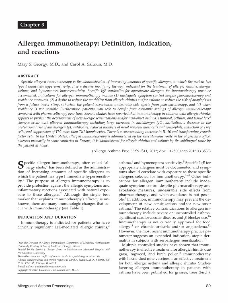
Specific allergen immunotherapy is the administration of increasing amounts of specific allergens to which the patient hastype I immediate hypersensitivity. It is a disease modifying therapy, indicated for the treatment of allergic rhinitis, allergicasthma, and hymenoptera hypersensitivity. Specific IgE antibodies for appropriate allergens for immunotherapy must bedocumented. Indications for allergen immunotherapy include (1) inadequate symptom control despite pharmacotherapy andavoidance measures, (2) a desire to reduce the morbidity from allergic rhinitis and/or asthma or reduce the risk of anaphylaxisfrom a future insect sting, (3) when the patient experiences undesirable side effects from pharmacotherapy, and (4) whenavoidance is not possible. Furthermore, patients may seek to benefit from economic savings of allergen immunotherapycompared with pharmacotherapy over time. Several studies have reported that immunotherapy in children with allergic rhinitisappears to prevent the development of new allergic sensitizations and/or new-onset asthma. Humoral, cellular, and tissue levelchanges occur with allergen immunotherapy including large increases in antiallergen IgG4 antibodies, a decrease in thepostseasonal rise of antiallergen IgE antibodies, reduced numbers of nasal mucosal mast cells and eosinophils, induction of Tregcells, and suppression of Th2 more than Th1 lymphocytes. There is a corresponding increase in IL-10 and transforming growthfactor beta. In the United States, allergen immunotherapy is administered by the subcutaneous route in the physician’s office,whereas primarily in some countries in Europe, it is administered for allergic rhinitis and asthma by the sublingual route bythe patient at home.
(Allergy Asthma Proc 33:S9–S11, 2012; doi: 10.2500/aap.2012.33.3533)
Specific allergen immunotherapy, often called “al-lergy shots,” has been defined as the administra-
tion of increasing amounts of specific allergens towhich the patient has type I immediate hypersensitiv-ity.1 The purpose of allergen immunotherapy is toprovide protection against the allergic symptoms andinflammatory reactions associated with natural expo-sure to these allergens.2 Although the single bestmarker that explains immunotherapy’s efficacy is un-known, there are many immunologic changes that oc-cur with immunotherapy (see Table 1).
INDICATION AND DURATIONImmunotherapy is indicated for patients who have
clinically significant IgE-mediated allergic rhinitis,3
asthma,4 and hymenoptera sensitivity.5 Specific IgE forappropriate allergens must be documented and symp-toms should correlate with exposure to those specificallergens selected for immunotherapy.6–8 Other indi-cations for allergen immunotherapy include inade-quate symptom control despite pharmacotherapy andavoidance measures, undesirable side effects frompharmacotherapy, and when avoidance is not possi-ble.8 In addition, immunotherapy may prevent the de-velopment of new sensitizations and/or new-onsetasthma.9 The relative contraindications to allergen im-munotherapy include severe or uncontrolled asthma,significant cardiovascular disease, and �-blocker use.10
Immunotherapy is not currently approved for foodallergy11 or chronic urticaria and/or angioedema.12
However, the most recent immunotherapy practice pa-rameter suggests an expanded indication, atopic der-matitis in subjects with aeroallergen sensitization.13
Multiple controlled studies have shown that immu-notherapy is effective treatment for allergic rhinitis duegrass, ragweed, and birch pollen.8 Immunotherapywith house-dust mite vaccines is an effective treatmentfor both allergic asthma and allergic rhinitis. Studiesfavoring allergen immunotherapy in patients withasthma have been published for grasses, trees (birch),
From the Division of Allergy–Immunology, Department of Medicine, NorthwesternUniversity Feinberg School of Medicine, Chicago, IllinoisFunded by the Ernest S. Bazley Grant to Northwestern Memorial Hospital andNorthwestern UniversityThe authors have no conflicts of interest to declare pertaining to this articleAddress correspondence and reprint requests to Carol A. Saltoun, M.D., # 14018, 676N. St. Clair St., Chicago, IL 60611E-mail address: [email protected] © 2012, OceanSide Publications, Inc., U.S.A.
Allergy and Asthma Proceedings S9
ragweed, cat, and fungi (Alternaria and Cladosporium).14
Patients with mild asthma are more likely to benefitfrom immunotherapy than patients with moderateor severe asthma who may be at increased risk foradverse reactions to immunotherapy. To appropri-ately choose allergens for cutaneous testing and im-munotherapy, it is important to be familiar with thesignificant aeroallergens in a patient’s geographiclocation.
In the United States, immunotherapy with inhalantallergens is initiated with once or twice weekly subcu-taneous injections (subcutaneous immunotherapy[SCIT]) of aqueous allergen vaccines at very low dosesthat are very unlikely to cause anaphylaxis.15 Doses arethen increased over several months until an efficaciousdose is reached. This dose is called the maintenancedose. The injection interval is then increased to bi-monthly and then monthly intervals. Approximately90% of appropriately selected patients receiving opti-mal dose maintenance immunotherapy for a year willnotice 50–75% improvement.14 In a controlled study inwhich immunotherapy for grass pollen allergy was discon-tinued after 3–4 years of successful treatment, seasonalsymptom scores and the use of rescue medication remainedlow for 3–4 years after the discontinuation of immunother-apy. Approximately 90% of patients who complete 4–5years of successful immunotherapy will maintain their im-provement.14
REACTIONS TO IMMUNOTHERAPYPostinjection reactions of erythema and induration of
�20 mm and lasting �2 days are known as local reac-tions, are common, and are of no consequence. Largerlocal reactions are best treated with antihistamines andice. Currently, there are no indisputable data to indi-cate that large local reactions predict subsequent sys-
temic reactions.10 Alternatively, it may be advisable torepeat the dosage for the next injection as patientsexpress concern about experiencing repeated large lo-cal reactions. Anaphylaxis manifested by urticaria, an-gioedema, injected conjunctiva, laryngeal edema, vom-iting, bronchospasm, hypotension, shock, and evendeath may occur after an injection of an allergen vac-cine.15 Most systemic reactions occur within 30 min-utes after an injection and, therefore, patients shouldremain in the physician’s office at least 30 minutes afteran injection.10 Administering an incorrect dose canresult in severe or fatal systemic reactions; the patient,vial, dilution, and immunotherapy schedule must becarefully identified before administration of an injec-tion of inhalant allergen. The condition and stability ofasthma should also be evaluated before administeringeach dose of immunotherapy.
SCIT IN PREGNANCYAllergen immunotherapy is effective in the pregnant
patient and may be continued during pregnancy ifmaintenance doses are well tolerated. According to theallergen immunotherapy practice parameters of 2011,the last highest achieved dose is temporarily used as amaintenance dose until delivery.13 However, if the pa-tient is not experiencing allergic reactions from immu-notherapy, the dosage can be increased as if she werenot pregnant.
SUBLINGUAL IMMUNOTHERAPYSublingual immunotherapy (SLIT) is commonly
used in Europe but not currently Food and Drug Ad-ministration approved in the United States. SLIT isadministered either as a rapidly dissolving tablet con-taining allergen extracts or in liquid form administeredwith a dropper; dosages studied are 20–400 times thetotal dose used in a course of SCIT.16 The regimentypically starts with a rapid build-up phase and thenthe treatment is taken either daily or three times perweek.16 The exact mechanism of action of SLIT remainsto be elucidated, but SLIT may reduce symptoms andrescue medication use by 30–40%.16 However, SLIT is�2⁄3 as effective as SCIT.16 The most common adverseevent reported is local itching and swelling and sys-temic side effects are relatively rare.9,16
CLINICAL PEARLS
• Specific allergen immunotherapy is indicated for thetreatment of allergic rhinitis, allergic asthma, andhymenoptera hypersensitivity. Allergen immuno-therapy is currently not indicated for the treatmentof eczema, food allergy, or chronic urticaria.
• Systemic reactions to immunotherapy usually occurwithin 30 minutes of treatment. The faster the onset
Table 1 Immunologic changes withimmunotherapy
Antibody changesIncrease in allergen-specific IgG (specifically IgG4)Early increase and late decrease in serum-specific
IgEDecrease in seasonal rise of specific IgE
Cellular changesDecreased mediator release from mast cells,
basophils, and eosinophilsReduction of tissue mast cells and eosinophilsInduction of regulatory T cells and suppression of
Th2 � Th1 cellsIncreased secretion of IL-10 and TGF-�Decrease in histamine-releasing factors
Source: Adapted from Refs. 8 and 17.TGF-� � transforming growth �.
S10 May–June 2012, Vol. 33, No. 3

of symptoms of a systemic reaction, the more severethe reaction is likely to be.
• SCIT is contraindicated in severe asthma.• SLIT, although not currently approved for use in the
United States, has been shown to be more effectivethan placebo and safer than SCIT.
REFERENCES1. Uzzaman A, and Cho SH. Classification of hypersensitivity
reactions. Allergy Asthma Proc 33:S96–S99, 2012.2. Shah R, and Grammer LC. An overview of allergens. Allergy
Asthma Proc 33:S2–S5, 2012.3. Uzzaman A, and Story R. Allergic rhinitis. Allergy Asthma Proc
33:S15–S18, 2012.4. Koterba AP, and Saltoun CA. Asthma classification. Allergy
Asthma Proc 33:S28–S31, 2012.5. Koterba AP, and Greenberger PA. Stinging insect allergy and
venom immunotherapy. Allergy Asthma Proc 33:S12–S14, 2012.6. Carr TF, and Saltoun CA. Skin testing in allergy. Allergy
Asthma Proc 33:S6–S8, 2012.7. Makhija M, and O’Gorman MRG. Common in vitro tests for
allergy and immunology. Allergy Asthma Proc 33:S108–S111,2012.
8. Grammer LC, and Harris KE. Principles of immunologic man-agement of allergic diseases due to extrinsic antigens. In Pat-terson’s Allergic Diseases, 7th ed. Grammer LC, and Green-
berger PA (Eds). Philadelphia, PA: Lippincott, Williams &Wilkins, 187–196, 2009.
9. Nelson HS. Immunotherapy for inhalant allergens. In Middleton’sAllergy Principles & Practice, 7th ed. Adkinson NF Jr, Bochner BS,Busse WW, et al. (Eds). Philadelphia, PA: Mosby, 1657–1677, 2009.
10. Joint Task Force on Practice Parameters. American Academy ofAllergy, Asthma and Immunology; Joint Council of Allergy,Asthma and Immunology. Allergen immunotherapy: A practiceparameter second update. J Allergy Clin Immunol 120(suppl):S25–S85, 2007.
11. Robison RG, and Pongracic JA. Food allergy. Allergy AsthmaProc 33:S77–S79, 2012.
12. Carr TF, and Saltoun CA. Urticaria and angioedema. AllergyAsthma Proc 33:S70–S72, 2012.
13. Cox L, Nelson H, Lockey R, et al. Allergen immunotherapy: Apractice parameter third update. J Allergy Clin Immunol127(suppl):S1–S55, 2011.
14. Radtke M, and Grammer LC. Subcutaneous administration ofallergen vaccines. In Allergens and Allergen Immunotherapy,4th ed., revised and expanded. Lockey RF, and Ledford DK(Eds). New York, NY: Marcel Dekker, Inc., 321–332, 2008.
15. Greenberger PA, and Ditto AM. Anaphylaxis. Allergy AsthmaProc 33:S80–S83, 2012.
16. Frew A. Allergen Immunotherapy. J Allergy Clin Immunol125:S306–S313, 2010.
17. Akdis CA, and Akdis M. Mechanisms and treatment of allergicdisease in the big picture of regulatory T cells. J Allergy ClinImmunol 123:735–746, 2009. e
Allergy and Asthma Proceedings S11

Chapter 4
Stinging insect allergy and venom immunotherapy
Alan P. Koterba, M.D., and Paul A. Greenberger, M.D.
ABSTRACT
The Hymenoptera order is divided into three families: Apids, Vespidae, and Formicidae. Apids include the honeybee,bumblebee, and sweat bee, which are all docile and tend to sting mostly on provocation. The Africanized killer bee, a productof interbreeding between the domestic and African honeybee, is very aggressive and is found mostly in Mexico, CentralAmerica, Arizona, and California. The yellow jacket, yellow hornet, white (bald)-faced hornet, and paper wasp all belong to theVespidae family. The Formicidae family includes the harvester ant and the fire ant. When a “bee” sting results in a large localreaction, defined as �5 in. and lasting �24 hours, the likelihood of anaphylaxis from a future sting is �5%. For comparison,when there is a history of anaphylaxis from a previous Hymenoptera sting and the patient has positive skin tests to venom, atleast 60% of adults and 20–32% of children will develop anaphylaxis with a future sting. Both patient groups should beinstructed about avoidance measures and carrying and knowing when to self-inject epinephrine, but immunotherapy (IT) withHymenoptera venom is indicated for those patients with a history of anaphylaxis from the index sting and not for patients whohave experienced a large local reaction. IT is highly effective in that by 4 years of injections, the incidence of subsequentsting-induced anaphylactic reactions is 3%. This incidence may increase modestly after discontinuation of injections but hasnot been reported �10% in follow-up.
(Allergy Asthma Proc 33:S12–S14, 2012; doi: 10.2500/aap.2012.33.3534)
An anaphylactic (systemic) reaction to a stinginginsect occurs in 0.3–3.0% of the general popula-
tion. Most reactions occur in those subjects who are�20 years of age, but fatalities tend to occur inadults.1,2 Hymenoptera anaphylaxis accounts for atleast 40–50 deaths/year in the United States.1 The mostcommon culprit is the yellow jacket.
CLASSIFICATIONThere are nine flying insects in the Hymenoptera
order that are known to cause anaphylactic reactions.They are divided into three families: Apidae, Vespidae,and Formicidae. The Apids include the honeybee,bumblebee, and sweat bee, which are all docile andtend to sting mostly on provocation. The Africanizedkiller bee (found mostly in Mexico, Central America,Arizona, and California) is a product of interbreedingbetween the domestic and African honeybee and isvery aggressive. The yellow jacket, yellow hornet,
white (bald)-faced hornet, and paper wasp all belongto the Vespidae family. Yellow jacket nests are locatedin the ground or in rock gardens and yellow jacketstend to be attracted to garbage, open soda cans, punchbowls, and picnic tables. The hornets are commonlyfound in shrubs, whereas paper wasps nest in theeaves of homes or inside of walls. The Formicidaefamily includes the harvester ant and the fire ant. Theyare found in the southeast/southwestern United Statesand are capable of causing systemic allergic reactions.The sting causes a painful, erythematous reaction fol-lowed by a wheal. Within 4 hours, a clear pustuleforms. The fluid can become cloudy and the pustulecan last some 3–10 days. The pustule is often mistakenfor cellulitis although secondary infections may occur.Immunotherapy (IT) is indicated in patients who areskin test positive to fire ant whole-body extract.
TYPES OF REACTIONSThere are five types of sting reactions: normal, local,
rare, toxic, and anaphylactic. A normal reaction is char-acterized by mild erythema (�2 in.), swelling, andpain. It is transient in nature and limited to the area ofthe sting. Treatment consists of cold compresses andanalgesics. A large local reaction (�5 in.) and lasting�24 hours has as high as a 7–17% incidence in thegeneral population. Local reactions are characterizedby extensive erythema and swelling and can last be-
From the Division of Allergy–Immunology, Department of Medicine, NorthwesternUniversity Feinberg School of Medicine, Chicago, IllinoisFunded by the Ernest S. Bazley Grant to Northwestern Memorial Hospital andNorthwestern UniversityThe authors have no conflicts of interest to declare pertaining to this articleAddress correspondence and reprint requests to Paul A. Greenberger, M.D., No.14018, 676 North St. Clair Street, Chicago, IL 60611E-mail address: [email protected] © 2012, OceanSide Publications, Inc., U.S.A.
S12 May–June 2012, Vol. 33, No. 3

tween 1 and 10 days.3 It is important to distinguishlarge local reactions from an anaphylactic reaction. Alarge local reaction is contiguous along a joint line (i.e.,entire arm swelling) whereas an anaphylactic reactionskips contiguous joint lines (i.e., sting on the hand andhive on the lip).4 The treatment for large local reactionsis analgesics, ice, and, rarely, prednisone (not evidencebased). Individuals who develop large local reactionstend to have a similar reaction on a subsequent sting.The risk of anaphylaxis is low in these individuals(�5%) and therefore does not require IT.2 Rare reac-tions include serum sickness for which IT may beindicated. In serum sickness, there is urticaria, arthral-gias, malaise, and fever about 7 days after an insectsting.5 There is a risk of anaphylaxis with a repeat stingin these individuals so IT should be considered whenskin testing is positive. Neurological, nephritic, vascu-litic, and encephalitic-like reactions are other rare re-actions that have been reported to occur up to 2 weeksafter the implicated sting. Toxic reactions occur aftermultiple simultaneous stings resulting in hypotension,cardiovascular collapse, and possibly death. IT is indi-cated for toxic reactions when patients have positiveskin test results.
TESTINGThe immediate-onset (anaphylactic) reactions are
mediated by IgE antibodies to particular venom. Thisresults in mast cell activation leading to mediator re-lease with cutaneous and systemic signs/symptoms ofanaphylaxis. Sixty percent of adult subjects and 20–32% of children with a history of anaphylaxis from aprevious sting will have anaphylaxis with a repeatsting.3 Moreover, IT for these individuals provides pro-tection from anaphylaxis in 97% of re-stings.2 Thus, ITis indicated for patients with a history of anaphylaxisand positive skin tests (showing venom-specific IgEantibody). Testing should be done at least 3 weeks afterthe suspected anaphylactic event because it may take2–3 weeks for venom-specific IgE to become detectable.Some patients have a convincing history of anaphy-laxis but negative skin testing. This may represent anonimmunologic reaction or perhaps a loss of reactiv-ity when there is a remote history of a sting reaction.2
If the initial skin is negative then repeat skin testing isadvisable after 3–6 months.2,6 Some have argued thatin vitro testing7 is advised in this setting, but the ad-ministration of IT should be with an extract that causesa positive skin test. Children �16 years of age withhistory of a cutaneous systemic reaction (hives/angio-edema8) do not necessarily require venom IT (VIT)because it is less likely they will experience severeanaphylactic shock with future stings.1 In particularcases, it may be appropriate to treat a child who hasexperienced acute urticaria and angioedema who has
positive skin tests. Baseline tryptase levels have beenfound to be elevated (�11 ng/mL) in a subset of pa-tients with known venom hypersensitivity. Interest-ingly, some of these patients were found to have occultindolent systemic mastocytosis or monoclonal mastcell activation syndrome.9 In addition, venom-associ-ated anaphylaxis may be the presenting finding ofindolent systemic mastocytosis or the monoclonal mastcell activation syndrome.
VIT DOSING AND SCHEDULESSkin testing is administered using five Hymenoptera
venom protein extracts—honeybee, yellow jacket, yel-low hornet, white-faced hornet, and wasp (Table 1).Typically, testing starts with a prick test at 0.01 �g/mLif the reaction was very severe with subsequent intra-dermal testing at 0.0001, 0.001, 0.1, and 1.0 �g/mL. Forfire ants, whole-body extracts are used for skin testing.In most cases, IT is administered with all of the venomsthat tested positive. Therapy for VIT begins at 0.05 �gwith incremental doses every week until a mainte-nance dosage of 100 �g (300 �g if mixed vespid isachieved). Once the patient has reached maintenance,they can gradually convert to monthly injections intheir 1st year and then perhaps every 6–8 weeks dur-ing the subsequent 3 years or until the skin test be-comes negative. VIT decreases risk of re-sting reactionin adults from 60 to 3%.2 A patient having undergoneor still undergoing VIT is still at risk of developing ananaphylactic (systemic) reaction if stung, so theyshould always carry an epinephrine autoinjector. Therate of systemic reactions to VIT is 3–12% per course ofIT, which is similar to aeroallergen IT.3,10,11
Table 1 Indications for VIT in patients withpositive skin tests
Reaction VIT
“Normal” transient pain, swelling NoExtensive local swelling (large local) NoMild anaphylaxis
generalized urticariaif �16 yr old No*if �16 yr old Yes
Moderate/severe anaphylaxis YesSerum sickness YesToxic reaction YesIndolent systemic mastocytosis Yes#Mast cell activation syndrome Yes#
*Controversial.#In some patients, an argument can be made even to treatpatients with negative skin tests to venom with IT.VIT � venom immunotherapy.
Allergy and Asthma Proceedings S13

TREATMENTAvoidance is a major therapeutic intervention in pre-
venting death from these insects. Measures includewearing long-sleeved light-colored clothing, shoes andhats, being cautious in picnic areas, covering food,avoiding drinking from open beverage cans, and notwearing perfume or cologne. Fire ant mounds are or-ange in color and can be found underground for up to80 ft. Thus, there is a case for wearing shoes or hardsandals and avoiding areas where there are fire antmounds. Along with teaching self-administration ofthe epinephrine autoinjector, it is important to empha-size the need to carry an epinephrine autoinjector at alltimes regardless of if the patient has received VIT ornot.12–14 It is also advisable to let the patient know thatanytime they do use an epinephrine autoinjector, theyshould go to the hospital to get further evaluation.Acute medical therapy for systemic reactions includesthe normal treatment for anaphylaxis including epi-nephrine, H1-receptor antagonists, corticosteroids, andsupportive therapy for shock. Discontinuation of VITusually occurs after 4–5 years despite a persistentlypositive skin test or when skin tests become negativeafter 4 years of injections.2 The most recent practiceparameter states that VIT should be continued for atleast 3–5 years.15 This is based on evidence that despitethe persistence of a positive skin test response, 80–90%of patients will not have a systemic reaction to an insectsting if VIT is stopped after 3–5 years; however, onemight consider indefinite IT in the patient with severeanaphylaxis.
IMMUNOLOGY
• IgG antibodies are capable of blocking in vitro ven-om-induced histamine release from basophils of al-lergic patients.
• Passively administered immunoglobulin from bee-keepers provides temporary immunity from venomanaphylaxis in sensitive patients.
• The concentration of IgG produced is proportionalto amount of exposure to venom.
CLINICAL PEARLS
• Appropriately treat anaphylactic reactions to venomstings as indicated such as with epinephrine.
• Teach patients avoidance measures such as wearinglight-colored clothing, no perfumes, and not walk-ing barefoot in the grass.
• Classification of an allergic reaction helps determinewhen IT is necessary.
• The “number needed to treat”16 for an adult patientreceiving IT for a history of anaphylaxis to stinginginsects and positive skin tests is 2 (1/0.60–0.03) or1/0.57 derived from 1/risk of anaphylaxis in un-treated group-risk of anaphylaxis in treated group.This is a remarkably low number. For childrenwhere the risk of a repeat anaphylactic reaction afterIT is �20%, the “number needed to treat” is 1/0.20–0.03 or 6, also a very low number.
REFERENCES1. Levine MI, and Lockey RF (eds.). Monograph on Insect Allergy.
Milwaukee, WI: American Academy of Allergy, Asthma andImmunology, 1–280, 2003.
2. Reisman RE. Allergy to stinging insects. In Patterson’s AllergicDiseases, 7th ed. Grammer LC, and Greenberger PA (Eds).Philadelphia, PA: Lippincott, Williams & Wilkins, 220–231,2009.
3. Gupta P, and Grammer LC. Administration of allergen vac-cines. In Allergens and Allergen Immunotherapy, 3rd ed., re-vised and expanded. Lockey RF, Bukantz SC, and Bousquet J(Eds). New York, NY: Marcel Dekker, Inc., 481–493, 2004.
4. Greenberger PA, and Ditto AM. Anaphylaxis. Allergy AsthmaProc 33:S80–S83, 2012.
5. Uzzaman A, and Cho SH. Classification of hypersensitivityreactions. Allergy Asthma Proc 33:S96–S99, 2012.
6. Carr TF, and Saltoun CA. Skin testing in allergy. AllergyAsthma Proc 33:S6–S8, 2012.
7. Makhija M, and O’Gorman MRG. Common in vitro tests forallergy and immunology. Allergy Asthma Proc 33:S108–S111,2012.
8. Carr TF, and Saltoun CA. Urticaria and angioedema. AllergyAsthma Proc 33:S70–S72, 2012.
9. Bonadonna P, Perbellini O, Passalacqua G, et al. Clonal mastcell disorders in patients with systemic reactions to Hymenop-tera stings and increased serum tryptase levels. J Allergy ClinImmunol 123:680–686, 2009.
10. Georgy MS, and Saltoun CA. Allergen immunotherapy: Defi-nition, indication, and reactions. Allergy Asthma Proc 33:S9–S11, 2012.
11. Cox L, Nelson H, Lockey R, et al. Allergen immunotherapy: Apractice parameter third update. J Allergy Clin Immunol127(suppl):S1–55, 2011.
12. DeMuth KA, and Fitzpatrick AM. Epinephrine autoinjectoravailability among children with food allergy. Allergy AsthmaProc 32:295–300, 2011.
13. Banerji A, Rudders SA, Corel B, et al. Repeat epinephrine treat-ments for food-related allergic reactions that present to theemergency department. Allergy Asthma Proc 31:308–316, 2010.
14. Amirzadeh A, Verma P, Lee S, and Klaustermeyer W. Epineph-rine auto-injector use and demographics in a Veterans Admin-istration population. Allergy Asthma Proc 31:304–307, 2010.
15. Golden DB, Moffitt J, Nicklas RA, et al. Stinging insect hyper-sensitivity: A practice parameter update 2011. J Allergy ClinImmunol 127:852–854.e1–23, 2011.
16. McAlister FA, Straus SE, Guyatt GH, and Haynes RB. Users’guides to the medical literature: XX. Integrating research evi-dence with the care of the individual patient. Evidence-BasedMedicine Working Group. JAMA 283:2829–2836, 2000. e
S14 May–June 2012, Vol. 33, No. 3
Chapter 5
Allergic rhinitis
Ashraf Uzzaman, M.D., and Rachel Story, M.D.
ABSTRACT
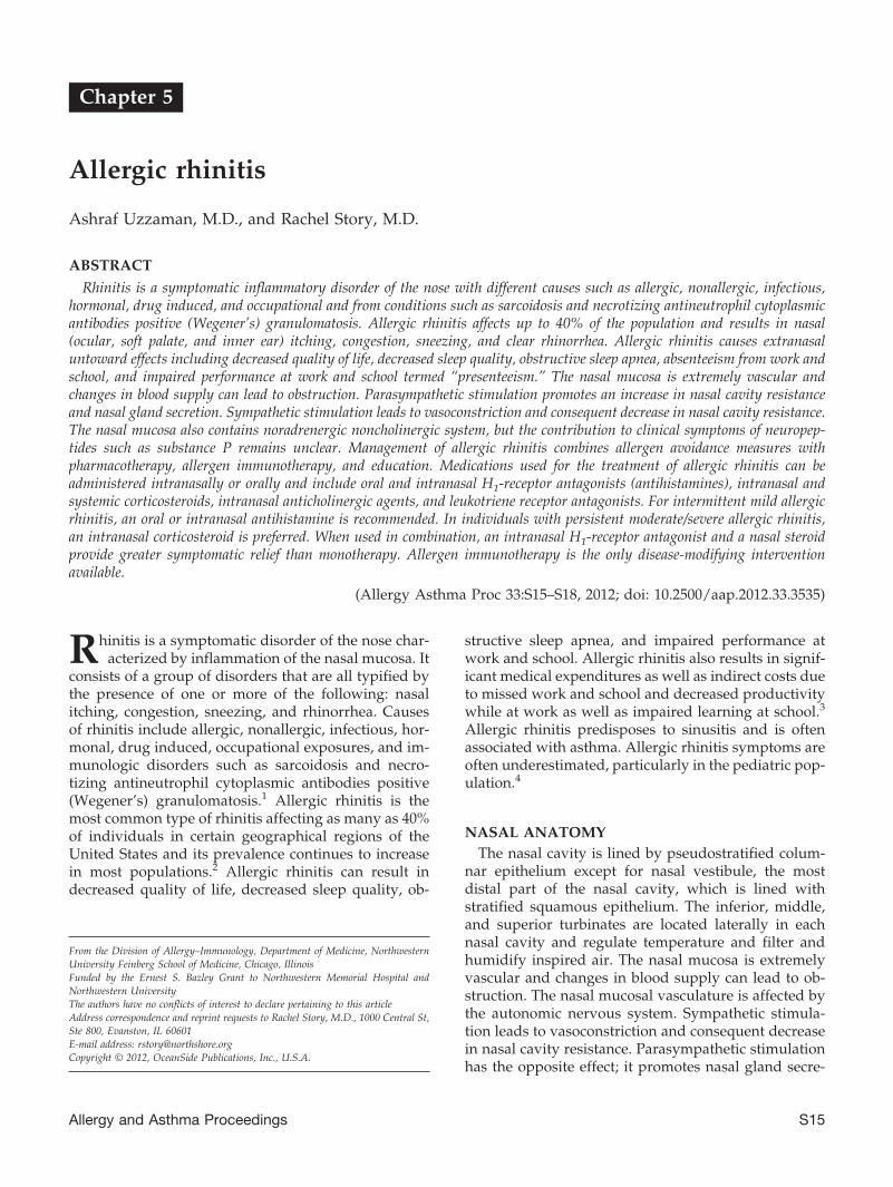
Rhinitis is a symptomatic inflammatory disorder of the nose with different causes such as allergic, nonallergic, infectious,hormonal, drug induced, and occupational and from conditions such as sarcoidosis and necrotizing antineutrophil cytoplasmicantibodies positive (Wegener’s) granulomatosis. Allergic rhinitis affects up to 40% of the population and results in nasal(ocular, soft palate, and inner ear) itching, congestion, sneezing, and clear rhinorrhea. Allergic rhinitis causes extranasaluntoward effects including decreased quality of life, decreased sleep quality, obstructive sleep apnea, absenteeism from work andschool, and impaired performance at work and school termed “presenteeism.” The nasal mucosa is extremely vascular andchanges in blood supply can lead to obstruction. Parasympathetic stimulation promotes an increase in nasal cavity resistanceand nasal gland secretion. Sympathetic stimulation leads to vasoconstriction and consequent decrease in nasal cavity resistance.The nasal mucosa also contains noradrenergic noncholinergic system, but the contribution to clinical symptoms of neuropep-tides such as substance P remains unclear. Management of allergic rhinitis combines allergen avoidance measures withpharmacotherapy, allergen immunotherapy, and education. Medications used for the treatment of allergic rhinitis can beadministered intranasally or orally and include oral and intranasal H1-receptor antagonists (antihistamines), intranasal andsystemic corticosteroids, intranasal anticholinergic agents, and leukotriene receptor antagonists. For intermittent mild allergicrhinitis, an oral or intranasal antihistamine is recommended. In individuals with persistent moderate/severe allergic rhinitis,an intranasal corticosteroid is preferred. When used in combination, an intranasal H1-receptor antagonist and a nasal steroidprovide greater symptomatic relief than monotherapy. Allergen immunotherapy is the only disease-modifying interventionavailable.
(Allergy Asthma Proc 33:S15–S18, 2012; doi: 10.2500/aap.2012.33.3535)
Rhinitis is a symptomatic disorder of the nose char-acterized by inflammation of the nasal mucosa. It
consists of a group of disorders that are all typified bythe presence of one or more of the following: nasalitching, congestion, sneezing, and rhinorrhea. Causesof rhinitis include allergic, nonallergic, infectious, hor-monal, drug induced, occupational exposures, and im-munologic disorders such as sarcoidosis and necro-tizing antineutrophil cytoplasmic antibodies positive(Wegener’s) granulomatosis.1 Allergic rhinitis is themost common type of rhinitis affecting as many as 40%of individuals in certain geographical regions of theUnited States and its prevalence continues to increasein most populations.2 Allergic rhinitis can result indecreased quality of life, decreased sleep quality, ob-
structive sleep apnea, and impaired performance atwork and school. Allergic rhinitis also results in signif-icant medical expenditures as well as indirect costs dueto missed work and school and decreased productivitywhile at work as well as impaired learning at school.3
Allergic rhinitis predisposes to sinusitis and is oftenassociated with asthma. Allergic rhinitis symptoms areoften underestimated, particularly in the pediatric pop-ulation.4
NASAL ANATOMYThe nasal cavity is lined by pseudostratified colum-
nar epithelium except for nasal vestibule, the mostdistal part of the nasal cavity, which is lined withstratified squamous epithelium. The inferior, middle,and superior turbinates are located laterally in eachnasal cavity and regulate temperature and filter andhumidify inspired air. The nasal mucosa is extremelyvascular and changes in blood supply can lead to ob-struction. The nasal mucosal vasculature is affected bythe autonomic nervous system. Sympathetic stimula-tion leads to vasoconstriction and consequent decreasein nasal cavity resistance. Parasympathetic stimulationhas the opposite effect; it promotes nasal gland secre-
From the Division of Allergy–Immunology, Department of Medicine, NorthwesternUniversity Feinberg School of Medicine, Chicago, IllinoisFunded by the Ernest S. Bazley Grant to Northwestern Memorial Hospital andNorthwestern UniversityThe authors have no conflicts of interest to declare pertaining to this articleAddress correspondence and reprint requests to Rachel Story, M.D., 1000 Central St,Ste 800, Evanston, IL 60601E-mail address: [email protected] © 2012, OceanSide Publications, Inc., U.S.A.
Allergy and Asthma Proceedings S15

tion and an increase in nasal cavity resistance. Thenasal mucosa also contains the noradrenergic noncho-linergic system, but the contribution to clinical symp-toms of neuropeptides such as substance P remainsunclear.
PATHOPHYSIOLOGYAllergic rhinitis is an immunoglobulin E (IgE)–me-
diated (or a type I, immediate5) reaction to the proteinor glycoprotein component of inhaled aeroallergens6
including pollens, molds, animal danders, dust-mitefecal particles, and cockroach residues.7 In the occupa-tional setting, small molecular weight chemicals canact as haptens that associate with self-proteins to formcomplete allergens.8 On inhalation, the allergen depos-its in the nasal mucus. After deposition, antigen-pre-senting cells in the nasal epithelial mucosa phagocy-tose and process the allergen and subsequently presentthe processed antigen to CD4� T cells in local lymphnodes. The allergen-stimulated T cells proliferate in aTh2 pathway and release cytokines including IL-3,IL-4, IL-5, IL-13, and others. These cytokines lead tolocal and systemic production of IgE antibodies byplasma cells. These antibodies bind to mast cells andbasophils. This process is referred to as sensitization.On reexposure, the allergen is recognized by IgE anti-bodies, which are bound to mast cells and basophils.The recognition and subsequent binding leads to de-granulation of mast cells and basophils that releasepreformed mediators including histamine and en-zymes such as tryptase and chymase. There is alsorapid de novo synthesis of other mediators such ascysteinyl leukotrienes (leukotriene D4) and prostaglan-din D2 (PGD2). The mediators lead to vasodilation ofarteriolar venous anastomosis, plasma leakage fromblood vessels, increased secretion of mucous, and stim-ulation of afferent nerves with consequent occlusion ofthe nasal passages. Histamine produces pruritus, rhi-norrhea, and sneezing and leukotrienes and PGD2 areassociated with the development of nasal congestion.This comprises the early or immediate-phase response.Cytokines released during the immediate-phase re-sponse mediate a cascade of events over the next 4–8hours, referred to the late-phase response. Clinicalsymptoms in early and late response are similar, butnasal predominates during the late phase. Mediatorsreleased at the postcapillary endothelial cells, duringthe early phase response, promote the expression ofadhesion molecules that assist in migration of eosino-phils, neutrophils, and basophils and, eventually, mac-rophages and CD4� Th2 cells into the superficial lam-ina propria of the nasal cavity. These cells becomeactivated and produce more mediators that are similarto those involved in the early response phase except formast cell–derived tryptase, chymase, and PGD2.9
On repeated exposure to an allergen, the nasal mu-cosa becomes more sensitive and there is a progressivedecrease in the amount of allergen required to elicitsymptoms, a phenomenon referred to as priming. Ad-ditionally, the priming effect may lead to increasedsensitivity of the nasal mucosa to nonallergic triggerssuch as cigarette smoke and strong odors.
DIFFERENTIAL DIAGNOSISThe constellation of nasal symptoms in individuals
with allergic rhinitis may also be present in personswith rhinitis from other causes. The occurrence of as-sociated ocular symptoms—itching, redness, and tear-ing make allergy a more likely cause of rhinitis. Aller-gic rhinitis must be distinguished from other causes ofrhinitis that may present with similar symptoms. Va-somotor rhinitis or nonallergic rhinitis without eosin-ophilia primarily manifests as nasal congestion andrhinorrhea and less commonly with nasal itching andsneezing.10 Symptoms occur in response to nonallergictriggers such as changes in temperature or relativehumidity, strong odors, cigarette smoke, and alcoholingestion. Nonallergic rhinitis with eosinophilia syn-drome is characterized by perennial nasal symptomsand primarily manifests as nasal congestion and is lessfrequently associated with nasal itching and sneezing,rhinorrhea, and loss of smell. It is unusual in the pe-diatric population. The nasal smears show 5–20% eo-sinophils and the skin-prick test and specific IgE levelsto environmental allergens are negative. Nasal symp-toms may result from hormonal changes such as thosethat occur during puberty, pregnancy, and with thy-roid disorders. Symptoms associated with pregnancyusually occur in the second trimester and typicallyresolve within 2 weeks of delivery. Rhinitis can de-velop due to the administration of intranasal and oralmedications. Rebound nasal congestion often occursafter discontinuing an intranasal adrenergic deconges-tant spray used for �4–7 days. This is referred to asrhinitis medicamentosa. Oral antihypertensive agentssuch as angiotensin-converting enzyme inhibitors and�-blockers may cause nasal symptoms. Nonsteroidalanti-inflammatory drugs such as aspirin and ibuprofenalso cause rhinitis in some individuals. Repeated nasaladministration of cocaine or amphetamines can lead torebound nasal congestion. Ingestion of ethanol in alco-holic beverages causes vasodilatation of nasal bloodvessels resulting in nasal congestion. Gustatory rhinitisis characterized by rhinorrhea and is associated withingestion of hot and spicy food. Atrophic rhinitis ischaracterized by atrophy of the nasal mucosa, nasaldryness, and foul-smelling nasal crusts often associ-ated with a constant sense of malodor. It may be pri-mary, because of infection, or secondary, associatedwith nasal surgery, irradiation, or trauma. Acute viral
S16 May–June 2012, Vol. 33, No. 3

upper respiratory tract infections typically manifestwith nasal symptoms, although nasal pruritus is typi-cally absent and constitutional symptoms are oftenpresent.11
CLASSIFICATION AND DIAGNOSISThe Allergic Rhinitis and Its Impact on Asthma
(ARIA) guidelines classify allergic rhinitis as intermit-tent or persistent. If one or more of the symptoms ofrhinitis such as nasal itching, rhinorrhea, sneezing, andcongestion are present for �4 days of the week or for�4 consecutive weeks, the disease is classified as in-termittent. If symptoms occur for �4 days of the weekand are present for �4 weeks, the disease is classifiedas persistent. Intermittent and persistent rhinitis arefurther categorized into mild or moderate/severe dis-ease based on the severity of the symptoms and qual-ity-of-life outcomes. Rhinitis is categorized as mild ifthe individual does not have sleep disturbance, there isno impairment of daily activities and leisure and/orsports, there is no impairment of school or work, andsymptoms are not troublesome. The disease may beclassified as moderate/severe if one or more of theaforementioned are present.12 The ARIA classificationhas largely replaced classifying allergic rhinitis as sea-sonal for individuals who have symptoms only duringparticular pollen (tree, grass, and weed) or mold sea-sons, or perennial for those with year-round symptomstypically caused by pet dander, house-dust mites, cock-roaches, and mold in some climates.
On physical exam, the nasal mucosa is commonlypale and boggy, the turbinates are swollen, and clearnasal secretions are often present. The diagnosis isestablished by performing a thorough history andphysical exam and correlating the patient’s historywith skin-prick test or allergen-specific serum IgE an-tibody results.12
A number of measures may provide an objectivemeasure of the severity of rhinitis and have been usedclinically. These include visual analog scale and objec-tive measures of nasal obstruction such as acousticrhinometry and rhinomanometry.
TREATMENTManagement of allergic rhinitis combines allergen
avoidance measures with pharmacotherapy, allergen-specific immunotherapy, and education.
Many environmental allergens are known to be as-sociated with allergic rhinitis. House-dust mite is animportant cause of allergic rhinitis and avoidance mea-sures have been shown to improve symptoms. There issome benefit of using impermeable covers to encasemattresses, pillows, box springs, and beddings. Somereduction of symptoms has been reported with wash-ing linen in hot water (120°F) and replacing carpets
with hardwood floors. Patients allergic to animals withdander benefit from avoidance at home; however, theymay encounter these allergens in school, at work, andin public transportation. Regular washing of the petand use of high-efficiency particulate air filters inhouseholds with pets has been shown to be of somebenefit in reducing symptoms in sensitized individu-als, but removal of the pet from the home is moreeffective. Moisture control and repair of areas allowingwater intrusion in the household will help preventgrowth of indoor mold.
The second step in treatment of allergic rhinitis in-volves pharmacotherapy. Physicians must take into ac-count safety, efficacy, cost-effectiveness, patient prefer-ence, severity of symptoms, and compliance whenrecommending medications. Medications used for thetreatment of allergic rhinitis can be administered intrana-sally or orally and include oral and nasal H1-receptorantagonists, intranasal and systemic corticosteroids, in-tranasal anticholinergic agents, and leukotriene receptorantagonists. Oral and intranasal decongestants effectivelytreat nasal congestion, but intranasal decongestantsshould only be used short term (�5–7 days) becauserhinitis medicamentosa may occur. The choice of phar-macotherapy is usually dictated by the severity and chro-nicity of symptoms. For intermittent mild rhinitis, an oralor intranasal antihistamine is recommended. In patientswith intermittent moderate/severe symptoms or in thosewith persistent mild symptoms an oral or intranasal an-tihistamine or an intranasal steroid is recommended. Inindividuals with persistent moderate/severe disease anintranasal corticosteroid is preferred. An intranasal H1-receptor antagonist may be added to control symptoms.When used in combination, a topical antihistamine and anasal steroid provides greater symptomatic relief thanmonotherapy.13,14 Leukotriene receptor antagonist canalso be used and are particularly beneficial for individu-als with allergic rhinitis and asthma, which may have anallergic component. Symptom control should be re-viewed 2–4 weeks after initiating treatment and if symp-toms persist, medications should be adjusted. If rhinor-rhea is the troubling symptom, an anticholinergic agentmay be added; and if nasal congestion is the disturbingsymptom a short course of oral steroids may be used. Forocular symptoms, an intraocular H1-receptor antagonistsometimes in combination with a mast cell stabilizer maybe used.
Allergen-specific immunotherapy15 should be con-sidered if symptoms are not adequately controlled byavoidance and pharmacotherapy or in individuals whocan not tolerate pharmacotherapy. Specific allergen im-munotherapy has been shown to modify the naturalhistory of allergic diseases, may prevent future sensi-tization to new environmental allergens, and has beenshown to reduce asthma when initiated early.16
Allergy and Asthma Proceedings S17

IMMUNOLOGY
• Allergic rhinitis is a Th2-mediated immune re-sponse.
• Synthesis of allergen-specific IgE is dependent onactivation of CD4� helper T cells and their secretionof IL-4 and IL-13.
• Mast cells and basophils express a high-affinity re-ceptor for the Fc portion of the IgE antibody calledFc�RI.
• Mast cells are activated by cross-linking of Fc�RIreceptors on mast cells by binding of multivalentantigens.
CLINICAL PEARLS
• Commonly occurring symptoms of allergic rhinitis arenasal itching, congestion, sneezing, and rhinorrhea.
• Sympathetic stimulation leads to decrease in nasalcavity resistance; parasympathetic stimulation hasthe opposite effect.
• Symptoms consist of an immediate response and adelayed response; nasal congestion is the predomi-nant symptom in delayed phase response.
• The ARIA guidelines classify allergic rhinitis intointermittent and persistent.
• Management combines allergen avoidance mea-sures with pharmacotherapy, allergen-specific im-munotherapy, and patient education.
REFERENCES1. Wallace DV, Dykewicz MS, Bernstein DI, et al. The diagnosis
and management of rhinitis: An updated practice parameter. JAllergy Clin Immunol 122:S1–S84, 2008.
2. Bernstein JA. Allergic and mixed rhinitis: Epidemiology andnatural history. Allergy Asthma Proc 31:365–369, 2010.
3. Blaiss MS. Allergic rhinitis: Direct and indirect costs. AllergyAsthma Proc 31:375–380, 2010.
4. Dykewicz MS. Rhinitis and sinusitis. J Allergy Clin Immunol111:S520–S529, 2003.
5. Uzzaman A, and Cho SH. Classification of hypersensitivityreactions. Allergy Asthma Proc 33:S96–S99, 2012.
6. Shah R, and Grammer LC. An overview of allergens. AllergyAsthma Proc 33:S2–S5, 2012.
7. Broide DH. Allergic rhinitis: Pathophysiology. Allergy AsthmaProc 31:370–374, 2010.
8. Sabin BR, and Grammer LC. Occupational immunologic lungdisease. Allergy Asthma Proc 33:S58–S60, 2012.
9. Shearer WT, and Leung DY. 2010 Primer on allergic and im-munologic diseases. J Allergy Clin Immunol 125:S1–S394, 2010.
10. Shah R, and McGrath KG. Nonallergic rhinitis. Allergy AsthmaProc 33:S19–S21, 2012.
11. Howarth PH. Allergic and nonallergic rhinitis. In Middleton’sAllergy: Principles and Practice, 7th ed. Adkinson NF Jr, Boch-ner BS, Busse WW, et al. (Eds). Philadelphia, PA: Mosby, 973–990, 2009.
12. Ricketti AJ. Allergic rhinitis. In Patterson’s Allergic Diseases:Diagnosis and Management, 7th ed. Adkinson NF Jr, BochnerBS, Busse WW, et al. (Eds). Philadelphia, PA: Lippincott, Wil-liams, and Wilkins, 466–484, 2009.
13. LaForce CF, Carr W, Tilles SA, et al. Evaluation of olopatadinehydrochloride nasal spray, 0.6%, used in combination with anintranasal corticosteroid in seasonal allergic rhinitis. AllergyAsthma Proc 31:132–140, 2010.
14. Kaliner MA. A novel and effective approach to treating rhinitiswith nasal antihistamines. Ann Allergy Asthma Immunol 99:383–390, 2007.
15. Georgy MS, and Saltoun CA. Allergen immunotherapy: Defi-nition, indication, and reactions. Allergy Asthma Proc 33:S9–S11, 2012.
16. Bousquet J, Khaltaev N, Cruz AA, et al. Allergic Rhinitis and ItsImpact on Asthma (ARIA) 2008 update. Allergy 86:S8–S160,2008. e
S18 May–June 2012, Vol. 33, No. 3

Chapter 6
Nonallergic rhinitis
Rachna Shah, M.D., and Kris G. McGrath, M.D.
ABSTRACT
Nonallergic rhinitis represents a non–IgE-mediated group of disorders that share the symptoms of nasal congestion, rhinorrhea, sneezing,and/or postnasal discharge but not pruritus that characterizes allergic rhinitis. Nonallergic rhinitis may be divided into two broad categories,inflammatory and noninflammatory etiologies. The inflammatory causes include postinfectious (viral and bacterial), rhinitis associated withnasal polyps, and nonallergic rhinitis with eosinophilia, where eosinophils are present in nasal smears but skin testing for aeroallergens isnegative. The noninflammatory causes include idiopathic nonallergic rhinitis (formerly referred to as vasomotor rhinitis or colloquially asan “overreactive nose”); rhinitis medicamentosa, which is medication-induced rhinitis; hormone related (pregnancy); systemic disease related(severe hypothyroidism); and structural defect related (deviated septum, head trauma causing cerebrospinal fluid rhinorrhea). The classicsymptoms of idiopathic nonallergic rhinitis are nasal congestion, postnasal drip, and sneezing triggered by irritant odors, perfumes, wine,and weather changes. The diagnosis of rhinitis begins with a directed history and physical exam. Examination of the nasal cavity withattention to appearance of the septum and inferior turbinates is recommended. Skin testing for seasonal and perennial aeroallergens is helpfulin establishing the presence or absence of IgE antibodies and to help differentiate nonallergic from allergic rhinitis. Topical H1-receptorantagonist (antihistamine) nasal sprays, intranasal steroids, intranasal anticholinergics, and oral decongestants are options for pharmaco-therapy. It is important to inquire about hypertension, arrhythmias, insomnia, prostate hypertrophy, or glaucoma to prevent undesirable sideeffects associated with the oral decongestant pseudoephedrine.
(Allergy Asthma Proc 33:S19–S21, 2012; doi: 10.2500/aap.2012.33.3536)
Nonallergic rhinitis represents a non–IgE-mediatedgroup of disorders that share the symptoms of
nasal congestion, rhinorrhea, sneezing, and/or postna-sal discharge. Symptoms are almost identical to aller-gic rhinitis, except there is absence of skin test reactiv-ity to common aeroallergens.1 An estimated 19 millionpersons in the United States suffer from pure nonaller-gic rhinitis.2 Women are more likely than men to havenonallergic rhinitis, and age of onset after age 40 yearsis more likely associated with nonallergic rhinitis.3
The pathogenesis of nonallergic rhinitis is incom-pletely understood. One theory is based on the conceptof entopy, which is localized allergy in the nose with-out positive skin prick or serum IgE specific to theallergen.4 Other studies have looked at nociceptivenerve dysfunction in the nose resulting in nonallergicrhinitis.4 Nasal mucosa of patients with nonallergicrhinitis showed an equal number of CD4� as those
found in patients with allergic rhinitis and nasal lavageshowed increase eosinophils in patients with nonaller-gic rhinitis as compared to controls.4
Nonallergic rhinitis may be divided into two broadcategories, inflammatory and noninflammatory etiolo-gies.5 Table 1 represents a differential diagnosis basedon this classification.
Vasomotor rhinitis, also referred to as idiopathic,nonallergic noninfectious rhinitis, is the most com-mon of these disorders and manifests with symptomof perennial nasal congestion, rhinorrhea, and post-nasal drip. Unlike allergic rhinitis, nasal pruritus israre. Classic “nonallergic” triggers include irritantssuch as smoke, perfumes, chemicals, weatherchange, and cold air.6 Subclassifications of vasomo-tor rhinitis include gustatory rhinitis (rhinitis pro-voked by eating, especially hot or spicy foods) andexercise induced.
In nonallergic rhinitis with eosinophilia syndrome(NARES), eosinophils are found in nasal cytology;however, skin testing for aeroallergens is negative.3
Some 5–20% eosinophils are observed on nasal smear.3
The etiology for this condition is unknown. NARESusually manifests in middle-aged adults and is veryuncommon in children. Patients may complain ofsneezing paroxysms, copious amounts of clear rhinor-rhea, nasal pruritus, and anosmia.2,7
From the Division of Allergy–Immunology, Department of Medicine, NorthwesternUniversity Feinberg School of Medicine, Chicago, IllinoisFunded by the Ernest S. Bazley Grant to Northwestern Memorial Hospital andNorthwestern UniversityThe authors have no conflicts of interest to declare pertaining to this articleAddress correspondence and reprint requests to Kris G. McGrath, M.D., Number14018, 676 North St. Clair Street, Chicago, IL 60611E-mail address: [email protected] © 2012, OceanSide Publications, Inc., U.S.A.
Allergy and Asthma Proceedings S19

Primary atrophic rhinitis occurs in young to middle-aged adults who present with nasal congestion andfoul smelling, thick dry crusts in the nares. This dis-ease, of unknown etiology, occurs rarely in westerncountries and is more prevalent in developing coun-tries with warm climates.7 Secondary atrophic rhinitismay result from granulomatous disease, nasal irradia-tion, aggressive nasal surgery, and trauma. It is lesssevere and progressive than primary atrophic rhinitis.
Infectious rhinitis or rhinosinusitis may present withpurulent nasal secretions, fever, and/or headacheamong other symptoms.8 Most commonly, this condi-tion follows an acute viral rhinosinusitis.
“Beefy red” turbinates on physical exam are classicallyseen in rhinitis medicamentosa.9 This syndrome refers tooveruse of topical �-adrenergic agents such as phenyl-ephrine and oxymetazoline or associated with cocaineuse. Use of these medications for �5–7 days may result inrebound congestion on stopping the medication. Othermedications that may cause symptoms of rhinitis includeangiotensin-converting enzyme inhibitors, phosphodies-terase type 5 inhibitors (sildenafil), nonsteroidal anti-in-flammatory drugs, and gabapentin.2
Nasal congestion and rhinorrhea are common duringpregnancy.9 It is sometimes related to organic condi-tions such as allergic rhinitis or sinusitis. Vasomotorrhinitis of pregnancy is sometimes referred to as “preg-nancy rhinitis.” It occurs in approximately one-fifth ofpregnant women and presents as nasal congestion inthe last 6 weeks of pregnancy and resolves within 2weeks of delivery.3 Historically, women taking oralcontraceptives or hormone replacement therapy occa-sionally reported nasal congestion and rhinorrhea. Therisk associated with current low-dose hormone medi-cations is unknown.
It is important to consider conditions that mimicnasal congestion and rhinitis. Anatomic abnormalities,such as a deviated septum, nasal tumors, and foreignbodies, especially in young children, are other causesof nasal obstruction. Cerebrospinal fluid rhinorrheacan present with symptoms of rhinitis, but the drain-age usually occurs from one side of the nasal passageand usually with the presence of a significant basalskull fracture, trauma, or surgery. Systemic diseasesassociated with rhinitis include severe hypothyroid-ism, diabetes mellitus, and granulomatous diseasessuch as necrotizing (ANCA�; Wegener’s) granuloma-tous vasculitis or midline lethal granuloma.
The diagnosis of rhinitis begins with a directed his-tory and physical exam. Careful attention should bepaid to the onset, duration, triggers of symptoms, andresponse to pharmacotherapy. Examination of the na-sal cavity with attention to appearance of the septumand inferior turbinates is recommended. Skin testingfor seasonal and perennial aeroallergens is helpful inestablishing the presence or absence of IgE antibodies
Table 1 Classification scheme of nonallergicrhinitis
InflammatoryInfectious rhinosinusitis
ViralBacterial
NARESRhinitis associated with nasal polyposis
NoninflammatoryIdiopathic nonallergic rhinitis or vasomotor rhinitisRhinitis medicamentosa caused by
Topical drugsPhenylephrineOxymetazoline
Topical illicit agentsCocaine
Systemic drugsAngiotensin-converting enzyme inhibitors and
angiotensin receptor blockers�-adrenergic blockersClonidineHydralazineHydrochlorothiazideGuanethidinePhosphodiesterase type 5 inhibitors (Sildenafil
and Tadalafil)PrazosinMethyldopaNSAIDS and aspirinReserpine (historical)
Psychotropic drugsThioridazineRisperidoneChlordiazepoxide-amitriptyline
Ovarian hormonal agentsOral contraceptives (high dose and older
generation)Hormone replacement therapy
Hormone-induced vasomotor instabilityPregnancy
Rhinitis associated with systemic diseaseSevere hypothyroidismDiabetes mellitus
Rhinitis associated with structural defectsDeviated septumHead trauma resulting in cerebrospinal fluid
rhinorrheaTumorsForeign bodies
NARES � nonallergic rhinitis with eosinophilia; NSAIDs �nonsteroidal anti-inflammatory drugs.
S20 May–June 2012, Vol. 33, No. 3

and to help differentiate nonallergic from allergic rhi-nitis.10 Rhinoscopy may be of value if an anatomic orpathological entity is suspected but not visualized onspeculum exam. CT scans can help diagnose unsus-pected rhinosinusitis or nasal polyps.
The treatment for nonallergic rhinitis is generally em-piric. Topical antihistamine nasal sprays,11,12 intranasalsteroids,13 and oral decongestants are the mainstays oftherapy. Topical antihistamines, e.g., azelastine hydro-chloride or olopatadine hydrochloride, decrease postna-sal drip and congestion.12 When used in combination, atopical antihistamine and a nasal steroid provides greatersymptomatic relief than monotherapy.14,15 Oral decon-gestants, such as pseudoephedrine, help with symptomsof congestion if side effects are tolerated. It is important totake a thorough history regarding hypertension, arrhyth-mias, insomnia, prostate hypertrophy, or glaucoma toprevent serious side effects associated with pseu-doephedrine. Nasal ipratropium bromide, an anticholin-ergic agent, can help treat rhinorrhea associated withnonallergic rhinitis, gustatory rhinitis, and cold air–in-duced rhinitis. Cessation of cigarettes, marijuana smok-ing, cocaine use, and topical nasal decongestants shouldbe recommended.
Patients with rhinitis medicamentosa attributable tooveruse of topical �-adrenergic agonist medicationsshould be weaned off these agents over 7–10 dayswhile using an intranasal steroid. The patient mayrequire a short course of oral prednisone tapered over7–10 days.3 NARES is treated with intranasal steroids.Topical antihistamines might be added to improvesymptoms. Treatment of rhinitis associated with a sys-temic disease should be directed at the underlyingdisease process. Management of atrophic rhinitis mayinclude nasal saline irrigation and antibiotics. Somepatients with either anatomic abnormalities or sus-pected foreign body should be referred to otolaryngol-ogy for further management.
In summary, nonallergic rhinitis constitutes a heter-ogeneous group of disorders with a common set ofsymptoms including nasal congestion, rhinorrhea,sneezing, and postnasal drip. Idiopathic nonallergicrhinitis is the most common of these conditions. Patienteducation, oral decongestants, intranasal steroids, andintranasal antihistamines are mainstays of treatment.
IMMUNOLOGY
• It has been reported that patients with nonallergicrhinitis have an equal number of CD4 T cells incomparison with patients with allergic rhinitis.
• It has been shown that the nasal lavage from pa-tients with nonallergic rhinitis have increased eosin-ophils compared with normal controls.
CLINICAL PEARLS
• Nonallergic rhinitis affects 19 million people in theUnited States and is more prevalent in women thanmen.
• The classic symptoms of nonallergic rhinitis are na-sal congestion, postnasal drip, and sneezing trig-gered by irritant odors, perfumes, wine, andweather changes.
• The mainstay for treatments are intranasal ste-roids, intranasal antihistamines, and oral decon-gestants. When used in combination, intranasalsteroids and intranasal antihistamines showed in-creased symptom relief in comparison with mono-therapy.
REFERENCES1. Wallace DV, Dykewicz MS, Bernstein DI, et al. The diagnosis
and management of rhinitis: An updated practice parameter. JAllergy Clin Immunol 122:S1–S84, 2008.
2. Settipane RA. Rhinitis: A dose of epidemiological reality. Al-lergy Asthma Proc 24:147–154, 2003.
3. Bernstein DI, and Epstein TG. Nasal polyposis, sinusitis, andnonallergic rhinitis. In Patterson’s Allergic Diseases, 7th ed.Grammer LC, and Greenberger PA (Eds). Philadelphia, PA:Lippincott, Williams & Wilkins, 487–497, 2009.
4. Pattanaik D, and Lieberman P. Vasomotor rhinitis. Curr AllergyAsthma Rep 10:84–91, 2010.
5. Orban N, Hesham S, and Durham S. Allergic and Non-allergicrhinitis. In Middleton’s Allergy Principles & Practice, 7th ed.Adkinson NF Jr, Bochner BS, Busse WW, et al. (Eds). Philadel-phia, PA: Mosby, 973–990, 2008.
6. Kim YH, Oh YS, Kim KJ, and Jang TY. Use of cold dry airprovocation with acoustic rhinometry in detecting nonspe-cific nasal hyperreactivity. Am J Rhinol Allergy 24:260 –262,2010.
7. Dykewicz M, and Hamilos D. Rhinitis and Sinusitis. J AllergyClin Immunol 125:S103–S115, 2010.
8. Revicki DA, Margolis MK, Thompson CL, et al. Major symptomscore utility index for patients with acute rhinosinusitis. Am JRhinol Allergy 25:99–106, 2011.
9. Settipane RA. Other causes of rhinitis: mixed rhinitis, rhinitismedicamentosa, hormonal rhinitis, rhinitis of the elderly, andgustatory rhinitis. Immunol Allergy Clin North Am 31:457–467,2011.
10. Carr TF, and Saltoun C. Skin testing in allergy. Allergy AsthmaProc 33:S6–S8, 2012.
11. Smith PK, and Collins J. Olopatadine 0.6% nasal spray protectsfrom vasomotor challenge in patients with severe vasomotorrhinitis. Am J Rhinol Allergy 25:e149–e152, 2011.
12. Lieberman P, Meltzer EO, LaForce CF, et al. Two-week com-parison study of olopatadine hydrochloride nasal spray 0.6%versus azelastine hydrochloride nasal spray 0.1% in patientswith vasomotor rhinitis. Allergy Asthma Proc 32:151–158, 2011.
13. Kalpaklioglu AF, and Kavut AB. Comparison of azelastine ver-sus triamcinolone nasal spray in allergic and nonallergic rhini-tis. Am J Rhinol Allergy 24:29–33, 2010.
14. LaForce CF, Carr W, Tilles SA, et al. Evaluation of olopatadinehydrochloride nasal spray, 0.6%, used in combination with anintranasal corticosteroid in seasonal allergic rhinitis. AllergyAsthma Proc 31:132–140, 2010.
15. Kaliner MA. A novel and effective approach to treating rhinitiswith nasal antihistamines. Ann Allergy Asthma Immunol 99:383–390, 2007. e
Allergy and Asthma Proceedings S21

Chapter 7
Nasal polyps
Mary S. Georgy, M.D., and Anju T. Peters, M.D.
ABSTRACT
Nasal polyps are inflammatory outgrowths of paranasal sinus mucosa caused by chronic mucosal inflammation that typicallyarise from the middle meatus and ethmoid region. The main symptoms of nasal polyps are perennial nasal congestion, nasalobstruction, and anosmia or hyposmia. Unlike patients with chronic rhinosinusitis (CRS) without nasal polyps who presentwith headache and facial pain, patients with nasal polyps typically do not complain of those symptoms. Nasal polyps appearas semitranslucent, pale gray growths in the nasal cavity in contrast to pink or erythematous adjacent mucosa. Nasal polypsoccur more frequently in patients with persistent asthma, aspirin-exacerbated respiratory disease (AERD), CRS, and cysticfibrosis. Children with nasal polyps should be evaluated for cystic fibrosis. Churg-Strauss syndrome and ciliary dyskinesia alsomay be associated with nasal polyps. Nasal polyps have increased numbers of activated eosinophils, mast cells, and IgE.Staphylococcal superantigens may play a role in the Th2 type of chronic eosinophilic inflammation observed in nasal polyps.Dysfunction of the epithelial barrier in nasal polyps causing reduced levels of antimicrobial proteins has been described. Topicalnasal steroids are the treatment of choice. They significantly decrease polyp size, nasal congestion, rhinorrhea, and increasenasal airflow. Short courses of oral steroids may be needed to reduce polyp size followed by maintenance therapy with intranasalsteroids. Surgery is reserved for cases when polyps cause severe obstruction, recurrent sinusitis, and for patients who have failedmedical therapy. Aspirin desensitization may decrease the requirement for polypectomies and sinus surgery in patients withAERD.
(Allergy Asthma Proc 33:S22–S23, 2012; doi: 10.2500/aap.2012.33.3537)
Nasal polyps are inflammatory outgrowths of para-nasal sinus mucosa caused by chronic mucosal
inflammation. Nasal polyps typically arise from themiddle meatus and ethmoid region. The prevalence ofnasal polyps increases with age and they are diagnosedmore often during the third and fourth decades of life.1
Nasal polyps are typically bilateral. Unilateral polypsare uncommon and should warrant evaluation of otherdiagnoses because they may be malignant.2 Nasal polypsoccur more frequently in patients with asthma, aspirin-exacerbated respiratory disease (AERD), and cystic fibro-sis.3 Children with nasal polyps should be evaluated forcystic fibrosis. Churg-Strauss syndrome and ciliary dys-kinesia may also be associated with nasal polyps.
The main symptoms of nasal polyps are perennialnasal congestion, nasal obstruction, and anosmia orhyposmia.1 Unlike patients with chronic sinusitis
(CRS) without nasal polyps who present with head-ache and facial pain, patients with nasal polyps typi-cally do not complain of those symptoms.3 Nasal pol-yps appear as semitranslucent, pale gray growths inthe nasal cavity.4
The etiology and pathogenesis of nasal polyps arenot well defined.5 Polyps are usually comprised ofeosinophils and there are elevated total IgE levels intissue. There is increased colonization of nasal polypsby Staphylococcus aureus with elevated specific IgE di-rected against staphylococcal exotoxins.6 Innate im-mune defects have also been indicated in the polyptissue. B-cell–activating factor (BAFF) of the TNF fam-ily levels are elevated in nasal polyp tissue.7 B-cell–activating factor of the TNF family is important inB-cell survival, proliferation, and antibody production.Recently, epithelial barrier dysfunction associated withdecreased levels of antimicrobial proteins such asS100A7 and S100A8/9 has been shown in sinonasalepithelium from nasal polyps.8
Aspirin hypersensitivity is quite common in patientswith nasal polyps, as high as 23% in a recent oralaspirin challenge study.9 In patients with associatedaspirin insensitivity and asthma (AERD), there are el-evated urinary levels of leukotriene E4 at baseline.There is no consistent association between atopy and
From the Division of Allergy–Immunology, Department of Medicine, NorthwesternUniversity Feinberg School of Medicine, Chicago, IllinoisFunded by the Ernest S. Bazley Grant to Northwestern Memorial Hospital andNorthwestern UniversityThe authors have no conflicts of interest to declare pertaining to this articleAddress correspondence and reprint requests to Anju T. Peters, M.D., Number 14018,676 North St. Clair Street, Chicago, IL 60611E-mail address: [email protected] © 2012, OceanSide Publications, Inc., U.S.A.
S22 May–June 2012, Vol. 33, No. 3

nasal polyps and nonallergic late-onset asthma is sig-nificantly linked more to polyps compared with aller-gic asthma.1,2 Recent studies, however, suggest that�50% of patients with nasal polyps are atopic.10
Nasal polyps may be visualized by nasal speculum;however, rhinoscopy may be required in some pa-tients. CT scans are used to investigate the extent of thedisease and are necessary before planning sinus sur-gery.11
Topical nasal steroids are the chronic treatment ofchoice. They significantly decrease polyp size, nasalcongestion, rhinorrhea, and increase nasal airflow.1
Short courses of oral steroids may be needed to reducepolyp size followed by maintenance therapy with in-tranasal steroids.1 Surgery is reserved for cases whenpolyps cause severe obstruction, recurrent sinusitis,and for patients who have failed medical therapy.12
Unfortunately, nasal polyps may recur after surgicalpolypectomy. For patients with AERD, functional en-doscopic sinus surgery is less effective than patientswithout aspirin intolerance. However, aspirin desensi-tization (performed by an experienced practitioner andin a monitored setting) has been reported to decreasethe requirement for polypectomies and sinus surgery.1
IMMUNOLOGY
• Nasal polyps have increased numbers of activatedeosinophils, mast cells, and IgE. Polyps from Chinahave been found to have increased numbers of neu-trophils in contrast to European polyps that tend tobe eosinophilic.
• Th2 type of inflammation is observed in nasal pol-yps.
• Staphylococcal superantigens may play a role in theTh2 type of chronic eosinophilic inflammation ob-served in nasal polyps.
• Defect in the epithelial barrier has been shown innasal polyps.
CLINICAL PEARLS
• Nasal polyps typically arise from the middle meatusand ethmoid region.
• Main symptoms of nasal polyps are congestion, na-sal obstruction, and hypo/anosmia.
• Intranasal steroids and intermittent oral steroids arethe mainstay of medical therapy.
• Functional endoscopic sinus surgery may be neededif patients with nasal polyps fail medical therapy.
REFERENCES1. Epstein TG, and Bernstein DI. Nasal polyposis, sinusitis, and
nonallergic rhinitis. In Patterson’s Allergic Diseases, 7th ed.Grammer LC, and Greenberger PA (Eds) Philadelphia, PA:Lippincott, Williams & Wilkins, 487–497, 2009.
2. Dykewicz MS, and Hamilos DL. Rhinitis and sinusitis. J AllergyClin Immunol 25:S103–S115, 2010.
3. Bachert C, Gevaert P, and van Cauwenberge P. Nasal polypsand rhinosinusitis. In Middleton’s Allergy Principles & Practice,7th ed. Adkinson NF Jr, Bochner BS, Busse WW, et al. (Eds).Philadelphia, PA: Mosby, 991–1004, 2009.
4. Hosemann W, Gode U, and Wagner W. Epidemiology, patho-physiology of nasal polyposis, and spectrum of endonasal sinussurgery. Am J Otolaryngol 15:85–98, 1994.
5. Hsu J, and Peters AT. Pathophysiology of chronic rhinosinusitiswith nasal polyp. Am J Rhinol Allergy 25:285–290, 2011.
6. Hellings PW, and Hens G. Rhinosinusitis and the lower air-ways. Immunol Allergy Clin North Am 29:733–740, 2009.
7. Kato A, Peters A, Suh L, et al. Evidence of a role for B cell-activating factor of the TNF family in the pathogenesis ofchronic rhinosinusitis with nasal polyps. J Allergy Clin Immu-nol 121:1385–1392, 2008.
8. Tieu DD, Peters AT, Carter RT, et al. Evidence for diminishedlevels of epithelial psoriasin and calprotectin in chronic rhino-sinusitis. J Allergy Clin Immunol 25:667–675, 2010.
9. Bavbek S, Dursun B, Dursun E, et al. The prevalence of aspirinhypersensitivity in patients with nasal polyposis and contrib-uting factors. Am J Rhinol Allergy 25:411–415, 2011.
10. Tan BK, Zirkle W, Chandra R, et al. Atopic profile of patientsfailing medical therapy for chronic rhinosinusitis. Int ForumAllergy Rhinol 1:88–94, 2011.
11. Leung R, Chaung K, Kelly JL, and Chandra RK. Advancementsin computed tomography management of chronic rhinosinus-itis. Am J Rhinol Allergy 25:299–302, 2011.
12. Aouad RK, and Chiu AG. State of the art treatment of nasalpolyposis. Am J Rhinol Allergy 25:291–298, 2011. e
Allergy and Asthma Proceedings S23

Chapter 8
Rhinosinusitis
Mary S. Georgy, M.D., and Anju T. Peters, M.D.
ABSTRACT
Rhinosinusitis is defined as inflammation of one or more of the paranasal sinuses and affects �16% of the population. Acuterhinosinusitis is defined as symptoms lasting �4 weeks and subacute rhinosinusitis is between 4 and 8 weeks. Chronicrhinosinusitis (CRS) is defined as symptoms lasting �8–12 weeks. CRS is divided into three groups: CRS with nasal polyps,CRS without nasal polyps, and allergic fungal rhinosinusitis. The sinus cavities are lined with pseudostratified ciliatedcolumnar epithelial cells interspersed with mucous goblet cells. Cilia continuously sweep the mucous toward the ostial openingsand are important in maintaining the proper environment of the sinus cavities. The frontal, maxillary, and anterior ethmoidsinuses drain into the ostiomeatal unit of the middle meatus. The posterior ethmoid sinuses and superior sphenoid sinuses draininto the sphenoethmoid recess of the superior meatus. Most acute sinus infections are caused by viruses and, therefore, it is notsurprising that the majority of patients improve within in 2 weeks without antibiotic treatment. A bacterial infection shouldbe considered if symptoms worsen or fail to improve within 7–10 days. Amoxicillin, trimethoprim-sulfamethoxazole, ordoxycycline are first-line therapy. The Joint Task Force on Practice Parameters for Allergy and Immunology suggests assessingresponse to symptoms after 3–5 days of therapy and continuing for an additional 7 days if there is improvement. Combiningan intranasal corticosteroid with an antibiotic reduces symptoms more effectively than antibiotics alone.
(Allergy Asthma Proc 33:S24–S27, 2012; doi: 10.2500/aap.2012.33.3538)
Rhinosinusitis is one of the most common medicalcomplaints and affects �16% of the population.1
Rhinosinusitis is defined as inflammation of one ormore of the paranasal sinuses. Acute rhinosinusitis isdefined as up to 4 weeks of symptoms; subacute rhi-nosinusitis is defined as symptoms persisting between4 and 8 weeks duration; and chronic rhinosinusitis(CRS) is defined as symptoms lasting at least 8–12weeks.2 CRS is divided into three groups, CRS withnasal polyps (CRSwNP), CRS without nasal polyps(CRSsNP), and allergic fungal rhinosinusitis (AFRS).2
The sinus cavities are lined with pseudostratifiedciliated columnar epithelial cells interspersed with mu-cous goblet cells. Cilia continuously sweep the mucoustoward the ostial openings and are important in main-taining the proper environment of the sinus cavities.The frontal, maxillary, and anterior ethmoid sinusesdrain into the ostiomeatal unit of the middle meatus.Posterior ethmoid sinuses and superior sphenoid si-
nuses drain into the sphenoethmoid recess of the su-perior meatus. Narrowing or complete obstruction ofthe ostia by various anatomic or inflammatory causesimpairs the ventilation of the sinuses, resulting in mu-cus accumulation, impaired mucociliary function, anddecreased oxygenation that may promote infections.
There are multiple predisposing factors for the de-velopment of rhinosinusitis (Table 1). Anatomic pa-thology such as a deviated nasal septum or nasal pol-yps may compromise the opening of the ostia. Theinflammation associated with both allergic and nonal-lergic rhinitis may increase the risk of rhinosinusitis.3
Infection is a major contributor to the development ofrhinosinusitis, which is predominately caused by vi-ruses. The transition from viral to bacterial rhinosinus-itis occurs in 0.5–2% of cases.2 Bacterial rhinosinusitisshould be suspected when symptoms persist beyond 7days.1 Organisms commonly found in acute bacterialrhinosinusitis include Streptococcus pneumoniae, Haemo-philus influenzae, and Moraxella catarrhalis.4 The role ofinfection in CRS is less certain, but studies have iden-tified the same organisms found in acute rhinosinus-itis, as well as, Pseudomonas aeruginosa, Staphylococcusaureus, coagulase-negative staphylococci, Gram-nega-tive enteric bacteria, and various anaerobes.1,2
Rhinosinusitis is associated with certain medical con-ditions (Table 1). For example, the incidence of rhino-sinusitis is higher among individuals with asthma5;
From the Division of Allergy–Immunology, Department of Medicine, NorthwesternUniversity Feinberg School of Medicine, Chicago, IllinoisFunded by the Ernest S. Bazley Grant to Northwestern Memorial Hospital andNorthwestern UniversityThe authors have no conflicts of interest to declare pertaining to this articleAddress correspondence and reprint requests to Anju T. Peters, M.D., Number 14018,676 North St. Clair Street, Chicago, IL 60611E-mail address: [email protected] © 2012, OceanSide Publications, Inc., U.S.A.
S24 May–June 2012, Vol. 33, No. 3

and increasing severity of asthma is associated withadvancing severity of CRS.6 Nasal polyps are rare inthe general population; however, 20–33% of CRS casesare associated with nasal polyps.2 Certain systemicconditions such as ANCA� necrotizing granulomatosis(Wegener’s), cystic fibrosis, cilia dysmotility syn-dromes, and various immunodeficiencies7 are associ-ated with the risk of developing rhinosinusitis. In ad-dition, defects in the local innate immune system mightpredispose to sinus infections.2
AFRS is CRS with the following characteristics: (1)allergic mucin that is thick and light tan to brown todark green in color, (2) fungal hyphae in the mucin butis not invasive, and (3) evidence of IgE-mediated fun-gal allergy (i.e., positive skin tests).1,2 Patients withAFRS are differentiated immunologically from CRSpatients by having increased total IgE and fungal-spe-cific IgE.8 AFRS is usually associated with nasal polypsand symptoms are similar to other forms of CRS (seelater in text).2 The symptoms are caused by intenseallergic inflammation directed against colonizedfungi.2
Symptoms of acute rhinosinusitis commonly includefrontal or maxillary head pain, fever, and mucopuru-lent or bloody nasal discharge (Table 2). Fever is lesscommon in children who more commonly present withcough, nasal discharge, and halitosis.1 CRS presentswith similar symptoms, but facial pain/pressure, head-ache, and postnasal drip are more common.1
On physical examination, mild percussion of themaxillary or frontal sinuses may elicit pain or tender-ness. Mucopurulent nasal discharge, erythema, andedema of the nasal turbinates may be visualized, but
these findings are nonspecific. Evaluation of the nasalpassageways may also reveal evidence of anatomicpathology associated with rhinosinusitis such as a de-viated septum, enlarged inferior turbinates, or nasalpolyps. Postnasal drainage may be present on exami-nation of the oropharynx. Finally, an evaluation forconditions associated with rhinosinusitis such as otitismedia or asthma is warranted.
In most cases, acute rhinosinusitis is diagnosed byclinical history and physical examination. However,sinus imaging may be warranted for patients withrhinosinusitis unresponsive to an initial course ofantibiotics or if the patient presents with symptomsor signs consistent with extrasinus involvement (seelater in text).1,2 Computed tomography (CT) is thestandard radiological method for identifying sinuspathology.1,9 Evidence of air–fluid levels, mucosalthickening, or opacification of the sinus cavities onimaging are consistent with but not specific for rhi-nosinusitis.
Laboratory tests are recommended in specific casesonly. Given the association between allergic rhinitisand rhinosinusitis, skin testing may be performed, es-pecially in patients with frequent or recurrent rhinosi-nusitis. Nasal cytology may help differentiate betweenallergic rhinitis and other forms of rhinitis such asvasomotor rhinitis or nonallergic rhinitis with eosino-phil syndrome, but this is not commonly performed inthe clinical setting. If cystic fibrosis is suspected, asweat chloride test should be performed. When thediagnosis is suspected, ciliary function studies shouldbe performed to evaluate for diseases such as Kart-agener syndrome. Recurrent rhinosinusitis associatedwith recurrent lower airway disease warrants evalua-
Table 1 Associated or predisposing conditions ofrhinosinusitis
Host factorsStructural anatomic variations (septal deviations
and neoplasm)Mucociliary abnormality (cystic fibrosis and ciliary
dyskinesia)ImmunodeficiencyAspirin-exacerbated respiratory diseaseGastroesophageal refluxGranulomatous disorders (sarcoid and ANCA�
necrotizing granulomatosis)Vasculitis (Churg-Straus syndrome)Hormonal (pregnancy and hypothyroidism)
Environmental factorsInfectionAllergic (allergic rhinitis and allergic fungal
sinusitis)Irritants (tobacco smoke, cocaine)
Source: Adapted from Ref. 11.
Table 2 Symptoms associated with the diagnosis ofchronic rhinosinusitis
Major symptomsFacial pain/pressureNasal congestion/fullnessNasal obstruction/blockageNasal discharge/purulence/postnasal dripHyposmia/anosmia
Minor symptomsCoughFatigueFeverHeadacheEar fullness/pressureHalitosisReferred tooth pain
Source: Adapted from Ref. 4.
Allergy and Asthma Proceedings S25
tion for common variable immunodeficiency or spe-cific antibody deficiency.10
As mentioned previously, most acute sinus infec-tions are caused by viruses and, therefore, it is notsurprising that the majority of patients improve withinin 2 weeks without antibiotic treatment.2 Therefore,antibiotics should be reserved for patients whosesymptoms may suggest a bacterial etiology. However,symptoms of bacterial rhinosinusitis do not typicallydiffer from a viral etiology. Symptoms that may sug-gest a bacterial rhinosinusitis include fever, generalmalaise, unilateral frontal headache, beginning of com-plications (see later in text), or patients at risk (immu-nodeficiency, advanced age, etc.).3 In addition, a bacte-rial infection should be considered if symptomsworsen or fail to improve within 7–10 days.1 Increasingpenicillin resistance to the main bacterial pathogensassociated with rhinosinusitis should be consideredwhen choosing an antibiotic. In patients who are not atrisk of resistant organisms, amoxicillin is first-line ther-apy. Other first-line alternatives include trimethoprim-sulfamethoxazole or doxycycline. If there is a lack ofresponse to first-line antibiotics, then the antibioticshould be switched to one with broader coverage. Sec-ond-line antibiotics include amoxicillin-clavulanicacid, cephalosporins, and macrolides.1 Another antibi-otic class that is used to treat rhinosinusitis is fluoro-quinolones but should be reserved for patients whohave failed other treatments. Practice parameters bythe Joint Task Force on Practice Parameters for Allergyand Immunology suggest assessing response in symp-toms after 3–5 days of therapy and continuing for anadditional 7 days if there is improvement. However, ifthere is no response, then the antibiotic should bechanged.11 Other treatments include topical steroids,sinus irrigations, and topical nasal vasoconstrictors.However, patients should be advised not to use topicalnasal vasoconstrictors for a prolonged period givenrisk of rhinitis medicamentosa. Finally, surgical inter-vention may be necessary in patients who fail medicaltherapy.1
The role of antibiotics for the treatment of CRS isquestionable.3 It is imperative to identify contribut-ing factors such as allergic rhinitis, structural abnor-malities, immunodeficiency, tobacco smoke, and en-vironmental or occupational factors.1 According tothe 2008 Working Group on CRS in Adults, antibi-otics should be reserved for patients with purulentsinus drainage.12 The length of antibiotic treatmentis controversial, but prolonged antibiotic treatmentfor 3– 6 weeks may be more effective than shortercourses.1 As in acute rhinosinusitis, other treatmentsinclude topical steroids and sinus irrigations. A shortcourse of oral steroids may be beneficial in treatingCRS especially CRSwNP. Further evaluation is war-
ranted in patients who fail to respond to medicaltherapy and surgical intervention may be necessary.
In AFRS, surgery is usually necessary to establish thediagnosis and remove the thickened mucous.2 Aftersurgical intervention, oral corticosteroids are usuallyprescribed with a gradual taper to the lowest dosenecessary to control symptoms.2 In addition, topicalcorticosteroid nasal sprays are used to control inflam-mation.2
The sinus cavities surround vital structures in theskull, and thus serious complications may occur if theinfection invades adjacent structures. Inadequatelytreated rhinosinusitis may result in periorbital celluli-tis, subperiosteal infections (Pott’s puffy tumor), cav-ernous sinus thrombosis, meningitis, and brain ab-scesses, although these complications are very uncommon.If there is any evidence of serious complications, im-mediate evaluation is necessary.
In summary, rhinosinusitis is one of the most com-mon presenting complaints in the United States. Theblockage of the ostiomeatal complex by inflammationor anatomic variations is a major pathogenic factor inthe development of rhinosinusitis. Frequently, inflam-mation related to a viral infection is the precipitatingcause for the development of bacterial rhinosinusitis.Certain other factors such as asthma or nasal polypsare also associated with a higher incidence of rhinosi-nusitis. Imaging and various laboratory tests are or-dered in only specific situations. Antibiotics may beused for the treatment of acute rhinosinusitis and CRS,although the treatment success may vary with the lat-ter.
IMMUNOLOGY
• Patients with recurrent rhinosinusitis should beevaluated for immunodeficiencies.
• CRS is characterized by basement membrane thick-ening, goblet cell hyperplasia, subepithelial edema,and mononuclear infiltration.
• The predominant inflammatory cell in tissue of pa-tients with CRSwNP differs from CRSsNP; eosino-phils predominate in CRSwNP and neutrophils pre-dominate in CRSsNP.
CLINICAL PEARLS
• Rhinosinusitis is inflammation of the paranasal si-nus cavities.
• Acute rhinosinusitis is defined as symptoms lasting�4 weeks, subacute rhinosinusitis is between 4–8weeks, and CRS is defined as symptoms lasting�8–12 weeks.
• Viral upper respiratory infection is a major contrib-uting factor in the development of acute rhinosinus-itis. The majority of the cases resolve with support-ive care and do not require antibiotics.
S26 May–June 2012, Vol. 33, No. 3

• Rhinosinusitis is associated with other conditionssuch as allergic rhinitis, asthma, and nasal polyps.
• CT scan of the sinuses is not recommended for thediagnosis of acute rhinosinusitis. CT scan of thesinuses is indicated for certain cases such as chronic,refractory, or recurrent rhinosinusitis.
REFERENCES1. Epstein TG, and Bernstein DI. Nasal polyposis, sinusitis, and
nonallergic rhinitis. In Patterson’s Allergic Diseases, 7th ed.Grammer LC, and Greenberger PA (Eds). Philadelphia, PA:Lippincott, Williams & Wilkins, 487–497, 2009.
2. Dykewicz MS, and Hamilos DL. Rhinitis and sinusitis. J AllergyClin Immunol 125:S103–S115, 2010.
3. Marple BF. Allergic rhinitis and inflammatory airway disease:Interactions within the unified airspace. Am J Rhinol Allergy24:249–254, 2010.
4. Bachert C, Gevaert P, and van Cauwenberge P. Nasal polypsand rhinosinusitis. In Middleton’s Allergy Principles & Practice,7th ed. Adkinson NF Jr, Bochner BS, Busse WW, et al. (Eds).Philadelphia, PA: Mosby, 991–1004, 2009.
5. Hellings PW, and Hens G. Rhinosinusitis and the lower air-ways. Immunol Allergy Clin North Am 29:733–740, 2009.
6. Lin DC, Chandra RK, Tan BK, et al. Association between sever-ity of asthma and degree of chronic rhinosinusitis. Am J RhinolAllergy 25:205–208, 2011.
7. Alqudah M, Graham SM, and Ballas ZK. High prevalence ofhumoral immunodeficiency patients with refractory chronicrhinosinusitis. Am J Rhinol Allergy. 24:409 – 412, 2010.
8. Hutcheson PS, Schubert MS, and Slavin RG. Distinctions be-tween allergic fungal rhinosinusitis and chronic rhinosinusitis.Am J Rhinol Allergy 24:405–408, 2010.
9. Leung R, Chaung K, Kelly JL, and Chandra RK. Advancementsin computed tomography management of chronic rhinosinus-itis. Am J Rhinol Allergy 25:299–302, 2011.
10. Carr TF, Koterba AP, Chandra R, et al. Characterization ofspecific antibody deficiency in adults with medically refractorychronic rhinosinusitis. Am J Rhinol Allergy 25:241–244, 2011.
11. Slavin RG, Spector SL, Bernstein IL, et al. American Academy ofAllergy, Asthma and Immunology; American College of Al-lergy, Asthma and Immunology; Joint Council of Allergy,Asthma and Immunology. The diagnosis and management ofsinusitis: A practice parameter update. J Allergy Clin Immunol116:S13–S47, 2005.
12. Marple BF, Stankiewicz JA, Baroody FM, et al. Diagnosis andmanagement of chronic rhinosinusitis in adults. Postgrad Med121:121–139, 2009. e
Allergy and Asthma Proceedings S27
Chapter 9
Asthma classification
Alan P. Koterba, M.D., Ph.D., and Carol A. Saltoun, M.D.
ABSTRACT
Asthma is a chronic inflammatory disorder of the airways resulting physiologically in hyperreactivity and clinically inrecurrent episodes of wheezing, chest tightness, or coughing. Airway inflammation, smooth muscle contraction, epithelialsloughing, mucous hypersecretion, bronchial hyperresponsiveness, and mucosal edema contribute to the underlying patho-physiology of asthma. Diagnostic tests such as methacholine or mannitol challenges or spirometry (pre- and postbronchodilatorresponses) help to identify such underlying pathophysiology via assessments of bronchial hyperreactivity and lung mechanicsbut are imperfect and ultimately must be viewed in the context of a patient’s clinical presentation including response topharmacotherapy. The National Asthma Education and Prevention Program Expert Panel Report (2007) classifies asthma intoeither intermittent or persistent, and the latter is either mild, moderate, or severe. Some patients change in either direction fromintermittent to persistent asthma. In addition, patients with asthma may be classified as allergic (IgE mediated), nonallergic(often triggered by viral upper respiratory tract infections or no apparent cause), occupational, aspirin-exacerbated respiratorydisease, potentially (near) fatal, exercise induced, and cough variant asthma. In the latter, the patients have a nonproductivecough that responds to treatment for asthma but not with antibiotics, expectorants, mucolytics, antitussives, beta2-adrenergicagonists, treatment for acid reflux, or rhinosinusitis. Thus, cough variant asthma is in the differential diagnosis of chroniccough.
(Allergy Asthma Proc 33:S28–S31, 2012; doi: 10.2500/aap.2012.33.3539)
Because of its complex pathophysiology, asthma isdifficult to define simplistically. The American
Thoracic Society and the National Asthma Educationand Prevention Program (NAEPP) define asthma indifferent ways but both concur that it is a chronicinflammatory disorder of the airways resulting physi-ologically in hyperreactivity and clinically in recurrentepisodes of wheezing, chest tightness, or coughing.1
Airway inflammation, smooth muscle contraction, ep-ithelial sloughing, mucous hypersecretion, bronchialhyperresponsiveness, and mucosal edema are all re-sponsible for the underlying pathophysiology ofasthma. Diagnostic tests such as methacholine or man-nitol challenges or spirometry (pre- and postbroncho-dilator responses) help to identify such underlyingpathophysiology via assessments of bronchial hyperre-activity and lung mechanics but are imperfect andultimately must be viewed in the context of a patient’s
clinical presentation. Once a diagnosis is made, how-ever, asthma is usually classified according to etiologyor severity.
Allergic asthma may occur at any time but is mostcommon between 4 and 40 years of age.2 Allergicasthma implies a temporal relationship that exists be-tween clinical reactivity and allergen exposure. As dis-cussed in another article of this journal,3 patients inhaleallergens such as pollen, mold, house-dust mite, andanimal proteins that trigger asthma symptoms bydimerizing (bridging) the high-affinity IgE receptorslocated on mast cells in the lungs. Both early andlate-phase reactions contribute to the disease state. Pos-itive skin tests for these allergens may corroborate thediagnosis but should be interpreted within the clinicalcontext.4 For example, patients who show positiveskin-prick testing to trees but who experience exacer-bations only after upper respiratory tract infectionsduring the winter would not likely have an allergiccomponent to their asthma whereas those who experi-ence exacerbations during tree pollination would. Pa-tients whose asthma displays variation according toseasons, locations, or exposures represent the moreclinically obvious allergic asthma patients. However,some patients with allergic asthma are more difficult toidentify. For instance, some pet-allergic patients dis-play perennial asthma symptoms but do not experi-
From the Division of Allergy–Immunology, Department of Medicine, NorthwesternUniversity Feinberg School of Medicine, Chicago, IllinoisFunded by the Ernest S. Bazley Grant to Northwestern Memorial Hospital andNorthwestern UniversityThe authors have no conflicts of interest to declare pertaining to this articleAddress correspondence and reprint requests to Carol A. Saltoun, M.D., Number14018, 676 North St. Clair Street, Chicago, IL 60611E-mail address: [email protected] © 2012, OceanSide Publications, Inc., U.S.A.
S28 May–June 2012, Vol. 33, No. 3
ence relief when at work, away from their pet. Thesepatients may display the chronic airway inflamma-tory changes that only resolve after prolonged phar-macotherapy and months of allergen avoidance.With all these scenarios, it is not surprising that�75% of patients with persistent asthma may havean allergic component. Allergen avoidance is, there-fore, the safest, most cost-effective therapy for aller-gen-induced asthma.
Nonallergic asthma is not associated with an allergiccause. Allergen-specific IgE is not detected and there isno temporal relationship between allergen exposureand symptoms. It is typically exacerbated by upperrespiratory tract or sinus infections, gastroesophagealreflux, and irritant exposures such as tobacco smoke,crying, or cold air. These same factors may also worsenother types of asthma. Nonallergic asthma is mostcommon in patients �4 years or �40 years of age andmay be persistent or intermittent.
Mixed asthma describes patients with a combinationof allergic and nonallergic asthma. For instance, amixed asthma patient might display perennial asthmasymptoms that worsen during the months of theirragweed allergy but have no positive skin test for aperennial allergen.
Occupational asthma may be either allergic or non-allergic and is estimated to account for as high as5–10% of all adult-onset asthma. The longer a patientremains exposed to the culprit allergen or chemical, themore likely they are to have long-term or irreversibledisease. Abnormal pulmonary function tests and in-creased bronchial hyperresponsiveness are also associ-ated with a worse prognosis. Multiple causes of occu-pational asthma exist including plicatic acid in westernred cedar, metal salts, pharmaceutical agents, animalor plant proteins, and industrial chemicals such astoluene diisocyanate, phthalic and trimellitic anhy-dride, and polyvinyl chloride. Prompt removal of thepatient from exposure to the causative agent is optimal.Occupational asthma is further discussed in anotherchapter of this syllabus.5
Exercise-induced asthma may exist in symptomaticpatients with persistent asthma or as the only manifes-tation of mild asthma.6,7 Symptoms including cough,wheeze, shortness of breath, and chest pain or discom-fort typically occurring 8–15 minutes after onset ofexercise in cold, dry environments. Exercise-relatedrespiratory symptoms limit physical activities and neg-atively impact daily lives.8,9 The mechanism of bron-choconstriction is not understood completely, althoughinflammatory mediators released in response to heatand water loss have been postulated.10 The diagnosis isusually clinical with a convincing history and an ap-propriate response to short-acting bronchodilators, al-though a �10% decline in forced expiratory volume in
1 second (FEV1) after an exercise challenge test can behelpful.6,7
Nonpharmacologic treatment includes warming up,covering the nose and mouth in cold weather, im-proved physical conditioning, and exercising in warm,moist environments. Pharmacologic therapy usuallyincludes an inhaled short-acting bronchodilator 10–20minutes before exercise. Use of additional agents suchas long-acting �-agonists (formoterol more than salme-terol because of shorter onset of action) or leukotrienereceptor antagonists also may be of benefit.10,11 Inhaledcorticosteroids taken regularly can help reduce symp-toms of exercise-induced asthma.
Cough variant asthma presents with the primarysymptoms of paroxysmal cough without wheeze andmay occur after upper respiratory tract infections, ex-ercise, allergic triggers, or nonspecific triggers. Diag-nosis and subsequent treatment of this condition areoften delayed because of alternate or coexisting causesof cough-like gastroesophageal reflux, rhinitis, and rhi-nosinusitis. Further complicating the diagnosis, the pa-tient’s FEV1 may be normal on office spirometry. Thecough is usually refractory to treatment with antibiot-ics, expectorants, antitussives, and �2-adrenergic ago-nists but often responds to inhaled steroids and, ifnecessary, oral steroids.
Aspirin-induced asthma (aspirin-exacerbated respi-ratory disease [AERD]) is characterized by severe bron-choconstriction, lacrimation, and rhinorrhea withinminutes or up to 3 hours after ingestion of aspirin orother nonselective nonsteroidal anti-inflammatorymedications. However, acetaminophen, nonacetylatedsalicylates such as salsalate, and cyclooxygenase 2 in-hibitors appear to be safe in these patients.12 Aspirinsensitivity occurs in 5–10% of patients with moderate-to-severe asthma and in up to 20–30% of patients withasthma who have nasal polyposis and chronic rhinosi-nusitis. These latter two conditions are further dis-cussed in other articles of this journal.13,14 Patients withthe triad of asthma, nasal polyposis, and aspirin sensi-tivity are classified as having AERD. These patientstend to have more severe asthma and usually presentwith aspirin sensitivity in the third or fourth decade oflife. It has been reported that the overexpression ofleukotriene C4 (LTC4) synthase and the consequenthigher production of inflammatory and bronchocon-strictive leukotrienes such as LTD4 may play a role inthe underlying pathogenesis. Aspirin desensitizationof AERD patients has provided benefit in asthma con-trol, reduced frequency of sinus infections, and needfor polyp removal surgery.12 Aspirin-sensitive patientsrequiring aspirin for ischemic heart disease also maybe desensitized. When considering aspirin desensitiza-tion, benefits must be weighed against risks, includinggastrointestinal complications, and strategies for theirmanagement.15,16
Allergy and Asthma Proceedings S29

Patients categorized with potentially (near) fatalasthma, discussed further in another article of thisjournal,17 must have a history of asthma resulting inone of the following: intubation, respiratory acidosis orfailure, two or more episodes of pneumothorax orpneumomediastinum, or two or more episodes ofacute severe asthma (status asthmaticus) despite ap-propriate therapy with oral corticosteroids. Malignantpotentially fatal asthma describes a patient with poten-tially fatal asthma who is noncompliant with medica-tions or follow-up. Sometimes depot methylpred-nisolone may be necessary to prevent hospitalizationsand help guarantee that the patient receives a neededcourse of corticosteroid therapy. Both classifications ofpotentially fatal and malignant potentially fatal asthmamay be applied to any other type of patient withasthma and may further serve to remind the healthcare provider to treat these patients aggressively, espe-cially in the face of an exacerbation.
Vocal cord dysfunction is an asthma mimic that isincreasingly recognized in patients with suspectedasthma. The diagnosis is identified when there is aflattened inspiratory flow volume loop or direct visu-alization of vocal cord adduction on inspiration duringlaryngoscopy. Treatment consists of breathing maneu-vers that can be taught with the assistance of a speechtherapist. In some patients, deep-seated psychopathol-ogy such as physical or sexual trauma has occurred butis not revealed in the course of treatment of asthma. Itis important to note that steroids (inhaled or oral)provide no benefit with this disorder and patients of-ten go unrecognized for long periods of time unneces-sarily treated with steroids.
As mentioned previously, asthma is a heterogeneousdisease and is not easily defined or categorized. At-tempts have been made to classify asthma based onphenotype. There are three asthma phenotypes thathave been identified in children: transient wheezer,nonatopic wheezer, and atopic wheezer (discussed indifferent article). The phenotypic spectrum of asthmain adults includes an eosinophilic, neutrophilic, andsevere asthmatic phenotype although they are still notwell defined.
It also has been proposed that asthma be recognizedby the endotype or distinct subtype such as AERD orallergic bronchopulmonary aspergillosis18 with dis-tinct pathophysiological processes instead of by phe-notype.
The types of asthma discussed previously are notmutually exclusive, and often patients have compo-nents of several different categories. As a result, appro-priate asthma treatment is often determined by theseverity of asthma. The NAEPP classifies asthma se-verity according to frequency of clinical symptoms andobjective measurements of lung function. Specifically,patients are classified into four categories based on
daytime symptoms, nocturnal symptoms, and FEV1
before therapy. Patients should always be placed in thecategory that describes their worst parameter.
Patients with mild intermittent asthma may be dis-tinguished from the other three categories because theydo not require daily, anti-inflammatory asthma ther-apy. These patients have daily symptoms less than twotimes per week, nighttime symptoms less than twotimes per month, and an FEV1 of �80%.
Patients with mild persistent asthma display day-time symptoms more than twice a week (but not daily),nighttime symptoms more than twice a month (but lessthan once a week), and an FEV1 of �80% while mod-erate persistent asthmatic patients have daily daytimesymptoms, nighttime symptoms more than once perweek, or an FEV1 between 61 and 79%. Patients withsevere persistent asthma display continuous symp-toms, frequent nighttime awakenings, or an FEV1 of�60%. The NAEPP applies these same categorizationsto children �5 years of age but does not include theFEV1 determinations given the difficulty in obtainingsuch measurements in younger children.
Over time, patients may be reclassified into differentasthma severity categories, but most should be well con-trolled once they are placed on appropriate therapy. It isimportant to routinely assess asthma control on eachclinic visit. Once therapy is initiated the goal should be tominimize asthma symptoms, functional impairments,and the risk of exacerbations. Therapy should be adjustedbased on the degree of control. A stepwise algorithm hasbeen published by the National Institutes of Health/NAEPP to guide the course of therapy.1
IMMUNOLOGY
• The early phase symptoms of allergic asthma occurafter allergen-specific IgE binds the high-affinity IgEreceptor on the surface of lung mast cells.
• The overexpression of LTC4 synthase may contrib-ute to aspirin-induced asthma associated with largeincreases in LTD4 production, which can be mea-sured as its metabolite, LTE4.
CLINICAL PEARLS
• An awareness of the asthma classification schemehelps guide diagnosis and treatment.
• Mild intermittent asthma patients may be treatedwith albuterol alone.
• Consider allergen immunotherapy in patients whohave mild-to-moderate allergic asthma.
• Patients with cough variant asthma may have nor-mal spirometry but may require inhaled corticoste-roids or prednisone for 1–2 weeks to relieve theirpersistent cough.
S30 May–June 2012, Vol. 33, No. 3

REFERENCES1. National Heart, Lung, and Blood Institute, National Asthma
Education and Prevention Program. Guidelines for the diagno-sis and management of asthma, full report 2007. Expert PanelReport 3, NIH Publication No. 07-0451, U.S. Department ofHealth and Human Services, Bethesda MD, 2007.
2. Greenberger PA. Asthma. In Patterson’s Allergic Diseases, 7thed. Grammer LC, and Greenberger PA (Eds). Philadelphia:Lippincott, Williams & Wilkins, 351–356, 2009.
3. Shah R, and Grammer LC. An overview of allergens. AllergyAsthma Proc 33:S2–S5, 2012.
4. Carr TF, and Saltoun CA. Skin testing in allergy. AllergyAsthma Proc 33:S6–S8, 2012.
5. Sabin BR, and Grammer LC. Occupational immunologic lungdisease. Allergy Asthma Proc 33:S58–S60, 2012.
6. Schwartz LB, Delgado L, Craig T, et al. Exercise-induced hyper-sensitivity syndromes in recreational and competitive athletes: APRACTALL consensus report (what the general practitionershould know about sports and allergy). Allergy 63:953–961, 2008.
7. Weiler JM, Anderson SD, Randolph C, et al. Pathogenesis,prevalence, diagnosis, and management of exercise-inducedbronchoconstriction: A practice parameter. Ann AllergyAsthma Immunol 105:S1–S47, 2010.
8. Parsons JP, Craig TJ, Stoloff SW, et al. Impact of exercise-relatedrespiratory symptoms in adults with asthma: Exercise-InducedBronchospasm Landmark National Survey. Allergy AsthmaProc 32:431–437, 2011.
9. Ostrom NK, Eid NS, Craig TJ, et al. Exercise-induced broncho-spasm in children with asthma in the United States: Resultsfrom the Exercise-Induced Bronchospasm Landmark Survey.Allergy Asthma Proc 32:425–430, 2011.
10. Lemanske RF Jr, and Busse WW. Asthma: Clinical expressionand molecular mechanisms. Primer on Allergic and Immuno-logic Disease. J Allergy Clin Immunol 125:S95–S102, 2010.
11. Wasfi YS, Kemp JP, Villaran C, et al. Onset and duration of attenua-tion of exercise-induced bronchoconstriction in children by single-dose of montelukast. Allergy Asthma Proc 32:453–459, 2011.
12. Woessner KM, Simon RA, and Stevenson DD. The safety ofcelecoxib in patients with aspirin-sensitive asthma. ArthritisRheum 46:2201–2206, 2002.
13. Goergy MS, and Peters AT. Nasal polyps. Allergy Asthma Proc33:S22–S23, 2012.
14. Georgy MS, and Peters AT. Rhinosinusitis. Allergy AsthmaProc 33:S24–S27, 2012.
15. White AA, and Stevenson DD. Side effects from daily aspirintreatment in patients with AERD: Identification and manage-ment. Allergy Asthma Proc 32:333–334, 2011.
16. Baker TW, and Quinn JM. Aspirin therapy in aspirin-exacerbatedrespiratory disease: A risk-benefit analysis for the practicing allergist.Allergy Asthma Proc 32:335–340, 2011.
17. Sabin BR, and Greenberger PA. Potentially (near) fatal asthma.Allergy Asthma Proc 33:S44–S46, 2012.
18. Greenberger PA. Allergic bronchopulmonary aspergillosis.Allergy Asthma Proc 33:S61–S63, 2012. e
Allergy and Asthma Proceedings S31

Chapter 10
Pediatric asthma: Principles and treatment
Rachel G. Robison, M.D., and Rajesh Kumar, M.D.
ABSTRACT
Approximately one-half of children with asthma present with symptoms before 3 years of age. The typical history describesrecurrent episodes of wheezing and/or cough triggered by a viral upper respiratory infection, activity, or changes in weatheror seasons. When symptoms occur after a viral respiratory infection, children with asthma often take longer than the usual weekto recover fully from their respiratory symptoms. Wheezing and coughing during exercise or during laughing or crying andepisodes triggered in the absence of infection suggest asthma. A trial of bronchodilator medication should show symptomaticimprovement. The goal of asthma therapy is to keep children “symptom free” by preventing chronic symptoms, maintaininglung function, and allowing for normal daily activities. Avoidance of triggers identified by history, such as second-handcigarette smoke exposure and allergens identified by skin-prick testing, can significantly reduce symptoms. According to theNational Asthma Education and Prevention Program 2007 report (Expert Panel Report 3 [EPR-3]. Guidelines for the diagnosisand management of asthma: Summary report 2007. J Allergy Clin Immunol 120:S94–S138, 2007.), if impairment symptomsare present �2 days/week or 2 nights/mo, the disease process is characterized as persistent, and in all age groupings, inhaledcorticosteroids (ICS) are recommended as the preferred daily controller therapy. Other controller medications such as cromolynmust be given three to four times a day and provides less efficacy than ICS. Montelukast is approved for children �12 monthsold and is often used for its ease of daily oral dosing. Long-acting beta2-adrenergic agonists should not be used as monotherapy(i.e., should only be used with ICS).
(Allergy Asthma Proc 33:S32–S35, 2012; doi: 10.2500/aap.2012.33.3540)
Asthma is the most common chronic disease ofchildhood, and approximately one-half of chil-
dren with asthma present with symptoms before 3years of age. However, nonasthmatic wheezing andcough are also common in pediatrics and frequentlycreate a diagnostic challenge to the practitioner.
Physicians must balance the importance of identify-ing asthma for therapy early in the course of diseasewith the responsibility to consider what else may causethe child’s respiratory symptoms. To diagnose asthmain a child, one must be familiar with a differentialdiagnosis for wheezing and episodic cough, as shownin Table 1. The history is of utmost importance in thediagnosis of childhood asthma. The typical history de-scribes recurrent episodes of wheezing and/or coughtriggered by a viral upper respiratory infection, activ-ity, or changes in weather or seasons. When symptoms
follow a viral respiratory infection, children withasthma often take longer than the usual week to re-cover fully from their respiratory symptoms. Wheez-ing and coughing during exercise or during laughingor crying and episodes triggered in the absence ofinfection suggest asthma. A trial of bronchodilatormedication should show symptomatic improvement.Asking about prior emergency room visits, hospitaliza-tions, or systemic steroid courses often reveals thefrequency and severity of these episodes. A history ofsecond-hand smoke exposure, gastroesophageal re-flux, or sinusitis may identify the primary cause ofrespiratory symptoms, but frequently these also aretriggers of underlying asthma. A personal or immedi-ate family history of atopic disease, such as allergicrhinitis, food allergy, or eczema, increases the likeli-hood a child will have persistent wheezing, especiallyif symptoms present in the 1st year of life.
Between asthma exacerbations, typical physicalexam findings may be normal or may have subtle signssuch as increased chest anterior–posterior diameter,prolongation of the expiratory phase of the respiratorycycle, or decreased air entry. When acutely symptom-atic, the child may not be hypoxic but is usually tachy-pneic with symmetric bilateral expiratory wheezing.Physical signs of overt respiratory distress are often
From the Division of Allergy–Immunology, Department of Medicine, NorthwesternUniversity Feinberg School of Medicine, Chicago, IllinoisFunded by the Ernest S. Bazley Grant to Northwestern Memorial Hospital andNorthwestern UniversityThe authors have no conflicts of interest to declare pertaining to this articleAddress correspondence and reprint requests to Rajesh Kumar, M.D., Box 60, 2300Children’s Plaza, Chicago, IL 60614E-mail address: [email protected] © 2012, OceanSide Publications, Inc., U.S.A.
S32 May–June 2012, Vol. 33, No. 3

less prominent in older children and teens comparedwith infants and younger children. Because the phys-ical exam is often normal between exacerbations, acomplete exam should be used to rule out diseases that
mimic asthma. The presence of persistent focal find-ings on repeat exams should not be attributed solely toasthma and deserves further investigation.
Traditional pulmonary function testing is difficult toperform in young children and is generally not recom-mended in children �5 years of age. However, othertests may prove useful when evaluating respiratorydisease. At least one baseline chest x ray should beperformed to rule out a structural chest abnormality.Additional studies may be ordered to rule out differentdiagnoses or factors complicating asthma, such as gas-troesophageal reflux, microaspiration, sinusitis, cysticfibrosis, sickle cell disease, tuberculosis, or immunode-ficiency. As discussed in another article of this journal,skin-prick testing to environmental allergens docu-ments the presence of sensitization and guides avoid-ance therapy in atopic children.1 After the age of 5years, traditional pulmonary function testing may behelpful to document airway resistance, flow volumeloops, and lung volumes. Methacholine challenge maybe helpful to document airway responsiveness but isnot specific for asthma alone.
The goal of asthma therapy is to keep children“symptom free” by preventing chronic symptoms,maintaining lung function, and allowing for normaldaily activities. Avoidance of triggers identified byhistory, such as second-hand cigarette smoke exposureand allergens2 identified by skin-prick testing, can sig-nificantly reduce symptoms and should be addressedat each physician encounter. In 2007, The NationalAsthma Education and Prevention Program publishedupdated clinical practice guidelines for the medicaltreatment of asthma with a new focus on impairmentand risk as the two key domains of severity and controlof asthma.3 The updated guidelines address children inthree groups separated by age: 0–4 years of age, 5–11years of age, and youths �12 years of age. In assess-ment of severity, if impairment symptoms are present�2 days/week or 2 nights/mo, the disease process ischaracterized as persistent, and in all age groupings,inhaled corticosteroids (ICSs) are recommended as thepreferred daily controller therapy. The addition of arisk assessment provides recommendations for therapyin children who may be at higher risk for exacerbationbut have low levels of impairment between exacerba-tions. Recommendations in this case include consider-ing daily long-term therapy initiation especially in chil-dren aged 0–4 years with risk factors for persistentasthma and more than four episodes of wheezing inthe past year lasting more than 1 day. Similarly, chil-dren aged 0–4 years with two exacerbations requiringoral corticosteroids in the past 6 months and childrenaged 5–11 years or youths �12 years of age with two ormore exacerbations requiring corticosteroids in thepast year would meet risk criteria to start or step uplong-term controller therapy.
Table 1 Differential diagnosis of asthmain children
InfantsBronchiolitisPneumonia and pertussisGastroesophageal refluxBronchopulmonary dysplasiaCystic fibrosisAspiration, with feeding or swallowing disorderCongenital cardiac diseaseCongenital airway defects
Subglottic stenosis (also prior intubation)LaryngotracheomalaciaLaryngeal webVascular ring, other extrinsic airway
compressionTracheoesophageal fistulaBronchopulmonary defects
Immunodeficiency syndromesCiliary dyskinesia syndromes
ToddlersViral airway infection, bronchiolitis, and croupPneumonia and pertussisGastroesophageal refluxAspiration, foreign bodyCystic fibrosisEpiglottitisAnaphylaxisImmunodeficiency syndromesCiliary dyskinesia syndromes
Older children (also consider toddlers’ diagnoses)Gastroesophageal refluxSinusitisOther infectious processes, including pertussis and
croupPneumonia, including Mycoplasma pneumoniaeAnaphylaxisTuberculosis
AdolescentsSinusitisGastroesophageal refluxPneumonia, including Mycoplasma pneumoniaeVocal cord dysfunctionHabit or psychogenic coughTuberculosisHuman immunodeficiency virusAnaphylaxisTumor, compressing airwayToxic inhalation
Allergy and Asthma Proceedings S33

The type, amount, and dosing of medication therapyfor asthma is determined by the initial assessment ofasthma severity and ongoing categorization of control.In general, low-dose ICS is the preferred medicationfor treatment of mild persistent disease. Alternativemedications in all ages include cromolyn and leukotri-ene receptor antagonists (montelukast). Nedocromil (ifavailable) and theophylline are included as alternativesin children �5 years of age. Cromolyn must be giventhree to four times a day and provides less efficacythan ICS. Montelukast is approved for children �12months old and is often used for its ease of daily oraldosing. Of note, the Food and Drug Administration(FDA) has instituted labeling of montelukast after re-ports of mood changes and suicidal ideation in a fewcases. Theophylline is used less because it requiresclose monitoring for potential toxicity and has little, ifany, anti-inflammatory effects.
For patients with moderate persistent disease, it ispreferred to start with a medium-dose ICS in children�4 years of age. However, for moderate persistentdisease in children 5–11 years of age, it is equallyacceptable to use low-dose ICS in combination witheither montelukast or a long-acting �-agonist (LABA)or to start the patient at a medium dose of ICS.4 Inchildren �12 years of age, addition of LABA to low-dose ICS is preferred over addition of montelukast inthe National Asthma Education and Prevention Pro-gram guidelines. However, LABAs carry a black boxwarning for possible contribution to asthma-relateddeaths and a new advisory was issued by the FDA in2010 regarding LABA use.
Although some FDA recommendations are consid-ered controversial, it is well accepted that LABAsshould not be used as a monotherapy (i.e., should onlybe used in conjunction with ICSs) and should not beused in patients with adequate asthma control on low-to medium-dose ICS. The beneficial effects of LABAuse should be weighed against the risk of exacerbationby the health care professional, and these risks andbenefits should always be discussed with parents be-fore initiation of therapy. In children, asthma educa-tion should include the regular reinforcement ofproper delivery device technique. For metered-doseinhalers, this must include a valved holding chamberto ensure consistent medication delivery to the air-ways. Dry-powder inhaler devices do not require avalved holding chamber, but instruction on propertechnique is equally important.
Many parents are hesitant to administer inhaled ste-roids for fear of side effects. Poor adherence to control-ler therapy is one of the largest barriers in consistentasthma care, so physicians must take time to addressthe benefit: risk ratio of ICSs with the family. To date,the only significant ICS side effect identified in chil-dren is an initial decrease in growth velocity resulting
in �1-cm difference in height from peers after 1 year ofuse. This difference does not progress after the initialyear of therapy,5 and some studies suggest a decreasein height difference over time. Studies in even youngerage groups over 2 years show similar effects on short-term growth, although effects on long-term growthwith dosing in the 1st years of life are still not known.6
Studies of bone mineral density in children haveshown a small decrease in bone mineral accretion inboys, but not in girls with cumulative use of ICS indi-cating a small potential for decreasing bone mineraldensity in boys through puberty.7 Epidemiologicalevaluations of cataract incidence have not been conclu-sive in finding an increased risk with ICS use alone.Despite these potential adverse effects, parents shouldunderstand that poor asthma control may lead to se-vere and potentially life-threatening exacerbations andmore frequent systemic steroid use, which can alsosuppress growth with additional unwanted side ef-fects.
Parents frequently ask, “Will my child outgrowasthma?” Most children who wheeze in infancy willnot have persistent wheezing in later childhood.Known risk factors for persistent wheezing includesmoke exposure, family history of asthma or atopy,and personal history of atopic markers (such as allergicrhinitis, eczema, or increased eosinophil counts). Thesubset of children who have persistent wheezing havenormal initial infant pulmonary functions but willshow decreased forced expiratory volume in 1 secondcompared with peers by 6 years of age. Longitudinaldata also reveal that decreased pulmonary function inmiddle childhood may persist into adult life and maybe correlated with severity.8 Therefore, early diseaseseverity may correlate with a decreased chance of qui-escent asthma or “outgrowing asthma,” later in life.Hence, airway remodeling appears to occur early in lifeas has been suggested by biopsy studies in preschoolchildren and may prove to be an important prognosticfactor for asthma.9
IMMUNOLOGY
• Bronchoalveolar lavage fluid from wheezing chil-dren has increased numbers of lymphocytes, poly-morphonuclear cells, macrophages, and monocytescompared with normal controls. Leukotriene B4, leu-kotriene C4, and prostaglandin E2 also are in-creased.10
• Even in young preschool children with recurrentwheezing, there is evidence for eosinophilic infil-tration and basement membrane thickening onbiopsy.9
• There is some evidence to suggest that there may bea deficit in interferon production in airway epithe-lium of asthmatic individuals.11
S34 May–June 2012, Vol. 33, No. 3

CLINICAL PEARLS
• Childhood wheezing and cough are common and donot always represent asthma.
• The physician should be familiar with an age-appro-priate differential diagnosis of asthma and what his-tory and physical exam markers predict a risk forpersistent wheezing.
• ICSs are preferred first-line controller therapy inchildhood persistent asthma. The only consistentside effect shown in children is an initial decrease ingrowth velocity, which does not appear to be pro-gressive. More data are needed to evaluate the long-term safety profile of inhaled steroids in young pre-school children.
• Asthma trigger avoidance, medication compliance,and medication administration technique should bereviewed at interval physician encounters. Valvedholding chambers should be used with metered-dose inhaler medicines in children.
REFERENCES1. Carr TF, and Saltoun CA. Skin testing in allergy. Allergy
Asthma Proc 33:S6–S8, 2012.2. Shah R, and Grammer LC. An overview of allergens. Allergy
Asthma Proc 33:S2–S5, 2012.
3. Expert Panel Report 3 (EPR-3). Guidelines for the diagnosis andmanagement of asthma: Summary report 2007. J Allergy ClinImmunol 120:S94–S138, 2007.
4. Berger WE, Leflein JG, Geller DE, et al. The safety and clinicalbenefit of budesonide/formoterol pressurized metered-dose in-haler versus budesonide alone in children. Allergy Asthma Proc31:26–39, 2010.
5. The Childhood Asthma Management Program Research Group.Long-term effects of budesonide or nedocromil in children withasthma. N Engl J Med 343:1054–1063, 2000.
6. Guilbert TW, Morgan WJ, Zeiger RS, et al. Long-term inhaledcorticosteroids in preschool children at high risk for asthma.N Engl J Med 354:1985–1997, 2006.
7. Kelly AW, Van Natta ML, and Covar RA. Effect of long-termcorticosteroid use on bone mineral density in children: A pro-spective longitudinal assessment in the Childhood AsthmaManagement Project (CAMP) study. Pediatrics 122:e53–e61,2008.
8. Oswald H, Phelan PD, Lanigan A, et al. Childhood asthma andlung function in mid-adult life. Pediatr Pulmonol 23:14–20,1997.
9. Saglani S, Payne DN, Zhu J, et al. Early detection of airway wallremodeling and eosinophilic inflammation in preschoolwheezers. Am J Respir Crit Care Med 176:858–864, 2007.
10. Guilbert TW, Moss MH, and Lemanske RF. Approach to infantsand children with asthma. In Middleton’s Allergy Principlesand Practice, 7th Ed. Adkinson NF Jr, Busse WW, Bochner BS,et al. (Eds). Philadelphia, PA: Mosby, 1319–1339, 2008.
11. Wark PAB, Johnston SL, Bucchieri F, et al. Asthmatic bronchialepithelial cells have a deficient innate immune response toinfection with rhinovirus. JEM 201:937–947, 2005. e
Allergy and Asthma Proceedings S35

Chapter 11
The infant and toddler with wheezing
Rachel Glick Robison, M.D., and Anne Marie Singh, M.D.
ABSTRACT
Recurrent wheezing is common in young infants and toddlers with 27% of all children having at least one wheezing episodeby the age of 9 years. The initial wheezing episodes in young children often are linked to respiratory infections due to viralpathogens such as respiratory syncytial virus, rhinovirus, human metapneumovirus, and influenza virus. Bacterial coloniza-tion of the neonatal airway also may be significant in the late development of recurrent wheeze and asthma. Some 60% ofchildren who wheeze in the first 3 years of life will have resolution of wheezing by age 6 years (“transient early wheezers”).Children who are “transient early wheezers” have reduced lung function, which remains low at age 6 years, although wheezinghas ceased when compared with children who have never wheezed. In contrast, “nonatopic wheezers” represent 20% ofwheezing toddlers �3 years of age. These children have more frequent symptoms during the 1st year of life and may continueto wheeze through childhood, but, typically, episodes become less frequent by early adolescence. Lung function in “nonatopicwheezers” is slightly lower than in control subjects from birth to 11 years of age, but they do not have bronchial hyperreactivityon methacholine challenge. The third phenotype refers to “atopic wheezing” or wheezing associated with IgE sensitization. Thisphenotype accounts for the last 20% of wheezing children �3 years of age. These “atopic wheezers” have normal lung functionin infancy; however, lung function is reduced by age 6 years and bronchial hyperreactivity typically is observed.
(Allergy Asthma Proc 33:S36–S38, 2012; doi: 10.2500/aap.2012.33.3543)
Recurrent wheezing is a common issue in younginfants and toddlers with 27% of all children
having at least one wheezing episode by the age of 9years.1 Early childhood wheezing accounts for fre-quent emergency room visits and 3% of all hospitaliza-tions among children.1 However, these infant and tod-dler wheezers represent a heterogeneous phenotype,and it can be challenging to characterize these patientsbecause of these multiple phenotypes within this agegroup. With an increase in asthma prevalence over thepast 20 years, the complicated nature of the early pre-sentation of wheezing deserves special attention.
WHEEZING PHENOTYPESAlthough cross-sectional analyses will identify many
wheezers in this age group, longitudinal follow-up hasled to a description of three common early life pheno-types: transient early wheezing, nonatopic persistentwheezing, and IgE-associated/atopic persistent wheezing.
The Tucson Children’s Respiratory Study followedover 1200 newborns for the development of wheezingand asthma. These authors found that 60% of childrenwho wheeze in the first 3 years of life will have reso-lution of wheezing by age 6 years.2 Children with thisphenotype are referred to as having transient earlywheezing, the most prevalent phenotype of earlywheezing. Transient early wheezers tend to wheezewith lower respiratory tract infections and often haveno family history of asthma or allergen sensitization.Interestingly, those with transient early wheezing arenoted to have reduced lung function that remains lowat age 6 years although wheezing has ceased whencompared with children who have never wheezed.2 Incontrast, nonatopic wheezing represents 20% ofwheezing toddlers �3 years of age. These childrenhave more frequent symptoms during the 1st year oflife and may continue to wheeze through childhood,but, typically, episodes become less frequent by earlyadolescence. Lung function in nonatopic wheezers isslightly lower than in control subjects from birth to 11years of age, but they do not have bronchial hyperre-activity on methacholine challenge. These findings arethought to be secondary to alterations in regulation ofairway motor tone.1 The final phenotype refers toatopic wheezing or wheezing associated with IgE sen-sitization. This phenotype accounts for the last 20% ofwheezing children �3 years of age. Symptoms in
From the Division of Allergy–Immunology, Department of Medicine, NorthwesternUniversity Feinberg School of Medicine, Chicago, IllinoisFunded by the Ernest S. Bazley Grant to Northwestern Memorial Hospital andNorthwestern UniversityThe authors have no conflicts of interest to declare pertaining to this articleAddress correspondence and reprint requests to Anne Marie Singh, M.D., Box 60,2300 Children’s Plaza, Chicago, IL 60614E-mail address: [email protected] © 2012, OceanSide Publications, Inc., U.S.A.
S36 May–June 2012, Vol. 33, No. 3

atopic wheezers often begin after age 1 year and areassociated with early food or aeroallergen sensitiza-tion. These atopic wheezers have normal lung functionin infancy; however, lung function is reduced by age 6years and bronchial hyperreactivity is often observed.1
RESPIRATORY TRACT INFECTIONS ANDEARLY WHEEZING
Often, the initial wheezing episodes in young chil-dren are linked to respiratory infections due to viralpathogens. Common pathogens associated with wheezinginclude respiratory syncytial virus, rhinovirus, humanmetapneumovirus, influenza, and others. The impor-tance of viral pathogens in the development of asthmahas been studied in several cohorts. Children hospital-ized with respiratory syncytial virus are at increasedrisk for the development of asthma. Using newer ad-vanced molecular techniques in children at high riskfor asthma development, symptomatic infant rhinovi-rus illnesses were the most significant predictor ofsubsequent development of asthma at age 6 years.3
Bacterial colonization of the neonatal airway also maybe significant in the late development of recurrentwheeze and asthma.4
Only a small subset of children who wheeze early inlife will develop persistent asthma, and parents oftenseek anticipatory guidance regarding future asthmarisk. To address this question, an active area of re-search involves identifying predictors for which chil-dren are at a higher risk to go on to develop asthma.The first asthma predictive index was developed basedon observations made in the Tucson Children’s Respi-ratory Study. This index had a positive predictivevalue of 47.5–51.5% for the development of asthmabetween ages 6 and 13 years and a negative predictivevalue of 91.6%.5 A modified asthma predictive indexwas used in a cohort of children at high risk for devel-opment of persistent asthma in the Prevention of EarlyAsthma in Kids study as shown in Table 1.5 A child
defined to be at risk has recurrent episodes of wheez-ing with one major criteria (parental history of asthma,physician-diagnosed atopic dermatitis, or sensitizationto at least one aeroallergen) or two of three minorcriteria (sensitization to milk, egg, or peanut; wheezingunrelated to viral illness; and/or serum eosinophilia�4%). Most ongoing research has now adopted thismodified asthma predictive index for classification ofchildren at risk for continued wheezing.
APPROACH TO THE INFANT AND TODDLERWITH EARLY WHEEZING
A detailed medical history should be obtained in anychild with recurrent wheezing. Special attentionshould be given to the timing and pattern of wheezingepisodes and if any symptoms of cough and limitationoccur outside of viral illness. Wheezing associated withfeeding, failure to thrive, or that has little response to�2-adrenergic agonist therapy should encourage theprovider to consider other diagnoses. Evaluation ofinfants and toddlers with recurrent wheezing shouldinclude assessment of comorbid gastroesophageal re-flux, microaspiration, and feeding disorders. A com-plete examination should be performed with focus onthe upper and lower respiratory tracts and chest. Anormal examination in between wheezing episodesdoes not exclude the diagnosis of recurrent wheezingor asthma.
Therapy for the infant and toddler with recurrentwheezing should be tailored to the patient based on the2007 National Asthma Education and Prevention Pro-gram guidelines.6 Children ages 0–4 years with inter-mittent wheeze with symptoms and need for short-acting �2-adrenergic agonist therapy �2 days/weekmay be symptomatically treated with short-acting �2-adrenergic agonist therapy. Low-dose inhaled cortico-steroids (ICSs) are the preferred therapy for childrenwith persistent symptoms �2 days per week in this agegroup. Physicians should consider risk when assessingdisease severity and consider a step up in care inpatients with two or more exacerbations in 6 monthsrequiring oral corticosteroids or in those with four ormore episodes of wheezing per year lasting �1 daywho have risk factors for persistent asthma.
Although ICSs appear to help with symptom andexacerbation control, data do not support that their usealters the natural course of asthma development.7 Al-though treatment with ICSs did decrease symptomsand exacerbations, it had no effect on the future devel-opment of asthma symptoms or lung function in the3rd treatment-free year. In a similar vein, ChildhoodAsthma Management of Program data revealed thatneither budesonide nor nedocromil is better than pla-cebo in terms of effects on lung function, but inhaledbudesonide improved control of asthma symptoms
Table 1 Modified asthma predictive index
History of four or more wheezing episodes with atleast one diagnosed by a physician
The child must meet at least one of the followingmajor criteria or two of the three minor criteria:
Major criteriaParental history of asthmaPhysician-diagnosed atopic dermatitisAllergic sensitization to one or more aeroallergen
Minor criteriaAllergic sensitization to milk, egg, or peanutWheezing unrelated to viral illnessSerum eosinophils �4%
Source: Adapted from Ref 5.
Allergy and Asthma Proceedings S37

better than placebo or nedocromil.8 Most recently, in asubset of children with recurrent wheezing episodes,daily low-dose ICS was not superior to an intermittenthigh-dose regimen in reducing asthma exacerbations.9
In summary, recurrent wheezing in infants and tod-dlers is common and represents a heterogeneous phe-notype. This variability in presentation and timecourse distinguishes early childhood wheezing fromadult asthma. Evaluation of patients in this age grouprequires a thorough history and physical with partic-ular focus on risk factors for persistence of wheezing.Through critical assessment of the impact on quality oflife and of risk factors for disease progression, in-formed and appropriate guidance and treatment canbe provided to the families of these children.
IMMUNOLOGY
• Children with a strong family history and/or evi-dence of allergic sensitization are at highest risk forpersistent asthma. These children may have sys-temic immune dysregulation (increased Th2 and de-creased regulatory and Th1 responses) related totheir asthma development.
• Lower respiratory tract infection early in life mayimpact the early development of the pulmonary sys-tem leading to an increased risk of asthma.
• Host-specific factors, such as antiviral responses, ge-netic predisposition in combination with develop-ment and environment are particularly important inthis age group.
CLINICAL PEARLS
• The majority of children (60%) with wheezing beforeage 3 years will have resolution of wheezing by age6 years.2
• Children who are at high risk of persistent asthmainclude those with history of parental asthma, per-sonal history of atopy and allergen sensitization,wheezing outside of illness, and elevated blood eo-sinophils.5
• Although early treatment with ICSs improves symp-toms and reduces exacerbations, it does not appearto affect the natural history and progression ofasthma.7,8
REFERENCES1. Guilbert TW, Moss MH, and Lemanske RF Jr. Approach to
infants and children with asthma. In Middleton’s Allergy Prin-ciples and Practice, 7th ed. Adkinson NF Jr, Busse WW, BochnerBS, et al. (Eds.). Philadelphia, PA: Mosby, 1319–1339, 2008.
2. Martinez FD, Wright AL, Taussig LM, et al. Asthma and wheez-ing in the first six years of life. N Engl J Med 332:133–138, 1995.
3. Jackson DJ, Gangnon RE, Evans MD, et al. Wheezing rhinovirusillnesses in early life predict asthma development in high-riskchildren. Am J Respir Crit Care Med 178:667–672, 2008.
4. Bisgaard H, Hermansen MN, Buchvald F, et al. Childhoodasthma after bacterial colonization of the airways in neonates.N Engl J Med 357:1487–1495, 2007.
5. Guilbert TW, Morgan WJ, Zeiger RS, et al. Atopic characteristicsof children with recurrent wheezing at high risk for the devel-opment of childhood asthma. J Allergy Clin Immunol 114:1282–1287, 2004.
6. Expert Panel Report 3 (EPR-3). Guidelines for the diagnosis andmanagement of asthma: Summary report 2007. J Allergy ClinImmunol 120:S94–S138, 2007.
7. Guilbert TW, Morgan WJ, Zeiger RS, et al. Long-term inhaledcorticosteroids in preschool children at high risk for asthma.N Engl J Med 354:1985–1997, 2006.
8. The Childhood Asthma Management Program Research Group.Long-term effects of budesonide or nedocromil in children withasthma. N Engl J Med 343:1054–1063, 2000.
9. Zeiger RS, Mauger D, Bacharier LB, et al. Daily or intermittentbudesonide in preschool children with recurrent wheezing.N Engl J Med 365:1990–2001, 2011. e
S38 May–June 2012, Vol. 33, No. 3

Chapter 12
Asthma: Principles of treatment
Tara F. Carr, M.D., and Anju T. Peters, M.D.
ABSTRACT
The goals of treatment are prevention of fatalities, hospitalizations, and emergency department visits, along with achievinggood long-term control of asthma, with reduction of symptoms, maintenance of normal activity level, prevention of exacerba-tions, and accelerated loss of pulmonary function (forced expiratory volume in 1 second [FEV1]) as well as avoiding harm fromtherapies. Treatment often is initiated based on severity of symptoms, physical examination findings, and, for some patients,the FEV1 or peak expiratory flow rates. Comorbidities such as gastroesophageal reflux disease and laryngopharyngeal reflux,rhinitis or rhinosinusitis, sleep apnea, recurrent infections, smoking, and substance abuse should be addressed. Two treatmentmodalities are indicated only for individuals with allergic asthma: allergen-specific immunotherapy, commonly known asallergy shots, and omalizumab. Allergen immunotherapy is effective in decreasing symptoms and medication use inselected patients with mild-to-moderate allergic asthma. In addition, patients receiving allergen immunotherapy forallergic rhinitis may have a decreased risk of developing asthma. Omalizumab, a recombinant humanized monoclonalanti-IgE antibody indicated for persistent moderate-to-severe allergic asthma, has been shown to improve asthma-relatedquality of life, decrease clinically significant exacerbation rates, number of courses of oral corticosteroids, and reduce theseverity of exacerbations. It is administered every 2– 4 weeks subcutaneously, and improvement should be ascertainedafter 4 – 6 months.
(Allergy Asthma Proc 33:S39 –S43, 2012; doi: 10.2500/aap.2012.33.3545)
Proper treatment of asthma requires an accuratediagnosis of the type of asthma (allergic versus
nonallergic) as well as its severity.1 Once a diagnosisis established, coexisting conditions that affectasthma severity such as rhinosinusitis and gastro-esophageal reflux disease (GERD) should be identi-fied and treated. Allergen avoidance is the first stepin the management of allergic asthma. In addition,nonallergic triggers of asthma must be avoided inindividuals with both allergic and nonallergic dis-ease. Avoidance of triggers is frequently inadequatefor the management of asthma, and medications of-ten are required. Medical therapy varies based on thetype and severity of asthma. Treatment of acute se-vere asthma (status asthmaticus) will be discussed ina later article of this journal.2
ALLERGIC VERSUS NONALLERGIC ASTHMA
Allergic AsthmaIndoor allergens such as molds (fungi), dust mites,
pet dander, and cockroach are most closely associatedwith increases in asthma symptoms and severity.1 Al-lergen avoidance is an essential component of treat-ment. Mold growth from water damage in the home orworkplace of an allergic asthmatic patient should beremediated. Dust-mite covers on the mattress and pil-lows and washing bedding will decrease exposure todust-mite antigens. If a patient with asthma is allergicto their pet, it is strongly recommended that they limitexposure to the pet and ideally find a new home for thepet. Cockroach eradication is recommended if cock-roach infestation is a problem.
Allergic bronchopulmonary aspergillosis is an im-munologic reaction to Aspergillus fumigatus that will bethe focus of a later article in this journal.3 It may bepresent in any patient with asthma who is allergic tothis fungus. All patients with persistent asthma shouldhave skin tests for A. fumigatus, and if positive, anappropriate evaluation should be performed.
Nonallergic AsthmaPatients with both allergic and nonallergic asthma
commonly react to nonallergic triggers. Cold air, infec-tions, and irritants such as diesel exhaust, indoor and
From the Division of Allergy–Immunology, Department of Medicine, NorthwesternUniversity Feinberg School of Medicine, Chicago, IllinoisFunded by the Ernest S. Bazley Grant to Northwestern Memorial Hospital andNorthwestern UniversityThe authors have no conflicts of interest to declare pertaining to this articleAddress correspondence and reprint rquests to Anju T. Peters, M.D., Number 14018,676 North St. Clair Street, Chicago, IL 60611E-mail address: [email protected] © 2012, OceanSide Publications, Inc., U.S.A.
Allergy and Asthma Proceedings S39

outdoor air pollution, perfume, and tobacco smokeoften trigger asthma. Simple measures such as avoid-ing tobacco smoke and perfume or other odors candecrease symptoms. Unfortunately, many commontriggers such as upper respiratory infections are diffi-cult to avoid. Influenza is frequently more severe inpatients with asthma. Therefore, it is recommendedthat all patients with asthma receive the influenzavaccination. Administration of the antipneumococ-cal polysaccharide vaccine also is recommended forall patients with asthma who are �19 years of age forthe prevention of invasive pneumococcal disease.4
CONCOMITANT MEDICAL CONDITIONSRhinosinusitis and GERD are common triggers of
asthma and/or cough. There is a detailed discussionof the diagnosis and management of acute andchronic rhinosinusitis in another article in thisjournal.5
GERD is known to trigger and exacerbate asthma.1 Athorough history will often elicit symptoms of GERD.However, because GERD can be silent, a trial of life-style modification and pharmacotherapy with a pro-ton-pump inhibitor may be of benefit in asymptomaticpatients with difficult to control asthma primarilywhen symptoms consist of both nocturnal cough andheartburn. Lifestyle modification includes weight loss,
low-fat diet, decreased caffeine and alcohol intake,raising the head of the bed 6 in., and avoiding meals for3 hours before reclining. Oral steroids may exacerbateGERD symptoms.
INITIATION OF THERAPYBecause asthma symptoms frequently persist despite
avoidance measures, medications often are needed.The goal of treatment is prevention of fatalities, hospi-talizations, and emergency department visits, alongwith long-term control of asthma, with reduction ofsymptoms, maintenance of normal activity level, pre-vention of exacerbations, and prevention of loss ofpulmonary function. Treatment is initiated based onseverity of symptoms, physical examination findings,and, for some patients, depending on the forced expi-ratory volume in 1 second (FEV1) or peak expiratoryflow rates. Table 1 contains details of asthma severityclassification as recommended by the 2007 NationalAsthma Education and Prevention Program ExpertPanel Report 3 (see Day, Intermittent asthma); Table 2contains details of asthma control assessment (see Day,Well controlled). Table 3 highlights the recommendedstepwise approach to therapy based on severity andsymptom control.6
Table 1 Guidelines for the classification of asthma severity (persons >12 yr of age)
Symptom Frequency PEF or FEV1 Interference withNormal Activity
Day Night
Intermittent asthma �2/wk �2 nights/mo �80% NoneMild persistent asthma �2/wk, �1/day 3–4 nights/mo �80% Minor limitationModerate persistent
asthmaDaily �1 night/wk, not every night �60 to �80% Some limitation
Severe persistentasthma
Throughout the day Often 7 times/wk �60% Extremely limited
Source: Adapted from Ref. 6.PEF � peak expiratory flow; FEV1 � forced expiratory volume in 1 s.
Table 2 Guidelines for the classification of asthma control (persons >12 yr of age)
Symptom Frequency PEF or FEV1 ACT
Day Night
Well controlled �2 days/wk �2 nights/mo �80% �20Not well controlled �2 days/wk 1–3 nights/wk 60–80% 16–19Very poorly controlled Throughout the day �4 nights/wk �60% �15
Source: Adapted from Ref. 6.ACT � asthma control test, a commonly used, validated questionnaire for assessing asthma control; PEF � peak expiratoryflow; FEV1 � forced expiratory volume in 1 s.
S40 May–June 2012, Vol. 33, No. 3

MEDICATIONSCommonly used asthma medications including
those recommended by the National Asthma Educa-tion and Prevention Program Expert Panel Report 3will be reviewed briefly. References for more compre-hensive reviews are included. Table 4 includes exam-ples of medications in each class.
Inhaled Rapid-Acting �2-Adrenergic AgonistsShort-acting �2 adrenergic agonists (SABAs) are
bronchodilators indicated for breakthrough symptoms
that occur despite daily controller medications or dur-ing an exacerbation. If these medications are needed�2 days a week or 2 nights a month, an increase indaily controller medication is recommended.6 Albu-terol is the most commonly used medication in thisclass. Its bronchodilator effects begin within 5 minutesof use, peak within 60 minutes, and last up to 6 hours.The maximum recommended dose is 2 puffs every 4hours. If a higher dose of �2-adrenergic agonist isrequired, patients should be instructed to increase theirinhaled corticosteroid (ICS) dose, begin an oral corti-
Table 3 Stepwise approach to asthma management for patients >12 years of age
Preferred Medication Alternative Therapy
Step 1 (intermittent only) SABA as needed For all patients;SABA as needed forsymptoms, addressenvironmental control,and managecomorbidities
Persistent asthma:Daily medication
Step 2 Low-dose ICS Cromolyn, LTRA,theophylline
Step 3 Low-dose ICS � LABAor medium-dose ICS
Low-dose ICS � eitherLTRA, theophyllineor zileuton
Step 4 Medium-doseICS � LABA
Medium-dose ICS � eitherLTRA, theophyllineor zileuton
Step 5 High-dose ICS � LABAand consider omalizumab
Step 6 High-dose ICS � LABA �oral corticosteroid andconsider omalizumab
Source: Adapted from Ref. 6.SABA � short acting �2-agonist; LABA � long acting �2-agonist; ICS � inhaled corticosteroid; LTRA � leukotriene receptorantagonist.
Table 4 Commonly used asthma medications
Class of Medication Examples (generic)
Inhaled short-acting �2-agonists Albuterol and levalbuterolICSs Beclomethasone dipropionate MDI, budesonide DPI and Respules,
ciclesonide MDI, fluticasone DPI, and mometasone DPI(LABA) Salmeterol, formoterolCombination ICS and LABA Fluticasone at 100, 250, or 500 �g with salmeterol at 50 �g
Budesonide at 80 and 160 �g with 4.5 �g of formoterolMometasone at 100 and 200 �g with 5 �g of formoterol
Systemic corticosteroids Prednisone and methylprednisoloneLTRAs and 5-lipoxygenase inhibitors Montelukast, zafirlukast, and zileuton
ICS � inhaled corticosteroids; LABA � long acting �2-agonist; LTRAs � leukotriene receptor antagonists; MDI �metered-dose inhaler; DPI � dry powder inhaler.
Allergy and Asthma Proceedings S41

costeroid, and/or call their health care professional foradvice; sometimes emergency department evaluationis needed. Common side effects of �2-adrenergic ago-nists include tremors, tachycardia, and palpitations.Asthma fatalities have been associated with excessiveuse of SABAs. The deaths are most likely caused byinadequate treatment of the underlying asthma. Thus,physicians should monitor closely the amount of SABAsthey prescribe for each patient. In addition to treatingacute symptoms of asthma, SABAs are effective forprophylaxis of exercise-induced asthma.
Inhaled CorticosteroidsICSs are the first-line treatment for persistent asthma.
ICSs reduce inflammation and improve asthma symp-tom scores, lung function, and quality of life. They alsoreduce the need for oral steroids, acute care visits, andhospitalizations. The most common ICS side effects areinfrequent and include oral candidiasis and voicechange (hoarseness). Proper inhaler technique, mouthrinsing, and gargling can decrease the incidence ofthrush. Systemic side effects from ICSs are thought tobe dose dependent. ICSs are not believed to cause anincrease in cataracts/glaucoma or a decrease in bonemineral density when administered in recommendeddoses. There is evidence that high-dose ICS can causeclinically evident effects on the hypothalamic-pitu-itary-adrenal axis in susceptible individuals. Thus, pa-tients should be treated with the lowest dose of ICSthat controls their asthma symptoms and lung func-tion.
Long-Acting �2-Adrenergic AgonistLABAs produce bronchodilation for �12 hours. Ad-
dition of an LABA to ICS improves lung function andasthma control in patients with moderate-to-severepersistent asthma. The administration of a LABA mayallow for a reduced dose of ICS.7 LABAs should not beused in the absence of an ICS because there are reportsof death when used as monotherapy for asthma, espe-cially in African Americans.8 Per 2010 Food and DrugAdministration restrictions, LABAs only should beused long term in patients whose asthma can not beadequately controlled on other asthma controller med-ications, should be used for the shortest duration oftime required to achieve control of asthma symptoms,and should be discontinued, if possible, once asthmacontrol is achieved.6
Oral CorticosteroidsOral corticosteroids have potent anti-inflammatory
effects and are the most effective drugs for the treat-ment of asthma.1 Long-term, high-dose oral corticoste-roids have many adverse side effects including loss ofbone density, weight gain, hyperglycemia, cataracts,
and hypothalamic-pituitary-adrenal axis suppression.Therefore, their use is recommended for patients withmoderate or severe persistent disease that is not con-trolled with other medications, for short-term use inacute exacerbations in patients with asthma, and as adiagnostic therapeutic trial for cough, improvement ofdyspnea, or airways obstruction. Fear of side effectsshould not limit the use of systemic corticosteroids inthe appropriate setting because they may be lifesavingfor many patients. Every-other-day dosing may de-crease side effects in patients requiring long-term ste-roid therapy.1 Most adult patients with an asthma flareare treated with prednisone at 0.5–1.0 mg/kg per dayfor 5 days; the dose in children is 1–2 mg/kg per day1;however, despite recommended dose ranges, observedpractice patterns are variable.9
Leukotriene Receptor Antagonists, LeukotrieneBiosynthesis Inhibitor
Leukotrienes are implicated in the inflammation andbronchoconstriction of asthma, including aspirin-intol-erant asthma. LTRAs block the effects of LTD4 and areapproved for use in asthma. LTRAs are not the pre-ferred medication for mild persistent asthma because asystematic review of the literature found them to beless effective than ICSs.6 In addition, they are not aseffective as LABAs when combined with ICS in mod-erate and severe persistent asthma.6 However, becauseLTRAs have almost no side effects, a therapeutic trial isappropriate if a patient has adverse effects fromLABAs or in an attempt to decrease ICS dose. A re-sponse to montelukast or zafirlukast occurs withindays to 4 weeks of initiating these medications.
Zileuton is an oral leukotriene synthesis inhibitor of5-lipoxygenase that inhibits from 26 to 86% of leuko-triene production. Reversible elevations of transami-nases occur and baseline and subsequent monitoring ofliver function tests is recommended.
Two treatment modalities are indicated only for in-dividuals with allergic asthma: allergen-specific immu-notherapy, commonly known as allergy shots, andomalizumab. Immunotherapy is effective in decreasingsymptoms and medication use in select patients withmild-to-moderate allergic asthma. In addition, patientsreceiving immunotherapy for allergic rhinitis mayhave a decreased risk of developing asthma.10–12
Omalizumab, a recombinant humanized monoclonalanti-IgE antibody currently indicated for moderate-to-severe allergic asthma, has been shown to improveasthma-related quality of life, decrease clinically signif-icant exacerbation rates and number of courses of oralcorticosteroids, and reduce the severity of exacerba-tions.13
S42 May–June 2012, Vol. 33, No. 3

CLINICAL PEARLS
• Avoidance is the cornerstone of management in al-lergic asthma.
• Accurate diagnosis of the type of asthma as well asits severity is essential for proper treatment.
• Coexisting conditions that affect asthma must beidentified and treated.
• Medical therapy will vary based on the type andseverity of asthma.
• Oral corticosteroids can be lifesaving and fear of sideeffects should not deter their use in appropriatepatients.
REFERENCES1. Greenberger PA. Asthma. In Patterson’s Allergic Diseases, 7th
ed. Grammer LC, and Greenberger PA (Eds). Philadelphia, PA:Lippincott, Williams & Wilkins, 333–388, 2009.
2. Shah R, and Saltoun CA. Acute severe asthma (status asthmati-cus). Allergy Asthma Proc 33:S47–S50, 2012.
3. Greenberger PA. Allergic bronchopulmonary aspergillosis. Al-lergy Asthma Proc 33:S61–S63, 2012.
4. Provisional Recommendations for Use of Pneumococcal Vaccines.Centers for Disease Control, October 2008. Available online at www.cdc.gov/vaccines/recs/provisional/downloads/pneumo-oct-2008-508.pdf.
5. Georgy MS, and Peters AT. Rhinosinusitis. Allergy AsthmaProc. 33:S24–S27, 2012.
6. National Heart, Lung, and Blood Institute. National asthmaeducation and prevention program. Expert panel report 3:Guidelines for the diagnosis and management of asthma.Summary Report 2007, NIH publication 08-5846, October2007.
7. Delea TE, Hagiwara M, Stempel DA, and Stanford RH. Addingsalmeterol to fluticasone propionate or increasing the dose offluticasone propionate in patients with asthma. Allergy AsthmaProc 31:211–218, 2010.
8. Nelson HS, Weiss ST, Bleecker ER et al. The Salmeterol Multi-center Asthma Research Trial: A comparison of usual pharma-cotherapy for asthma or usual pharmacotherapy plus salmet-erol. Chest 129:15–26, 2006.
9. Fuhlbrigge A, Jr RL, Rasouliyan L, et al. Practice patterns fororal corticosteroid burst therapy in the outpatient managementof acute asthma exacerbations. Allergy Asthma Proc 33:82–89,2012.
10. Georgy MS, and Saltoun CA. Allergen immunotherapy: Defi-nition, indication, and reactions. Allergy Asthma Proc 33:S9–S11, 2012.
11. Saltoun CA. Update on efficacy of allergen immunotherapy for aller-gic rhinitis and asthma. Allergy Asthma Proc 23:370–380, 2002.
12. Cox L, Nelson H, Lockey R, et al. Allergen immunotherapy: Apractice parameter third update. J Allergy Clin Immunol127(suppl):S1–S55, 2011.
13. Humbert M, Beasley R, Ayres J, et al. Benefits of omalizumab asadd-on therapy in patients with severe persistent asthma whoare inadequately controlled despite best available therapy(GINA 2002 step 4 treatment): INNOVATE. Allergy 60:309–316,2005. e
Allergy and Asthma Proceedings S43

Chapter 13
Potentially (near) fatal asthma
Bradley R. Sabin, M.D., and Paul A. Greenberger, M.D.
ABSTRACT
Potentially (near) fatal asthma (PFA) defines a subset of patients with asthma who are at increased risk for death from theirdisease. The diagnosis of PFA should motivate treating physicians, health professionals, and patients to be more aggressive inthe monitoring, treatment, and control of this high-risk type of asthma. A diagnosis of PFA is made when any one of thefollowing are present: (1) history of endotracheal intubation from asthma, (2) acute respiratory acidosis (pH � 7.35) orrespiratory failure from acute severe asthma, (3) two or more episodes of acute pneumothorax or pneumomediastinum fromasthma, (4) two or more episodes of acute severe asthma despite the use of long-term oral corticosteroids and other antiasthmamedications. There are two predominant phenotypes of near fatal exacerbations, the “subacute” exacerbation and the“hyperacute” exacerbation. The best way to “treat” acute severe asthma is 3–7 days before it occurs (i.e., at the onset ofsymptoms or change in respiratory function) and to optimize control of asthma by decreasing the number of symptomatic daysand days/nights requiring rescue therapy and increasing baseline respiratory status in “poor perceivers.” PFA is treated witha multifaceted approach; physicians should appreciate limitations of pharmacotherapy including combination inhaled cortico-steroid/long-acting beta-agonist products as well as addressing nonadherence, psychiatric, and socioeconomic issues thatcomplicate care.
(Allergy Asthma Proc 33:S44–S46, 2012; doi: 10.2500/aap.2012.33.3546)
Potentially (near) fatal asthma (PFA) defines a sub-set of patients with asthma who are at increased
risk for death from their disease. The diagnosis of PFAshould motivate treating physicians and patients to bemore aggressive in the monitoring, treatment, and con-trol of their disease. A diagnosis of PFA is made whenany one of the following are present1:1. History of endotracheal intubation from asthma.2. Acute respiratory acidosis (pH � 7.35) or respira-
tory failure from acute severe asthma.3. Two or more episodes of acute pneumothorax or
pneumomediastinum from asthma.4. Two or more episodes of acute severe asthma de-
spite the use of long-term oral corticosteroids andother antiasthma medications.
Asthma exacerbations are considered near fatal if thepatient presents with any of the following: acute respi-ratory arrest, hypercapnia with a PCO2 of at least 50mmHg, or impaired level of consciousness.1 Severe
exacerbations in the urgent or emergency care settingare defined as a forced expiratory volume in 1 second(FEV1) or peak expiratory flow (PEF) �40% predicted.2
A patient with asthma may have potentially fatal dis-ease by fulfilling the third or fourth criterion, withoutever having a near fatal exacerbation.
The mortality of asthma appears to be decreasingwith better therapeutics and recognition of disease se-verity, but 3613 deaths due to asthma remained in2006.3 Patient factors implicated in asthma-relateddeaths include nonadherence with office visits, majorpsychiatric illness, refusal to take prednisone, noncom-pliance with prescribed medications, being frightenedof one’s asthma, sleep apnea, and refusal to remove ananimal from the home as instructed.1 Some physicianrisk factors for PFA are described in Table 1. Riskfactors for PFA in childhood are less well established,but food allergy was recently added to the list of pov-erty, residence in an urban area, race/ethnicity, dura-tion of asthma, male gender, and multiple emergencyroom visits in the year before an intensive care unitadmission.4 The Expert Panel Report 3 guidelines in2007 emphasized accounting for risk, including thelikelihood of future asthma exacerbations and hospi-talizations, when managing asthma.2 Patients withPFA have a “more aggressive” form of asthma associ-ated with longer hospitalizations, a shorter intervalbetween recognition of symptoms and admission to
From the Division of Allergy–Immunology, Department of Medicine, NorthwesternUniversity Feinberg School of Medicine, Chicago, ILFunded by the Ernest S. Bazley Grant to Northwestern Memorial Hospital andNorthwestern UniversityThe authors have no conflicts of interest to declare pertaining to this articleAddress correspondence and reprint requests to Paul A. Greenberger, M.D., Number14018, 676 North St. Clair Street, Chicago, IL 60611E-mail address: [email protected] © 2012, OceanSide Publications, Inc., U.S.A.
S44 May–June 2012, Vol. 33, No. 3

the hospital, and lower PEF rates.1 The ability to iden-tify adults and children at high risk for death fromtheir asthma allows physicians to intervene with moreaggressive treatment and hopefully prevent fatalities.
The majority (80–85%) of near fatal asthma exacer-bations are “subacute” with symptom progressionover days to weeks. “Hyperacute” exacerbations withacute respiratory failure within 2 hours are much lesscommon and may be associated with massive allergenexposure or possibly emotional distress.5 Airway re-modeling, considered present when the FEV1/forcedvital capacity ratio is �75% after 1 year of antiasthmamedications, was found to be a risk factor for near fatalasthma attacks.6 Other reported risk factors includerepeated hospitalizations, multiple emergency roomvisits, previous intubations, a history of hypercapnia,use of more than two canisters a month of �2-agonist,medical comorbidities, and sensitivity to Alternariaspecies. A complete list is published elsewhere.5 It isnot proved that poor perception of hypercapnia con-tributes to near fatal asthma episodes; however, abnor-mal response to hypoxemia has been identified.1
Nonadherent patients with PFA are at particularlyhigh risk of death from asthma and are described ashaving malignant PFA. Table 2 lists frequent reasonsfor nonadherence in patients with PFA.1
It is worth noting that adherent patients should beconsidered steroid resistant if they have a �12% in-crease in FEV1 after 1 week of 20 mg of prednisoneb.i.d. or 40 mg daily for 1 week and 20 mg daily for the
2nd week. Optimal treat regimens for these patientsremain under investigation.1
PFA is treated with a multifaceted approach, andsome fatalities can be prevented if patients are man-aged effectively. The previous chapter details the ap-propriate diagnosis and evaluation of a patient withasthma, avoidance measures, treatment of coexistingmedical conditions that affect the severity of asthma,and treatment with medications, immunotherapy, oran immunomodulator based on asthma severity. Pa-tients with PFA often require alternate-day oral ste-roids to control their disease with occasional courses ofdaily prednisone during exacerbations. Nonadherencemust be addressed directly by the managing physician;psychiatric evaluation and management should beused, if necessary. If patients are nonadherent despitepsychiatric intervention or refuse psychiatric care, ju-dicious use of depot corticosteroids should be consid-ered such as 40–80 mg of depot methylprednisoloneadministered as a deep intramuscular injection. Al-though long-acting parenteral corticosteroids have ahigher incidence of steroid side effects than alternate-day oral steroids, they can be lifesaving in nonadherentpatients with PFA.1
PULMONARY PEARLS
• “Subacute” near fatal exacerbations are associatedwith airway plugging from tenacious mucus, epithe-lial damage, mucosal edema, and predominately eo-sinophilic infiltrate.
• “Hyperacute” near fatal exacerbations lack mucusplugging and are associated with a predominatelyneutrophilic infiltrate.
• Hyperinflation (elevated functional residual capac-ity and residual volume) after acute severe asthmapersists up to 6 weeks, even after FEV1 and PEF havereturned to normal.
Table 1 Some physician factors associated withsuboptimal management of PFA
Failure to appreciate the limitations of �2-agonists,ICS, ICS/LABA combinations, leukotrienemodifiers, theophylline, or their additive effects insevere asthma
Fear of prednisone or failure to initiate prednisone orincrease the dosage when indicated for an asthmaexacerbation, such as during an upper respiratorytract infection
Poor understanding of the diagnosis, treatment, andcontrol of severe life-threatening asthma
Failure to recognize the clinical implications of aquiet chest in an acutely dyspneic patient
Lack of availabilityPrescription of excessively demanding regimensFailure to achieve asthma controlFailure to refer to an allergist immunologist for
consultation
Source: Ref. 1.ICS � inhaled corticosteroids; LABA � long-acting �-ago-nist; PFA � potentially (near) fatal asthma.
Table 2 Some reasons for non-adherence inpatients with PFA
Major psychiatric disease (schizophrenia, bipolardisorder, depression, and personality disorder)
Denial of diseaseAdolescent nonadherenceFear of prednisone (prednisone phobia)Inadequate cognitive ability or lack of insightChaotic dysfunctional familyChild abuse by proxy—parent’s failure to permit
essential medications such as prednisone inchildren with history of acute severe asthma
Source: Ref. 1.PFA � potentially (near) fatal asthma.
Allergy and Asthma Proceedings S45
• Patients with PFA have increased prominence ofcentrilobular findings on high-resolution CT indica-tive of intrabronchiolar mucoid impaction, peribron-chiolar inflammation, or bronchiolar wall remodel-ing compared with patients with mild-to-severeasthma.7 This may be in part from differential ex-pression of proteoglycans and small airway remod-eling.8
CLINICAL PEARLS
• PFA identifies patients with asthma at increased riskof death from their disease.
• There are two predominant phenotypes of near fatalexacerbations, the “subacute” exacerbation and the“hyperacute” exacerbation.
• Patients with PFA should be treated aggressively toavoid a fatality. In fact, the best way to “treat” acutesevere asthma is 3–7 days before it occurs (i.e., at theonset of symptoms or change in respiratory function).
• Nonadherence may be an issue in PFA.• Poor perceivers are patients who tolerate a reduction
in FEV1 without a sense of dyspnea. They haveimpaired responses to hypoxemia but do increasetheir respiratory rate during times of hypercapnia.
REFERENCES1. Greenberger PA. Asthma. In Patterson’s Allergic Disease, 7th
ed. Grammer LC, and Greenberger PA (Eds). Philadelphia:Wolters Kluwer, Lippincott, Williams, & Wilkins, 333–388, 2009.
2. National Heart, Lung, and Blood Institute. National AsthmaEducation and Prevention Program. Expert Panel Report 3.Guidelines for the diagnosis and management of asthma, FullReport 2007. NIH Publication No. 07-0451, U.S. Department ofHealth and Human Services, Bethesda MD, 2007.
3. Heron MP, Hoyert DL, Murphy SL, et al. Deaths: Final data for2006. National Vital Statistics Reports, Vol. 57, No. 14. NationalCenter for Health Statistics, Hyattsville, MD, 2009.
4. Vogel NM, Katz HT, Lopez R, and Lang DM. Food allergy isassociated with potentially fatal childhood asthma. J Asthma45:869–866, 2008.
5. Restrepo RD, and Peters J. Near-fatal asthma: Recognition andmanagement. Curr Opin Pulm Med 14:13–23, 2008.
6. Jang AS, Lee JH, Park SW, et al. Risk factors related to fixedairway obstruction in patients with asthma after antiasthmatreatment. Ann Allergy Asthma Immunol 99:408–412, 2007.
7. Lee YM, Park JS, Hwang JH, et al. High-Resolution CT findingsin patients with near-fatal asthma: Comparison of patients withmild-to-severe asthma and normal control subjects and changesin airway abnormalities following steroid treatment. Chest 126:1840–1848, 2004.
8. de Medeiros Matsushita M, Ferraz da Silva LF, dos Santos MA,et al. Airway proteoglycans are differentially altered in fatalasthma. J Pathol 207:102–110, 2005. e
S46 May–June 2012, Vol. 33, No. 3
Chapter 14
Acute severe asthma (status asthmaticus)
Rachna Shah, M.D., and Carol A. Saltoun, M.D.
ABSTRACT
Acute severe asthma, formerly known as status asthmaticus, is defined as severe asthma unresponsive to repeated courses ofbeta-agonist therapy such as inhaled albuterol, levalbuterol, or subcutaneous epinephrine. It is a medical emergency thatrequires immediate recognition and treatment. Oral or parenteral corticosteroids should be administered to all patients withacute severe asthma as early as possible because clinical benefits may not occur for a minimum of 6–12 hours. Approximately50% of episodes are attributable to upper respiratory infections, and other causes include medical nonadherence, nonsteroidalanti-inflammatory exposure in aspirin-allergic patients, allergen exposure (especially pets) in severely atopic individuals,irritant inhalation (smoke, paint, etc.), exercise, and insufficient use of inhaled or oral corticosteroids. The patient historyshould be focused on acute severe asthma including current use of oral or inhaled corticosteroids, number of hospitalizations,emergency room visits, intensive-care unit admissions and intubations, the frequency of albuterol use, the presence of nighttimesymptoms, exercise intolerance, current medications or illicit drug use, exposure to allergens, and other significant medicalconditions. Severe airflow obstruction may be predicted by accessory muscle use, pulsus paradoxus, refusal to recline below 30°,a pulse �120 beats/min, and decreased breath sounds. Physicians’ subjective assessments of airway obstruction are ofteninaccurate. More objective measures of airway obstruction via peak flow (or forced expiratory volume in 1 second) and pulseoximetry before oxygen administration usually are helpful. Pulse oximetry values �90% are less commonly associated withproblems although CO2 retention and a low Pao2 may be missed.
(Allergy Asthma Proc 33:S47–S50, 2012; doi: 10.2500/aap.2012.33.3547)
Acute severe asthma, formerly known as statusasthmaticus, is defined as severe asthma unre-
sponsive to repeated courses of �-agonist therapy suchas inhaled albuterol, levalbuterol, or subcutaneous epi-nephrine. It is a medical emergency and requires im-mediate recognition and treatment.
PREDISPOSING FACTORSApproximately 50% of patients diagnosed with acute
severe asthma have a concomitant respiratory tractinfection.1 Other exacerbating factors leading to thislife-threatening condition in asthmatic patients includemedical noncompliance, nonsteroidal anti-inflamma-tory exposure in aspirin-allergic patients, allergen ex-posure (especially pets2) in severely atopic individuals,irritant inhalation (smoke, paint, etc.), exercise, andinsufficient use of inhaled or oral corticosteroids.
Genetic polymorphisms also can be associated withseverity of asthma. IL-4 is a cytokine that is responsiblefor B-cell class switching from IgM to IgE. The IL4*-589T allele has been noted to be a risk factor for life-threatening asthma.3 The B16-Arg/Arg allele at the16th amino acid residue of the �2-adrenergic receptoris associated with decline in pulmonary function withroutine use of �2-adrenergic agonists.4
PATHOLOGY AND IMMUNOLOGYAutopsy evaluation of patients who die from asthma
have shown anatomic changes that include airway nar-rowing, extensive plugging of the airways with mucusand inflammatory infiltrates, hyperinflation, and atel-ectasis.4 Pulmonary infiltrates contain mostly eosino-phils, neutrophils, plasma cells, and lymphocytes.4
Bronchial lavage analyzed from intubated patientswith acute severe asthma and stable asthma showed asignificant increase in neutrophils in patients with se-vere acute asthma.5 There was, however, no significantdifference in bronchoalveolar lavage eosinophil countbetween severe acute asthma and stable asthma.5 Cy-tokine and chemokine profiles showed an increase inproinflammatory cytokines, eosinophil recruitment(IL-5 and RANTES), and remodeling (IL-11).4–6
From the Division of Allergy–Immunology, Department of Medicine, NorthwesternUniversity Feinberg School of Medicine, Chicago, IllinoisFunded by the Ernest S. Bazley Grant to Northwestern Memorial Hospital andNorthwestern UniversityThe authors have no conflicts of interest to declare pertaining to this articleAddress correspondence and reprint requests to Carol A. Saltoun, M.D., Number14018, 676 North St. Clair Street, Chicago, IL 60611E-mail address: [email protected] © 2012, OceanSide Publications, Inc., U.S.A.
Allergy and Asthma Proceedings S47

PATIENT ASSESSMENTThe patient history should be focused on acute se-
vere asthma. Appropriate questions would elicit infor-mation about the patient’s past or current use of corti-costeroids, hospitalizations, emergency room (ER)visits, intensive-care unit admissions and intubations,the frequency of albuterol use, the presence of night-time symptoms, exercise intolerance, current medica-tions or illicit drug use, exposure to allergens, andother significant medical conditions.
The physical exam should focus on assessing theseverity of the patient’s asthma and possible causativeor complicating conditions such as pneumonia, pneu-mothorax, pneumomediastinum, or atelectasis. Severeairflow obstruction may be predicted by accessorymuscle use, pulsus paradoxus, refusal to recline below30°, and a pulse �120 beats/min. The presence ordegree of wheezing is less valuable as a predictor ofsevere airflow obstruction.
Physicians’ subjective assessments of airway ob-struction are often inaccurate. More objective measuresof airway obstruction via peak flow (or forced expira-tory volume in 1 second) and pulse oximetry beforeoxygen administration usually are helpful. Pulse oxi-metry values �90% are less commonly associated withproblems although CO2 retention and a low Pao2 maybe missed. If there are any doubts, an arterial blood gasshould be obtained to assess a patient’s true stage ofseverity (see Table 1). Other indications for obtainingan arterial blood gas include a peak flow �45% ofpredicted and declining peak flow or oxygen satura-tion despite treatment.
DIAGNOSTIC TESTSDiagnostic testing should be tailored to the individ-
ual situation and generally will include a completeblood count with differential (especially in febrile pa-tients and those with a productive cough), chest x ray,EKG, electrolytes (with a focus on the potassium levelgiven both steroids and �2-adrenergic agonists maylower serum levels), peak flow rate (pre- and postbron-chodilator therapy), arterial blood gas, and serum the-ophylline concentration (when relevant).
All patients who have acute severe asthma should beadmitted to the hospital or specialized treatment units.Those patients at increased risk for respiratory failure,such as those displaying a stage III or IV arterial bloodgas, should be monitored in the intensive care unit andintubated if necessary. The goal of acute therapy is notto restore patients to their baseline pulmonary func-tion, which may take days to weeks. The goal is tostabilize them as quickly as possible, maintain ade-quate oxygenation, and improve bronchial obstructionwith a minimum of side effects.
Short-acting �2-adrenergic agonists such as albuterol(most commonly), levalbuterol, pirbuterol, or terbuta-line represent the first-line treatment for acute severeasthma, especially because they provide almost fourtimes more bronchodilatation than methylxanthines oranticholinergics. Salmeterol, a long-acting bronchodi-lator, has a later onset of action (20 minutes) and for-moterol (5 minutes) are not recommended for the acutetreatment of asthma. Levalbuterol, the (R)-enantiomerof albuterol that does not contain the nonbronchodilat-ing and possibly proinflammatory (S)-isomer, has beenshown to provide equal bronchodilation with fewer�2-adrenergic–mediated side effects than racemic albu-terol.7 In addition, levalbuterol may also be a cost-saving medication, because early ER use of levalbu-terol versus racemic albuterol in acute severe asthmaleads to fewer hospitalizations.7 Interestingly, thosepatients admitted to the hospital for acute severeasthma had no change in hospital course or days untildischarge if given levalbuterol versus racemic albu-terol.7
Different methods of albuterol administration—viametered-dose inhaler, dry powder inhaler, and nebu-lizer—provide the same benefit if given properly andin the same dosage, which is 7.5 mg in an hour for thenebulizer treatment. Metered-dose inhalers combinedwith a spacer may provide this same improvement inforced expiratory volume in 1 second more quickly andwith fewer side effects. In adults, nebulized albuteroltreatments are generally dosed at 2.5 or 5 mg (0.15mg/kg per dose in children) and can be given every 20minutes for three doses. Then, 2.5–10 mg (0.15–0.3
Table 1 Arterial blood gas stages in acute severe asthma patients
Stage pH (nl 7.35–7.43) pCO2 (nl 35–40 mmHg) pO2 (nl 90–100 mmHg) Clinical Implications
I Respiratory alkalosis 2 Normal Asthma exacerbationII Respiratory alkalosis 22 2 Common emergency room
findingIII Normal Normal 22 Impending respiratory
failureIV Respiratory acidosis 11 222 Impending respiratory
arrest
S48 May–June 2012, Vol. 33, No. 3

mg/kg up to 10 mg in children) may be given every1–4 hours as needed or 10–15 mg/hour (0.5 mg/kg/hour in children) may be given continuously via neb-ulizer. Intramuscular epinephrine should be given insituations where the asthma attack is part of an ana-phylactic reaction or administration of aerosolized al-buterol is either ineffective or not possible. Give 0.3–0.5mg of a 1/1000 dilution in an adult (0.01 mg/kg up to0.3 mg in a child) every 20 minutes for three doses.
Oral or parenteral corticosteroids should be given toall patients with acute severe asthma as early as pos-sible because clinical benefits may not occur for 6–12hours. The recommended minimum doses for methyl-prednisolone are 80 mg in one or two divided doses aday for adults and 1–2 mg/kg in two divided doses(maximum, 60 mg/day) in children. This dosageshould be continued until the peak flow reaches 70% ofpredicted.8 Duration of therapy with corticosteroidsafter the acute attack should be 3–10 days.7 Consensushas not been established as to the exact dosing ofcorticosteroids after an acute attack.9
METHYLXANTHINESAminophylline and theophylline are bronchodilators
that may have anti-inflammatory properties, but havebeen shown to be much less effective and associatedwith more serious side effects than �2-adrenergic ago-nists in the treatment of acute asthma. Side effectsusually occur at blood concentrations of �20 mg/Land include headache, nausea, restlessness, cardiac ar-rhythmia, and seizure. Because of these potential sideeffects, aminophylline and theophylline are not recom-mended for use in patients with acute severe asthma.8
ANTICHOLINERGICSIpratropium bromide and atropine block acetylcho-
line’s effects on several muscarinic receptors andthereby help to decrease bronchoconstriction and mu-cous secretion. Although ipratropium bromide has fewside effects, slow onset of action (60–90 minutes), andaverage potency (an �15% increase in peak flow), it isrecommended for use in the ER but not for patientsadmitted to the hospital.8
HELIOXHeliox is a mixture of helium and oxygen that re-
duces airway resistance by promoting laminar airflow.It is an evidence B recommendation by the ExpertPanel Report 3 to use in severe, life-threatening acuteasthma exacerbations.8 Studies on this topic have hadmixed results; however, one study showed mild-to-moderate benefit within 1 hour of use and less appar-ent benefit after 1 hour of use.10
MAGNESIUM SULFATEHypermagnesemia can cause the relaxation of respi-
ratory smooth muscle whereas hypomagnesemia maycause its contraction. Although there is insufficientevidence to support the widespread use of i.v. magne-sium in acute asthma, it is an evidence B recommen-dation by the Expert Panel Report 3 in patients who arehaving a severe, life-threatening asthma exacerbation.8
If one chooses to administer magnesium, 1 dose ofmagnesium sulfate at 2 g i.v. in adults and 25 mg–2 gin children, should be given after 1 hour of conven-tional therapy has been administered.8 Side effects,such as muscle weakness, should be monitored.
OTHER TREATMENTSHypoxemic patients should receive enough supple-
mental oxygen to keep their saturations above 90%(95% in children), and sedatives should be avoided toavoid any respiratory depressant effects. Antibioticsshould only be given when the clinical setting supportstheir use—even purulent sputum and fever may befrom viral infections.
Most patients with acute severe asthma recoverwhen treated appropriately. On discharge, it is recom-mended that patients be educated about their medica-tions and the life-threatening risks of asthma andscheduled for an outpatient follow-up appointment,preferably within a week.
IMMUNOLOGY
• Genetic factors such as IL4*-589T allele and B16-Arg/Arg allele may be associated with severity ofasthma.
• The airways of patients with severe asthma showinfiltration by eosinophils, neutrophils, degranu-lated mast cells, and occlusion of the bronchial lu-men by mucus.
• Significant increase in bronchoalveolar lavage neu-trophil count is seen in patients with acute severeasthma versus stable asthma.
• Increased proinflammatory cytokines, eosinophil re-cruitment (IL-5 and RANTES) and remodeling (IL-11) have been found in patients with acute severeasthma versus stable asthma.
CLINICAL PEARLS
• When obtained in the setting of a hypoxemic asth-matic patient, an arterial blood gas that reveals anormal pH and CO2 indicates impending respira-tory failure.
• Short-acting �2-adrenergic agonists and oral or i.v.corticosteroids should be administered to patientswith acute severe asthma. Observe for hypokalemiaand hyperglycemia.
Allergy and Asthma Proceedings S49

• Magnesium sulfate and Heliox can be used if life-threatening symptoms of acute severe asthma arepresent but are not a substitute for early intubation.
REFERENCES1. Greenberger PA. Asthma. In Patterson’s Allergic Diseases, 7th
ed. Grammer LC, and Greenberger PA (Eds). Philadelphia, PA:Lippincott, Williams & Wilkins, 378–381, 2009.
2. Ownby DR. Pet dander and difficult-to-control asthma: Theburden of illness. Allergy Asthma Proc 31:381–384, 2010.
3. Sandford AJ, Chagani T, Zhu S, et al. Polymorphisms in the IL4,IL4RA, and FCER1B genes and asthma severity. J Allergy ClinImmunol 106:135–140, 2000.
4. Kaza V, Bandi V, and Guntupalli K. Acute severe asthma:Recent advances. Curr Opin Pulm Med 13:1–7, 2007.
5. Tonel AB, Gosset P, and Tillie-Leblond I. Characteristics of theinflammatory response in bronchial lavage fluids in patients
with status asthmaticus. Int Arch Allergy Immunol 124:267–271, 2001.
6. McFadden ER. Acute severe asthma. Am J Respir Crit Care Med168:740–759, 2003.
7. Carl JC, Myers TR, Kirchner HL, et al. Comparison of racemic albu-terol and levalbuterol for treatment of acute asthma. J Pediatrics143:731–736, 2003.
8. National Heart, Lung and Blood Institute. Expert Panel Report 3:Guidelines for the diagnosis and management of asthma (EPR-32007). NIH Item No. 08-4051, National Institutes of Health,Bethesda, MD, 2007. Available online at www.nhlbi.nih.gov/guidelines/asthma/asthgdln.pdf; accessed June, 2011.
9. Fuhlbrigge AL, Lemanske RF Jr, Rasouliyan L, et al. Practicepatterns for oral corticosteroid burst therapy in the outpatientmanagement of acute asthma exacerbations. Allergy AsthmaProc 33:82–89, 2012.
10. Ho AM, Lee A, Karmakar MK, et al. Heliox vs Air-oxygen mixturesfor the treatment of patients with acute asthma. Chest 123:882–890,2003. e
S50 May–June 2012, Vol. 33, No. 3

Chapter 15
Lessons learned from clinical trials of asthma
Bradley R. Sabin, M.D., Pedro C. Avila, M.D., Leslie C. Grammer, M.D.,and Paul A. Greenberger, M.D.
ABSTRACT
The preponderance of clinical data suggest that inhaled corticosteroids (ICSs) are the preferred therapy for the long-termmanagement of asthma, whereas oral or parental corticosteroids and short-acting beta2-adrenergic agonists remain the mainstaytreatment of acute exacerbations. Allergen and tobacco avoidance are tenets to the practice of allergy–immunology and arebeneficial in the treatment of asthma. Failure to avoid animal danders or fungi to which a patient with asthma is allergic is arisk factor for a fatal attack. First introduced in the 1970s, ICSs are the mainstay of pharmacotherapy to control airwayinflammation and bronchial hyperresponsiveness in children and adults with asthma. ICSs reduce symptoms, exacerbations,hospitalizations, and deaths while improving quality of life and lung function. When used in combination with an ICS,essentially all clinical trials have indicated that long-acting beta2-adrenergic agonists are effective and safe. Leukotrienemodifiers (LTMs) are effective in the treatment of persistent asthma, exercise-induced asthma, and aspirin-induced asthma but,in general, are less efficacious than ICSs when used as monotherapy to control asthma symptoms. Nevertheless, some patientsrespond to LTMs better than ICSs so a personalized approach to asthma pharmacotherapy is recommended. Not only isconventional (subcutaneous) allergen immunotherapy effective in patients with allergic asthma, immunotherapy (subcutaneousor sublingual) administered for rhinoconjunctivitis in children has been shown to reduce the development of asthma.
(Allergy Asthma Proc 33:S51–S54, 2012; doi: 10.2500/aap.2012.33.3548)
Numerous clinical investigations have been pub-lished regarding the care of patients with
asthma. Interpretation and understanding of select tri-als are critical to the practice of evidence-based medi-cine by the allergist–immunologist.
EVIDENCE REGARDING THE LONG-TERMMANAGEMENT OF ASTHMA
Quick-Relief MedicationsShort-acting �2-adrenergic receptor agonists (SABAs)
are the most effective medication for rapid relief ofacute bronchoconstriction but are not effective for thelong-term control of asthma. In the �-Agonist for MildAsthma trial, 255 patients aged 12–55 years with mildasthma showed no benefit or deleterious effects whenassigned to inhaled albuterol (Alb) q.i.d. for 16 weekscompared with as-needed therapy.1 The �-AdrenergicResponse by Genotype trial reported that patients ho-
mozygous for arginine as opposed to glycine in the16th amino acid of the �2-adrenergic receptor had adecrement of their morning peak expiratory flow (PEF)rate of 10 L/min compared with placebo when takingscheduled Alb q.i.d.1 Admittedly, this change is small.Preferably, SABAs should be used intermittently forrelief of asthma symptoms.
Long-Term Control Medications
Inhaled Corticosteroids. First introduced in the 1970s,inhaled corticosteroids (ICSs) have become the main-stay of pharmacotherapy to control airway inflamma-tion and bronchial hyperresponsiveness in childrenand adults with asthma. ICSs reduce symptoms, exac-erbations, hospitalizations, and deaths while improv-ing quality of life (QOL) and lung function. In 1991,Haahtela et al. found that compared with terbutaline(Ter), adults (n � 103) with mild asthma treated with600 �g of inhaled budesonide (Bud) had improvedairway hyperresponsiveness, asthma symptoms, andmorning PEF.1 Although a 3-year follow-up suggestedreduced efficacy if initiation of Bud was delayed by 2years,1 a 13-year follow-up evaluation of these subjectsshowed no difference in symptom severity and lungfunction whether or not initiation of Bud therapy wasdelayed in patients with recent onset of mildasthma.2The Inhaled Steroid Treatment as Regular
From the Division of Allergy–Immunology, Department of Medicine, NorthwesternUniversity Feinberg School of Medicine, Chicago, IllinoisFunded by the Ernest S. Bazley Grant to Northwestern Memorial Hospital andNorthwestern UniversityThe authors have no conflicts of interest to declare pertaining to this articleAddress correspondence and reprint request to Paul A. Greenberger, M.D., Number14018, 676 North St. Clair Street, Chicago, IL 60611E-mail address: [email protected] © 2012, OceanSide Publications, Inc., U.S.A.
Allergy and Asthma Proceedings S51

Therapy in Early Asthma study described a largerpatient population (n � 7241) and wider age range(5–66 years) over 3 years. The key conclusion was thatinitiation of Bud within 2 years of the diagnosis ofasthma was superior to placebo regarding asthma con-trol and risk for severe asthma exacerbations.1 TheChildhood Asthma Management Program researchgroup study included 1041 children (aged 5–12 years)for 4–6 years and showed superiority of Bud at 200 �gb.i.d. compared with nedocromil and placebo regard-ing forced expiratory volume in 1 second (FEV1; only atyear 1 not year 4), symptoms, and exacerbation rate.1 Itis important to note that in the 1st year of ICS therapy,growth in children was reduced by 1.1 cm in the Child-hood Asthma Management Program research groupstudy and 1.3 cm in Inhaled Steroid Treatment as Reg-ular Therapy in Early Asthma study. However, a long-term follow-up of children treated with Bud suggeststhat they reach normal adult height.3
ICS in combination with a bronchodilator is alsoefficacious if used on an as-needed basis in addition tomaintenance daily therapy (this approach is not ap-proved by the Food and Drug Administration [FDA]).The Symbicort Maintenance and Reliever Therapy trialcompared Bud/formoterol (For) at 80/4.5 �g b.i.d. andas needed with Bud/For at 80/4.5 �g bid � Ter asneeded and Bud at 320 �g b.i.d. � Ter as needed in2760 patients aged 4–80 years over 2 years.1 Bud/Formaintenance and reliever showed decreased exacerba-tion risk by 46% and improved symptoms and lungfunction compared with the other two groups. A ret-rospective analysis of five studies with similar designsshowed equivalent control of asthma in all groups andsupported the decreased exacerbation rate in the main-tenance and reliever group.4 Additionally, the Improv-ing Asthma Control Trial investigated Bud at 200 �g asa 10-day course as needed rather than daily in 225adults with mild persistent asthma followed for 1year.1 Despite mild worsening in markers of airwayinflammation, as-needed Bud provided equivalent out-comes to daily Bud regarding QOL, symptoms, andFEV1.1 Of note, research subjects received 10 days ofscheduled treatment with prednisone, Bud, and zaf-irlukast at the beginning and end of the study. TheBeclomethasone Plus Salbutamol Treatment trial ex-amined the combination of beclomethasone dipropi-onate � Alb at 250/100 �g used as needed in 455adults with mild persistent asthma and found no-infe-riority regarding PEF and exacerbation and a lowercumulative dose of ICS compared with daily ICS ther-apy.1 Questions remain if ICSs used on an as-neededbasis are effective to prevent severe exacerbations anddeaths, and use of an ICS in this fashion has not beenFDA approved.
Long-Acting �2-Agonists. The use of a long-acting �2-agonists (LABA) as monotherapy to maintain asthmacontrol is contraindicated. In the Salmeterol or Corti-costeroids trial 164 patients with mild intermittentasthma controlled on inhaled triamcinolone (Tri) wererandomized to placebo, continuation of Tri or salmet-erol (Sal) at 42 �g b.i.d., and at 16 weeks inflammationworsened and the risk of treatment failure and exacer-bations increased in the Sal group.1 These data weresupported by the Salmeterol � Inhaled Corticosteroidstrial, which showed that in 175 adults with moderatepersistent asthma controlled with Tri/Sal, the Tri couldnot be weaned off without an increase in treatmentfailure rate from 13.7 to 46.3%.1 In the Salmeterol Mul-ticenter Asthma Research Trial, Sal at 42 �g b.i.d. orplacebo was added to usual asthma care in 26,355patients, but the trial was stopped early because of anincrease in respiratory-related deaths in the Sal group(24 versus 11) largely accounted for by events in theAfrican American population; particularly those whowere not using an inhaled corticosteroid.1 In response,the FDA placed a boxed warning alerting physiciansthat LABAs may increase the risk of asthma-relateddeaths.
When used in combination with an ICS, many trialshave established LABAs to be effective and safe. TheFormoterol and Corticosteroids Establishing Therapytrial evaluated the addition of For at 12 �g b.i.d. to Bud(100 and 400 �g b.i.d.) in 852 patients with asthma andan FEV1 � 50% and showed that adding For to low-dose Bud better controlled symptoms, and increasing thedose of the Bud better prevented exacerbations.1 TheGaining Optimal Asthma Control trial supported the For-moterol and Corticosteroids Establishing Therapy trialand reported that the addition of Sal to fluticasone (Flu)was superior to Flu alone in establishing asthma con-trol, especially in patients with more severe disease.1
The Best Add-on Therapy Giving Effective Responsestrial showed similar findings in children (aged 6–17years).5 Subgroups of the 182 patients who were notcontrolled on Flu at 100 �g b.i.d. had improved controlwith increasing the Flu to 250 �g b.i.d., adding mon-telukast at 5–10 mg or adding Sal at 50 �g BID, but Salwas the step-up therapy most likely to improve con-trol. A meta-analysis by Bateman et al. in 2008 of 66GlaxoSmithKline sponsored trials involving 20,966 pa-tients showed no increased risk of hospitalizationwhen Sal was combined with an ICS. The event rate forintubation or death (n � 2) was too low to draw firmconclusions, but no increased risk was identified in theICS � Sal group.6 Taken together, these data show thatLABAs are excellent at controlling symptoms, are thebest first add-on therapy to ICS, and should never beused as a single controller therapy. However, the risksand benefits of LABA therapy must be contemplatedfor patients on an individual basis.
S52 May–June 2012, Vol. 33, No. 3
Leukotriene Modifiers. Leukotriene modifiers are ef-fective in the treatment of asthma and exercise-inducedand aspirin-induced bronchospasm but, in general, areless efficacious than ICSs when used as monotherapyto control asthma symptoms. Altman et al. evaluatedmontelukast in 343 adult patients with chronic asthmaand found 10% improvement in FEV1, 1 inhalation lessuse of a SABA per day, and improved QOL.7 However,when compared with ICSs, Malmstrom et al. reportedsuperiority of beclomethasone dipropionate at 200 �gb.i.d. compared with montelukast at 10 mg q.d. inimproving FEV1 (13.1% versus 7.4%), daytime symp-tom score, nocturnal awakenings, asthma control, andin reducing asthma exacerbations.1
Monoclonal Antibodies: Anti-IgE. Omalizumab is a re-combinant humanized monoclonal antibody againstIgE and is effective adjunctive therapy for moderate-to-severe asthma. Milgrom et al. evaluated omali-zumab in 317 patients aged 11–50 years who weresensitized to a perennial allergen and required fre-quent SABA rescue therapy despite receiving �400�g/day of an ICS.1 Compared with placebo, omali-zumab provided greater reduction in corticosteroids,improved symptoms, and decreased exacerbations.The benefit of omalizumab for patients already requir-ing high doses of ICS and LABA is described. Omali-zumab carries a small risk of anaphylaxis and requiressubcutaneous administration at a doctor’s office.
Allergen and Irritant Avoidance. Allergen and tobaccoavoidance are tenets to the practice of allergy–immu-nology and beneficial in the treatment of asthma. Fail-ure to avoid animal dander or fungi to which a patientwith asthma is allergic is a risk factor for a fatal attack.Morgan et al. reported that tailored intervention basedon allergen skin testing and history aiming at remov-ing or dramatically reducing exposure to allergen andtobacco smoke from the environments of children withasthma improved asthma control compared with shamintervention.1 Environmental intervention reducedsick days, missed school days, and unscheduled officevisits for asthma.
Evidence Regarding the Acute Exacerbation ofAsthma
SABAs and early administration of systemic cortico-steroids are critical for the management of acute exac-erbations of asthma. Four to 12 inhalations of an SABAmetered-dose inhaler with a spacer are equivalent toone 2.5-mg nebulized dose of Alb. In the emergencysetting these treatments usually are administered every20 minutes for the 1st hour (or there is continuousnebulization over an hour). Systemic corticosteroids inacute exacerbations have been noted in meta-analyses
to decrease the relapse rate in the weeks after theexacerbation, decrease asthma mortality, and shortenthe time to recovery.
The role of ICSs in acute asthma exacerbations islimited. Two double-blind randomized controlled tri-als showed no benefit of doubling the dose of an ICS inan acute exacerbation. Oborne et al. randomized pa-tients (n � 403) in a blinded, placebo-controlled trial toquadruple the dose of the ICS in an exacerbation andfound a nonsignificant trend toward less oral cortico-steroid use in treatment population (9% versus 14%).8
In the subset of patients (n � 94) who experienced anexacerbation, there was significant decrease in oralcorticosteroid use in the treatment group (21% versus50%). Rowe et al. evaluated high-dose Bud after emer-gency room discharge and stabilization of asthma withsystemic corticosteroids.1 At 3 weeks follow-up, Buddecreased the exacerbation relapse rate 48% comparedwith placebo.
Evidence for Therapies to Alter the Development ofAsthma
Current evidence shows that pharmacotherapy doesnot prevent the progression of asthma in children. Inthe Prevention of Early Asthma in Kids trial, childrenaged 2–3 years with a positive asthma predictive indexwere randomized to Flu at 88 �g b.i.d. or placebo for 2years of treatment and 1 year of follow-up.1 Flu im-proved symptoms during the first 2 years of therapy,but by year 3, there was no difference between thegroups suggesting that Flu did not alter the naturalhistory of the disease.
Specific allergen immunotherapy (SIT) may reducethe development of asthma in children with allergicrhinoconjunctivitis. In the PAT study, 205 children(aged 6–14 years) with rhinoconjunctivitis were ran-domized to SIT or an open control group and moni-tored over 3 years for the development of asthma.After excluding patients who had asthma at the outsetof the trial, significantly fewer patients developedasthma in the treatment group (19 of 79) comparedwith control (32 of 72). On 10-year follow-up, a statis-tically significant difference persisted with fewer newasthma cases in the treatment group.9
CLINICAL PEARLS
• Evidence-based medicine informs clinical decisionmaking, but limitations of studies should be recog-nized, including generalizability, and managementshould be individualized for each patient.
• The preponderance of clinical data suggests ICS tobe the preferred therapy for the long-term man-agement of asthma, whereas oral corticosteroidsand SABAs remain the mainstay treatment ofacute exacerbations.
Allergy and Asthma Proceedings S53

• The Expert Panel Report 3 guidelines advise to con-sider SIT in patients with persistent asthma and a clearallergic trigger. The evidence is strongest for subcuta-neous immunotherapy in monosensitized patients.10
REFERENCES1. Avila PC. Asthma clinical trials. In Patterson’s Allergic Dis-
eases, 7th ed. Grammer LC, and Greenberger PA (Eds). Phila-delphia, PA: Wolters Kluwer, Lippincott, Williams & Wilkins,457–465, 2009.
2. Haahtela T, Tamminen T, Kava T, et al. Thirteen year follow upof early intervention with an inhaled corticosteroid in patientswith asthma. J Allergy Clin Immunol 124:1180–1185, 2009.
3. Fanta CH. Asthma. N Engl J Med 360:102–114, 2009.4. Bateman ED, Reddel HK, Eriksson G, et al. Overall asthma
control: The relationship of current control and future risk. JAllergy Clin Immunol 125:600–608, 2010.
5. Lemanske RF Jr, Mauger DT, Sorkness CA, et al. Step-up ther-apy for children with uncontrolled asthma receiving inhaledcorticosteroids. N Engl J Med 362:975–985, 2010.
6. Bateman E, Nelson H, Bousquet J, et al. Meta-analysis: Effects ofadding salmeterol to inhaled corticosteroids on serious asthma-related events. Ann Intern Med 149:33–42, 2008.
7. Altman LC, Munk Z, Seltzer J, et al. A placebo controlled,dose-ranging study of montelukast, a cysteinyl- leukotriene-receptor antagonist. Montelukast asthma study group. J AllergyClin Immunol 102:50–56, 1998.
8. Oborne J, Mortimer K, Hubbard RB, et al. Quadrupling thedose of inhaled corticosteroid to prevent asthma exacerba-tions. A randomized, double-blind, placebo-controlled, par-allel group clinical trial. Am J Respir Crit Care Med 180:598 –602, 2009.
9. Jacobsen L, Niggemann B, Dreborg S, et al. Specific immuno-therapy has long-term preventative effect of seasonal and pe-rennial asthma: 10-Year follow-upon the PAT study. Allergy62:943–948, 2007.
10. National Heart, Lung, and Blood Institute, National AsthmaEducation and Prevention Program. Guidelines for the diag-nosis and management of asthma, full report 2007. ExpertPanel Report 3, NIH Publication No. 07-0451, U.S. Departmentof Health and Human Services, 2007. e
S54 May–June 2012, Vol. 33, No. 3

Chapter 16
Asthma in pregnancy
Sara Bealert, P.A.-C., and Paul A. Greenberger, M.D.
ABSTRACT
The course of asthma during pregnancy may be affected by maternal physiological changes and triggers of asthma such asviral infections, exposure to allergens, and nonadherence with therapy. If asthma is uncontrolled, there are recognized harmfuleffects not only to the mother but also to the fetus. However, with effective asthma control, most women have outcomes, at ornear that of the general population. Many medications are considered appropriate for use in pregnancy including inhaledcorticosteroids (ICSs) such as budesonide, beclomethasone dipropionate, and fluticasone and the leukotriene receptor antago-nists montelukast and zafirlukast. When ICSs or ICS/long-acting beta2-adrenergic agonist combinations are not effectiveduring exacerbations of asthma, short courses of oral corticosteroids should be administered earlier rather than later. Spirometryand flow volume loop tracings are useful measures of pulmonary function for gravidas. Results may be compared withnonpregnant reference values. Vocal cord dysfunction may be suspected when the inspiratory loop is truncated. The gravidadoes not reject the fetus because of lack of vascular continuity, a trophoblast layer causing separation, and suppressivemechanisms at the placental interface. The secretion of IL-10 increases in pregnancy and is lower in women with recurrentspontaneous abortions. Only immunoglobulin G (IgG) subclasses are transported across the placenta, especially IgG1, IgG3,and IgG4. Fetal B cells can produce endogenous IgE by 20 weeks of gestation.
(Allergy Asthma Proc 33:S55–S57, 2012; doi: 10.2500/aap.2012.33.3549)
Asthma is the most common obstructive pulmonarydisease affecting pregnant women and is one of
the most common potentially serious medical condi-tions in pregnant patients. According to a nationalsurvey, the prevalence of asthma in pregnant womenhas increased to 8.4–8.8% as of 2006.1 This elevationmay be attributable to the increasing prevalence ofasthma, especially among the younger age groups.Physiological changes during pregnancy, asthma trig-gers such as viral infections and exposure to allergens,and adherence/nonadherence with therapy often af-fect the course of asthma. If asthma is uncontrolled, itcan cause detrimental effects not only to the motherbut also to the fetus. However, with effective asthmacontrol, most women have outcomes, at or near that ofthe general population. Therefore, it is important tomonitor and treat the pregnant patient with asthma.2
The literature reports varying degrees of improve-ment, worsening, or no change in asthma during preg-
nancy. It can be averaged that asthma improves inone-third, worsens in one-third, and remains un-changed in one-third of gravidas. Generally, the moresevere the asthma prepregnancy, the more likely she isto have an exacerbation or worsen during pregnancy.3
It also has been reported that asthma is similar inseverity to the year preceding pregnancy, providedmedications are continued. If medications are stopped,asthma of any severity may worsen.4 The course ofasthma tends to be similar in successive gestations.3
Regardless of the severity of asthma prepregnancy, thegravida should be monitored closely to ensure thehealth of the mother and fetus.
Normal physiological changes during pregnancy af-fect the respiratory system. Before significant enlarge-ment of the uterus, tidal volume and minute ventila-tion increase. Minute ventilation can rise initially by19% and up to 50% by late pregnancy. This ventilationincrease is attributable to high levels of progesteroneand carotid body sensitivity to hypocarbia, causing acompensated respiratory alkalosis.2 Physiological dys-pnea of pregnancy is experienced by 75% of pregnantwomen. It is defined as shortness of breath at rest orwith mild exertion thought to be caused by increaseddrive to breath and increased respiratory load.5 Totallung capacity remains unchanged or can be decreasedby 4–6%. The residual volume may be decreasedslightly, causing the vital capacity to remain stable or
From the Division of Allergy–Immunology, Department of Medicine, NorthwesternUniversity Feinberg School of Medicine, Chicago, IllinoisFunded by the Ernest S. Bazley Grant to Northwestern Memorial Hospital andNorthwestern UniversityThe authors have no conflicts of interest to declare pertaining to this articleAddress correspondence and reprint request to Paul A. Greenberger, M.D., Number14018, 676 North St. Clair Street, Chicago, IL 60611E-mail address: [email protected] © 2012, OceanSide Publications, Inc., U.S.A.
Allergy and Asthma Proceedings S55

slightly increase. As the uterus enlarges, functionalresidual capacity decreases by 10–25%, decreasing fur-ther in the supine position. Chest wall compliance alsodecreases by 35–40% because of the uterus causing anincrease in abdominal pressure. The diaphragm ele-vates �4 cm, and the circumference of the lower ribcage increases �5 cm. Despite these respiratory andanatomic changes, pregnancy has no significant effecton forced expiratory volume in 1 second (FEV1) orFEV1/forced vital capacity. Peak expiratory flow ratesremain unchanged throughout the majority of preg-nancy; however, it can be slightly decreased, if mea-sured supine in the advanced gravid.2,5 Of course, peakflows, FEV1, and FEV1/forced vital capacity will bedecreased with acute exacerbations of asthma.
Severe and uncontrolled asthma may lead to adverseoutcomes in pregnancy. Some complications may includepreterm delivery, which is delivery before 37 weeks’ ges-tation; newborns small for gestational age, which is birthweight of �10th percentile for gestational age and gen-der; low birth weight, which is �2500 g; or fetal demise.These outcomes are attributed to fetal hypoxia. Maternalcomplications from uncontrolled asthma may include an-tepartum or postpartum hemorrhage, gestational hyper-tension, preeclampsia, oligohydramnios, hyperemesisgravidarum, increased need for cesarean delivery, or ma-ternal death. Poor outcomes were observed in pregnantwomen whose asthma was more severe or who had morefrequent exacerbations.2,6 It has been reported that grav-idas with moderate-to-severe asthma are at a higher riskof having newborns that are small for gestational age.7
Prevention and appropriate treatment of acute and se-vere asthma leads to a decreased risk of adverse eventsand outcomes similar to healthy gravidas. Therefore, it isimportant that gravidas with asthma be treated appro-priately to maintain good asthma control.2,6
The evaluation and treatment of gravidas withasthma should start with avoidance measures to sen-sitized allergens. If skin testing has not previously beencompleted, it may be done to evaluate allergens con-tributing to symptoms. Main avoidance measures in-clude pets, dust, indoor molds, and cockroaches. Also,it is prudent to counsel regarding the avoidance ofirritants such as tobacco or illicit drug use.2
It is advisable to monitor lung function and explore thelevel of control of asthma during pregnancy. Spirometryis a useful measure of asthma control and should becompleted monthly according to the National AsthmaEducation and Prevention Program.8 If this is not possi-ble, peak flow measurements are found to be sufficient.8
It is acceptable to use nonpregnant reference values toevaluate lung function in the pregnant patient.5
Medication remains an integral part of treating thegravida with asthma to prevent potential complicationsof pregnancy because of fetal oxygen impairment; how-ever, many pregnant patients are fearful of taking medi-
cations because of potential harm to their fetus. Educa-tion is vital to ensure an understanding of the diseaseprocess, potential complications of uncontrolled asthma,and safety of medications. A stepwise approach to treat-ment is recommended according to asthma severity. Theeffectiveness of medications is assumed to be similar inpregnant patients compared with nonpregnant women.Budesonide is the most studied inhaled corticosteroid(ICS) and therefore is first-line therapy for the gravidwith persistent asthma. Leukotriene receptor antagonistsare considered appropriate in pregnancy, but data oneffectiveness during pregnancy are minimal. Limiteddata are available regarding safety and efficacy of usingcombination ICS with long-acting �-agonist (LABA) inthe pregnant patient.2 However, there is justification toexpect LABA’s safety to be similar to short-acting �-ago-nists.2,8 It is the recommendation of the American Con-gress of Obstetricians and Gynecologists Practice Bulletinon asthma in pregnancy to add an LABA to ICS if asthmais uncontrolled on medium-dose ICS.4 For patients whoseasthma continues to be uncontrolled on high-dose com-bination therapy, treatment may require the addition ofregular oral corticosteroids.2,6,8 Acute asthma exacerba-tions can lead to severe consequences for the fetus; therefore,it is imperative to treat quickly and aggressively often withsystemic steroids, because doubling the ICS often is notadequate. Comorbid conditions, such as allergic rhinitis,gastroesophageal reflux disease, and rhinosinusitis shouldbe treated to ensure adequate control of asthma.2
Finally, special considerations are given to the preg-nant patient with asthma. Allergen immunotherapymay be continued or even initiated during pregnancy.2
It has been shown in clinical practice to be safe as longas the patient does not experience severe systemic re-actions. During labor and delivery, the goal is that thegravida should have no limitations to vaginal delivery.When the gravida has used moderate- to high-doseICSs or systemic corticosteroids during gestation, it isadvised to initiate hydrocortisone at 100 mg i.v. every8 hours until postpartum and give prednisone preop-eratively if cesarean section is planned.2
In summary, the course of asthma in the pregnant patientshould be monitored closely to avoid complications in preg-nancy. Treatment should not be limited because of fear ofharming the fetus (Table 1). It has been shown that appro-priate management can ensure a pregnancy similar to thenormal population for nearly all gravidas.
IMMUNOLOGY
• The mother does not reject the fetus because of lackof vascular continuity, a trophoblast layer causingseparation, and suppressive mechanisms at the pla-cental interface.3 The secretion of IL-10 increases inpregnancy and is lower in women with recurrentspontaneous abortions.
S56 May–June 2012, Vol. 33, No. 3

• Only immunoglobulin G (IgG) subclasses are trans-ported across the placenta, especially IgG1, IgG3,and IgG4.
• Fetal B cells can produce endogenous IgE by 20weeks of gestation.
• Prostaglandin D2 and F2� cause bronchoconstriction,whereas prostaglandin E mediates bronchodilation.
• Adaptive immunity remains stable during preg-nancy. Influenza immunization is reliable duringpregnancy.3
• Pregnancy does not affect the quantity of CD4 andCD8 cells; however, in late pregnancy, there can bea decrease in the circulation of Th1 cells and increasein Th2 cells.3
• Antibody-dependent cell-mediated cytotoxicity isreported to be normal to decreased.3 The clinicalimplication is not clear.
CLINICAL PEARLS
• The course of asthma may improve, remain thesame, or worsen during pregnancy. In gravidas withmoderate and severe persistent asthma, frequentmonitoring with examinations and pulmonary func-tion should be performed.
• Asthma should be treated aggressively to preventcomplications of pregnancy.
• If asthma is well controlled during pregnancy, out-comes can be similar to the general population.
• When ICSs or ICS/long-acting �2-agonist combina-tion are inadequate during exacerbations of asthma,short courses of oral corticosteroids should be ad-ministered early rather than later.
• Spirometry and flow volume loop tracings are use-ful measures of pulmonary function for gravidas.Results may be compared with nonpregnant refer-ence values. Vocal cord dysfunction may be sus-pected when the inspiratory loop is truncated.
REFERENCES1. Kwon H, Triche E, Belanger K, and Bracken M. The epidemi-
ology of asthma during pregnancy: Prevalence, diagnosis, andsymptoms. Immunol Allergy Clin North Am. 26:29–62, 2006.
2. Greenberger P. Allergic disorders and pregnancy. In Patterson’sAllergic Diseases, 7th ed. Grammer LC, and Greenberger PA(Eds). Philadelphia, PA: Wolters Kluwer, Lippincott, Williams& Wilkins, 622–632, 2009.
3. Schatz M, Zeiger R, Falkoff R, et al. Asthma and Allergic dis-eases during pregnancy. In Middleton’s Allergy: Principles andPractice, 7th ed. Adkinson NF, Bochner BS, Busse WW, et al.(Eds). Philadelphia, PA: Mosby, 1424–1425, 1433–1434, 2009.
4. Belanger K, Hellenbrand M, Holford T, and Bracken M. Effectof pregnancy on maternal asthma symptoms and medicationuse. Obstet Gynecol 115:559–567, 2010.
5. Wise R, Polito A, and Krishnan V. Respiratory physiologic changes inpregnancy. Immunol Allergy Clin North Am 26:1–12, 2006.
6. Dombrowski MP, Schartz M. ACOG Committee on PracticeBulletin-Obstetrics: Clinical management guidelines for obstet-ric-gynecologists, number 90, February 2008. Asthma in preg-nancy. Obstet Gynecol 111:457–464, 2008.
7. Firoozi F, Lemiere C, Ducharme FM, et al. Effect of maternalmoderate to severe asthma on perinatal outcomes. Respir Med104:1278–1287, 2010. (DOI:10.1016/j.rmed.2010.03.010.)
8. National Institutes of Health, National Heart, Lung and BloodInstitute. National Asthma Education and Prevention ProgramWorking Group Report on Managing asthma during pregnancy:Recommendations for pharmacologic treatment. U.S. Depart-ment of Health and Human Services, Update 2004, NIH Publi-cation No. 05-5236, 2005. e
Table 1 Appropriate drug therapies during gestation
AsthmaAlbuterolLevalbuterolSalmeterol*Formoterol*BudesonideFluticasoneBeclomethasone dipropionatePrednisoneMethylprednisoneHydrocortisoneMontelukastZafirlukastCromolynTheophyllineEpinephrineIpratropium bromide
Tri-valent inactivated influenza vaccine (second andthird trimester#)
RhinitisBudesonideBeclomethasone dipropionateLoratadineCetirizineLevocetirizineDiphenhydramineChlorpheniramineIpratropium bromide
Gastroesophageal reflux diseaseLansoprazoleEsomeprazoleRabeprazoleCimetidineRanitidineFamotidine
AntibioticsAzithromycinPenicillin derivativesCephalosporinsClindamycin
*For add-on therapy, with inhaled corticosteroid.#May be administered in first trimester.
Allergy and Asthma Proceedings S57

Chapter 17
Occupational immunologic lung disease
Bradley R. Sabin, M.D., and Leslie C. Grammer, M.D.
ABSTRACT
Occupational immunologic lung disease is characterized by an immunologic response in the lung to an airborne agent inhaledin the work environment and can be subdivided into immunologically mediated occupational asthma (OA) and hypersensitivitypneumonitis (HP). Irritant-induced OA, a separate nonimmunologic entity, can be caused by chronic exposure to inhaledirritants or reactive airways dysfunction syndrome, defined as an asthma-like syndrome that persists for �3 months and occursabruptly after a single exposure to a high concentration of an irritating industrial agent. High-risk fields for OA includefarmers, printers, woodworkers, painters, plastic workers, cleaners, spray painters, electrical workers, and health care workers.OA can be triggered by high molecular weight (HMW) proteins that act as complete allergens or low molecular weight (LMW)sensitizers that act as haptens. HMW proteins (�10 kDa) are generally derived from microorganisms (such as molds andbacteria, including thermophilic actinomycetes), plants (such as latex antigens and flour proteins), or animals (such as animaldander, avian proteins, and insect scales) and are not specifically regulated by the Occupational Safety and Health Admin-istration (OSHA). LMW haptens that bind to proteins in the respiratory mucosa include some OSHA-regulated substancessuch as isocyanates, anhydrides, and platinum. HP can present in an acute, a chronic, or a subacute form. The acute, subacute,and early chronic form is characterized by a CD4� TH1 and CD8� lymphocyte alveolitis. Classically, the bronchoalveolarlavage will show a CD4/CD8 ratio of �1.
(Allergy Asthma Proc 33:S58–S60, 2012; doi: 10.2500/aap.2012.33.3550)
Occupational immunologic lung disease (OILD) ischaracterized by an immunologic response in
the lung to an airborne agent inhaled in the workenvironment and can be subdivided into immunolog-ically mediated occupational asthma (OA) and hyper-sensitivity pneumonitis (HP).1,2 Irritant-induced OA, aseparate nonimmunologic entity, can be caused bychronic exposure to inhaled irritants or reactive air-ways dysfunction syndrome, defined as an asthma-likesyndrome that persists for �3 months and occursabruptly after a single exposure to a high concentrationof an irritating industrial agent. Diagnostic criteria forreactive airways dysfunction syndrome have beenpublished elsewhere.3,4 OILD must also be differenti-ated from “work-exacerbated asthma” in which preex-isting or concurrent asthma is worsened by workplaceconditions such as cold air or dust exposure rather than
caused by an immune response to a workplace sensi-tizer.4
The incidence and prevalence of OILD has remaineddifficult to estimate. Affected workers may avoid orleave a profession before coming for medical attention(the healthy worker effect). Two percent of all asthmacases in industrialized nations and 9–15% of all U.S.adult asthma cases have been estimated to be occupa-tional in origin. High-risk fields for OA include farm-ers, printers, woodworkers, painters, plastic workers,cleaners, spray painters, electrical workers, and healthcare workers. Tables detailing professions and the spe-cific associated sensitizer have been described else-where.3,4 Over 250 substances have been implicated inOA with the most common being isocyanates, flourand grain dust, airborne particles from food includingfish, colophony, latex, animal dander, aldehydes, andwood dust.5 The incidence of OA varies by the level,duration of exposure, and the specific agent impli-cated. Genetic risk has also been identified throughpositive associations with various HLA alleles and glu-tathione S-transferase polymorphisms, but this has notbeen shown to be helpful in diagnosis.3
OA can be triggered by high molecular weight(HMW) proteins that act as complete allergens or lowmolecular weight (LMW) sensitizers that act as hap-tens.6 HMW proteins (�10 kDa) are generally derived
From the Division of Allergy–Immunology, Department of Medicine, NorthwesternUniversity Feinberg School of Medicine, Chicago, IllinoisFunded by the Ernest S. Bazley Grant to Northwestern Memorial Hospital andNorthwestern UniversityThe authors have no conflicts of interest to declare pertaining to this articleAddress correspondence and reprint requests to Leslie C. Grammer, M.D. Number14022, 676 North St. Clair Street, Chicago, IL 60611E-mail address: [email protected] © 2012, OceanSide Publications, Inc., U.S.A.
S58 May–June 2012, Vol. 33, No. 3
from microorganisms (such as molds and bacteria, in-cluding thermophilic actinomycetes), plants (such aslatex antigens and flour proteins), or animals (such asanimal dander, avian proteins, and insect scales) andare not specifically regulated by the Occupational Safetyand Health Administration (OSHA). LMW haptens thatbind to proteins in the respiratory mucosa include someOSHA-regulated substances such as isocyanates, anhy-drides, and platinum.4 HMW sensitizers and some LMWsensitizers, such as trimellitic anhydride, phthalic anhy-dride, platinum, chromium, nickel, epoxy amines, andpenicillin, act through an IgE-mediated mechanism.3 Intype I hypersensitivity reactions,7 allergen crosslinksIgE on sensitized mast cells or basophils in the airwaymucosa leading to the release mediators including his-tamine and leukotrienes, as well as cytokines, such asIL-3, IL-4, and IL-5, and chemokines. The bioactivemediators contribute to immediate-type asthma via thedevelopment of allergic inflammation involving thelarge airways. Wheezing is the predominant symptomduring the acute stage. The late asthmatic responseoccurs several hours later and may result from a typeIVa2 hypersensitivity reaction in which TH2 cells areinvolved.7 The late response presents as a small airwayobstruction and lasts several hours. Patients frequentlycomplain of a cough and dyspnea, but less commonlyof wheezing. Other responses have been described,including the repetitive asthmatic responses in whichpatients have recurrent bronchospastic events overseveral days after a single inhalation challenge. Thismay be mediated by a type IVb hypersensitivity reac-tion involving CD8� T cells.7 A latent period of asymp-tomatic inhalation exposure of months to years usuallyoccurs before the onset of any respiratory symptoms.Occupational rhinitis or conjunctivitis often predatesthe development of OA.8 Once symptoms have started,removal of the worker from the offending environmenttypically results in improvement or termination ofsymptoms.4
Occupational HP results from occupational dusts de-rived from fungal spores or bacteria, such as moldyhay and avian dust, or from chemicals, such as isocya-nates. HP can present in an acute, chronic, or subacuteform. The acute, subacute, and early chronic form ischaracterized by a CD4� TH1 and CD8� lymphocytealveolitis. Classically, the bronchoalveolar lavage willshow a CD4/CD8 ratio of �1. Later in the course of thedisease there may be a skewing toward TH2 lympho-cytes and IL-4 production.9 The acute form resultsfrom short-term, high-level exposure to a workplaceantigen. Symptoms occur 4–8 hours after inhalationexposure and include fever, chills, nonproductivecough, dyspnea, and myalgias. The chronic form re-sults from persistent low-level exposure to an antigen.Patients present with cough, dyspnea, weight loss, andfatigue. In the subacute form, patients usually have an
insidious onset of symptoms after several days toweeks of exposure. Symptoms in the subacute forminclude exertional dyspnea, productive cough, andmalaise. All three forms can progress to irreversiblelung disease with severe pulmonary fibrosis. Earlyrecognition of HP and avoidance of further exposure tothe causative antigen are instrumental in preventingthe progression to irreversible disease.4
Tools for the diagnosis of OILD include the medicalhistory and examination, immunologic testing, pulmo-nary function studies, and bronchial provocation chal-lenge. The history should include the clinical featuresof airway disease, temporal relationships betweensymptoms and workplace exposure, a detailed job his-tory, a review of work processes, and the presence ofconcomitant rhinitis and conjunctivitis. Review of thematerial safety data sheets may be helpful for work-place exposure but they may be incomplete, lackinginformation about potential for respiratory sensitiza-tion.3 Validated skin tests10 for HMW antigens have ahigh negative predictive value and thus can rule out aspecific antigen as the culprit. Conjugated extracts forLMW antigens are not commercially available, and invitro IgE assays11 are of low sensitivity.3 Double gelimmunodiffusion techniques can detect the presence ofprecipitating antibodies against antigens present in theworkplace and suggest a causal relationship in HP.5
Serial measurements of forced expiratory volume in 1second or peak expiratory flow every 4 hours during a1-week period of work absenteeism followed by a 3- to4-week period of work attendance can establish a re-lationship to a workplace antigen. Generally, patientsshow a trans-shift decrement in expiratory flow thateither worsens or stabilizes during the workweek andimproves on the weekend. Specific allergen inhalationchallenges are regarded as the gold standard for thediagnosis of OILD; however, these studies require spe-cialized equipment and are only performed in select cen-ters. Natural challenges can be performed by exposure ofthe patient to the work environment with measurementof pulmonary functions before and after exposure. Non-specific bronchoprovocation with methacholine is highlysensitive but not specific for OA. A threefold improve-ment in PC20 after 10–14 days of absenteeism comparedwith the end of a workweek suggests OA.3
The prognosis of OILD appears to be good if thedisease is detected early and additional exposure isavoided. However, patients diagnosed with late-stagedisease usually do not recover completely. Poor prog-nosis of OA is associated with duration of symptoms of�1–2 years, abnormal pulmonary function test results,persistently elevated specific IgE levels, and a highdegree of bronchial hyperreactivity.4
The first step in the management of OILD involvespreventing further exposure to the causative antigen.Complete avoidance of the antigen is ideal; however,
Allergy and Asthma Proceedings S59

environmental measures can be instituted at the work-place to control the worker’s exposure. These measuresinclude transferring the worker to another department,respiratory protection devices, or improving the ven-tilation of the workplace. Inhaled bronchodilators,cromolyn, and corticosteroids are useful in the treat-ment of OA but are not efficacious in the setting ofongoing exposure and should not be used as a substi-tute for complete avoidance. A short course of oralcorticosteroids may be useful in acute HP. Immuno-therapy12 with allergens causing OA have been used inoyster gatherers, bakers, and laboratory animal work-ers with some success. However, prevention of OILDthrough education, improved workplace ventilation,and adherence to established limits of exposure is theultimate goal.4 A good example of this is the nearresolution of farmer’s lung with the elimination ofanimal feed (hay, grain, etc.) storage in silos on dairyfarms.5
IMMUNOLOGY
• HMW sensitizers are complete allergens whereasLMW sensitizers behave as haptens.
• The immediate immunologic response occurs in thelarge airways through classic IgE crosslinking andrelease of histamine; leukotrienes; cytokines such asIL-3, IL-4, and IL-5; and chemokines.
• The late asthmatic response in OA occurs in thesmall airways and may result from a type IVa2 hy-persensitivity reaction in which TH2 cells are in-volved.
• The bronchoalveolar lavage in HP shows a CD4/CD8 ratio of �1.
CLINICAL PEARLS
• The earlier OILD is diagnosed and the sooner theworker is removed from the environment, the betterthe prognosis.
• The most common sensitizers include isocyanates,flour and grain dust, airborne particles from foodincluding fish, colophony, latex, animal dander, al-dehydes, and wood dust.
• Occupational rhinitis or conjunctivitis often pre-cedes asthma and may provide a warning for at riskworkers.
• The primary goal should lie in prevention of thedisease, rather than management.
• Pharmacologic treatment of occupational lung dis-ease should not be used as a substitute for completeavoidance of the causative agent.
REFERENCES1. Koterba AP, and Saltoun CA. Asthma classification. Allergy
Asthma Proc 33:S28–S31, 2012.2. Blatman KH, and Grammer LC. Hypersensitivity pneumonitis.
Allergy Asthma Proc 33:S64–S66, 2012.3. Dykewicz MS. Occupational asthma: Current Concepts in
pathogenesis, diagnosis, and management. J Allergy Clin Im-munol 123:519–528, 2009.
4. Grammer LC, and Harris KE. Occupational immunologic lungdisease. In Patterson’s Allergic Diseases, 7th ed. Grammer LC,and Greenberger PA (Eds). Philadelphia, PA: Lippincott, Wil-liams & Wilkins, 457–465, 2009.
5. Peden D, and Reed CE. Environmental and occupational aller-gies. J Allergy Clin Immunol 125:S150–S160, 2010.
6. Shah R, and Grammer LC. An overview of allergens. AllergyAsthma Proc 33:S2–S5, 2012.
7. Uzzaman A, and Cho SH. Classification of hypersensitivityreactions. Allergy Asthma Proc 33:S96–S99, 2012.
8. Slavin RG. Update on occupational rhinitis and asthma. AllergyAsthma Proc 31:437–443, 2010.
9. Greenberger PA, and Grammer LC. Pulmonary disorders, in-cluding vocal cord dysfunction. J Allergy Clin Immunol 125:S248–S254, 2010.
10. Carr TF, and Saltoun CA. Skin testing in allergy. AllergyAsthma Proc 33:S6–S8, 2012.
11. Makhija M, and O’Gorman MRG. Common in vitro tests forallergy and immunology. Allergy Asthma Proc 33:S108–S111,2012.
12. Georgy MS, and Saltoun CA. Allergen immunotherapy: Defi-nition, indication, and reactions. Allergy Asthma Proc 33:S9–S11, 2012. e
S60 May–June 2012, Vol. 33, No. 3

Chapter 18
Allergic bronchopulmonary aspergillosis
Paul A. Greenberger, M.D.
ABSTRACT
Allergic bronchopulmonary aspergillosis (ABPA) occurs in patients with asthma or cystic fibrosis resulting in pulmonaryinfiltrates, tenacious mucus plugs that harbor hyphae of Aspergillus fumigatus, elevations of total serum IgE concentrationand peripheral blood, and sputum eosinophilia. Bronchiectasis is an irreversible complication of ABPA. The key to earlydiagnosis is considering ABPA in anyone with asthma or cystic fibrosis and a positive skin test to Aspergillus and /orrecurrent infiltrates on radiographs. The differential diagnosis for ABPA in patients with asthma includes diseases in whichthere is an overlap of asthma, peripheral blood eosinophilia, and radiographic infiltrates. Examples include chronic eosinophilicpneumonia, Churg-Strauss syndrome, drug-induced pulmonary infiltrates, infection with a parasite, asthma with atelectasis,and lymphoma. Mucus plugging causing a “tree in bud” pattern on CT examination of the lungs may be from ABPA or otherconditions such as nontuberculous (atypical) mycobacteria (Mycobacteria avium–intracellulare complex). Prednisone isindicated to clear pulmonary infiltrates, and a usual course is for 3 months. Itraconazole and voriconazole are adjunctive anddrug–drug interactions must be considered as azoles decrease elimination of various medications. Although not familial in mostpatients, presentation of Asp f1 antigen is restricted to specific major histocompatability complex class II molecules, HLA-DR2and HLA-DR5. There is an increased number of CD4� Th2 lymphocytes in bronchoalveolar lavage, and Aspergillusfumigatus can serve as a growth factor of eosinophils potentiating the effects of IL-3, IL-5, and granulocyte colony-stimulatingfactor.
(Allergy Asthma Proc 33:S61–S63, 2012; doi: 10.2500/aap.2012.33.3551)
Allergic bronchopulmonary aspergillosis (ABPA) isa disease that occurs when the pulmonary im-
mune system reacts to antigens of Aspergillus fumigatus,a ubiquitous thermophilic fungus that colonizes bron-chial mucosa. It is thought that Aspergillus sporeswithin the bronchial tree activate the immune systemto cause tissue damage including proximal bronchiec-tasis and bronchiolitis obliterans distally. Patients atrisk for ABPA typically are those with asthma or cysticfibrosis.
This disease occurs in 1–2% of patients with persis-tent asthma and 2–15% of patients with cystic fibrosis.1
ABPA should be suspected in any patient with asthmaand a positive skin test to A. fumigatus, worseningasthma, recurrent infiltrates, expectoration of goldenbrown mucous plugs, or in cystic fibrosis patients whobegin to wheeze regularly or have an elevated IgE
concentration of �500 kU/L. Patients with ABPA havelung disease that ranges from mild intermittent asthmato corticosteroid-dependent asthma. ABPA exacerba-tions correlate with increased fungal spore counts;therefore, in Chicago, exacerbations predominantly oc-cur from June to November, and in Great Britain, theyoccur during the months of October through February.
The differential diagnosis for ABPA includes dis-eases in which there is an overlap of asthma, peripheralblood eosinophilia, and radiographic infiltrates and inpatients with asthma and sensitization to fungal anti-gens. The former may include chronic eosinophilicpneumonia, Churg-Strauss syndrome, drug-inducedpulmonary infiltrates, infection with a parasite, asthmawith atelectasis, and lymphoma. Mucus plugging caus-ing a “tree in bud” pattern on CT examination of thelungs may be from ABPA or other conditions such asnontuberculous (atypical) mycobacteria (Mycobacteriaavium–intracellulare complex) and may occur in pa-tients with asthma, especially during exacerbations.The latter refers to one of two populations, either the25% or more of patients with persistent asthma whohave immediate skin test reactivity to Aspergillusspecies or other fungal antigens or patients withsevere asthma with fungal sensitization.2 Patientswith severe asthma with fungal sensitization should
From the Division of Allergy–Immunology, Department of Medicine, NorthwesternUniversity Feinberg School of Medicine, Chicago, IllinoisFunded by the Ernest S. Bazley Grant to Northwestern Memorial Hospital andNorthwestern UniversityThe authors have no conflicts of interest to declare pertaining to this articleAddress correspondence and reprint requests to Paul A. Greenberger, M.D., Number14018, 676 North St. Clair Street, Chicago, IL 60611E-mail address: [email protected] © 2012, OceanSide Publications, Inc., U.S.A.
Allergy and Asthma Proceedings S61

not meet criteria for ABPA but should have a totalserum IgE of �1000 kU/L and immediate skin testreactivity or in vitro IgE antibodies to at least onefungal antigen.2
One approach to diagnose ABPA, however, is byfulfilling the following minimal essential diagnosticcriteria:1. Asthma.2. Immediate cutaneous reactivity on skin-prick test-
ing (1:10 wt/vol of A. fumigatus); nearly all patientshave reactivity on prick (epicutaneous) testing.
3. Elevated total serum IgE (�417 kU/L).4. Elevated serum IgE and IgG to A. fumigatus (2�
asthma controls).5. Proximal (central) bronchiectasis on radiograph (in-
ner 2⁄3 of lung on CT scan).
Patients with criteria 1 through 4 are labeled ABPA-S(seropositive), and if 5 is present, they are labeledABPA-CB (central bronchiectasis). Patients with classicABPA may also have radiographic infiltrates, periph-eral blood eosinophilia if not treated with prednisone,precipitating antibodies, and mucous plugs with A.fumigatus.
Radiographs of patients with ABPA can show tran-sient or permanent infiltrates, and when advanced, canshow pulmonary fibrosis, cavitation, contracted upperlobes, and lobar collapse.1,3
ABPA is classified into five stages.1,3 Stage I (acute) isdiagnosed when a patient first appears with the essentialcriteria of ABPA including elevated serologies of total IgEconcentration, IgE–A. fumigatus, IgG–A. fumigatus, and apulmonary infiltrate. The chest radiograph or CT (highresolution) examination may or may not have bronchiec-tasis. Stage II (remission) occurs when the total IgE con-centration has fallen to 50–75% of the peak IgE; it may notnormalize. Stage III (exacerbation) occurs when there arenew radiographic infiltrates by chest radiograph, CT ex-amination and in most patients, the total serum IgE dou-bles. Stage IV (corticosteroid-dependent asthma) oc-curs when a patient’s oral prednisone can not betapered without causing an increase in the IgE level orworsening of the asthma or recurrent pulmonary infil-trates. Stage V (fibrotic) is the most severe stage of thedisease. In this stage, the lungs are fibrotic, cavitary, orboth. Radiographic images will show fibrosis, and pul-monary function tests will show restrictive physiologywith irreversible obstruction. Lung transplant has beenattempted in a few patients and in one patient has beenassociated with recurrence of ABPA within a year. Itshould be noted that patients in stage V are neverclassified as ABPA-S, and if patients are diagnosed andtreated early and aggressively, few if any will progressto stage V.
ABPA is treated with avoidance measures (remedia-tion of fungal sources in the home or workplace), man-
agement of asthma (and comorbidities such as rhinitisor rhinosinusitis), and prednisone.1 Initially, 0.5mg/kg of prednisone is administered every day for 2weeks. This dose is converted to every other day for6–8 weeks and then discontinued. The total serum IgEconcentration is measured at the time of diagnosis andrepeated at 4 and 8 weeks and then every 8 weeks for1 year. If the IgE concentration has decreased by one-third and radiographs are clear of infiltrates at 8 weeks,the prednisone is tapered by 5 mg every 2 weeks.However, if the total serum IgE concentration doubles,this change may be indicative of an indolent exacerba-tion, and the prednisone is increased. Also, if after 2months of treatment the radiographic infiltrates do notclear or the IgE does not decrease, one must also con-sider medication nonadherence, a continuing ABPAexacerbation, or the incorrect diagnosis. A repeat high-resolution CT scan of the lung could be helpful in theestablishing a diagnosis.
Patients with ABPA need to have spirometry mea-sured yearly and treated for current comorbiditieslike allergic rhinitis, gastroesophageal reflux, andrhinosinusitis. They should also avoid places that areharbingers for mold including leaky basements andmulch because this exposure can exacerbate theircondition.
The use of itraconazole and voriconazole in the man-agement of ABPA is adjunctive. Short-term studieswith itraconazole have reported significant reductionsin symptoms but not prevention of infiltrates or pres-ervation of lung function.4 Also, itraconazole and vori-conazole have adverse effects including inhibition ofhepatic metabolism of medications via effects onCYP3A4. Voriconazole can cause reversible photosen-sitivity or unpleasant alterations in perception of colorsand can reduce metabolism of prednisolone.
Omalizumab has not been found to be harmful, andoccasional patients are described who have improved,at least in terms of management of persistent asthma.Formal studies are lacking.
IMMUNOLOGY
• Presentation of Asp f1 antigen is restricted to specificmajor histocompatability complex class II molecules,HLA-DR2 and HLA-DR5.
• In ABPA, there is an increased number of CD4 (Th2phenotype) T cells in the bronchoalveolar lavage.1
• There is gain of function of IL-4 causing activation ofCD20 B cells and up-regulation of CD23 (the low-affinity IgE receptor, Fc�RII)5
• A. fumigatus can serve as a growth factor of eosino-phils potentiating the effects of IL-3, IL-5, and gran-ulocyte colony-stimulating factor.
S62 May–June 2012, Vol. 33, No. 3

CLINICAL PEARLS
• Early treatment of ABPA prevents the progressionof disease to fibrotic lung disease.
• The key to early diagnosis is considering this diseasein anyone with asthma and a positive skin test toAspergillus and /or recurrent infiltrates on radio-graphs.
• Prednisone is indicated to clear pulmonary infil-trates, and a usual course is for 3 months, notindefinitely because the total serum IgE concen-tration remains �417 kU/L after 3 months oftreatment with prednisone. Avoid prednisone forABPA when there are no pulmonary infiltrates(consolidations and significant mucus plugging)or corticosteroid-dependent asthma or end-stagefibrocavitary ABPA.
• Peripheral blood eosinophilia (in the absence ofsystemic corticosteroids) often is from 10 to
25% (in contrast to 40 –90% that occurs with par-asitism).
REFERENCES1. Greenberger PA. Allergic bronchopulmonary aspergillosis. In
Patterson’s Allergic Diseases, 7th ed. Grammer LC, and Green-berger PA (Eds). Philadelphia, PA: Wolters Kluwer, Lippincott,Williams & Wilkins, 439–456, 2009.
2. Denning DW, O’Driscoll BR, Powell G, et al. Randomizedcontrolled trial of oral antifungal treatment for severe asthmawith fungal sensitization (SAFS), the Fungal Asthma Sensi-tization Trial (FAST) study. Am J Respir Crit Care Med179:11–18, 2009.
3. Greenberger PA. Allergic bronchopulmonary aspergillosis. JAllergy Clin Immunol 110:685–692, 2002.
4. Wark P. Pathogenesis of allergic bronchopulmonary aspergillo-sis and an evidence-based review of azoles in treatment. RespirMed 98:915–923, 2004.
5. Knutsen AP, Kariuki B, Consolino JD, et al. IL-4 alpha chainreceptor (IL-4alpha) polymorphisms in allergic bronchopulmo-nary aspergillosis. Clin Mol Allergy 4:3, 2006. (DOI:10.1186/1476-7961-4-3). e
Allergy and Asthma Proceedings S63

Chapter 19
Hypersensitivity pneumonitis
Karen Hsu Blatman, M.D., and Leslie C. Grammer, M.D.
ABSTRACT
Hypersensitivity pneumonitis (HP), also referred to as extrinsic allergic alveolitis, is characterized by non-IgE–mediatedinflammation of the parenchyma, alveoli, and terminal airways of the lung initiated by inhaled antigens in a susceptible host.Etiologic agents of HP are either organic high molecular weight compounds such as bacteria, fungi, amoebae, plant, and animalproteins or inorganic low molecular weight haptens such as isocyanate and drugs including amiodarone, nitrofurantoin, andminocycline. Six significant predictors have been identified that provide �95% diagnostic accuracy. These six predictors are(1) exposure to a known offending allergen, (2) positive precipitating antibodies to the offending antigen, (3) recurrent episodesof symptoms, (4) inspiratory crackles on lung auscultation, (5) symptoms occurring 4–8 hours after exposure, and (6) weightloss. HP is staged into acute, subacute, and chronic. In the acute stage after direct exposure to the antigen, there is fever, chills,nonproductive cough, dyspnea, malaise, and myalgias, all resembling influenza. However, if obtained, a chest radiograph showsnodular infiltrates, and pulmonary function testing is restrictive (unless the cause is avian in which obstruction or obstructionwith restriction is present). In the chronic stage, fever and chills are absent, but weight loss can occur. The immunologicresponse includes activated macrophages and CD8� cytotoxic lymphocytes, and bronchoalveolar lavage fluid reveals markedlymphocytosis with a ratio of CD4� cells to CD8� cells �1. Activated macrophages have increased expression of CD80/CD86,and T cells have increased expression of its counter-ligand CD28, evidence for heightened antigen presentation.
(Allergy Asthma Proc 33:S64–S66, 2012; doi: 10.2500/aap.2012.33.3552)
Hypersensitivity pneumonitis (HP), also referred toas extrinsic allergic alveolitis, is characterized by
non-IgE–mediated inflammation of the parenchyma,alveoli, and terminal airways of the lung initiated byinhaled antigens in a susceptible host. Historically,since the 1930s, HP was primarily associated with oc-cupational exposures such as Thermophilic actinomy-cetes on hay resulting in farmer’s lung. Residentialcontact with agents responsible for HP now accountsfor most new cases of the disease with pet birds, con-taminated heating/cooling units, and household in-door molds the most frequent sources of exposure.1
The mechanism of inflammation in HP is not eluci-dated fully. The inflammation is non-IgE mediated andseems to be driven by activated alveolar macrophagesand CD8� cytotoxic lymphocytes that are unable tosuppress macrophage activity.2 The Gell and Coombstype III immune complex and IV cell-mediated reac-
tion are the best way to explain the mechanisms thatresult in HP. Elevated antibody production, primarilyIgG, is seen in HP and is thought to be secondary toCD4� T-helper lymphocyte stimulation of plasmacells.1
Etiologic agents of HP are either organic high molec-ular weight compounds such as bacteria, fungi, amoe-bae, plant, and animal proteins or inorganic low mo-lecular weight haptens such as isocyanate and drugsincluding amiodarone, nitrofurantoin, and minocy-cline. Table 1 shows some etiologic agents, however itis not a comprehensive list, because �200 agents havebeen identified.1–3 Examples include Thermophilic acti-nomyces found in moldy hay causing farmer’s lung,Naegleria gruberi in contaminated humidifiers causingventilation pneumonitis, avian protein in bird drop-pings causing bird-fancier’s lung, and acid anhydridesin plastics causing plastic worker’s lung (Table 1).
The epidemiology of HP varies with the etiologicagent, population, and environment. Although Thermo-philic actinomyces and avian proteins cause most casesof HP in the United States, the incidence varies byregion and season with farmer’s lung more common inhumid regions and seasons. In Japan, summer timemold contamination of homes with Trichosporon cuta-neum or Cryptococcus albidus is the most common causeof HP. However, new cases of HP and new causative
From the Division of Allergy–Immunology, Department of Medicine, NorthwesternUniversity Feinberg School of Medicine, Chicago, IllinoisFunded by the Ernest S. Bazley Grant to Northwestern Memorial Hospital andNorthwestern UniversityThe authors have no conflicts of interest to declare pertaining to this articleAddress correspondence and reprint requests to Leslie C. Grammer, M.D. Number14022, 676 North St. Clair Street, Chicago, IL 60611E-mail address: [email protected] © 2012, OceanSide Publications, Inc., U.S.A.
S64 May–June 2012, Vol. 33, No. 3

agents continue to emerge. In recent literature, biolog-ics have been linked to HP. Several case reports haveidentified new cases of HP induced by rituximab.4
HP presents in an acute, subacute, or chronic formdepending on the amount and duration of exposureand the level of host reactivity. The acute form typ-ically occurs within 6 –12 hours of an intense expo-sure with symptoms of fever, dyspnea, and nonpro-ductive cough. The chronic form is associated with
continuous low-level exposure and presents withinsidious onset of shortness of breath that particu-larly occurs with exertion, productive cough, andweight loss. Fever is not typical for the chronic HP.The subacute form has features of both the acute andthe chronic forms. Table 2 has additional informationdistinguishing the three forms of HP.2
A high index of suspicion is required to make adiagnosis of HP. There is no single confirmatory test
Table 1 Some Antigens of hypersensitivity pneumonitis
Disease or Condition Source of Antigens Antigen
Farmer’s lung Moldy hay Thermophilic actinomycetesBird fancier’s disease, pigeon breeder’s
disease, and budgerigar diseaseDroppings and feather bloom Avian proteins
Summer-type hypersensitivitypneumonitis
Moldy homes in Japan Trichosporon cutaneum
Humidifier lung Air conditioner and humidifier Bacillus, Klebsiella, and CytophagaChemical worker’s lung Paint/chemical catalyst, varnish,
and lacquerIsocyanates
Yacht-maker’s lung Chemicals used in makingyachts
Dimethyl phthalate or styrene
Saxophonist’s lung Moldy reed CandidaLab worker’s lung Rats and other mammals Urine antigensCheese worker’s lung Moldy cheese PenicilliumHot tub lung Hot tub MycobacteriumDrug-induced hypersensitivity
pneumonitisDrugs such as amiodarone, gold,
sulfonamide, nitrofurantoin,minocycline, leflunomide,methotrexate, fluoxetine,sirolimus, and HMG-CoAreductase inhibitor
Table 2 Clinical presentations of hypersensitivity pneumonitis
Feature Acute Subacute Chronic
Fever and chills � � �Dyspnea � � �Cough Non-productive Productive ProductiveMalaise and myalgia � � �Weight loss � � �Rales � � �Chest film Nodular infiltrates Nodular infiltrates FibrosisPFTs Restrictive Mixed MixedDLCO Decreased Decreased DecreasedHigh-resolution chest CT May have normal chest CT
Or some ground glassopacities with centrilobularmicronodules
Ground glass opacitieswith centrilobularmicronodularpattern (�5 mm)and mosaic pattern
Parenchymal fibrosis, includinghoneycombing and tractionbronchiectasis
Source: Adapted with permission from Ref. 2.PFTs � pulmonary function tests; DLCO � diffusion capacity of the lung for carbon monoxide.
Allergy and Asthma Proceedings S65

for HP. Physicians must identify exposure to an agentcapable of causing HP with an appropriate temporalrelation to symptoms. Diagnostic tests are used to sup-port the diagnosis. Six significant predictors have beenidentified that provide �95% diagnostic accuracy.These six predictors include (1) exposure to a knownoffending allergen, (2) positive precipitating antibodiesto the offending antigen, (3) recurrent episodes ofsymptoms, (4) inspiratory crackles on lung ausculta-tion, (5) symptoms occurring 4–8 hours after exposure,and (6) weight loss.5 For the acute form of HP, aninhalation challenge can clearly establish exposure andan appropriate temporal relation to symptoms, but it israrely done because severe respiratory reactions mayoccur.
Skin testing is not useful because HP is not an IgE-mediated disease. The presence of serum-precipitatingIgG antibodies to the offending antigen are found inHP but are not diagnostic because many individualswith exposure, but no disease, may have precipitatingantibodies. In addition, the absence of precipitatingantibodies does not exclude the diagnosis. The resultsof physical examination, chest x ray, and pulmonaryfunction tests differ depending on the form of HP.Details of the differences are shown in Table 2. Lungbiopsy is not diagnostic but is often characterized bypoorly formed granulomas, alveolar wall infiltrationwith lymphocytes, plasma cells and neutrophils, and,in the chronic form of HP, fibrosis. Usual interstitialpneumonia patterns may be seen.2
The cornerstone of management in HP is early diag-nosis and avoidance of the offending agent. Avoidanceoften requires a change in occupation or parting with abeloved pet bird. Therefore, there are frequently emo-tional, psychological, social, and economic factors thatlead to noncompliance. Prednisone at a dose of 0.5mg/kg per day can decrease symptoms in the acuteand subacute forms but has no benefit in terms ofdisease progression. It is recommended not to treat apatient with more than a brief course of oral steroids inthe setting of continued antigen exposure.
The majority of patients with HP enjoy a completerecovery once the offending agent is identified andavoided. However, deaths have been reported if expo-sure continues.2
IMMUNOLOGY
• The inflammation in HP is not IgE mediated.• Activated macrophages and CD8� cytotoxic lym-
phocytes are key mediators of inflammation in HP.
• Bronchoalveolar lavage fluid typically reveals a lym-phocytosis with a ratio of CD4�cells to CD8� cells �1.
• Activated macrophages have increased expressionof CD80/CD86 and T cells have increased expres-sion of its counter-ligand CD28.
• Although HP is classically thought of as a Th1 dis-ease, a Th2 profile may be involved in those withchronic HP.
• Levels of Th1 cytokines are increased, including IL-12, IL-18, and TNF-�.
• IL-17, a proinflammatory cytokine that increases thenumber and activation of neutrophils, may also bean important participant in HP.
CLINICAL PEARLS
• HP is a clinical diagnosis that requires a high indexof suspicion to detect.
• Levels of precipitating antibodies to the inciting an-tigen are identified; however, the absence of precip-itating antibodies does not exclude the diagnosis.
• Residential exposures to causative agents are re-sponsible for some new cases of HP.
• HP can result from medications.• Clinical features and diagnostic test results differ in
the acute, subacute, and chronic forms of HP.• Skin testing is not useful because HP is not an IgE-
mediated disease.• The cornerstone of management in HP is early di-
agnosis and avoidance of the offending agent.• Prednisone will decrease symptoms in the acute and
subacute phases but will not impact disease progres-sion in most cases.6
REFERENCES1. Hanak V, Golbin JM, and Ryu JH. Causes and presenting fea-
tures in 85 consecutive patients with hypersensitivity pneumo-nitis. Mayo Clin Proc 82:812–816, 2007.
2. Grammer LC. Occupational allergic alveolitis. Ann AllergyAsthma Immunol 83:602–606, 1999.
3. Zacharisen MC, and Fink JN. Hypersensitivity pneumonitis. InPatterson’s Allergic Diseases, 7th ed. Grammer LC, and Green-berger PA (Eds). Philadelphia, PA: Lippincott, Williams andWilkins, 427–438, 2009.
4. Tonelli AR, Lottenberg R, Allan RW, and Siriam PS. Rituximab-induced hypersensitivity pneumonitis. Respiration 78:225–229,2009.
5. Lacasee Y, Saelman M, Costabel U et al. Clinical diagnosis ofhypersensitivity pneumonitis. Am J Respir Crit Care Med 168:952–958, 2003.
6. Barrera L, Mendoza F, Zuniga J, et al. Functional diversity ofT-cell subpopulations in subacute and chronic hypersensitivitypneumonitis. Am J Respir Crit Care Med 77:44–55, 2008. e
S66 May–June 2012, Vol. 33, No. 3

Chapter 20
Atopic dermatitis
Bradley R. Sabin, M.D., Neill Peters, M.D., and Anju T. Peters, M.D.
ABSTRACT
Atopic dermatitis (AD), also known as atopic eczema, is a chronic relapsing inflammatory dermatosis characterized bypruritus, xerosis, and a close association with IgE-mediated sensitization to aeroallergens and foods. More than 60% of childrenwith AD are at risk to develop allergic rhinitis or asthma (the atopic march). The distribution of lesions varies by age. Infantstend to have lesions on the cheeks and scalp, and very young children typically have involvement over the extremities cheeks,forehead, and neck. Rash in the diaper area of infants is rarely AD. Lesions in older children and adults are usually located inflexural areas, such as the antecubital and popliteal fossae, along with the head and neck. Acute lesions of AD begin aserythematous papules and serous exudates. Secondary lesions include excoriations and crusted erosions due to scratching.Subacute lesions appear as erythematous scaling papules and plaques. If the itch and rash progress uncontrolled, chroniclichenified AD develops featuring accentuated skin markings with hyperpigmentation. Trigger avoidance, skin hydration, andtopical steroids are the first steps for improvement. In acute lesions of AD, the Th2 cells produce IL-4, IL-13, and IL-31, whichmay potentiate barrier dysfunction and contribute to pruritus. In chronic lesions, the Th1 cells predominate and secreteinterferon gamma and IL-12. Barrier dysfunction from filaggrin predisposes patients to AD. Skin superinfection, particularlywith Staphylococcus aureus, is common, and cultures of affected lesions help guide therapy. Eczema herpeticum from herpessimplex virus can be life-threatening in AD patients.
(Allergy Asthma Proc 33:S67–S69, 2012; doi: 10.2500/aap.2012.33.3553)
Atopic dermatitis (AD), also known as atopic ec-zema, is a chronic relapsing inflammatory der-
matosis characterized by pruritus, xerosis, and a closeassociation with IgE-mediated sensitization to aeroal-lergens and foods.1 Sensitization to house-dust mitesmay contribute to the presentation of eczema, andapproximately one-third of moderate-to-severe ADcases in children may be exacerbated by at least oneimplicated food.2,3 More than 60% of children with ADare at risk to develop allergic rhinitis or asthma (theatopic march). There is also a genetic component to ADshown by fraternal and identical twins with a threefoldand sevenfold increased concordance rate compared withthe general population.4 The prevalence of AD has sub-stantially increased in industrialized nations, now affect-ing 15–30% of children and 2–10% of adults.5
Acute lesions of AD begin as erythematous papulesand serous exudates. Secondary lesions include exco-
riations and crusted erosions due to scratching. Sub-acute lesions appear as erythematous scaling papulesand plaques. If the itch and rash progress uncontrolled,chronic lichenified AD develops featuring accentuatedskin markings with hyperpigmentation. Depending onthe course, acute and chronic lesions may be presentsimultaneously. The distribution of lesions varies byage. Infants tend to have lesions on the cheeks andscalp and very young children typically have involve-ment over the extremities, cheeks, forehead, and neck.Rash in the diaper area of infants is rarely AD andshould point to another diagnosis. Lesions in olderchildren and adults are usually located in flexural ar-eas, such as the antecubital and popliteal fossae, alongwith the head and neck. Severe AD may present withlesions anywhere on the body, especially the hands,feet, and trunk.4,5
The diagnosis of AD is made by history and physicalexamination. If a biopsy specimen is obtained it willshow spongiosis (intracellular edema in the epidermallayer), and a perivascular infiltrate of monocytes, lym-phocytes, and rare eosinophils. As the disease pro-gresses, the epidermis becomes more thickened andhypertrophied. Forty-five percent of cases begin withinthe first 6 months of life with 85% of cases beginningbefore the age of 5 years.5 Eighty percent of AD casesare characterized as mild, 18% are moderate, and 2%
From the Division of Allergy–Immunology, Department of Medicine, NorthwesternUniversity Feinberg School of Medicine, Chicago, IllinoisFunded by the Ernest S. Bazley Grant to Northwestern Memorial Hospital andNorthwestern UniversityThe authors have no conflicts of interest to declare pertaining to this articleAddress correspondence and reprint requests to Anju T. Peters, M.D., Number 14018,676 North St. Clair Street, Chicago, IL 60611E-mail address: [email protected] © 2012, OceanSide Publications, Inc., U.S.A.
Allergy and Asthma Proceedings S67

are severe. Seventy percent of patients outgrow AD bypuberty. Persistent AD usually occurs in patients withmoderate-to-severe disease. A minority of AD patientshave disease onset in adulthood, and they tend to benonatopic.5 New onset of lesions in adulthood shouldraise suspicion of another disorder. Diagnostic criteriawith good sensitivity and specificity were described bythe United Kingdom’s Working Party in 1994.4 Theseguidelines include pruritus, the pattern of skin in-volvement, personal or family history of atopic disease,and young age of onset. Laboratory evaluation of pa-tients with AD may reveal elevated serum IgE concen-trations and total eosinophil counts, especially in chil-dren with asthma. Skin-prick and in vitro IgE tests forsuspected foods or common food allergens may beperformed,6–8 but the positive predictive value of thesetests is only 50%.4 However, especially in young chil-dren with severe eczema, these results may direct atrial elimination diet while monitoring for clinical im-provement. Gradual reintroduction of suspected foodsas oral challenges should be performed under the su-pervision of a physician aware of anaphylaxis risks.4
The pathogenesis of AD includes skin barrier dys-function and altered innate and adaptive immune re-sponses. Some patients harbor null mutations in thefilaggrin gene, which lead to altered barrier functionand persistent severe eczema. Keratinocytes of AD pa-tients show diminished up-regulation of innate antimi-crobial peptides such as defensins and cathelicidins,which contribute to defective killing of Staphylococcusaureus and subsequent colonization in 90% of patients.S. aureus exotoxin augments skin inflammation byserving as a superantigen to initiate antigen-indepen-dent proliferation of T cells, increase cutaneous lym-phocyte homing, and provoke the production of S.aureus exotoxin-specific IgE. An elevation of total IgEin AD is correlated with bacterial superinfection.9
Acute AD lesions show a Th2 phenotype with in-creased IL-4, IL-13, and IL-31 (which is pruritogenic)perhaps secondary to accentuated thymic stromal lym-phopoietin production by keratinocytes. Th2 inflam-mation decreases filaggrin gene expression and con-tributes to barrier dysfunction. Chronic AD lesionsdeviate toward TH1 inflammation with predominantIL-12, IL-18, and interferon (IFN) �.1
The differential diagnosis of AD is broad and hasbeen outlined in other sources.4 Common mimickersinclude seborrheic dermatitis, contact dermatitis, pso-riasis, keratosis pilaris, and pityriasis rosea, along withinfections such as scabies, candidiasis, and human im-munodeficiency virus–associated eczema, and neo-plastic conditions, such as cutaneous T-cell lymphoma.In children, AD is a component of various immunode-ficiencies such as hyper-IgE syndrome, Wiskott-Al-drich syndrome, and severe combined immunodefi-ciency disease.4
The severity of the signs and symptoms of AD maybe assessed by validated scoring systems including theEczema Area and Severity Index or scoring atopic der-matitis (SCORAD). Eczema Area and Severity Indexevaluates total body surface involved with AD andfocuses on signs of inflammation such as erythema,induration, excoriation, and lichenification, whereasSCORAD also accounts for subjective symptomssuch as pruritus. These measures help quantify dis-ease burden, tailor treatment intensity, and gaugeresponse to therapy and are used primarily in clini-cal trials of AD.10
Initial management of AD includes education, trig-ger avoidance, restoration of the epidermal barrierwith emollients, itch control, and topical corticoste-roids. Patient education to maximize compliance isessential.11 Avoidance of irritants and identified aller-gens that worsen eczema should be assessed duringmedical evaluations. Clothes with harsh fabrics, suchas wool, should be avoided. Cleansers that are syndets(Dove, Cetaphil, etc.) are better tolerated than surfac-tants (Ivory, etc.) because they do not remove cer-amides as readily from skin and thus reduce transepi-dermal water loss. Exposure to secondhand smoke,temperature extremes, and excessive sunlight shouldbe avoided. In dust-mite allergic patients, preliminaryevidence suggests some efficacy of immunotherapy inthe treatment of AD, but, currently, immunotherapy isreserved for AD patients with concomitant respiratoryallergies.4 Skin hydration is facilitated by short, luke-warm baths once a day, followed by immediate appli-cation of a topical emollient. Although ointments aremore occlusive and keep evaporation to a minimum,moisturizing creams may be preferred in hot, humidconditions to prevent sweat from irritating AD le-sions.4
Topical corticosteroids are the first-line therapy tocontrol inflammation in AD flares.4 These are appliedonce to twice a day, preferably immediately after abath and before an emollient is applied. They are clas-sified into seven potency ratings with the highest po-tency products having the lowest Roman numeral.Mild AD and moderate-to-severe AD usually requireslow-potency topical corticosteroids (groups VI andVII), and midpotency topical corticosteroids (groupsIV and V), respectively. Steroid potency may be in-creased to control inflammation. Wet wraps with top-ical corticosteroids increase skin penetration and canbe used for severe AD by experienced physicians. Lo-cal adverse effects of topical steroids may include skinatrophy, striae, and telangiectasias. The risk of signifi-cant systemic absorption and hypothalamic–pituitary–adrenal axis suppression increases with potency andduration of topical steroid use. Local and systemicadverse effects are limited by using weaker strengthsand nonfluorinated preparations on the face and inter-
S68 May–June 2012, Vol. 33, No. 3
triginous areas and by using stronger preparations onother areas for short courses. Topical calcineurin inhib-itors, tacrolimus, and pimecrolimus have been ap-proved in AD management but carry a Food and DrugAdministration “black box” warning for possible de-velopment of malignancies with long-term use. Theyare commonly used twice daily in atrophy-prone areassuch as the face, groin, and axillae.4
Other medical therapies may assist with AD man-agement. In children with bacterial skin infection, thecombination of bleach baths twice weekly and intrana-sal mupirocin twice daily for 5 consecutive days eachmonth after a 14-day course of cephalexin has beenshown to decrease severity of AD on water-exposedskin and not on the face.12 For bleach baths, it is sug-gested that a 1⁄4 cup of bleach be added to a 3⁄4 filled tuband the patient should soak for 5–10 minutes. Immu-nosuppressants such as oral corticosteroids used onalternate days, cyclosporine A, azathioprine, metho-trexate, or mycophenolate mofetil may be of benefit.Phototherapy may be helpful in severe refractory casesunder the supervision of a specialist.4
Skin superinfection in AD, particularly with S. au-reus, is quite common, and cultures of affected lesionshelp guide therapy. Eczema herpeticum due to herpessimplex virus can be life-threatening in AD patients.Systemic acyclovir should be administered promptly.Eczema vaccinatum is a life-threatening reaction fromthe vaccinia virus in the small pox vaccine. Small poxvaccination should be avoided both in patients withhistory of AD and in their household contacts. Ocularcomplications of AD include keratoconus, atopic kera-toconjunctivitis, and recurrent conjunctivitis. AD pa-tients may suffer psychological and emotional compli-cations by dealing with a chronically uncomfortablecondition. The itch of AD profoundly affects sleep.This is not histamine related, and first-generation an-tihistamines are efficacious because of their sedativeeffect.4
Additional studies on methods to decrease a pa-tient’s risk of AD are needed; however, in 2008 theAmerican Academy of Pediatrics recommended exclu-sive breastfeeding for at least 4 months in high-riskinfants to decrease their risk of AD.13
IMMUNOLOGY
• Barrier dysfunction related to filaggrin may predis-pose patients to AD.
• In acute lesions of AD, the Th2 cell produce IL-4,IL-13, and IL-31, which may potentiate barrier dys-function and contribute to pruritus.
• In chronic lesions, the Th1 cell predominates andsecretes IFN-� and IL-12.
• Keratinocytes produce insufficient antimicrobialpeptides in AD, which contribute to S. aureus colo-
nization. S. aureus endotoxin potentiates inflamma-tion within the lesion.
• Polymorphisms in Toll-like receptor 2 may contrib-ute to recurrent bacterial infections in AD.1
• The interaction of the IgE receptor FC�RI and Toll-like receptor 9 on plasmacytoid dendritic cells di-minishes IFN-� and IFN-� production and may con-tribute to viral infection.1
CLINICAL PEARLS
• AD is characterized by chronic, relapsing periods ofintense itching with typical lesions induced byscratching. The pattern of disease varies by age.
• AD has a strong association with a personal or fam-ily history of asthma or allergic rhinitis.
• Trigger avoidance, skin hydration, and topical ste-roids should be the first steps for improvement.
• Use of topical corticosteroids requires knowledge ofdifferences in preparations, appropriate duration ofapplication, and risks of long-term use.
• There is evidence that exclusive breastfeeding for 4months in high-risk infants decreases risk for devel-opment of AD.
REFERENCES1. Fonacier LS, Dreskin SC, and Leung DYM. Allergic skin dis-
eases. J Allergy Clin Immunol 125:S138–S149, 2010.2. Greenhawt M, McMorris M, and Baldwin J. Carmine hypersen-
sitivity masquerading as azithromycin hypersensitivity. Al-lergy Asthma Proc 30:95–101, 2009.
3. Lieberman JA, and Sicherer SH. The diagnosis of food allergy.Am J Rhinol Allergy 24:439–443, 2010.
4. Oug PY, and Leung DYM. Cutaneous allergic disease. In Pat-terson’s Allergic Diseases, 7th ed. Grammer LC, and Green-berger PA (Eds). Philadelphia, PA: Lippincott, Williams, &Wilkins, 519–525, 2009.
5. Bieber T. Atopic dermatitis. N Engl J Med 358:1483–1494, 2008.6. Carr TF, and Saltoun CA. Skin testing in allergy. Allergy
Asthma Proc 33:S6–S8, 2012.7. Makhija M, and O’Gorman MRG. Common in vitro tests for allergy
and immunology. Allergy Asthma Proc 33:S108–S111, 2012.8. Bernstein IL, Blessing-Moore J, Cox LS, et al. Allergy diagnostic
testing: An updated practice parameter. Ann Allergy 121:S1–S148, 2008.
9. Simpson AB, Yousef E, and Hossain J. Evaluation of the rela-tionship between IgE level and skin superinfection in childrenwith atopic dermatitis. Allergy Asthma Proc 31:232–237, 2010.
10. Boguniewicz M, and Leung DYM. Atopic dermatitis. In Middle-ton’s Allergy: Principles and Practice, 7th ed. Adkinson NF Jr,Yunginger JW, Busse WW, et al. (Eds). Philadelphia, PA:Mosby, 1063–1079, 2008.
11. Chou JS, Lebovidge J, Timmons K, et al. Predictors of clinicalsuccess in a multidisciplinary model of atopic dermatitis treat-ment. Allergy Asthma Proc 32:377–383, 2011.
12. Huang JT, Abrams M, Tlougan B, et al. Treatment of Staphylo-coccus aureus colonization in atopic dermatitis decreases diseaseseverity. Pediatrics 123:e808–e814, 2009.
13. Greer FR, Sicherer SH, Burks W, et al. Effects of early nutritionalinterventions on the development of atopic disease in infantsand children: The role of maternal dietary restriction, breast-feeding, timing of introduction of complementary foods, andhydrolyzed formulas. Pediatrics 121:183–191, 2008. e
Allergy and Asthma Proceedings S69

Chapter 21
Urticaria and angioedema
Tara F. Carr, M.D., and Carol A. Saltoun, M.D.
ABSTRACT
Urticaria, also known as hives, may affect up to 20% of the population at some time in their lives. Urticaria is characterizedby extreme pruritus and described as erythematous, raised, circumscribed lesions with central pallor that blanch with pressure.The pathogenesis of urticaria involves mast cell activation, with subsequent release of histamine and other vasoactive mediators,leading to increased vascular permeability of postcapillary venules and development of edema, erythema, and pruritus.Urticaria is closely associated with angioedema in 40% of individuals; �10% of patients experience angioedema withouturticaria. Urticarial lesions often are generalized with multiple lesions in no specific distribution; angioedema tends to belocalized, commonly affecting the face (periorbital and perioral regions), tongue, uvula, soft palate or larynx, extremities, andgenitalia. Urticaria is subdivided into acute and chronic urticaria based on duration of symptoms. Acute urticaria lasts �6weeks and an identifiable cause may be discovered such as food products, medications (aspirin, nonsteroidal anti-inflammatorydrugs, and antibiotics), or insect stings. Urticaria lasting �6 weeks is designated as chronic urticaria, and an etiology is seldomidentified and thus considered idiopathic. Chronic urticaria may have an autoimmune basis. There is a well-documentedassociation between autoimmune hypothyroidism (Hashimoto’s disease) and urticaria and angioedema with higher incidence ofantithyroid (antithyroglobulin and antiperoxidase) antibodies in these usually euthyroid patients. Furthermore, studies haverevealed a circulating IgG antibody directed against the IgE receptor (FC�RI�) or IgE in 40–60% of patients with chronicurticaria. Histamine 1–receptor antagonists (antihistamines) are initial therapy.
(Allergy Asthma Proc 33:S70–S72, 2012; doi: 10.2500/aap.2012.33.3554)
Urticaria, also known as hives, may affect 20% ofthe population at some time in their lives.1 Ur-
ticaria is characterized by extreme pruritus and de-scribed as erythematous, raised, circumscribed lesionswith central pallor that blanch with pressure. An indi-vidual lesion may enlarge, coalesce with other lesions,and typically disappears within 24 hours. The patho-genesis of urticaria involves mast cell activation, withsubsequent release of histamine and other vasoactivemediators, leading to increased vascular permeabilityof postcapillary venules and development of edema,erythema, and pruritus. Urticaria is closely associatedwith angioedema in 40% of individuals; �10% of pa-tients experience angioedema without urticaria.1,2 Al-though urticaria involves the superficial dermis, angio-edema is a similar pathophysiological process thatoccurs in the deep dermis and subcutaneous tissues,
resulting in ill-defined, nonpitting swelling. Becausethere are fewer mast cells and sensory nerve endings inthe deeper skin layers, patients with angioedema ex-perience minimal or no pruritus but may report aburning or swollen sensation instead. Urticarial lesionsoften are generalized with multiple lesions in no spe-cific distribution; angioedema tends to be localized,commonly affecting the face (periorbital and perioralregions), extremities, and genitalia.
Urticaria is subdivided into acute and chronic urti-caria based on duration of symptoms. Acute urticariatypically lasts �6 weeks and an identifiable cause isfrequently discovered such as food products, medica-tions, or insect stings. Urticaria lasting �6 weeks isdesignated as chronic urticaria, and an etiology is sel-dom identified and thus considered idiopathic. Someidiopathic cases of chronic urticaria may have an au-toimmune basis. There is a well-documented associa-tion between autoimmune hypothyroidism (Hashimoto’sdisease) and urticaria and angioedema,1,2 with higherincidence of antithyroid antibodies in these patients.Furthermore, studies have revealed a circulating IgGantibody directed against the IgE receptor (FC�RI�) orIgE in 40–60% of patients with chronic urticaria.2,3
There are various causes of urticaria and angio-edema, many of which are listed in Table 1. Foods such
From the Division of Allergy–Immunology, Department of Medicine, NorthwesternUniversity Feinberg School of Medicine, Chicago, IllinoisFunded by the Ernest S. Bazley Grant to Northwestern Memorial Hospital andNorthwestern UniversityThe authors have no conflicts of interest to declare pertaining to this articleAddress correspondence and reprint requests to Carol A. Saltoun, M.D., Number14018, 676 North St. Clair Street, Chicago, IL 60611E-mail address: [email protected] © 2012, OceanSide Publications, Inc., U.S.A.
S70 May–June 2012, Vol. 33, No. 3
as nuts, shellfish, milk, and eggs are frequent culpritswith IgE-mediated reactions often accompanied bygastrointestinal symptoms.4 Medications includingpenicillins, cephalosporins, and sulfonamides are com-mon triggers of IgE-mediated acute urticaria and an-gioedema. Skin contact with allergens such as latex, petsaliva, or grass may cause allergic contact urticaria.Angiotensin-converting enzyme inhibitors, possiblysecondary to the effects on the bradykinin cascade, areassociated with angioedema and less commonly urti-caria. Certain drugs or diagnostic agents cause non–IgE-mediated mast cell degranulation, which includesradiocontrast media, opiates, nonsteroidal anti-inflam-matory drugs, and aspirin.
There are several nonimmunologic causes of urti-caria. Physical urticaria may be generalized or local-ized to the area of exposure and include cold induced,pressure, solar, cholinergic, or exercise induced. Der-
matographism, which literally means “write on skin,”is characterized by a “wheal and flare” reaction afterstroking the skin and may occur alone or coexist withchronic urticaria.1 Some patients have multiple differ-ent types of urticaria, one example being chronic idio-pathic urticaria with cholinergic urticaria and der-matographism.
Urticaria and angioedema may be associated withunderlying disorders, such as during the prodrome tosome infections, especially viral (hepatitis A and B andEpstein- Barr virus), or during infection with Helicobac-ter pylori and parasitic diseases. Malignancy, althoughvery rare in patients with chronic urticaria, should beconsidered in patients who are older or complain ofsystemic symptoms. Persistent, red–brown maculo-papular lesions that urticate with stroking, known asDarier’s sign, characterize urticaria pigmentosa. A pro-portion of patients with urticaria pigmentosa are diag-nosed eventually with indolent systemic mastocytosis.Urticarial vasculitis, with or without an associated con-nective tissue disorder, should be suspected if the le-sions last �24 hours or residual ecchymoses remainafter the urticaria subsides.
A focused clinical history is necessary to differentiatethe many possible triggers of urticaria and angio-edema. Establishing a temporal course of exposure andreaction is important, because exposure to an offend-ing food or medication generally causes symptomswithin 2–3 hours, and often within 30 minutes of in-gestion. In addition, the duration of individual urticar-ial lesions and each episode or attack is helpful. Ageneral review of systems may be useful for eliminat-ing or raising suspicion of infectious processes, malig-nancy, and connective tissue disorders.
On physical exam, urticarial lesions are typically er-ythematous, raised, circumscribed lesions that blanchwith pressure. The extent and distribution of urticariaand angioedema should be noted. Examine the skin forevidence of bruising or urticaria pigmentosa. Check fordermatographism by stroking the skin with a tonguedepressor three times in the same location and observein 15 minutes. Finally, evaluate the patient for anysigns or symptoms of underlying systemic disorders(i.e., thyroid disorders, lymphoproliferative neoplasms,and connective tissue disorders).
For acute urticaria, a trigger may be identified, andfurther workup is unnecessary. If urticaria is chronicand there is suspicion for a systemic disorder, addi-tional screening tests may be warranted. Blood teststhat are considered include CBC (with platelets anddifferential), chemistry panel, liver function tests, TSH,ESR, and ANA. Complement levels (specifically C4 forscreening) are considered for angioedema.5,6 It is notrecommended that laboratory work be obtained on allindividuals, but ordered based on clinical suspicion. Askin biopsy may be necessary to identify neutrophil-
Table 1 Causes of urticaria and angioedema
Food productsMost commonly shellfish, nuts, legumes, milk, and
eggsMedications/therapeutic agents
IgE mediated: penicillins, cephalosporins, andsulfonamides
Immune complex and complement mediated:transfusion products
Bradykinin mediated: angiotensin-convertingenzyme inhibitors
Other: nonselective NSAIDs, aspirin, opiates, andradiocontrast media
Insect stingsPhysical
Cold, cholinergic, solar, delayed pressure,dermatographism, vibratory, and aquagenic
InfectionViral, fungal, bacterial, and helminthic
Underlying medical diseaseUrticaria pigmentosa (systemic mastocytosis),
cutaneous vasculitis associated with connectivetissue disorder, serum sickness, malignancy, andacquired angioedema
HereditaryHereditary angioedema, hereditary vibratory
angioedema,and familial cold urticaria,C3b inactivator deficiencyMuckle-Wells syndrome (amyloidosis, nerve
deafness, and urticaria)Idiopathic
Note: an autoimmune etiology has been identifiedin some of these cases and activation bythrombin in others
NSAIDs � nonsteroidal anti-inflammatory drugs.
Allergy and Asthma Proceedings S71

rich urticaria or to distinguish chronic urticaria frommore serious conditions such as urticarial vasculitis orindolent systemic mastocytosis. Skin testing or in vitrotesting for anti-aeroallergen IgE antibody is not recom-mended.
Treatment begins with avoidance if a trigger is iden-tified. Aspirin and nonselective nonsteroidal anti-in-flammatory drugs are frequent causes, as well asknown aggravators of urticaria/angioedema, and thusshould be avoided. H1-receptor antagonists are themainstay of therapy for urticaria and angioedema.Older, first-generation H1-receptor antagonists such asdiphenhydramine and hydroxyzine are effective andare frequently prescribed; however, their use may belimited by sedation. One or more of the second-gener-ation H1-receptor antagonists often is preferred by thepatient because these medications are either nonsedat-ing or less sedating. At times, using two to four timesthe Food and Drug Administration–approved dose ofthese medications may be required to control symp-toms.2 However, some cases of chronic urticarial maybe resistant to antihistamine treatment.7 Furthermore,other medications may be used individually or in con-junction with antihistamines. Doxepin, in a dose of10–30 mg, is both an H1- and H2-receptor antagonistand often is successful, but its use is also limited bysedation.8 Montelukast, a leukotriene inhibitor, is help-ful in a few cases within 1 month of administration.Ketotifen (2 mg t.i.d.), a mast cell stabilizer not avail-able in the United States, can be prescribed as adjunc-tive therapy.1 Refractory or severe cases may requireshort-term or chronic use of corticosteroids. Alternate-day dosing, if it can be achieved, is preferable. Othermedications that may be effective include zileuton,colchicine, dapsone, azathioprine, tacrolimus, cromo-lyn sodium, and cyclosporine.9,10 Omalizumab may beeffective in some patients with recalcitrant chronic id-iopathic urticarial, but use is not Food and Drug Ad-ministration approved. If there is development of life-threatening symptoms (upper airway obstruction,acute wheezing dyspnea, lightheadedness with a 30-mmHg drop in blood pressure) in a patient withchronic idiopathic urticaria, the patient has subse-quently developed anaphylaxis, and epinephrineshould be administered immediately followed by med-ical evaluation.
IMMUNOLOGYBiopsy patterns of urticarial and angioedema lesions1,2
are as follows:
• Acute urticaria/angioedema—dilation of small venulesand capillaries in superficial dermis (urticaria) or subcu-taneous tissue (angioedema), flattening of rete pegs,edematous collagen fibrils.
• Chronic idiopathic urticaria—nonnecrotizing perivascu-lar mononuclear cell infiltrate consisting of activated Tlymphocytes (predominantly CD4�), monocytes, andgranulocytes (neutrophils, eosinophils, and basophils).
• Urticarial vasculitis—neutrophil infiltration withvessel wall necrosis and occasional deposition ofimmunoglobulin and complement.
CLINICAL PEARLS
• Urticaria is characterized by extremely pruritic, er-ythematous, raised migratory lesions that typicallylast �24 hours.
• Angioedema is a similar pathological process char-acterized by deeper swelling and is associated withurticaria in 40% of cases.
• Laboratory workup is based on clinical suspicionand includes some or all of the following: CBC withplatelet and differential, chemistry, liver functionpanel, ESR, ANA, TSH, and complement studies.
• If individual urticarial lesions last �24 hours orleave residual ecchymoses or pigmentation, con-sider a skin biopsy to exclude urticarial vasculitis.Also consider skin biopsies in severe refractory casesor multiple systemic complaints.
• Treatment of severe chronic idiopathic urticaria usu-ally requires a combination of or higher doses ofsecond-generation (cetirizine, fexofenadine, and lo-ratadine) H1-receptor antagonists and in severe re-fractory cases, oral steroids.
REFERENCES1. Saltoun CA. Urticaria, angioedema and hereditary angioedema.
In Patterson’s Allergic Diseases, 7th ed. Grammer LC, andGreenberger PA (Eds). Philadelphia, PA: Lippincott, Williams& Wilkins, 539–553, 2009.
2. Powell RJ, DuToit GL, Siddique N, et al. BSACI guidelines forthe management of chronic urticaria and angio-oedema. ClinExp Allergy 37:631–650, 2007.
3. Kaplan AP. Urticaria and angioedema. In Middleton’s AllergyPrinciples and Practice, 7th ed. Adkinson NF Jr, Yunginger JW,Busse WW, et al (Ed). Philadelphia, PA: Mosby, 1063–1079,2008.
4. Lieberman JA, and Sicherer SH. The diagnosis of food allergy.Am J Rhinol Allergy 24:439–443, 2010.
5. Zuraw BL, and Christiansen SC. Pathophysiology of hereditaryangioedema. Am J Rhinol Allergy 25:373–378, 2011.
6. Bernstein JA. Update on angioedema: Evaluation, diagnosis,and treatment. Allergy Asthma Proc 32:408–412, 2011.
7. Magen E, Mishal J, Zeldin Y, and Schlesinger M. Clinical andlaboratory features of antihistamine-resistant chronic idiopathicurticaria. Allergy Asthma Proc 32:460–466, 2011.
8. Yasharpour MR, and Randhawa I. Antidepressants in chronicidiopathic urticaria. Allergy Asthma Proc 32:419–424, 2011.
9. Morgan M, and Khan DA. Therapeutic alternatives for chronicurticaria: An evidence based review, part 1. Ann AllergyAsthma Immunol 100:403–412, 2008.
10. Morgan M, and Khan DA. Therapeutic alternatives for chronicurticaria: An evidence based review, part 2. Ann AllergyAsthma Immunol 100:517–526, 2008. e
S72 May–June 2012, Vol. 33, No. 3

Chapter 22
Hereditary and acquired angioedema
Mary S. Georgy, M.D., and Jacqueline A. Pongracic, M.D.
ABSTRACT
Hereditary angioedema (HAE) is an autosomal dominant disorder defined by a deficiency of functional C1 esterase inhibitor(C1-INH). Acquired angioedema (AAE) is caused by either consumption (type 1) or inactivation (type 2) of CI-INH. Both HAEand AAE can be life-threatening. The screening test for both conditions is complement component C4, which is low to absentat times of angioedema or during quiescent periods. A useful test to differentiate HAE from AAE is C1q protein, which isnormal in HAE and low in AAE. There are three types of HAE: type 1 HAE is most common, occurring in �85% of patientsand characterized by decreased production of C1-INH, resulting in reduced functional activity to 5–30% of normal. In type 2,which occurs in 15% of cases, C1-INH is detectable in normal or elevated quantities but is dysfunctional. Finally, type 3, whichis rare and almost exclusively occurs in women, is estrogen dependent and associated with normal CI-INH and C4 levels.One-third of these patients have a gain-of-function mutation in clotting factor XII leading to kallikrein-driven bradykininproduction. Although the anabolic steroid, danazol, is useful in increasing the concentration of C4 and reducing the episodesof angioedema in HAE and AAE, it has expected adverse effects. Fortunately, disease-specific therapies are available and includeC1-INH enzyme for i.v. infusion either acutely or empirically, ecallantide, an inhibitor of kallikrein, and icatibant, a bradykininB2-receptor antagonist, both approved for acute angioedema and administered, subcutaneously.
(Allergy Asthma Proc 33:S73–S76, 2012; doi: 10.2500/aap.2012.33.3555)
Hereditary angioedema (HAE) and acquired angio-edema (AAE) account for only a small fraction
of cases of angioedema, but they are important consid-erations because of life-threatening implications. HAEis an autosomal dominant disorder defined by a defi-ciency of functional C1 esterase inhibitor (C1-INH).Acquired angioedema is caused by either increasedmetabolism or increased destruction of CI-INH.1–3
There are three types of HAE: type 1 HAE is mostcommon, occurring in �85% of patients and character-ized by decreased production of C1-INH, resulting inreduced functional activity to 5–30% of normal. In type2, which occurs in 15% of cases, C1-INH is detectable innormal or elevated quantities but is dysfunctional. Fi-nally, type 3, which is rare and primarily occurs inwomen, is estrogen dependent and associated withnormal CI-INH levels.4 One-third of these patientshave a defect in clotting factor XII resulting in gain-of-
function and others have abnormalities related to bra-dykinin metabolism.1
Types 1 and 2 HAE patients are heterozygotes andpossess one normal C1-INH gene, which is encoded onchromosome 11q11-q13.1 and contains multiple Alurepeat sequences.4,5 There are �100 different muta-tions responsible for HAE. Type 1 HAE is caused bydeletions or insertions of nucleotides and type 2 HAEresults from mutations in the C1-INH reactive site,arginine 444, as well as from mutations coding for thehinge region of the protein.5
AAE results from increased consumption (type 1) orinactivation (type 2) of C1-INH.6 Decreased serum C1qconcentrations differentiate AAE from HAE (Table 1).Type 1 AAE typically occurs in patients with rheuma-tologic disorders and B-cell lymphoproliferative dis-eases and usually becomes clinically apparent duringor after the fourth decade of life. These individualshave circulating anti-idiotypic antibodies against im-munoglobulins on B cells, which combine to form im-mune complexes. In turn, these immune complexesactivate C1. C1-INH is consumed as it attempts toinactivate these large quantities of C1, but inevitablysynthesis can not meet the demands of increased con-sumption and the result is angioedema. In type 2 AAE,autoantibodies against C1-INH are produced. The au-toantibody, typically IgG, binds to C1-INH and inacti-vates it. An inactive fragment is then cleaved, which is
From the Division of Allergy–Immunology, Department of Medicine, NorthwesternUniversity Feinberg School of Medicine, Chicago, IllinoisFunded by the Ernest S. Bazley Grant to Northwestern Memorial Hospital andNorthwestern UniversityThe authors have no conflicts of interest to declare pertaining to this articleAddress correspondence and reprint requests to Jacqueline A. Pongracic, M.D., Box60, 2300 Children’s Plaza, Chicago, IL 60614E-mail address: [email protected] © 2012, OceanSide Publications, Inc., U.S.A.
Allergy and Asthma Proceedings S73

detectable as “normal” C1-INH. Hence, as in type 2HAE, quantitative measurement of C1-INH may benormal but functional analysis reveals its inactivity.
C1-INH belongs to the serine protease inhibitor fam-ily and is synthesized primarily by hepatocytes. Themajor functions of C1-INH include inhibition of acti-vated C1r and C1s, activated Hageman factor (XIIa),Hageman factor fragment (XIIf), activated factor XI,tissue plasminogen activator, and activated kallikrein.1
A brief discussion of these pathways follows.The classic pathway of the complement cascade is
initiated by an antibody–antigen complex, activatingC1, which is a trimolecular complex composed of C1q,C1r, and C1s. C1q initially binds to the antibody (IgMor IgG), recruiting C1r first and later C1s, which be-comes activated. Activated C1qrs cleaves C4, the prod-uct of which cleaves C2, which leads to the classiccomplement cascade, activating C3, which eventuallygenerates the membrane attack complex (C5–C9) re-sponsible for cell lysis. C1-INH inhibits C1r and C1s,keeping the classic complement pathway quiescent.Because C1-INH is consumed when acting as an inhib-itor, the product of two active genes is required foreffective inhibition.
C1-INH also inactivates plasma kallikrein and factorXIIA in the bradykinin-producing contact system. Theprimary mediator of HAE is bradykinin, generatedthrough the contact system, and plasma bradykininconcentrations are elevated in acute HAE andAAE.1,4,5,7 However, the specific trigger that initiatesangioedema via this pathway is unknown.
Clinical manifestations of HAE consist of localizedsubcutaneous or submucosal edema that may be spon-taneous or triggered by trauma, medical or dental pro-cedures, emotional stress, menstruation, infections,oral contraceptive use, or use of other medications.Patients may have a prodrome of tightness or tinglingin the affected area several hours before the onset ofangioedema.1 In addition, some attacks are preceded
by erythema marginatum, a flat, nonpruritic rash.1
Typically, the angioedema worsens over �1.5 days andlasts for 2–5 days before resolving spontaneously. Theedema is nonpitting, tensely swollen, and often painfulbut not erythematous, warm, or pruritic. HAE is notassociated with urticaria.8 Areas most commonly af-fected include the face (lips, eyelids, and tongue), ex-tremities, and genitals. Cutaneous angioedema of anextremity is the first presenting symptom in 75% ofpatients with HAE.5 Symptoms begin in childhood andbecome more severe during adolescence. However, on-set of symptoms in infants and in the elderly has beenreported. The frequency of attacks is variable; somepatients may have weekly episodes, whereas yearsmay lapse between episodes in others.1
The gastrointestinal system also is affected becauseof visceral edema resulting in intestinal obstruction,anorexia, vomiting, and severe abdominal pain. Diar-rhea can occur on resolution of visceral edema. Theabdomen is typically tender to palpation but withoutguarding. When cutaneous symptoms are absent, thispicture mimics an acute abdomen, which may lead tounnecessary surgical exploration. Some fatalities haveoccurred when surgery was performed for an “acuteabdomen” in undiagnosed patients with HAE.
The most ominous symptoms of HAE relate to theairway with edema of the oropharynx and larynx, re-sulting in airway obstruction. More than 50% of pa-tients with HAE will have laryngeal involvement atsome point in their disease course. Early symptomsinclude buccal or lingual edema, throat tightness, orvoice changes. These symptoms may progress to diffi-culty swallowing and frank stridor within 20–30 min-utes. Mortality can be as high as 15–33%.9 Asphyxia-tion can occur at any age.
The diagnosis of HAE is dependent on the laboratoryhallmarks of a low (�30% normal) functional C1-INHtiter and profoundly depressed C4 level.10,11 Antigenic(or quantitative) levels of C1-INH are low in type 1
Table 1 Examples of laboratory findings in HAE and AAE syndromes
HAE AAE
Type 1 Type 2 Type 3 Type 1 Type 2
C1-INH antigenic level 22 Normal or 1 Normal 2 “Normal” or 2C1-INH functional level 22 22 Normal 2 2C4 22 22 Normal 22 22C1q Normal Normal Normal 2 2
Source: Adapted from Ref. 5.Excluding type 3 HAE, the concentration of C4 is reduced during acute and quiescent periods and is the test of choice to confirmor exclude these conditions. The concentration of C3 is normal, somewhat paradoxically. The concentration of C2 can be reducedduring acute episodes of HAE. If the C4 concentration is �10 mg/dL, the C1q concentration should be obtained to helpdifferentiate HAE from AAE.AAE � acquired angioedema; HAE � hereditary angioedema.
S74 May–June 2012, Vol. 33, No. 3

HAE patients. The absolute concentrations of C4 orC1-INH function do not change with acute symptoms.Hence, it is not necessary to await an attack of angio-edema to obtain a diagnostic specimen; the C4 concen-tration will be �10 mg/dL even during quiescent pe-riods. In type 2 HAE, quantitative measurement ofC1-INH is normal, but functional analysis reveals re-duced activity. Patients with type 3 HAE have normalC4 concentrations; CI-INH levels and function are alsonormal (Table 1). In addition to low C4 concentrations,patients with AAE have low C1q concentrations.6
C1-INH is available for both prophylaxis and treat-ment of acute attacks.12,13 The products available in theUnited States for the treatment of acute attacks areplasma C1-INH (Berinert; CSL Behring, King of Prus-sia, PA), a kallikrein inhibitor, ecallantide (Kalbitor;Dyax, Cambridge, MA), and a bradykinin receptor B2-antagonist, icatibant (FIRAZYR; Shire Orphan Thera-pies, Inc, Lexington, MA).14 Anabolic steroids, such asdanazol (200 mg q.i.d.) or stanozolol (4 mg q.i.d.), haveslower onset of action for treatment of acute attacks.Intubation may be indicated and a tracheostomy can bepotentially lifesaving because the swelling does notextend below the larynx.15 H1-receptor antagonists,glucocorticoids, and epinephrine have not been provento be efficacious. Intravenous administration of freshfrozen plasma (FFP), which contains C1-INH, is con-troversial because it also contains kininogen and there-fore may lead to increased bradykinin production.Therefore, FFP is not routinely used to treat acuteexacerbations.
Chronic prophylactic therapy may be indicated inthe following scenarios: diminished quality of life,more than one attack per month, previous intubationor intensive care unit stay, previous laryngeal swelling,�10 days lost from school or work per year, narcoticdependence, limited access to health care, and/orrapid onset of attacks.9,12 Purified C1-INH (Cinryze;Viropharma, Inc., Exton, PA) is approved in the UnitedStates for prophylaxis of HAE; it is administered i.v.every 3–4 days.9,12 Attenuated androgens (17-� alky-lated androgens), such as danazol or stanozolol (ifavailable), are commonly used for prophylaxis. Theseagents are thought to stimulate synthesis of C1-INHdirected by the one normal gene. Side effects mayinclude weight gain, altered libido, hirsutism, liverenzyme abnormalities, hypercholesterolemia, and mi-croscopic hematuria. These medications are absolutelycontraindicated in pregnancy because of concerns ofpotential virilization to the fetus, during lactation, andin children who have not attained sexual maturity orfull growth potential (because of premature closure ofbony epiphyses). Use of these medications is a relativecontraindication in men undergoing treatment forprostate cancer. Dosing ranges for danazol and stano-zolol (not available in the United States) are 200–800
mg/day and 2–12 mg/day, respectively. The lowesteffective dose should be used for maintenance. Short-term prophylaxis for surgical and dental proceduresmay also be achieved with 1 week of androgen ther-apy.
FFP, although not generally used for acute attacks,may be useful for short-term prophylaxis 12–24 hoursbefore surgical and dental procedures. However, pre-treatment for 4 days with anabolic attenuated andro-gens is preferable unless a patient receives C1-INH.
Antifibrinolytic agents (plasmin inhibitors), such astranexamic acid and �-aminocaproic acid, may be usedfor prophylaxis in children as well as adults who havefailed attenuated androgens. These agents result in theinhibition of kallikrein and bradykinin production.9
Side effects include myalgias, muscle weakness, hypo-tension, fatigue, and elevation of serum creatine ki-nase. Thus, the indication for these medications hasdecreased unless the more expensive alternatives arenot available.
Management of AAE differs from HAE. With regardto type 1 AAE, treatment of the underlying disordermay result in resolution of abnormalities. Therapy withattenuated androgens may be of little benefit in type 2AAE and labeled indications of C1-INH and ecallan-tide do not include AAE. Infusion of C1-INH, althoughseemingly logical, is not as effective because the auto-antibodies also inactivate the concentrate.
Products on the horizon include a recombinant C1-INH.10
IMMUNOLOGY
• Most cases of HAE are autosomal dominant disor-ders resulting in a deficiency of functional C1-INH.
• AAE occurs due to increased consumption or inac-tivation of C1-INH.
• Bradykinin is the primary mediator of HAE.
CLINICAL PEARLS
• HAE and AAE are uncommon but very importantcauses of angioedema.
• HAE tends to present earlier in life than AAE.• C1q concentrations distinguish AAE from HAE;
they are low in AAE but normal in HAE.• C4 is low during both acute episodes and quiescent
periods. Normal C4 concentrations virtually excludethe diagnosis of HAE (as well as AAE; except fortype 3 HAE).
• C1-INH is available for i.v. administration for boththe treatment of acute attacks and the prophylaxis ofHAE.
• Ecallantide, an kallikrein inhibitor, is available forsubcutaneous administration for acute attacks ofHAE.
Allergy and Asthma Proceedings S75
• Icatibant, a bradykinin B2-receptor antagonist, isavailable for subcutaneous administration for acuteattacks of HAE.
REFERENCES1. Frank M. Complement disorders and hereditary angioedema. J
Allergy Clin Immunol 125:S262–S271, 2010.2. Zuraw BL, and Christiansen SC. Pathophysiology of hereditary
angioedema. Am J Rhinol Allergy 25:373–378, 2011.3. Banerji A. Hereditary angioedema: Classification, pathogenesis,
and diagnosis. Allergy Asthma Proc 32:403–407, 2011.4. Kaplan AP. Urticaria and angioedema. In Middleton’s Allergy
Principles and Practice, 7th ed. Adkinson NF Jr, Bochner BS,Busse WW, et al. (Ed.). Philadelphia, PA: Mosby, 1073–1075,2009.
5. Nzeako UC, Frigas E, and Tremaine WJ. Hereditary angio-edema: A broad review for clinicians. Arch Intern Med 161:2417–2429, 2001.
6. Frazer-Abel A, and Giclas PC. Update on laboratory tests for thediagnosis and differentiation of hereditary angioedema andacquired angioedema. Allergy Asthma Proc 32(suppl 1):S17–S21, 2011.
7. Saltoun C. Urticaria, angioedema, and hereditary angioedema.In Patterson’s Allergic Diseases, 7th ed. Grammer LC, andGreenberger PA (Eds). Philadelphia, PA: Wolters Kluwer, Lip-pincott, Williams & Wilkins, 544–546, 2009.
8. Carr TF, and Saltoun CA. Urticaria and angioedema. AllergyAsthma Proc 33:S70–S72, 2012.
9. Dagen C, and Craig T. Treatment of Hereditary Angioedema:Items that need to be addressed in practice parameter. AllergyAsthma Clin Immunol 6:11, 2010.
10. Bernstein JA. Update on angioedema: Evaluation, diagnosis,and treatment. Allergy Asthma Proc 32:408–412, 2011.
11. Khan DA. Hereditary angioedema: Historical aspects, classifi-cation, pathophysiology, clinical presentation, and laboratorydiagnosis. Allergy Asthma Proc 32:1–10, 2011.
12. Frank MM. Update on preventive therapy (prophylaxis) ofhereditary angioedema. Allergy Asthma Proc 32:17–21, 2011.
13. Riedl MA. Update on the acute treatment of hereditary angio-edema. Allergy Asthma Proc 2011 32:11–16, 2011.
14. Banta E, Horn P, and Craig TJ. Response to ecallantide treat-ment of acute attacks of hereditary angioedema based on timeto intervention: results from the EDEMA clinical trials. AllergyAsthma Proc 32:319–324, 2011.
15. Christiansen SC, and Zuraw BL. Hereditary angioedema: Manage-ment of laryngeal attacks. Am J Rhinol Allergy 25:379–382, 2011. e
S76 May–June 2012, Vol. 33, No. 3
Chapter 23
Food allergy
Rachel G. Robison, M.D., and Jacqueline A. Pongracic, M.D.
ABSTRACT
The onset of IgE-mediated food allergy is usually within minutes to 2 hours of food ingestion. Risk factors for fatalfood-induced anaphylaxis include presence of asthma (which is a risk factor for anaphylaxis in general), failure to useepinephrine autoinjectors promptly, history of prior severe reactions, known food allergy, denial of symptoms, and adolescent/young adult age. The most commonly implicated foods are cow’s milk, eggs, peanuts, soy, tree nuts, fish, shellfish, and wheat.Allergies to peanut, tree nuts, and seafood are the most common food allergens in adults. The major food allergens areglycoproteins that are generally water soluble and stable to the effects of heat, proteases, and acids. Food proteins that escapeproteolysis are taken up by intestinal epithelial cells and presented to primed T cells. This process leads to the generation ofT-helper type 2 (Th2) cells that produce IL-4, IL-5, and IL-13. Recent studies have found that tolerance can be acquired with�70% of children becoming tolerant to cow’s milk and eggs by age 16 years. Allergies to peanuts, tree nuts, and seafood arefrequently lifelong. Food-allergic patients or their care givers should be taught when and how to administer injectableepinephrine. In terms of prevention, the American Academy of Pediatrics concluded that there is no convincing evidence thatdelaying the introduction of solid foods, including common allergens, beyond 4–6 months of age has a protective effect on thedevelopment of atopic disease.
(Allergy Asthma Proc 33:S77–S79, 2012; doi: 10.2500/aap.2012.33.3556)
Food hypersensitivity is defined as an aberrant im-mune response induced by exposure to a partic-
ular food protein. Generally, such responses are char-acterized as IgE-mediated, cell-mediated, or mixedreactions. IgE-mediated (or type I hypersensitivity) re-actions account for the majority of food reactions. Theoverall prevalence of food allergy in the general pop-ulation is �1–2%. Three million (3.9%) children �18years of age are estimated to have food allergy with an18% increase over the previous decade noted in the2008 National Center for Health Statistics assessment.1
Although any food protein may elicit an IgE-mediatedreaction, the most commonly implicated foods arecow’s milk, eggs, peanuts, soy, tree nuts, fish, shellfish,and wheat. These foods cause �85% of food allergy inchildren. Allergies to peanuts, tree nuts, and seafoodare the most common food allergens in adults.
Multiple research initiatives are focused on elucidat-ing the pathophysiology of food allergy and character-
istics of food allergens. An overview of allergens isprovided in another article of this journal.2 The majorfood allergens are glycoproteins that are generally wa-ter soluble and stable to the effects of heat, proteases,and acids. A failure to establish oral tolerance to thesefood proteins or a breakdown in existing toleranceallows for disproportionate production of food-specificIgE antibodies. Route of exposure, genetic susceptibil-ity, patient age, and antigen properties such as solubil-ity are factors thought to contribute to the develop-ment of tolerance.3 Antigen dose, frequency ofexposure, and preparation of the food protein alsoaffect allergenicity and tolerance.4
Typical IgE-mediated food reactions often are char-acterized by cutaneous reactions, such as urticaria orangioedema, which are further discussed in anotherarticle of this journal;5 however, these are not requiredfor diagnosis because anaphylactic reactions may occurwithout cutaneous findings in up to 20% of cases.6
Gastrointestinal manifestations, such as nausea, vom-iting, diarrhea, and abdominal pain, also may be seen,especially in children. Oral symptoms may includetongue edema, lip or perioral edema, or pruritus of thepalate or lips. Patients with pollen–food allergy syn-drome (oral allergy syndrome) in particular will havetingling; pruritus; or mild angioedema of the lip,tongue, and oropharynx. Isolated respiratory symp-toms such as bronchoconstriction and wheezing are
From the Division of Allergy–Immunology, Department of Medicine, NorthwesternUniversity Feinberg School of Medicine, Chicago, IllinoisFunded by the Ernest S. Bazley Grant to Northwestern Memorial Hospital andNorthwestern UniversityThe authors have no conflicts of interest to declare pertaining to this articleAddress correspondence and reprint requests to Jacqueline A. Pongracic, M.D., Box60, 2300 Children’s Plaza, Chicago, IL 60614E-mail address: [email protected] © 2012, OceanSide Publications, Inc., U.S.A.
Allergy and Asthma Proceedings S77

infrequent and more commonly occur as part of ageneralized anaphylactic reaction. Symptoms such assneezing, rhinorrhea, and nasal pruritus may occur inconjunction with the other symptoms noted previouslybut are not suggestive of food allergy when occurringin isolation.
Food allergy is the most common cause of general-ized anaphylaxis seen in hospital emergency depart-ments.6 Anaphylaxis is discussed in greater detail inanother article of this journal.7 Peanut and tree nutingestions account for �85% of food-induced anaphy-laxis fatalities in the United States.8 The onset of symp-toms is usually within minutes of food ingestion butmay occur up to 2 hours after ingestion. Even afterinitial treatment, symptoms of anaphylaxis may recuragain several hours after the initial reaction. Thesebiphasic or late-phase reactions can occur up to one-third of the time and may be just as severe as the initialreaction.6,8 It is important to note that food-inducedanaphylaxis may be fatal. Risk factors for fatal food-induced anaphylaxis include presence of asthma, fail-ure to use epinephrine autoinjectors promptly, historyof prior severe reactions, known food allergy, denial ofsymptoms, and adolescent/young adult age.8
As emphasized in food allergy guidelines, a thor-ough history is essential for proper assessment of IgE-mediated food allergy.9 The temporal association (bestwithin 2 hours of ingestion), reproducibility (symp-toms occur on every exposure), and clinical featuresare important to ascertain. Diet history and ingredientlabels should be reviewed carefully, although occultingredients may be the cause. Confirmation of the di-agnosis of an IgE-mediated food allergy can be pro-vided by skin-prick tests (SPT) or in vitro tests to assessthe presence of food-specific IgE as further discussedin the diagnostic testing practice parameter and otherarticles of this journal.10–12 The positive predictivevalue of food SPT is �50%, but the negative predictiveaccuracy is �95%. Thus, negative SPTs essentially ruleout an IgE-mediated process. Intradermal tests shouldnot be used in the assessment of food hypersensitivitybecause of a high false positive rate and a risk ofsystemic reaction, which could be fatal. In vitro tests,although more expensive, are more accessible to thegeneral practitioner. These tests quantify the level offood-specific IgE present in the patient’s serum and arevery sensitive but have a high false positive rate. Sim-ilar to SPT, food-specific IgE testing can be difficult tointerpret, particularly when positive with only a vaguehistory of reaction, and is more useful when yielding anegative result. Both SPT wheal diameter and food-specific IgE levels do not correlate with the severity offood reactions, but rather higher results are associatedwith a greater likelihood of a reaction occurring if theitem is ingested.
Oral food challenges may be indicated for diagnosiswhen history and testing are inconclusive or whenevaluating for the development of tolerance. Double-blind placebo-controlled food challenges are consid-ered to be the gold standard for the diagnosis of foodallergies because they are least susceptible to bias.13
However, open challenges often are used in the aller-gist’s office. Medications and supplies for emergencyresuscitation must be immediately available to the su-pervising physician in such settings.
IgE-dependent food hypersensitivities are character-ized as acute responses, but there are other subacuteand chronic immunologic aberrancies that may affectthe gastrointestinal tract or skin, particularly in chil-dren. Atopic dermatitis can be exacerbated by a foodallergen; up to 35% of children with moderate-to-se-vere atopic dermatitis have been confirmed to havefood allergy as a contributory factor.4,14 Pollen–foodallergy occurs when plant proteins, especially those inraw fruit that are cross-reactive with aeroallergens,induce immediate symptoms on contact with the oralmucosa causing local mast cell activation.15 Eosino-philic gastroenteropathies are considered to be mixedIgE, non-IgE processes, and in some patients can beattributed to food allergens.16 Symptoms depend onthe site of eosinophilic infiltration and can includedysphagia, gastroesophageal reflux, gastric outlet ob-struction, postprandial emesis, weight loss, and failureto thrive. Several non–IgE-mediated food reactions aredescribed as well. Food protein-induced enterocolitissyndrome has the most dramatic presentation of cell-mediated gastrointestinal food allergy in infants and isusually triggered by exposure to cow’s milk or soy.Patients present with delayed onset (�2 hours afteringestion) of profuse vomiting, abdominal cramps,and/or diarrhea, resulting in dehydration, metabolicacidosis, and possibly hypotension and shock. Somecases may have chronic diarrhea and poor growth,which resolve on elimination of the antigen. Dietaryprotein-induced proctitis or proctocolitis, in contrast, isa relatively benign disorder resulting in bloody, mu-cousy stools in infants caused by cow’s milk exposureand usually resolves by 1 year of age. Dietary proteinenteropathy, also usually attributable to cow’s milk, isa malabsorptive disorder that presents with diarrheaand poor weight gain. This disorder is transient anddoes not require permanent elimination diets.
Available treatment options at this time for IgE-medi-ated food reactions include avoidance of food allergensand prompt treatment of reactions caused by accidentalingestions. Treatment of acute reactions is dependent onthe severity of symptoms with isolated, nonprogressivecutaneous symptoms being treated with H1-receptor an-tagonists and severe or multisystem anaphylactic reac-tions requiring epinephrine. Autoinjectors containingepinephrine, such as EpiPen (Dey, Napa, CA) or Twinject
S78 May–June 2012, Vol. 33, No. 3
(Shionogi Pharma, Inc., Atlanta, GA) should be pre-scribed to facilitate rapid administration, after which acall to emergency medical services or transport to thenearest hospital should transpire.17,18 Observation for aminimum of 4 hours after resolution of the immediateresponse is advisable because of the risk of a biphasicreaction. Regular follow-up with an allergy–immunologyspecialist on a yearly basis, especially in childhood, andassessments of food-specific IgE should be performed todetermine if tolerance has developed and food challengecan be recommended.
While previous data suggested that the majority ofchildren outgrow allergies to foods such as cow’s milkand eggs by age 5 years, recent studies have found thattolerance is acquired later with �70% of children becom-ing tolerant to cow’s milk and eggs by age 16 years. 19,20
On the other hand, allergies to certain foods such aspeanuts, tree nuts, and seafood are frequently lifelong.Although currently there are no data that food allergycan be prevented, recommendations from the AmericanAcademy of Pediatrics for prevention of atopic disease inthose infants with a strong family history include breast-feeding for at least 4–6 months or substituting exten-sively or partially hydrolyzed formulas for cow’s milk–based formula in those infants who are not breastfed.21
The American Academy of Pediatrics also states thatthere is no convincing evidence that delaying the intro-duction of solid foods, including common allergens, be-yond 4–6 months of age has a protective effect on thedevelopment of atopic disease.
IMMUNOLOGY
• Food hypersensitivity develops due to a failure toestablish oral tolerance or due to a breakdown inexisting tolerance.
• Food proteins that escape proteolysis are taken upby intestinal epithelial cells and presented to primedT cells. This process leads to the generation of Thelper type 2 (Th2) cells that produce IL-4, IL-5, andIL-13, which can lead to the responses of IgE- medi-ated hypersensitivity reactions.4
• Dose of antigen is the primary factor in determiningwhether oral tolerance will be induced via active sup-pression by regulatory T cells (favored by low dose) orby clonal anergy or deletion (favored by high dose).3
CLINICAL PEARLS
• Food-induced anaphylaxis may occur without skinmanifestations, so health care providers need tohave a high index of suspicion when treating pa-tients with possible IgE-mediated food reactions.
• Common food allergens in children include eggs,peanuts, cow’s milk, soy, tree nuts, fish, shellfish,and wheat.
• Recent studies indicate that it is taking longer forchildren to outgrow allergies to foods such as cow’smilk and eggs and these allergies may not be out-grown until adolescence.19,20 Allergies to peanuts,tree nuts, fish, and shellfish often are presentthrough adulthood.
• It is advisable that any patient with a food allergy carryan epinephrine autoinjector at all times and family andfriends must be instructed on its proper use.
REFERENCES1. Branum AM, and Lukacs. Food allergy among U.S. children:
Trends in prevalence and hospitalizations. NCHS Data Brief10:1–8, 2008.
2. Shah R, and Grammer LC. An overview of allergens. AllergyAsthma Proc 33:S2–S5, 2012.
3. Burks AW, Laubach S, and Jones SM. Oral tolerance, foodallergy, and immunotherapy: Implications for future treatment.J Allergy Clin Immunol 121:1344–1350, 2008.
4. Sicherer SH, and Sampson HA. Food allergy. J Allergy ClinImmunol 125:S115–S125, 2010.
5. Carr TF, and Saltoun CA. Urticaria and angioedema. AllergyAsthma Proc 33:S70–S72, 2012.
6. Wang J, and Sampson HA. Food Anaphylaxis. Clin Exp Allergy37:651–660, 2007.
7. Greenberger PA, and Ditto AM. Anaphylaxis. Allergy AsthmaProc 33:S80–S83, 2012.
8. Sampson HA, and Burks AW. Adverse reactions to foods. InMiddleton’s Allergy Principles and Practice, 7th ed. AdkinsonNF, Busse WW, Bochner BS, et al. (Eds). Philadelphia, PA:Mosby, 1139–1168, 2008.
9. Boyce JA, Assa’ad A, Burks W, et al. Guidelines for the diag-nosis and management of food allergy in the United States:Summary of the NIAID-Sponsored expert panel report. J Al-lergy Clin Immunol 126:1105–1118, 2010.
10. Bernstein IL, Blessing-Moore J, Cox LS, et al. Allergy diagnostic test-ing: An updated practice parameter. Ann Allergy 121:S1–S148, 2008.
11. Carr TF, and Saltoun CA. Skin testing in allergy. AllergyAsthma Proc 33:S6–S8, 2012.
12. Makhija M, and O’Gorman MRG. Common in vitro tests forallergy and immunology. Allergy Asthma Proc 33:S108–S111,2012.
13. Lieberman JA, and Sicherer SH. The diagnosis of food allergy.Am J Rhinol Allergy 24:439–443, 2010.
14. Greenhawt M, McMorris M, and Baldwin J. Carmine hypersen-sitivity masquerading as azithromycin hypersensitivity. Al-lergy Asthma Proc 30:95–101, 2009.
15. Asero R, Mistrello G, and Amato S. The nature of melon allergyin ragweed-allergic subjects: A study of 1000 patients. AllergyAsthma Proc 32:64–67, 2011.
16. Assa’ad A. Eosinophilic gastrointestinal disorders. AllergyAsthma Proc 30:17–22, 2009.
17. DeMuth KA, and Fitzpatrick AM. Epinephrine autoinjectoravailability among children with food allergy. Allergy AsthmaProc 32:295–300, 2011.
18. Banerji A, Rudders SA, Corel B, et al. Repeat epinephrine treat-ments for food-related allergic reactions that present to theemergency department. Allergy Asthma Proc 31:308–316, 2010.
19. Skripak JM, Matsui EC, Mudd K, and Wood RA. The naturalhistory of IgE-mediated cow’s milk allergy. J Allergy Clin Im-munol 120:1172–1177, 2007.
20. Savage JH, Matsui EC, Skripak JM, and Wood RA. The naturalhistory of egg allergy. J Allergy Clin Immunol 120:1413–1417, 2007.
21. Greer FR, Sicherer SH, and Burks AW, Committee on Nutrition andSection on Allergy and Immunology. Pediatrics 121:183–191, 2008.e
Allergy and Asthma Proceedings S79

Chapter 24
Anaphylaxis
Paul A. Greenberger, M.D., and Anne M. Ditto, M.D.
ABSTRACT
Anaphylaxis is a sudden-onset, immediate reaction that implies a risk of death. Think of a “rule of 2’s” for anaphylaxisimplying that reactions usually begin within 2 minutes to 2 hours after injection, infusion, ingestion, contact, orinhalation. Fatalities can be from asphyxiation from laryngeal or oropharyngeal swelling, collapse from hypotensive shock,cardiac arrest, or acute severe bronchoconstriction causing respiratory failure and arrest. When there is activation of mastcells and basophils in anaphylaxis, chemical mediators are detectable. The preformed mediators from mast cells includehistamine, tryptase, carboxypeptidase A, and proteoglycans (heparin and chondroitin sulfates). Newly synthesizedmediators include prostaglandin D2, leukotriene D4, and platelet-activating factor. Crucial actions of the mediatorsinclude an abrupt increase in vascular permeability, vascular smooth muscle relaxation, and bronchial smooth musclecontraction. Anaphylaxis can be classified into immunologic, nonimmunologic, or idiopathic based on the associatedmechanism. For example, immunologic causes of anaphylaxis are those mediated by IgE antibodies acting through theFc�R I (foods, insect venom, and beta-lactam antibiotics) whereas non-IgE immunologic anaphylaxis is mediated withoutpresence of antiallergen IgE antibodies or via Fc�RI activation (radiographic contrast material). Nonimmunologicanaphylaxis involves mast cell mediator release such as occurs with exercise, cold temperature exposure, or frommedications such as opioids or vancomycin. Idiopathic anaphylaxis involves mast cell activation (acutely elevated urinehistamine or serum tryptase) and activated lymphocytes. Because anaphylaxis is a medical emergency, the drug of choiceis epinephrine, not H1-receptor antagonists.
(Allergy Asthma Proc 33:S80 –S83, 2012; doi: 10.2500/aap.2012.33.3557)
Anaphylaxis is a sudden-onset, immediate reac-tion that is potentially fatal.1 Most patients do
not succumb from anaphylaxis but when they do, inhalf the cases, death occurs within the 1st hour afteronset of anaphylaxis.2 From a pathophysiologicalbasis, fatalities can be from asphyxiation from laryn-geal or oropharyngeal swelling, collapse from hypo-tensive shock, cardiac arrest, or acute severe bron-choconstriction causing respiratory failure andarrest.2 When any one of the following three criteriaare present, the diagnosis of anaphylaxis is consid-ered “highly likely”:3
1. Acute onset of an illness (over minutes to hours)involving skin (generalized hives), mucosal tissue(swollen lips–tongue–uvula) or both and at leastone of the following: (a) respiratory compromise or
(b) reduced blood pressure or associated symptomsof end organ dysfunction (collapse, syncope, or in-continence).
2. Two or more of the following that occur rapidlyafter exposure to a likely allergen (minutes to sev-eral hours): (a) involvement of the skin–mucosaltissue (generalized hives, swollen lips, tongue, oruvula), (b) respiratory compromise (acute dyspnea,wheezing, stridor, or hypoxemia), (c) reduced bloodpressure or associated symptoms (collapse or syn-cope), (d) persistent gastrointestinal symptoms (ab-dominal cramping or vomiting).
3. Reduced blood pressure after exposure to a knownallergen for that patient (minutes to several hours),for infants age-specific declines or �30% decrease;for adults, systolic blood pressure �90 mmHg or�30% decrease from a patient’s baseline.
Anaphylaxis can be classified into immunologic,nonimmunologic or idiopathic based on the associatedmechanism.4 For example, immunologic causes of ana-phylaxis are those mediated by IgE antibodies actingthrough the Fc�R I whereas nonimmunologic anaphy-laxis is mediated without presence of antiallergen IgEantibodies or via Fc�RI activation.4 Idiopathic anaphy-laxis involves mast cell activation (acutely elevated
From the Division of Allergy–Immunology, Department of Medicine, NorthwesternUniversity Feinberg School of Medicine, Chicago, IllinoisFunded by the Ernest S. Bazley Grant to Northwestern Memorial Hospital andNorthwestern UniversityThe authors have no conflicts of interest to declare pertaining to this articleAddress correspondence and reprint requests to Anne M. Ditto, M.D., Number 14018,676 North St. Clair Street, Chicago, IL 60611E-mail address: [email protected] © 2012, OceanSide Publications, Inc., U.S.A.
S80 May–June 2012, Vol. 33, No. 3

urine histamine or serum tryptase) and activated lym-phocytes.5 The term “anaphylactoid” describing typesof non—IgE-mediated anaphylaxis, such as with radio-graphic contrast materials, is not recommended cur-rently. Such reactions are categorized as non-IgE im-munologic anaphylaxis.4
Type I immediate or IgE-mediated reactions arediscussed in another article of this journal, whichfocuses on the classification of hypersensitivity dis-orders.6 Examples of IgE-mediated immunologicanaphylaxis include foods7,8; stings, or bites frominsects9; medications (penicillins and cephalospo-rins)10; latex; allergen immunotherapy injections11,12;and some food additives such as spices, bee pollen,colorants (carmine),13 dust mites contaminatingwheat used for baked goods (beignets), and nema-tode (Anisakis simplex) contamination of mackerel. Inchildren, the common foods are eggs, milk, peanuts,soy, shellfish, tree nuts, wheat, and fish. In adults,common food allergens are peanuts, tree nuts, shell-fish, and fish.
Some examples of non-IgE immunologic anaphy-laxis, besides radiographic contrast material, are attrib-utable to dextran and oversulfated chondroitin sulfate(a contaminant in heparin) that caused activation ofcomplement-derived anaphylatoxins, C3a and C5a,and bradykinin from the contact system.5
Nonimmunologic anaphylaxis involves mast cell me-diator release such as occurs with exercise, cold tem-perature exposure, or from medications such as opi-oids or vancomycin.
Exercise-induced anaphylaxis (EIA) is a uniquesubset of anaphylactic reactions of which there arethree main types: (1) food specific, (2) postprandial,and (3) food independent. Patients with food-spe-cific EIA have symptoms when exercising within4 – 6 hours after ingestion of a particular food. Thesepatients are nearly always skin-test or in vitro–testpositive to the specific food. The mechanism of thistype of reaction is poorly understood but is thoughtto be IgE mediated and fairly reproducible, meaningthat ingestion of a particular food with exercise willalways cause some degree of anaphylaxis; however,consumption of the food independent of exercisedoes not produce symptoms and neither does exer-cise alone. In postprandial and food-independentEIA, patients develop anaphylaxis after ingestion offood followed by exercise or after exercise alone.These reactions often are not reproducible, and vary-ing degrees of exercise may precipitate a reaction.Treatment of patients with the food-specific or post-prandial EIA entails avoidance of food within 6hours of exercise. All patients with EIA should carryself-injectable epinephrine and exercise with a“buddy” who knows about the patient’s conditionand can administer epinephrine, if needed.
When there is activation of mast cells and baso-phils in anaphylaxis, chemical mediators are detect-able.14 The preformed mediators from mast cells in-clude histamine, tryptase, carboxypeptidase A, andproteoglycans (heparin and chondroitin sulfates).Newly synthesized mediators include prostaglandinD2, leukotriene D4, and platelet-activating factor.Crucial actions of the mediators include an abruptincrease in vascular permeability, vascular smoothmuscle relaxation, and bronchial smooth muscle con-traction.
A focused history is needed to diagnose anaphylaxis.The temporal relation of anaphylaxis after the inges-tion of foods or medications should be ascertained asthe majority of these reactions occur within minutes tohours after exposure. Most reactions occur within 2hours. Think of a “rule of 2’s” for anaphylaxis implyingthat reactions usually begin within 2 minutes to 2hours after injection, infusion, ingestion, contact, orinhalation.
Laboratory tests can be considered according to theclinical status. In the acute setting, serum tryptase canbe obtained in the first 6 hours but preferably from 15minutes to 3 hours (a spot sample of urine might beobtained if there is access to an assay for histamine ora metabolite, N-methylhistamine). In the office settingwhen the patient is stable, a serum tryptase can beobtained if it is necessary to exclude indolent systemicmastocytosis and skin (or potentially in vitro) tests foranti-food, anti-stinging insect venom or medication IgEantibodies.15 To exclude hereditary angioedema or ac-quired C1 inhibitor deficiency syndrome, the comple-ment factor C4 is the critical screening test.16 A normalvalue excludes these two conditions. It remains uncer-tain how to diagnose type III hereditary angioedemabecause complement concentrations are normal andtests for activation of factor XII are not availablewidely.
The differential diagnosis of anaphylaxis includessyncope (such as a faint or vasodepressor syncopeimmediately after an injection of a potential aller-gen), acute severe asthma with facial erythema andswelling just before a patient has a respiratory arrestfrom asthma,17 cardiac arrhythmia or flash pulmo-nary edema after injection of radiographic contrastmaterial, systemic capillary leak syndrome (extremecapillary hyperpermeability occurring over severalhours with marked hypotension and hemoconcen-tration, often with monoclonal gammopathy), panicattack, neurological-based seizure in the setting of aninfusion of an antibiotic or radiographic contrastmaterial, indolent systemic mastocytosis, urticariapigmentosa, vocal cord dysfunction, postprandialepisode of poisoning from spoiled fish harboringhistamine-generating bacteria,8 Munchausen syn-drome (nonorganic production of loud or terrifying
Allergy and Asthma Proceedings S81

sounds that mimic stridor or frighten medical per-sonnel who fear infectious epiglottitis or hereditaryangioedema), and undifferentiated somatoform idio-pathic anaphylaxis (upper airway obstruction, hypo-tensive collapse, wheezing dyspnea, generalizedflushing, or limited urticaria that are not confirmedobjectively but whose description by the patient ap-pears completely consistent with anaphylaxis).18
Munchausen anaphylaxis is true anaphylaxis thathas been produced deliberately either by the subject(ingestion of aspirin or a food in a subject known tobe allergic to such) or by proxy (parent or caregiverto subject). The perpetrator–patient lies when que-ried about the ingestion of a known allergen.18
Because anaphylaxis is a medical emergency, thedrug of choice is epinephrine, not H1-receptor antag-onists. Epinephrine (0.3 mL of a 1:1000 solution foradults and 0.01 mL/kg maximum of 0.3 mL in chil-dren) should be administered i.m. in any patientsuspected of having anaphylaxis and repeated if nec-essary, as frequently as 5–15 minutes. In such a case,emergency assistance should be sought. A recentstudy suggests that in 17% of cases of food anaphy-laxis, a second dose of epinephrine may be neces-sary.19 An H1-receptor antagonist and corticosteroidalso should be administered because the former canrelieve some symptoms and the latter may reduce orprevent a late reaction (no studies are available inthis regard). Patients should be taught how andwhen to self-administer epinephrine such as in pa-tients with peanut or tree nut anaphylaxis, EIA, id-iopathic anaphylaxis, and patients with stinging orbiting insect anaphylaxis. Self-injectable epinephrinedevices contain autoinjectors with 0.15 or 0.30 mg ora device with an autoinjector and preloaded syringeas backup.
For idiopathic anaphylaxis18,20 or non-IgE immuno-logic anaphylaxis from radiographic contrast mate-rial,21 corticosteroid pretreatment has been found to beeffective in reduction of either repeated episodes ofidiopathic anaphylaxis or immediate reactions to ra-diographic contrast material. In the latter case, pre-treatment of at-risk patients with prednisone at 50 mgorally at �13, �7, and �1 hour along with diphenhy-dramine at 50 mg at �1 hour before radiographiccontrast material infusion has reduced the incidence ofrepeated anaphylactic reactions to �0.5%.
It is advisable that many patients who experienceanaphylaxis be evaluated by an allergist–immunolo-gist. Some specific areas of expertise include the con-firmation of the diagnosis and determination of thelevel of risk of future episodes, advice on avoidanceand cross-reacting allergens, reassurance and teachingon when to self-inject epinephrine, clarification if im-munotherapy for stinging or biting insect anaphylaxisis indicated, recommending empiric treatment of pa-
tients with idiopathic anaphylaxis, and being a re-source for patients who otherwise may live in fear ofthe next episode.
CLINICAL PEARLS
• An anaphylactic reaction should be suspected, diag-nosed, and treated with epinephrine (for adults, 0.3–0.5 mg; for children, 0.01 mL/kg up to 0.3 mg) i.m.to prevent further clinical deterioration.
• A detailed history is important in attempting toidentify the cause of anaphylaxis. If a cause is found,the agent can be avoided.
• An emergency regimen including self-injectable epi-nephrine, prednisone, and cetirizine should be car-ried by patients.
• An atopic history is associated with increased riskof anaphylaxis from latex, exercise, radiographiccontrast media, foods, and that which is idio-pathic. (Note absence of penicillin and stinginginsects.)
IMMUNOLOGY
• The preformed mediators of anaphylaxis includeneutral proteases, some of which activate other in-flammatory cascades such as the kinin system, che-motactic factors, histamine, and proteoglycans (hep-arin).
• Newly synthesized mediators include prostaglandinD2, leukotriene D4, and platelet-activating factor.These are derived from lipids such as arachidonicacid. Cytokines that are newly generated includeTNF-�.
• Susceptibility factors for anaphylaxis may includedecreased levels of platelet-activating factor acetyl-hydrolase, increased baseline histamine concentra-tions, and low serum angiotensin-converting en-zyme activity.
REFERENCES1. Lieberman P, Nicklas RA, Oppenheimer J, et al. The diagno-
sis and management of anaphylaxis practice parameter: 2010Update. J Allergy Clin Immunol 126:477– 480.e1– 42, 2010.
2. Greenberger PA, Rotskoff BD, and Lifschultz B. Fatal anaphy-laxis: Postmortem findings and associated comorbid diseases.Ann Allergy Asthma Immunol 98:252–257, 2007.
3. Sampson HA, Munoz-Furlong A, Campbell RL, et al. Secondsymposium on the definition and management of anaphy-laxis: summary report—Second National Institute of Allergyand Infectious Diseases/Food Allergy and AnaphylaxisNetwork symposium. J Allergy Clin Immunol 117:391–397,2006.
4. Simons FER. Anaphylaxis. J Allergy Clin Immunol 125:S161–S181, 2010.
5. Kishimoto TK, Viswanathan K, Ganguly T, et al. Contaminatedheparin associated with adverse clinical events and activationof the contact system. N Engl J Med 358:2457–2467, 2008.
6. Uzzaman A, and Cho SH. Classification of hypersensitivityreactions. Allergy Asthma Proc 33:S96–S99, 2012.
S82 May–June 2012, Vol. 33, No. 3

7. Lieberman JA, and Sicherer SH. The diagnosis of food allergy.Am J Rhinol Allergy 24:439–443, 2010.
8. Robison RG, and Pongracic JA. Food allergy. Allergy AsthmaProc 33:S77–S79, 2012.
9. Koterba AP, and Greenberger PA. Stinging insect allergy andvenom immunotherapy. Allergy Asthma Proc 33:S12–S14, 2012.
10. Greenberger PA. Drug allergy. Allergy Asthma Proc 33:S103–S107, 2012.
11. Georgy MS, and Saltoun CA. Allergen immunotherapy: Def-inition, indication, and reactions. Allergy Asthma Proc 33:S9 –S11, 2012.
12. Phillips JF, Lockey RF, Fox RW, et al. Systemic reactions tosubcutaneous allergen immunotherapy and the response toepinephrine. Allergy Asthma Proc 32:288–294, 2011.
13. Greenhawt M, McMorris M, and Baldwin J. Carmine hypersen-sitivity masquerading as azithromycin hypersensitivity. Al-lergy Asthma Proc 30:95–101, 2009.
14. Ghandhi C, and Wasserman SI. Biochemical mediators of aller-gic reactions. In Patterson’s Allergic Diseases, 7th ed. Grammer
LC, Greenberger PC (Eds). Philadelphia, PA: Wolters Kluwer/Lippincott Williams & Wilkins, 31–40, 2009.
15. Makhija M, and O’Gorman MRG. Common in vitro tests forallergy and immunology. Allergy Asthma Proc 33:S108 –S111, 2012.
16. Georgy MS, and Pongracic JA. Hereditary and acquired angio-edema. Allergy Asthma Proc 33:S73–S76, 2012.
17. Shah R, and Saltoun CA. Acute severe asthma (status asthmati-cus). Allergy Asthma Proc 33:S47–S50, 2012.
18. Greenberger PA. Idiopathic anaphylaxis. Immunol Allergy ClinNorth Am 27:273–293, 2007.
19. Banerji A, Rudders SA, Corel B, et al. Repeat epinephrine treat-ments for food-related allergic reactions that present to theemergency department. Allergy Asthma Proc 31:308–316, 2010.
20. Blatman KH, and Ditto AM. Idiopathic anaphylaxis. AllergyAsthma Proc 33:S84–S87, 2012.
21. Greenberger PA, and Patterson R. The prevention of immediategeneralized reactions to radiographic contrast media in high-risk patients. J Allergy Clin Immunol 87:867–872, 1991. e
Allergy and Asthma Proceedings S83

Chapter 25
Idiopathic anaphylaxis
Karen Hsu Blatman, M.D., and Anne Marie Ditto, M.D.
ABSTRACT
Idiopathic anaphylaxis (IA) is defined as anaphylaxis without any identifiable precipitating agent or event. The clinicalmanifestations of IA are the same as allergen-associated (immunologic) anaphylaxis and include urticaria, angioedema,hypotension, tachycardia, wheezing, stridor, pruritus, nausea, vomiting, flushing, diarrhea, dysphagia, light-headedness, andloss of consciousness. Patients usually tend to have the same manifestations on repeated episodes. IA is a prednisone-responsivedisease that is ultimately a diagnosis of exclusion. Approximately 40% of patients are atopic. Serum tryptase (or urinehistamine or its metabolite) will be elevated acutely but if elevated in the absence of anaphylaxis, should suggest alternativediagnoses including indolent systemic mastocytosis. A focused history, examination, and follow-up will dictate whether apatient’s symptoms may be attributable to disorders that mimic anaphylaxis, such as indolent systemic mastocytosis, carcinoidsyndrome, pheochromocytoma, hereditary angioedema acquired C1 esterase inhibitor deficiency, or panic attacks. The presenceof urticaria may help limit the differential because they do not usually accompany any of the aforementioned disorders, exceptfor indolent systemic mastocytosis. IA is classified according to the symptoms as well as the frequency of attacks. Patients whoexperience six or more episodes in a year or two or more episodes in 2 months are classified as IA-frequent (IA-F). Patients whoexperience fewer episodes are classified as IA-infrequent (IA-I). This distinction is important because IA-F patients initially willrequire prednisone as disease-modifying therapy whereas most IA-I patients will not. Patients with IA must carry and knowwhen and how to self-administer epinephrine.
(Allergy Asthma Proc 33:S84–S87, 2012; doi: 10.2500/aap.2012.33.3558)
Idiopathic anaphylaxis (IA), defined as anaphylaxiswithout any identifiable precipitating agent or
event, was first described in 1978 by Bacal et al. whoreported 11 patients whose episodes of anaphylaxiscould not be explained.1 This series has been expandedto include �335 patients who have been followed forover 1100 patient years without an external allergenbeing implicated.2 Despite the fact that much is stillunknown regarding the etiology of IA, treatment isassociated with good prognosis.
Multiple theories on the pathogenesis of IA havebeen proposed, but none have successfully explainedwhy these patients experience episodic release of bio-active mediators from mast cells and basophils.3 Todate, studies have not shown that basophils or mastcells exhibit a lower response threshold when releasinghistamine or other mediators. No hidden culprit aller-
gens have been found despite investigations into meta-bisulfite (a common food additive/preservative) orprogesterone sensitivity. Although anti-IgE autoanti-bodies have been shown to play a role in idiopathicurticaria—by activating mast cells via cross-linkingsurface IgE (or Fc�RI)—preliminary studies at North-western University were unable to detect any signifi-cant differences in anti-IgE levels between IA patientsand controls.4 Grammer et al. published data to showthat IA patients displayed more activated B cells intheir peripheral blood than controls and that symptom-atic IA patients showed a higher percentage of acti-vated T lymphocytes than IA patients in remission.5
The role of these activated T and B cells in the patho-genesis of IA still has to be determined.
The clinical manifestations of IA are the same as aller-gen-associated (immunologic) anaphylaxis and includeurticaria, angioedema, hypotension, tachycardia, wheez-ing, stridor, pruritus, nausea, vomiting, flushing, diar-rhea, dysphagia, light-headedness, and loss of conscious-ness.6 Patients may experience different combinations ofthe mentioned symptoms but usually tend to have thesame manifestations on repeated episodes. Progressionfrom hives and pruritus to the life-threatening symptomsof syncope, wheezing, and laryngeal edema may varyfrom 10 minutes to hours, depending on the individual.
From the Division of Allergy–Immunology, Department of Medicine, NorthwesternUniversity Feinberg School of Medicine, Chicago, IllinoisFunded by the Ernest S. Bazley Grant to Northwestern Memorial Hospital andNorthwestern UniversityThe authors have no conflicts of interest to declare pertaining to this articleAddress correspondence and reprint requests to Anne M. Ditto, M.D., Number 14018,676 North St. Clair Street, Chicago, IL 60611E-mail address: [email protected] © 2012, OceanSide Publications, Inc., U.S.A.
S84 May–June 2012, Vol. 33, No. 3
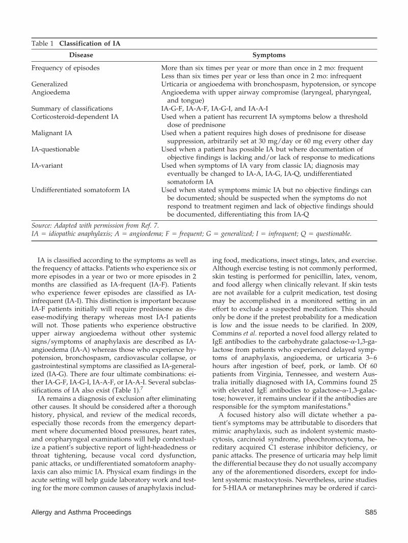
IA is classified according to the symptoms as well asthe frequency of attacks. Patients who experience six ormore episodes in a year or two or more episodes in 2months are classified as IA-frequent (IA-F). Patientswho experience fewer episodes are classified as IA-infrequent (IA-I). This distinction is important becauseIA-F patients initially will require prednisone as dis-ease-modifying therapy whereas most IA-I patientswill not. Those patients who experience obstructiveupper airway angioedema without other systemicsigns/symptoms of anaphylaxis are described as IA-angioedema (IA-A) whereas those who experience hy-potension, bronchospasm, cardiovascular collapse, orgastrointestinal symptoms are classified as IA-general-ized (IA-G). There are four ultimate combinations: ei-ther IA-G-F, IA-G-I, IA-A-F, or IA-A-I. Several subclas-sifications of IA also exist (Table 1).7
IA remains a diagnosis of exclusion after eliminatingother causes. It should be considered after a thoroughhistory, physical, and review of the medical records,especially those records from the emergency depart-ment where documented blood pressures, heart rates,and oropharyngeal examinations will help contextual-ize a patient’s subjective report of light-headedness orthroat tightening, because vocal cord dysfunction,panic attacks, or undifferentiated somatoform anaphy-laxis can also mimic IA. Physical exam findings in theacute setting will help guide laboratory work and test-ing for the more common causes of anaphylaxis includ-
ing food, medications, insect stings, latex, and exercise.Although exercise testing is not commonly performed,skin testing is performed for penicillin, latex, venom,and food allergy when clinically relevant. If skin testsare not available for a culprit medication, test dosingmay be accomplished in a monitored setting in aneffort to exclude a suspected medication. This shouldonly be done if the pretest probability for a medicationis low and the issue needs to be clarified. In 2009,Commins et al. reported a novel food allergy related toIgE antibodies to the carbohydrate galactose-�-1,3-ga-lactose from patients who experienced delayed symp-toms of anaphylaxis, angioedema, or urticaria 3–6hours after ingestion of beef, pork, or lamb. Of 60patients from Virginia, Tennessee, and western Aus-tralia initially diagnosed with IA, Commins found 25with elevated IgE antibodies to galactose-�-1,3-galac-tose; however, it remains unclear if it the antibodies areresponsible for the symptom manifestations.8
A focused history also will dictate whether a pa-tient’s symptoms may be attributable to disorders thatmimic anaphylaxis, such as indolent systemic masto-cytosis, carcinoid syndrome, pheochromocytoma, he-reditary acquired C1 esterase inhibitor deficiency, orpanic attacks. The presence of urticaria may help limitthe differential because they do not usually accompanyany of the aforementioned disorders, except for indo-lent systemic mastocytosis. Nevertheless, urine studiesfor 5-HIAA or metanephrines may be ordered if carci-
Table 1 Classification of IA
Disease Symptoms
Frequency of episodes More than six times per year or more than once in 2 mo: frequentLess than six times per year or less than once in 2 mo: infrequent
Generalized Urticaria or angioedema with bronchospasm, hypotension, or syncopeAngioedema Angioedema with upper airway compromise (laryngeal, pharyngeal,
and tongue)Summary of classifications IA-G-F, IA-A-F, IA-G-I, and IA-A-ICorticosteroid-dependent IA Used when a patient has recurrent IA symptoms below a threshold
dose of prednisoneMalignant IA Used when a patient requires high doses of prednisone for disease
suppression, arbitrarily set at 30 mg/day or 60 mg every other dayIA-questionable Used when a patient has possible IA but where documentation of
objective findings is lacking and/or lack of response to medicationsIA-variant Used when symptoms of IA vary from classic IA; diagnosis may
eventually be changed to IA-A, IA-G, IA-Q, undifferentiatedsomatoform IA
Undifferentiated somatoform IA Used when stated symptoms mimic IA but no objective findings canbe documented; should be suspected when the symptoms do notrespond to treatment regimen and lack of objective findings shouldbe documented, differentiating this from IA-Q
Source: Adapted with permission from Ref. 7.IA � idiopathic anaphylaxis; A � angioedema; F � frequent; G � generalized; I � infrequent; Q � questionable.
Allergy and Asthma Proceedings S85

noid or a pheochromocytoma is suspected, respec-tively. Skin or bone marrow biopsies may be needed toconfirm a diagnosis of mastocytosis, especially if a totaltryptase concentration is �20 ng/mL. A tryptase con-centration also may be ordered to help confirm that apatient’s symptoms are truly caused by anaphylaxis andmast cell release. Given that peak tryptase concentrationsoccur about 2 (to 4) hours after an episode and decreasethereafter, optimal timing for drawing a tryptase concen-tration would be 1–2 hours after the event and not be-yond 6 hours.9 Other laboratory work would includecomplete blood count, kidney panel, liver functions, and
complement levels (C4 as screening to exclude hereditaryangioedema or acquired C1 inhibitor deficiency).10
In 15 patients with indolent systemic mastocytosiscompared with 15 patients with IA, Koterba and Akinreported that the baseline tryptase concentrations werehigher in the patients with indolent systemic mastocy-tosis and all bone marrow examinations displayedmarkers of clonal mast cell disease (CD25�, c-kitD816V mutation).11 None of the indolent systemic mas-tocytosis patients experienced urticaria during theiranaphylactic episodes, whereas those with IA reportedurticaria frequently.11
Patients presents with compatible history
Evaluate for all known causes of anaphylaxis
IA-F Therapy 1. Emergency plan 2. Prednisone 40-60 mg daily x 1 week 3. Daily H1 anti-histamine (e.g. cetirizine 10 mg daily) 4. Followup, initially every 1-2 weeks 5. Treat with daily prednisone until symptoms controlled. Then after 1-2 weeks of controlled symptoms, taper to every other day dosing. If doing well, then taper by 5 mg every other day every 1-2 weeks.6. If episode of IA occurs during taper, evaluate for objective findings, increase dose, then attempt taper again 7. If another episode occurs, other medications such as oral albuterol, montelukast, oral cromolyn, or ketotifen may allow taper off prednisone 8. Consider alternative diagnosis if unable to taper prednisone
Acute Treatment/Emergency Plan 1. Self-inject epinephrine 2. Prednisone 60 mg 3. Hydroxyzine 25 or 50 mg 4. Go to nearest Emergency Dept
IA-I Therapy 1. Emergency Plan 2. No chronic medication 3. Follow-up with appropriate frequency to evaluate course and need for medication 4. If symptoms are severe, can treat as IA-F
IA-F and IA-I Management 1. Review emergency plan 2. Insure that patient keeps emergency medications immediately available 3. If patient lives at a distance, enlist local physician 4. Education -Emergency Plan -Check patient carries epinephrine auto-injector at all times
-Patient can demonstrate proper use of epinephrine auto-injector -Give patient information on IA -Prognosis is usually good Figure 1. Algorithm for the manage-
ment of idiopathic anaphylaxis (IA).
S86 May–June 2012, Vol. 33, No. 3

Although treatment for IA patients should be indi-vidualized, there are some guidelines that may be ap-plied (Figure 1). All IA patients should be educatedabout their disease and taught how to manage an acuteattack. At the first signs of anaphylaxis, adult patientsshould inject 0.3 mL (1/1000) of aqueous epinephrineintramuscularly followed by oral intake of prednisoneat 50–60 mg and either cetirizine at 10 mg or hy-droxyzine at 25–50 mg. In cases where a patient has aprodrome, e.g., where lip tingling precedes lip andpharyngeal angioedema, the patient may take an anti-histamine immediately and be prepared to self-admin-ister epinephrine. The emergency kit should be carriedon the person at all times and occasionally monitoredfor expiration. All patients who use epinephrineshould go immediately to the nearest emergency de-partment for further evaluation.
Patients with IA-F need to be started on disease-modifying suppressive therapy, which should include40–60 mg of prednisone daily as well as an antihista-mine, either cetirizine at 10 mg or hydroxyzine at 25–50mg daily. Patients should be given at least 1 week ofdaily prednisone although some may require 2 weeks.It is so rare that IA does not respond to daily systemicsteroids that the diagnosis should be called into ques-tion if a patient is not controlled after 2–3 weeks ofdaily prednisone or is having symptoms (hypotension,upper airway angioedema, or generalized urticaria) onhigher doses of prednisone, e.g., 40–60 mg daily after 2weeks. Once symptoms resolve for 7 days, patientsmay be tapered to every-other-day dosing. If no re-lapses occur, the patient may be further weaned by 5mg every other day every 1–2 weeks. The antihista-mines may be tapered once the prednisone is discontin-ued but patients are usually maintained on a daily anti-histamine, preferably nonsedating, for at least a year.
Steroid-dependent IA, requiring at least 30 mg ofprednisone daily or 60 mg every other day is uncom-mon but alternative medications such as ketotifen, oralcromolyn, oral albuterol, or montelukast can be tried inthose patients for whom prednisone can not be discon-tinued. To avoid complex and costly medication regi-mens, these second-line agents should be discontinuedif they do not clearly decrease the patient’s prednisonerequirement. There have been case reports of success-ful anti-IgE therapy with omalizumab for IA.12 How-ever, if a patient is not responsive to prednisone or cannot be tapered, diagnosis of IA should be reconsidered.
Patients with IA should be offered a Medic Alertbracelet that states their diagnosis. They should also beeducated and frequently reminded about how to ad-minister their epinephrine, prednisone, and an antihis-tamine. Although the potential life-threatening nature oftheir disease should be reinforced, physicians should reas-sure their patients that compliance with the prescribed reg-imen offers a remission rate in excess of 80%.2
IMMUNOLOGY
• Symptoms of IA occur as a result of episodic activation ofmast cells and basophils via an unknown mechanism.
• IA patients display more activated B cells in theirperipheral blood.
• IA patients experiencing an acute episode display ahigher percentage of activated T lymphocytes thanthose patients in remission.
• It is uncertain whether anti-IgE autoantibodies playa role in the pathogenesis of IA.
CLINICAL PEARLS
• IA is a steroid-responsive disease that is ultimately adiagnosis of exclusion.
• Its underlying pathogenesis is unknown but may beautoimmune in nature.
• All IA patients should carry an emergency kit thatincludes epinephrine.
• If symptoms are not responsive to systemic steroidsadministered over several weeks, one should recon-sider the diagnosis of IA.
• Serum tryptase (or urine histamine or its metabolite)will be elevated acutely but if elevated in the ab-sence of anaphylaxis, should suggest alternative di-agnoses including indolent systemic mastocytosis.
REFERENCES1. Bacal E, Patterson R, and Zeiss CR. Evaluation of severe (ana-
phylactic) reactions. Clin Allergy 8:295–304, 1978.2. Ditto A, Harris K, Krasnick J, et al. Idiopathic anaphylaxis: A series of
335 cases. Ann Allergy Asthma Immunol 77:285–291, 1996.3. Lieberman P, Nicklas RA, Oppenheimer J, et al. The diagnosis
and management of anaphylaxis practice parameter: 2010 Up-date. J Allergy Clin Immunol 126(3):477–480.e1–42, 2010.
4. Gruber BL, Baeza ML, Marchese MJ, et al. Prevalence andfunctional role of anti-IgE autoantibodies in urticarial syn-dromes. J Invest Dermatol 90:213–217, 1988.
5. Grammer LC, Shaughnessy MA, Harris KE, et al. Lymphocyte sub-sets and activation markers in patients with acute episodes of idio-pathic anaphylaxis. Ann Allergy Asthma Immunol 85:368–371, 2000.
6. Greenberger PA, and Ditto AM. Anaphylaxis. Allergy AsthmaProc 33:S80–S83, 2012.
7. Patterson R, Stoloff RS, Greenberger PA, et al. Algorithms forthe diagnosis and management of idiopathic anaphylaxis. AnnAllergy 71:40–44, 1993.
8. Commins SP, James H, Tran N, et al. Testing for IgE antibody tocarbohydrate galactose-a(alpha)-1,3-galactose (alpha-gal) in pa-tients with recurrent, idiopathic anaphylaxis: How many casesare we missing? J Allergy Clin Immunol 125:S119, 2009 (Abs).
9. Makhija M, and O’Gorman MRG. Common in vitro tests for allergyand immunology. Allergy Asthma Proc 33:S108–S111, 2012.
10. Georgy MS, and Pongracic JA. Hereditary and acquired angio-edema. Allergy Asthma Proc 33:S73–S76, 2012.
11. Koterba AP, and Akin C. Differences in the clinical presentationof anaphylaxis in patients with indolent systemic mastocytosis(ISM) versus idiopathic anaphylaxis (IA). J Allergy Clin Immu-nol 121:S68–S69, 2008 (Abs).
12. Warrier P, and Casale TB. Omalizumab in idiopathic anaphy-laxis. Ann Allergy Asthma Immunol 102:257–258, 2009. e
Allergy and Asthma Proceedings S87

Chapter 26
Eosinophilic esophagitis
Karen Hsu Blatman, M.D., and Anne Marie Ditto, M.D.
ABSTRACT
Eosinophilic esophagitis (EoE) is distinguished from gastroesophageal reflux disease (GERD) by persistent esophagealeosinophilia despite medical therapy with proton-pump inhibitors for 4–6 weeks. In children, symptoms vary by age groupssuch as feeding disorders in 2 year olds; vomiting in 8 year olds; and abdominal pain, dysphagia, and/or food impaction inadolescents. Most adults present with dysphagia, food impaction, heartburn or chest pain. Common endoscopic features inadults with EoE include linear furrows (creases that orient longitudinally), mucosal rings (esophageal “trachealization”),small-caliber esophagus, white plaques or exudates (which are microabscesses of eosinophils), and strictures. Children oftenpresent with similar endoscopic features, but one-third of pediatric patients with EoE have normal endoscopy. Histologicalfeatures of EoE include increased intramucosal eosinophils in the esophagus (�15 eosinophils/high-power field) without similarfindings in the stomach or duodenum. There also may be eosinophilic microabscesses. In addition to evidence of mast cellactivation, mucosa from patients with EoE have increased IL-5, supporting eosinophilia, and up-regulation of gene expressionof eotaxin-3, a chemokine important in eosinophil migration. The majority of patients have evidence of either aeroallergen and/orfood sensitization. An elemental/amino acid–based formula diet has shown to be effective in children but may not be welltolerated by adults because of taste and volume or high expense. Topical corticosteroids improve esophageal eosinophilia andsymptoms and have become the “gold standard” of pharmacotherapy.
(Allergy Asthma Proc 33:S88–S90, 2012; doi: 10.2500/aap.2012.33.3559)
Eosinophilic esophagitis (EoE) is a clinicopathologi-cal disease characterized by esophageal eosino-
philia. EoE was first described in 1977 by Dobbins et al.as a variant of eosinophilic gastroenteritis.1 The esoph-agus is normally devoid of eosinophils and their pres-ence in the esophagus was previously thought to beprimarily related to gastroesophageal reflux disease(GERD). In 1993, EoE became recognized as a separateclinicopathological entity when 11 of 12 adults present-ing with dysphagia and eosinophils in their esophagealbiopsy specimens (�20 eosinophils/high-power field)were found to have normal esophageal acid exposureon 24-hour pH monitoring. Esophageal manometryshowed a nonspecific motility disturbance in 10 ofthem.2 EoE is now generally distinguished from GERDby persistent esophageal eosinophilia despite medicaltherapy with proton-pump inhibitors.3 Since recog-
nized, the incidence of EoE has increased,3 and this isnot explained by increased awareness alone.
EoE has a male predilection and affects both childrenand adults although many of the clinicopathologicalfeatures differ between these two age groups includingage at presentation and symptoms. Most adults do notcome to clinical attention until the third decade of life,4
presenting most commonly with dysphagia, food im-paction, heartburn, or chest pain. Although adoles-cents often present with similar symptoms, infants andtoddlers present with feeding difficulties, failure tothrive, food aversion, and regurgitation. School-agechildren often present with vomiting and abdominalpain.4,5 In a case series of 103 pediatric patients, symp-toms varied by age groups. These included feedingdisorders (median age, 2.0 years), vomiting (medianage, 8.1 years), abdominal pain (median age, 12.0years), dysphagia (median age, 13.4 years), and foodimpaction (median age, 16.8 years).6
The most common endoscopic features in adultswith EoE include linear furrows (creases that orientlongitudinally), mucosal rings (esophageal “tracheal-ization”), small-caliber esophagus, white plaques orexudates (which are microabscesses of eosinophils),and strictures. Children often present with similar en-doscopic features, but the findings may be more subtle,and one-third of pediatric patients with EoE have a
From the Division of Allergy–Immunology, Department of Medicine, NorthwesternUniversity Feinberg School of Medicine, Chicago, IllinoisFunded by the Ernest S. Bazley Grant to Northwestern Memorial Hospital andNorthwestern UniversityThe authors have no conflicts of interest to declare pertaining to this articleAddress correspondence and reprint requests to Anne M. Ditto, M.D., Number 14108,676 North St. Clair Street, Chicago, IL 60611E-mail address: [email protected] © 2012, OceanSide Publications, Inc., U.S.A.
S88 May–June 2012, Vol. 33, No. 3
normal endoscopy.4 None of the features is pathogno-monic. The diagnosis of EoE ultimately requires a bi-opsy revealing the presence of eosinophils. Histologi-cal features include increased intramucosal eosinophilsin the esophagus (�15 eosinophils/high-power field)without similar findings in the stomach or duodenum.There may also be basal layer hyperplasia and eosino-philic microabscesses.7,8 Some patients previously hadbeen misdiagnosed with candidal esophagitis. Accord-ing to a retrospective study of adult patients, it hasbeen reported that eosinophilic infiltration of theesophagus may not be distributed evenly.7 Therefore,it is recommended that both proximal and distal bi-opsy specimens be obtained for diagnosis with a min-imum of five biopsies to maximize the sensitivity.7
Recent consensus recommendations have led to thecurrent diagnostic criteria: (1) presence of symptomsincluding but not limited to dysphagia and food im-paction in adults and feeding intolerance and GERDsymptoms in children; (2) at least 15 eosinophils/high-power field in the esophageal tissues; and (3) exclusionof other disorders associated with similar clinical, his-tological or endoscopic features such as GERD witheither the use of high-dose acid suppression 4–6 weeksbefore biopsy procurement or normal pH monitoring.9
Acid suppression is important because case reportshave shown high esophageal eosinophilia in associa-tion with GERD, which were completely responsive toproton-pump inhibitors.10
EoE is thought to be allergic, but the allergen remainselusive. The majority of adults and older children havebeen found to be sensitized to aeroallergens.6,11 Aeroal-lergens may act locally after being swallowed, directlycausing allergic inflammation in the esophagus, or theymay act systemically.10 In 1995, 10 children with esoph-ageal eosinophilia who initially were diagnosed withGERD but failed treatment (including Nissan fundo-plication in 6 of the children) improved on an elemen-tal diet.12 However, unlike most patients with IgE-mediated food allergy, patients with EoE rarely have ahistory of food-induced anaphylaxis. In addition, iden-tification of food triggers is difficult because symptomssuch as dysphagia and food impaction may be causedby strictures. Foods also may be involved because oftheir relationship to aeroallergens and positive skintests to food may be caused by cross-reactivity of theseallergens. However, studies to address this notion arelacking.13 Some research suggests that this may be adelayed reaction to foods as described in atopic der-matitis and therefore food patch testing may be help-ful. However, patch testing is not currently validatedor standardized.14 Because of the association of atopicdisease with EoE, evaluation by an allergist–immu-nologist is recommended. Allergy testing may alsoguide choices for food elimination or reintroduction.5
Topical corticosteroids improve esophageal eosino-philia and symptoms9,15 and have become the “goldstandard” of therapy. Studies have used either “puffand swallow” aerosolized fluticasone propionate orswallowed budesonide suspension respules mixedwith sucralose (Splenda [Tate & Lyle PLC, London,United Kingdom]). There is no recommended length oftime to treat before discontinuing therapy. Symptomrecurrence often is reported after topical steroids arediscontinued.3 Side effects include esophageal candidi-asis and unpleasant taste of budesonide with sucralose.
Removal of food antigens has been shown to im-prove symptoms in patients with EoE. Consultationwith a dietician is recommended. An elemental/aminoacid–based formula diet has shown to be effective butmay not be well tolerated by adults because of tasteand volume. Food elimination diets may be empiricelimination diets, often referred to as the six-food elim-ination diet (SFED), which entails avoiding the eightmost common allergenic foods (milk, wheat, eggs, soy,peanuts, tree nuts, shellfish, and fish),15 or may bedirected by allergy testing. Other interventions includeendoscopic esophageal dilation for adults with EoEwho present with strictures.16 Although dilation ishighly effective in providing symptom relief and canbe performed safely, it does not change the underlyinginflammation.17
EoE is an emerging disease. The understanding ofthe etiology, natural history, diagnosis, and manage-ment will continue to evolve. More long-term studiesare needed to determine the efficacy of treatment.
IMMUNOLOGY
• Patients with EoE have marked up-regulation of thegene expression of eotaxin-3,6,14 a chemokine impor-tant in eosinophil migration.
• Mucosa from patients with EoE have increased Th2proinflammatory cytokines such as IL-5.14
• Mucosa from patients with EoE have increased mastcell infiltration.6
• Mast cell associated genes were up-regulated inesophageal tissue and responded to both corticoste-roids and diet.18
CLINICAL PEARLS
• In adults, EoE often presents as dysphagia or foodimpaction.
• In children, EoE often presents with failure to thrive,reflux, vomiting, or abdominal pain.
• EoE has an association with a personal or familyhistory of atopy.
• The majority of patients have evidence of eitheraeroallergen and/or food sensitization. Isolatedesophageal eosinophilia is inadequate for the diag-nosis of EoE. Other causes, such as GERD, must be
Allergy and Asthma Proceedings S89

excluded and EoE should be associated with clinicalsymptoms.
REFERENCES1. Dobbins JW, Sheahan DG, and Behar J. Eosinophilic gastroen-
teritis with esophageal involvement. Gastroenterology 72:1312–1316, 1977.
2. Attwood SE, Smyrk TC, Demeester TR, et al. Esophageal esoi-nophilia with dysphagia. A distinct clinicopathologic syn-drome. Dig Dis Sci 38:109–116, 1993.
3. Hirano I, Gonsalves N, and Ditto AM. Eosinophilic esophagitis.In Patterson’s Allergic Diseases, 7th ed. Grammer LC, andGreenberger PA (Eds). Philadelphia, PA: Lippincott Williams &Wilkins, 633–645, 2009.
4. Liacouras CA, Furuta GT, Hirano I, et al. Eosinophilic esoph-agitis: Updated consensus recommendations for children andadults. J Allergy Clin Immunol 128:3–20, 2011.
5. Liacouras CA, Spergel JM, Rucelli E, et al. Eosinophilic esoph-agitis: A 10-year experience in 381 children. Clin GastroenterolHepatol 3:1198–1206, 2005.
6. Noel RJ, Putnam PE, and Rothenberg ME. Eosinophilic esoph-agitis. N Engl J Med 351:940–941, 2004.
7. Gonsalves N, Policarpio-Nicholas M, Zhang Q, et al. Histo-pathologic variability and endoscopic correlates in adults witheosinophilic esophagitis in adults. Gastrointest Endosc 64:313–319, 2006.
8. Genevay M, Rubbia-Brandt L, and Rougemont AL. Do eosino-phil numbers differentiate eosinophilic esophagitis from gastro-esophageal reflux disease? Arch Lab Med 134:815–825, 2010.
9. Furuta GT, Liacouras CA, Collins MH, et al. Eosinophilic esoph-agitis in children and adults: A systematic review and consen-
sus recommendations for diagnosis and treatment. Gastroenter-ology 133:1342–1363, 2007.
10. Ngo P, Furuta GT, Antonioli DA, et al. Eosinophils in theesophagus—Peptic or allergic eosinophilic esophagitis? Caseseries of three patients with esophageal eosinophilia. Am JGastroenterol 101:1666–1670, 2006.
11. Roy-Ghanta S, Larosa DF, and Katzka DA. Atopic characteris-tics of adult patients with eosinophilic esophagitis. Clin Gastro-enterol Hepatol 6:531–535, 2008.
12. Kelly KJ, Lazenby AJ, Rowe PC, et al. Eosinophilic esophagitisattributed to gastroesophageal reflux: Improvement with anamino acid-based formula. Clin Exp Allergy 35:1421–1422, 2005.
13. Simon D, Straumann A, Wenk A, et al. Eosinophilic esophagitisin adults—No clinical relevance of wheat and rye sensitizations.Allergy 61:1480–1483, 2006.
14. Rothenberg ME. Biology and treatment of eosinophilic esoph-agitis. Gastroenterology 137:1238–1249, 2009.
15. Konikoff MR, Noel RJ, Blanchard C, et al. A randomized, dou-ble-blind, placebo-controlled trial of fluticasone propionate forpediatric eosinophilic esophagitis. Gastroenterology 131:1381–1391, 2006.
16. Kagalwalla AF, Sentongo TA, Ritz S, et al. Effect of six-foodelimination diet on clinical and histologic outcomes in eosino-philic esophagitis. Clin Gastroenterol Hepatol 4:1097–1102,2006.
17. Schoepfer AM, Gonsalves N, Bussman C, et al. Esophagealdilation in eosinophilic esophagitis: Effectiveness, safety, andimpact on the underlying inflammation. Am J Gastroenterol105:1062–1070, 2010.
18. Hsu Blatman KS, Gonsalves N, Hirano I, et al. Expression ofmast-cell associated genes is upregulated in adult eosinophilicesophagitis and responds to steroid or dietary therapy. J Al-lergy Clin Immunol 127:1307–1308, 2011. e
S90 May–June 2012, Vol. 33, No. 3

Chapter 27
Approach to primary immunodeficiency
Ashraf Uzzaman, M.D., and Ramsay L. Fuleihan, M.D.
ABSTRACT
Primary immunodeficiency diseases (PID) are inherited defects of the innate or adaptive arms of the immune system that leadto an increase in the incidence, frequency, or severity of infections. There may be defects in the adaptive arm of the immunesystem that include combined immunodeficiency and antibody deficiency syndromes or by abnormalities in innate immunitysuch as disorders of phagocytes, the complement pathway, or Toll-Like receptor (TLR) mediated signaling. Recurrentsinopulmonary infections with encapsulated bacteria such as Haemophilus influenza type B or Streptococcus pneumoniaemay be characteristic of an IgG antibody deficiency or dysfunction. Frequent viral, fungal, or protozoal infections may suggestT lymphocyte dysfunction. Multiple staphylococcal skin infections and fungal infections may imply neutrophil dysfunction orthe hyper-IgE syndrome, and recurrent neisserial infection is a characteristic manifestation of late complement component(C5–9, or the membrane attack complex) defects. Recurrent viral or pyogenic bacterial infections often without the presence ofa significant inflammatory response suggest a defect in TLR signaling. Mycobacterial infections are characteristic of defects ininterleukin (IL)-12, interferon (IFN) gamma, or their receptors. Screening of newborns for T-cell lymphopenia using apolymerase chain reaction to amplify T-cell receptor excision circles (TRECs), which are formed when a T cell rearranges thevariable region of its receptor, serves as a surrogate for newly synthesized naïve T cells. Because of very low numbers of TRECs,severe combined immunodeficiency, DiGeorge syndrome, and other causes of T-cell lymphopenia have been identified innewborns.
(Allergy Asthma Proc 33:S91–S95, 2012; doi: 10.2500/aap.2012.33.3560)
Primary immunodeficiency diseases (PID) are inher-ited defects of the innate or adaptive arms of the
immune system that lead to an increase in the inci-dence, frequency, or severity of infections.1–3 In gen-eral, PIDs are uncommon, but the associated morbidityand mortality may be high. The overall prevalence ofPID is not known but is estimated to be 1:10,000 livebirths. However, IgA deficiency occurs more frequently;as many as 1:333 individuals may be affected. An earlydiagnosis of PID is essential because prompt treatmentmay help prevent associated morbidity and mortality.
PRIMARY IMMUNE DEFICIENCY DISEASESPIDs may be caused by defects in the adaptive arm of
the immune system that include combined immunode-ficiencies (CID) and antibody deficiency syndromes or
by abnormalities in innate immunity such as disordersof phagocytes, the complement pathway or Toll-likereceptor (TLR) mediated signaling.
Severe CID (SCID), results from the absence of Tlymphocytes or T lymphocyte function with the pres-ence or absence of B cells and/or natural killer (NK)cells. Even if B cells are present, antibody production isimpaired because of lack of T cell help. SCID is char-acterized by early onset of infections caused by bacte-rial, viral, or fungal pathogens; chronic diarrhea; andfailure to thrive. Pneumocystis jiroveci pneumonia iscommonly observed in these patients as well as pul-monary infections by adenovirus, RSV, CMV, andparainfluenza viruses. Isolated CD4 T-cell deficiencycaused by MHC class II deficiency or idiopathic CD4lymphopenia and isolated CD8 T-cell deficiency fromabsence of MHC class I molecule expression or causedby ZAP 70 also cause a severe T-cell immunodefi-ciency.
DiGeorge syndrome results from a developmentaldefect in the third and fourth pharyngeal pouches re-sulting in impaired formation of the thymus, parathy-roid glands, and the heart. Affected patients have aspectrum of T-cell deficiency from mild or moderate inpartial DiGeorge syndrome to severe T-cell deficiencyin complete DiGeorge syndrome, which is a condition
From the Division of Allergy and Immunology, Department of Pediatrics, Children’sMemorial Hospital, Northwestern University Feinberg School of Medicine, ChicagoIllinoisFunded by the Ernest S. Bazley Grant to Northwestern Memorial Hospital andNorthwestern UniversityThe authors have no conflicts of interest to declare pertaining to this articleAddress correspondence and reprint requests to Ramsay L. Fuleihan, M.D., Box 60,2300 Children’s Plaza, Chicago, IL 60614E-mail address: [email protected] © 2012, OceanSide Publications, Inc., U.S.A.
Allergy and Asthma Proceedings S91

similar to SCID. Patients may also have congenitalheart disease and hypocalcemia.
B-cell activation and immunoglobulin isotype switch-ing and affinity maturation requires interaction of CD40ligand (CD40L), which is expressed on activated CD4 Tcells and CD40, which is present on B cells, monocytes,and other antigen-presenting cells. A mutation in CD40Lcauses X-linked hyper-IgM syndrome with normal orincreased IgM but decreased or absent levels of otherimmunoglobulin isotypes. Patients are at risk for oppor-tunistic infection such as P. jiroveci pneumonia and cryp-tosporidium, may have neutropenia, and have a higherincidence of liver disease, sclerosing cholangitis, and tu-mors of the liver and biliary tree. A rapidly progressiveneuroendocrine carcinoma has also been reported in thiscondition. Defects in the CD40 gene cause an autosomalrecessive form of hyper-IgM syndrome with a similarphenotype to X-linked hyper-IgM. However, mutationsin activation-induced cytidine deaminase and uracyl N-glycosylase, which are enzymes involved in class switchrecombination and affinity maturation, also result in theautosomal recessive hyper-IgM syndrome but do nothave an associated T-cell immunodeficiency or suscepti-bility to opportunistic infections but develop lymphoidhyperplasia and may be more susceptible to autoimmunedisease.
Antibody deficiency syndromes may result from de-fects in B-cell development, maturation, or function.An arrest in the development of pro- to pre-B cells inthe bone marrow may be caused by a defect in thepre–B-cell receptor or by defects in the pre–B-cell re-ceptor–associated intracytoplasmic proteins such as B-cell linker protein (BLNK) or Bruton tyrosine kinase(BTK). Defects in BTK lead to X-linked agammaglobu-linemia (XLA), the most commonly observed early on-set agammaglobulinemia. These defects manifest acharacteristic absence (�1%) of B cells in the peripheralblood. XLA patients may have a small number of ma-ture B cells. Individuals with antibody deficiency syn-dromes are susceptible to infection by encapsulatedbacteria and suffer recurrent otitis media, sinusitis, andpneumonia. They are also at risk for enteroviral infec-tions, which may lead to meningoencephalitis, vaccine-associated poliomyelitis, and infection by mycoplasma,which may cause arthritis.
Common variable immune deficiency (CVID) ischaracterized by impaired antibody production in re-sponse to vaccinations and infections and low concen-trations of IgG and one or more other immunoglobulinisotypes. The diagnosis is usually not entertained inchildren �2–4 years of age. Most individuals sufferrecurrent episodes of otitis media, sinusitis, and pneu-monia by encapsulated bacterial organisms, and somemay have an increased incidence of lymphoid hyper-plasia, granulomatous lesions, lymphoma, and autoim-mune manifestations such as cytopenias and inflam-
matory bowel disease. CVID may be several clinicallydistinct subtypes, and the genetic cause in most pa-tients remains unknown. Selective IgA deficiency is themost common asymptomatic PID; however, a smallnumber may manifest recurrent sinopulmonary infec-tions, autoimmunity, and allergy. A number of indi-viduals may have associated IgG subclass deficiency,most commonly of IgG2. IgG subclass deficiency also isknown to occur independent of IgA deficiency andmay be associated with recurrent infections, especiallyif associated with a specific antibody deficiency usuallyto polysaccharide antigens such as Streptococcus pneu-moniae. Specific antibody deficiency is characterized bynormal level of B cells and immunoglobulins associ-ated with impaired antibody production to polysac-charide antigens.4 Transient hypogammaglobulinemiaof infancy is characterized by low-serum IgG concen-tration with good antibody responses to protein anti-gens such as tetanus and spontaneous resolution of thehypogammaglobulinemia, sometime after 2 years ofage. Although, most children remain asymptomatic,some may have recurrent upper respiratory tract viralinfections.
A defect in phagocyte numbers or function maycause recurrent bacterial and fungal infections. Recur-rent oral stomatitis and infections of the respiratorytract and skin and deep-seated abscesses commonlyare observed. Chronic granulomatous disease (CGD)results from a defect of phagocyte function. Reducedlevels of nicotinamide adenine dinucleotide phosphateoxidase complex in phagocytes leads to defective kill-ing of catalase-positive bacteria and fungi. The chronicinflammatory response to the presence of these organ-isms leads to granuloma formation especially in hol-low organs, which may cause outlet obstruction in thegastrointestinal and urinary tracts.
Impaired trafficking of phagocytes from the circula-tion into the tissues results in leukocyte adhesion de-fect (LAD). Clinical and laboratory manifestations ofan LAD include delayed umbilical cord separation�4–6 weeks and a very elevated white blood cell countof neutrophils (up to 100,000/�L during an infection).
Defects in proteins that comprise the complementsystem also lead to disease. Lack of early complementcomponents C2 and C4 predispose to autoimmunediseases such as systemic lupus erythematosus. A de-ficiency in C2 and C3 may lead to an increased risk ofinfection by encapsulated bacteria. Recurrent and in-vasive neisserial infections occur in individuals withdefects in the terminal components of the complementpathway, C5-C9. Absence of alternate complementpathway factors such as factor D and properdin mayresult in a similar increase in risk of neisserial infec-tions. Recurrent bacterial infections in early life mayresult from a defect in the mannose binding lectin, a
S92 May–June 2012, Vol. 33, No. 3

component of the mannose binding lectin complementpathway.
TLRs, which are present on cell surface and intracel-lularly on endosomal membranes, recognize pathogen-associated molecular patterns, which are present onbacteria and viruses such as lipopolysaccharide onGram-negative bacteria and single- or double-strandedRNA on viruses. A defect in TLR-mediated signalingleads to reduced production of proinflammatory cyto-kines such as interleukin (IL)-1, IL-6, TNF-�, and IL-12and results in an increased risk of severe and recurrentviral or pyogenic bacterial infections often without thepresence of a significant inflammatory response.
APPROACH TO DIAGNOSISThe diagnosis of a PID must be entertained when an
individual has a family history of PID or a history ofsevere, unusual, frequent, or difficult-to-treat infec-tions. A detailed history of infections should be ex-plored including the sites and frequency of infectionsas well as the organisms involved. Recurrent sinopul-monary infections with encapsulated bacteria such asHaemophilus influenza type B or S. pneumoniae may becharacteristic of an antibody deficiency.5 Frequent vi-ral, fungal, or protozoal infections may suggest T-lym-phocyte dysfunction. Multiple staphylococcal skin in-fections and fungal infections may imply neutrophildysfunction or the hyper-IgE syndrome, and recurrentneisserial infection is a characteristic manifestation oflate complement component (C5–9, or the membraneattack complex) defects. Recurrent viral or pyogenicbacterial infections often without the presence of asignificant inflammatory response suggest a defect inTLR signaling. Mycobacterial infections are character-istic of defects in IL-12, interferon �, or their receptors.
The age of onset of infections is also important. CIDdiseases commonly cause symptoms before 6 monthsof age, whereas B-cell dysfunction often becomessymptomatic between 6 and 12 months of age whenmaternal antibody has waned. Delayed umbilical cordseparation, �4–6 weeks and a very elevated whiteblood cell count may indicate a LAD. X-linked reces-sive diseases should be considered in male patients,whereas autosomal recessive conditions affect maleand female patients equally and should be entertainedwhen consanguinity is present.
The physical exam should begin with assessment ofgrowth parameters. Some infants with SCID may pres-ent with failure to thrive. The exam might be signifi-cant for sequela of recurrent infections such as digitalclubbing, wheezing, or rhonchi as signs of pulmonaryinjury. Certain immunodeficiencies are associated withspecific findings on physical exam. Patients with Di-George syndrome may have facial dysmorphisms andmay present with congenital heart disease. Patients
with Chediak-Higashi may have partial albinism. Theskin exam may reveal multiple telangiectasias onthe skin and bulbar conjunctiva in ataxia-telangiecta-sia. A severe eczematous rash may occur as part ofhyper-IgE syndrome or Wiskott-Aldrich syndrome(WAS). Attention to the presence or absence of lym-phoid tissue may help to narrow the diagnosis. Pa-tients with XLA may lack lymph nodes revealed byabsent tonsils on physical exam, whereas autosomalrecessive hyper-IgM is associated with lymphoid hy-perplasia.
Laboratory testing should be ordered and inter-preted within the context of the individual patient’shistory and physical exam. Normal ranges are usuallyage specific, and age-adjusted norms must be used;results may be affected by current infections, medica-tions, and flares of autoimmune diseases. A completeblood count with differential can show lymphopenia inSCID, elevated white cell counts in LAD even in theabsence of infection, or thrombocytopenia in WAS oras a complication of autoimmunity in CVID or IgAdeficiency. Neutropenia is found in primary neutrope-nia as a manifestation of autoimmune disease or inassociation with the X-linked hyper-IgM syndrome.Morphological exam of neutrophils can be helpful be-cause it may reveal giant granules in Chediak-Higashisyndrome.
Antibody deficiency is assessed with quantitativeserum immunoglobulin concentrations and the func-tional adequacy of humoral immunity is determinedby serum antibody concentrations to common vaccinessuch as tetanus, H. influenza type B, and pneumococcalvaccination, which evaluate antibody responses to pro-tein, conjugate, and polysaccharide antigens, respec-tively. Flow cytometry is used to enumerate B-cellnumbers, which are absent or severely reduced inagammaglobulinemia and some forms of SCID, andmemory B cells, which are reduced in CVID and hyper-IgM syndrome.
T-cell numbers and subsets are enumerated by flowcytometry. T cells are absent in SCID and the presenceor absence of B cells and NK cells helps differentiatedifferent causes of SCID. In addition, flow cytometrycan identify naïve and memory T cells and activationmarkers on the T-cell surface as well as intracellularproteins such as WASP in WAS, FoxP3 in regulatory Tcells, and cytokines in different effector T-cell popula-tions. The ability of T cells to proliferate in response tomitogens helps rule out SCID and shows normal sig-naling pathways including the production of IL-2 andthe expression of the IL-2 receptor on T cells. In vitrofunctional competence of T cells is assessed by theirproliferative response to commonly encountered anti-gens such as Candida or tetanus antigens, indicating thepresence of antigen-specific memory T cells. A normalantibody response to protein antigens such as tetanus
Allergy and Asthma Proceedings S93

is an in vivo method of establishing T-cell helper func-tion.
Phagocyte function may be assessed with flow cy-tometry. The diagnosis of LAD-1 may be confirmed bythe absence of CD11b/C18 marker on leukocytes. CGDis best diagnosed by fluorescent detection of the oxi-dative burst using flow cytometry. This assay uses asmall dye called dihydrorhodamine that fluoresceswhen exposed to reactive oxygen species.
An initial evaluation of complement components isperformed with a CH50 level for the classic comple-ment pathway and an AP50 level for the alternatepathway. If low, individual complement levels mustthen be evaluated. The TLR signaling pathways can beassessed by responses to engagement by a variety ofTLR ligands.
A further review of laboratory testing to assess theimmune system is discussed in another article in thisjournal.6
TREATMENTTreatment of immunodeficiency should begin
quickly and aggressively after the diagnosis is madeto prevent complications from infections. Humoralimmunity disorders require treatment with immuno-globulin replacement i.v. at doses of 400 – 600 mg/kgper month or subcutaneously at 100 –200 mg/kg perweek,7 avoidance of live polio vaccine, and mayrequire antibiotic prophylaxis. Milder cases of hu-moral immunodeficiency such as specific antibodydeficiency syndrome may be managed with antibi-otic prophylaxis. Cellular disorders such as SCIDrequire prompt evaluation for a stem cell trans-plant,8 avoidance of live viral vaccines includingrotavirus, and any blood product transfusion needsto be irradiated, leukoreduced, and CMV�. X-linkedhyper-IgM, WAS, and other severe immunodefi-ciency diseases also can be treated with stem celltransplantation. X-linked SCID, adenosine deami-nase (ADA)– deficient SCID, and WAS can also betreated by gene therapy, especially if the patientdoes not have a matched donor for stem cell trans-plantation.9 ADA-deficient SCID may be treatedwith enzyme replacement with PEG-ADA. Patientswith complete DiGeorge syndrome may benefit fromthymic transplant. Prophylaxis against infectionswith antibiotics (trimethoprim/sulfamethoxazoleand itraconazole) and interferon � may be used inpatients with CGD. It is important to keep in mindthat patients with an underlying immunodeficiencymay require prolonged and aggressive therapy forinfections.
NEWBORN SCREENINGNewborn screening for T-cell lymphopenia has
been initiated in Wisconsin and in other states using
a polymerase chain reaction to amplify T-cell recep-tor excision circles, which are formed when a T cellrearranges the variable region of its receptor andserves as a surrogate for newly synthesized naïve Tcells. SCID, DiGeorge syndrome, and other causes ofT-cell lymphopenia have been identified in the new-born period allowing curative treatment by stem celltransplantation before the development of seriousand life-threatening infections and complications.
SUMMARYIn summary, although immunodeficiency disorders
are uncommon, physicians must remain suspicious inthe setting of frequent severe infections or infections byunusual organisms. Prevention of associated morbidityand mortality is dependent on early diagnosis andtreatment.
IMMUNOLOGY
• B-cell disorders are characterized by decreasednumbers of B cells and low-serum quantitative im-munoglobulins and poor response to polysaccharideantigens.
• Brutons agammaglobulinemia, an example of a B-cell disorder, is caused by a defect in B- cell tyrosinekinase, which results in decreased serum immuno-globulins.
• T-cell disorders are characterized by decreasednumbers of T cells and/ or decreased T-cell function.An example is SCID, which may be caused by num-ber of genetic defects.
• Defects of innate immunity also cause severe, re-current, and life-threatening infections and can becaused by defects in complement proteins, phago-cytic cells, or signaling defects in TLRs.
CLINICAL PEARLS
• Immunodeficiency should be suspected in pa-tients with recurrent, unusual, or difficult-to-treatinfections or with a family history of immunode-ficiency.
• Consider humoral immunity defect in patients withrecurrent encapsulated bacterial infections.
• Consider T-cell defects in patients with frequentviral, fungal, or protozoal infections or recurrentinfections and failure to thrive.
• In patients with recurrent neisserial infections, a latecomplement defect should be considered.
• Newborn screening can identify patients with T-cell lymphopenia before recurrent infections de-velop.
• Early and aggressive therapy can reduce morbid-ity and mortality and result in an improved out-come.
S94 May–June 2012, Vol. 33, No. 3

REFERENCES1. Notarangelo LD, Fischer A, Geha RS, et al. International Union
of Immunological Societies Expert Committee on Primary Im-munodeficiencies: Primary immunodeficiencies: 2009 Update. JAllergy Clin Immunol 124:1161–1178, 2009.
2. Buckley RH. Primary immunodeficiency diseases. In Middleton’sAllergy Principles & Practice, 7th ed. Adkinson NF, Bochner BS,Busse WW, et al. (Eds). Philadelphia, PA: Elsevier, 801–929, 2009.
3. Berger M. Evaluation and management of immune deficiency inallergy practice. In Patterson’s Allergic Diseases, 7th ed. Grammer LC,and Greenberger PA (Eds). Philadelphia, PA: Walters Kluwer Lip-pincott Williams & Wilkins, 41–56, 2009.
4. Carr TF, Koterba AP, Chandra R, et al. Characterization ofspecific antibody deficiency in adults with medically refractorychronic rhinosinusitis. Am J Rhinol Allergy 25:241–244, 2011.(PubMed PMID DOI: 21819760.)
5. Alqudah M, Graham SM, and Ballas ZK. High prevalence ofhumoral immunodeficiency patients with refractory chronic
rhinosinusitis. Am J Rhinol Allergy 24:409–412, 2010. (PubMedPMID DOI: 21144219.)
6. Makhija M, and O’Gorman MRG. Common in vitro tests forallergy and immunology. Allergy Asthma Proc 33:S108 –S111, 2012.
7. Yong PL, Boyle J, Ballow M, et al. Use of intravenous immuno-globulin and adjunctive therapies in the treatment of primaryimmunodeficiencies: A working group report of and study bythe Primary Immunodeficiency Committee of the AmericanAcademy of Allergy Asthma and Immunology. Clin Immunol135:255–263, 2010.
8. Griffith LM, Cowan MJ, Notarangelo LD, et al.; WorkshopParticipants. Improving cellular therapy for primary immunedeficiency diseases: Recognition, diagnosis, and management. JAllergy Clin Immunol 124:1152–1160, 2009.
9. Fischer A, Hacien-Bey-Abina S, and Cavazanna-Calvo M. Genetherapy for primary immunodeficiencies. Immunol AllergyClin North Am 30:237–248, 2010. e
Allergy and Asthma Proceedings S95

Chapter 28
Classification of hypersensitivity reactions
Ashraf Uzzaman, M.D., and Seong H. Cho, M.D.
ABSTRACT
The original Gell and Coomb’s classification categorizes hypersensitivity reactions into four subtypes according to the typeof immune response and the effector mechanism responsible for cell and tissue injury: type I, immediate or IgE mediated; typeII, cytotoxic or IgG/IgM mediated; type III, IgG/IgM immune complex mediated; and type IV, delayed-type hypersensitivityor T-cell mediated. The classification has been improved so that type IIa is the former type II and type IIb is antibody-mediatedcell stimulating (Graves Disease and the “autoimmune” type of chronic idiopathic urticaria). Type IV has four major categories:type IVa is CD4�Th1 lymphocyte mediated with activation of macrophages (granuloma formation and type I diabetes mellitus);type IVb is CD4�Th2 lymphocyte mediated with eosinophilic involvement (persistent asthma and allergic rhinitis); type IVcis cytotoxic CD8� T lymphocyte with involvement of perforin-granzme B in apoptosis (Stevens-Johnson syndrome and toxicepidermal necrolysis); type IVd is T-lymphocyte–driven neutrophilic inflammation (pustular psoriasis and acute generalizedexanthematous pustulosis). Some diseases have multiple types of immunologic hypersensitivity.
(Allergy Asthma Proc 33:S96–S99, 2012; doi: 10.2500/aap.2012.33.3561)
Although, the immune system is primarily used toprotect against microbes such as bacteria, vi-
ruses, and fungi, unexpected excesses of the immuneresponse may lead to disease states. The excessive im-mune responses are usually referred to as hypersensi-tivity reactions. The original Gell and Coomb’s classi-fication categorizes hypersensitivity reactions into foursubtypes according to the type of immune responseand the effector mechanism responsible for cell andtissue injury: type I, immediate or IgE mediated; typeII, cytotoxic or IgG/IgM mediated; type III, IgG/IgMimmune complex mediated; and type IV, delayed-typehypersensitivity or T-cell mediated.1 In clinical prac-tice, however, patients often display a constellation ofsymptoms that usually overlap several of these mech-anisms. For example, individuals who are allergic topenicillin may exhibit symptoms that suggest a type Ior IgE-mediated reaction such as anaphylaxis; theymay also exhibit a serum sickness such as a disease thatsuggests a type III or an IgG/IgM immune complex–mediated reaction (Table 1).2
TYPE I—IMMEDIATE OR IgE-MEDIATEDREACTIONS
IgE antibodies are produced in response to parasiticinfections and, along with eosinophil activation, pri-marily serve protective functions and help eradicatethe parasites. Atopic individuals may form allergen-specific IgE antibodies in response to allergens,3 suchas present in the environment, in foods,4 and in drugs.5
The IgE antibodies thus formed attach to high-affinityIgE receptors (Fc�RI), which are present on mast celland basophil surfaces. On reexposure, the allergen isrecognized by IgE antibodies bound to mast cells andbasophils and leads to triggering of these cells result-ing in an immediate hypersensitivity reaction. The im-mediate hypersensitivity reaction usually consists oftwo phases; an immediate response that occurs withinminutes and is caused by histamine, prostaglandin D2,leukotriene D4, and kinins (and tryptase) and a delayedreaction that occurs after 4–8 hours and is effected bycytokines such as IL-1, tumor necrosis factor, IL-4, IL-5,IL-13, and various colony-stimulating factors such asgranulocyte monocyte colony-stimulating factor.6
In addition to mast cells and basophils, eosinophilsand neutrophils may also be involved in producing thecomplete spectrum of the immediate hypersensitivityreaction.
Examples of immediate hypersensitivity reactionsare anaphylaxis,7 which is characterized by dilatationof the blood vessels and constriction of the airways andmay occur in response to allergens present in foods—nuts and milk; bronchial asthma,8 which may be
From the Division of Allergy–Immunology, Department of Medicine, NorthwesternUniversity Feinberg School of Medicine, Chicago, IllinoisFunded by the Ernest S. Bazley Grant to Northwestern Memorial Hospital andNorthwestern UniversityThe authors have no conflicts of interest to declare pertaining to this articleAddress correspondence and reprint requests to Seong Cho, M.D., Number 14108, 676North St. Clair Street, Chicago, IL 60611E-mail address: [email protected] © 2012, OceanSide Publications, Inc., U.S.A.
S96 May–June 2012, Vol. 33, No. 3

caused by repeated immediate hypersensitivity andlate-phase reactions in the lung tissue; and skin mani-festations such as urticaria,9 which is a wheal and flare(erythema) reaction.
TYPE II—CYTOTOXIC OR IgG/IgM-MEDIATEDREACTIONS
Immune responses that usually afford protectionagainst infections and eradication of malignant cellsmay sometimes cause damage to tissues. The im-mune responses commonly involve IgG and IgMand, to a lesser extent, IgA antibodies. The antibod-ies usually are directed against cell surface antigenssuch as those present on red blood cells, neutrophils,and platelets; those present on epithelial cells ofglandular and mucosal surfaces; or against thosepresent on tissues such as basement membranes.Three underlying mechanisms commonly accountfor the tissue damage.10 First, antibodies may di-rectly coat or opsonize cells or they may activate thecomplement system, which leads to the productionof activated complement components that may thencoat or opsonize the cells. These opsonized cells arephagocytosed and are destroyed by phagocytes thatexpress receptors for antibodies and complementproteins. The underlying mechanism in autoimmunehemolytic anemia and autoimmune thrombocytope-nic purpura is an example. Second, antibodies de-posited in tissues subsequently recruit neutrophilsand macrophages, which leads to tissue injury andinflammation. This is the mechanism of injury in anti-body-mediated glomerulonephritis. The third mecha-
nism of immune response is antibody-dependent cell-mediated cytotoxicity, which occurs when eosinophilsbind to IgE-bound helminths and release their granulecomponents. Type II reactions can also be divided intotwo different subtypes. Type IIa reactions are charac-terized by cytolytic reactions produced by antibodiescausing autoimmune hemolytic anemia, whereastype IIb reactions are characterized by cell-stimulat-ing antibodies in patients with Graves disease (along-acting thyroid stimulator, thyroid-stimulatinghormone receptor antibodies) or antibodies to thehigh-affinity mast cell receptor (Fc�RI�) or IgE inchronic idiopathic urticaria.9
TYPE III—IgG/IgM IMMUNE COMPLEXMEDIATED
In type II immune response, the mechanism of tissueinjury involves the formation of IgG or IgM antibodiesto self or foreign antigens and then the formation ofcomplexes. The complexes deposit and activate thecomplement pathway, with concomitant fall in serumcomplement levels. The activated complement compo-nents recruit and activate neutrophils, which results ininflammation and tissue injury. The constellation ofsymptoms is determined by the site of immune com-plex deposition and not by the source of the antigen.The antigen–antibody complexes are usually depositedin small arteries, renal glomeruli, and synovial of jointsand the symptoms usually are vasculitis, nephritis, andarthritis, respectively.10 One example is serum sick-ness-like disease, which may be acute or may have aprolonged or chronic course. This prototypical immune
Table 1 Classification of hypersensitivity reactions
Classification Immunoreactants Clinical Presentation
Type I Mast cell mediated, IgE dependent(anaphylactic, and IgE independent)
Anaphylaxis, urticaria, angioedema, asthma, andallergic rhinitis
Type IIa Antibody-mediated cytotoxic reactions(IgG and IgM antibodies complementoften involved)
Immune cytopenias
Type IIb Antibody-mediated cell-stimulatingreactions
Graves disease and chronic idiopathic urticaria
Type III Immune complex–mediated reactionscomplement involved
Serum sickness and vasculitis
Type IVa Th1 cell-mediated reactions macrophageactivation
Type 1 diabetes and contact dermatitis (withIVc)
Type IVb Th2 cell–mediated reactions eosinophilicinflammation
Persistent asthma and allergic rhinitis
Type IVc Cytotoxic T cell-mediated(perforin/granzyme B involved)
Stevens-Johnson syndrome and TEN
Type IVd T-cell-mediated neutrophilic inflammation AGEP and Behcet disease
Source: Adapted from Ref. 2.AGEP � acute generalized exanthematous pustulosis; TEN � toxic epidermal keratinocytes.
Allergy and Asthma Proceedings S97

complex–mediated disease, however, was first observedin individuals with diphtheria infections. These individ-uals were being treated with sera containing antibodies todiphtheria antitoxin (passive immunization) from horsesthat had been immunized with the diphtheria toxin. Anumber of autoimmune diseases may also be caused bytissue deposition of antigen–antibody complexes. Sys-temic lupus erythematosus is an autoimmune disease inwhich a large number of antibodies to DNA and nucleo-proteins are produced, which complex with antigens andthen deposit in the tissues and lead to an inflammatoryresponse.
TYPE IV—DELAYED-TYPE HYPERSENSITIVITYOR T-CELL MEDIATED
Type IV reactions involve sensitized T cells. Thetype IV reaction that Gell and Coombs described,also called delayed-type hypersensitivity, is medi-ated by CD4� T helper cells and is a Th1 type ofresponse.2 This response is currently called type IVa.The tissue injury is primarily caused by lysosomalenzymes, reactive oxygen intermediates, nitric oxide,and proinflammatory cytokines that are secreted byactivated macrophages. The secretion of cytokinesand growth factors often lead to tissue fibrosis. Adelayed hypersensitivity response may be involvedin the pathogenesis of a number of diseases. Exam-ples include, type I diabetes, in which destruction ofinsulin-producing islet cells may be affected by lym-phocytes and macrophages; multiple sclerosis, anautoimmune disorder affecting the central nervoussystem, in which T cells react against myelin anti-gens; and rheumatoid arthritis, in which a T-cell–mediated inflammation is suspected.
Enumeration of the T-cell subsets has allowed fur-ther categorization of the type IV immune responses.This categorization into four subtypes—type IVa,IVb, IVc, and IVd—is based on the distinct cytokineprofile, types of cells involved, and pathogenesis.11
An example of type IVa response is contact derma-titis due to poison ivy Rhus antigen. This reactioninvolves Th1 type T cells that activate macrophagesby secreting large amounts of cytokines such as in-terferon-� and tumor necrosis factor-�. Type IVbreactions follow a Th2 type immune response. CD4�
Th2 cells produce IL-4, IL-5, and IL-13, which pro-mote IgE production from B cells, deactivation ofmacrophage, and mast cell and eosinophil responses.Type IVb reactions may be involved in the late-phaseallergic inflammations of the bronchi or nasal mu-cosa (i.e., asthma and allergic rhinitis). Type IVcreactions are mainly mediated by cytotoxic CD8� Tcells. Type IVc reactions seem to be the major mech-anism of bullous skin reactions such as Stevens-Johnson’s syndrome and toxic epidermal necrolysis,
where activated CD8� T cells induce apoptosis ornecrosis of keratinocytes. Type IVd reactions areneutrophilic inflammation via T lymphocytes. Sterileneutrophilic inflammation of the skin in acute gen-eralized exanthematous pustulosis is a typical exam-ple. Acute generalized exanthematous pustulosis ischaracterized by appearance of superficial pustulesafter drug ingestion or infection. In this disease,T-cell– derived CXCL-8 recruits neutrophils to thelesion and granulocyte monocyte colony-stimulatingfactor from T cells prevents apoptosis of the re-cruited neutrophils. In addition, IL-17 and IL-22stimulate production of IL-8, which supports theaccumulation of neutrophils in lesions. Behcet dis-ease and pustular psoriasis are other examples oftype IVd reactions.
IMMUNOLOGY
• Type I reactions are effected by mediators releasedfrom mast cells and basophils.
• Type II reactions result from formation of antibodiesthat are usually directed against cellular or matrixantigens and lead to localized disease.
• Type III reactions result from the deposition of an-tigen-antibody complexes that activate the comple-ment pathway and then recruit and activate neutro-phils and result in tissue injury.
• Type IV reactions are mediated by T lymphocytes;there are four subtypes. Some conditions involvemore than one subtype.
CLINICAL PEARLS
• Penicillin can cause all types of reactions; type I,anaphylaxis and urticaria; type II, hemolytic ane-mia; type III, serum sickness-like reaction; andtype IV, delayed type drug rash or contact derma-titis.
• An anaphylactic reaction to radiocontrast media isnon-IgE–mediated hypersensitivity reaction andcan be prevented by pretreatment with corticoste-roid and antihistamine, whereas IgE-mediatedanaphylaxis is not blocked by corticosteroid pre-treatment.
REFERENCES1. Coombs RRA, and Gell PGH. Classification of allergic reactions
responsible for clinical hypersensitity and disease. In ClinicalAspect of Immunology, 3rd ed. Gell PGH, Coombs RRA, andLachman PJ (Eds). Oxford, U.K.: Blackwell Scientific Publica-tions, 575–596, 1975.
2. Ditto AM. Drug allergy. Part A. Introduction, epidemiology,classification of adverse reactions, immunochemical basis, riskfactors, evaluation of patients with suspected drug allergy,patient management considerations. In Patterson’s Allergic Dis-eases, 7th ed. Grammer LC, and Greenberger PA (Eds). Phila-
S98 May–June 2012, Vol. 33, No. 3
delphia, PA: Wolters Kluwer, Lippincott, Williams & Wilkins,238–275, 2009.
3. Shah R, and Grammer LC. An overview of allergens. AllergyAsthma Proc 33:S2–S5, 2012.
4. Robison RG, and Pongracic JA. Food allergy. Allergy AsthmaProc 33:S77–S79, 2012.
5. Greenberger PA. Drug allergy. Allergy Asthma Proc. 33:S103–S107, 2012.
6. Abbas AK, Lichtman AH, and Pillai S. Immediate hypersensi-tivity. In Cellular and Molecular Immunology, 6th ed. AbbasAK, Lichtman AH, and Pillai S (Eds). Philadelphia, PA: Saun-ders, 441–461, 2007.
7. Greenberger PA, and Ditto AM. Anaphylaxis. Allergy AsthmaProc 33:S80–S83, 2012.
8. Koterba AP, and Saltoun CA. Asthma classification. AllergyAsthma Proc 33:S28–S31, 2012.
9. Carr TF, and Saltoun CA. Urticaria and angioedema. AllergyAsthma Proc 33:S70–S72, 2012.
10. Chinen J, Fleisher TA, and Shearer WT. The Immune system:An overview. In Middleton’s Allergy Principles & Practice, 7thed. Adkinson NF Jr, Bochner BS, Busse WW, et al. (Eds). Phil-adelphia, PA: Mosby, 3–17, 2009.
11. Pichler WJ. Delayed drug hypersensitivity reactions. Ann InternMed 139:683–693, 2003. e
Allergy and Asthma Proceedings S99

Chapter 29
Unproved and controversial methods and theories inallergy–immunology
Rachna Shah, M.D., and Paul A. Greenberger, M.D.
ABSTRACT
Unproved methods and controversial theories in the diagnosis and management of allergy–immunology are those that lack scientificcredibility. Some definitions are provided for perspective because in chronic medical conditions, frequently, nonscientifically based treatmentsare developed that can have a very positive psychological effect on the patients in the absence of objective physical benefit. Standard practicecan be described as “the methods of diagnosis and treatment used by reputable physicians in a particular subspecialty or primary carepractice” with the understanding that diagnosis and treatment options are consistent with established mechanisms of conditions or diseases.3
Conventional medicine (Western or allopathic medicine) is that which is practiced by the majority of MDs, DOs, psychologists, RNs, andphysical therapists. Complementary medicine uses the practice of conventional medicine with complementary and alternative medicine suchas using acupuncture for pain relief in addition to opioids. Alternative medicine implies use of complementary and alternative practices inplace of conventional medicine. Unproved and controversial methods and theories do not have supporting data, validation, and sufficientscientific scrutiny, and they should not be used in the practice of allergy–immunology. Some examples of unproven theories about allergicimmunologic conditions include allergic toxemia, idiopathic environmental intolerance, association with childhood vaccinations, and adrenalfatigue. Unconventional (unproved) diagnostic methods for allergic–immunologic conditions include cytotoxic tests, provocation–neutral-ization, electrodermal diagnosis, applied kinesiology assessments, and serum IgG or IgG4 testing. Unproven treatments and interventionmethods for allergic–immunologic conditions include acupuncture, homeopathy (“likes cure likes”), halotherapy, and autologous urineinjections.
(Allergy Asthma Proc 33:S100–S102, 2012; doi: 10.2500/aap.2012.33.3562)
Unproved methods and controversial theories in thediagnosis and management of allergy–immunol-
ogy are those that lack scientific credibility. There contin-ues to be widespread interest in “alternative or com-plementary medicine” with estimates of 34 – 40%consumption of alternative medicine/products in theUnited States.1,2 These methods and treatments may notbe based on scientific evidence or controlled trials; in fact,when trials are performed, the results are not supportivefor efficacy or diagnostic usefulness.3 To put unprovedand controversial methods and theories in context, somedefinitions are provided. Standard practice can be de-scribed as “the methods of diagnosis and treatment usedby reputable physicians in a particular subspecialty orprimary care practice” with the understanding that diag-nosis and treatment options are consistent with estab-
lished mechanisms of conditions or diseases.3 Thesemethods and treatments have “stood the test of time”through studies of efficacy and safety.3 It should be men-tioned that standard practice may change from informa-tion learned from scientific studies or expert opinion.Conventional medicine (Western or allopathic medicine) isthat which is practiced by the majority of MDs, DOs,psychologists, RNs, and physical therapists.2 Complemen-tary medicine uses the practice of conventional medicinewith complementary and alternative medicine such asusing acupuncture for pain relief in addition to opioids.2
Alternative medicine implies use of complementary andalternative practices in place of conventional medicine.3
The following unproved and controversial (“alternativeand complementary”) methods and theories do not havesupporting data, validation, and sufficient scientific scru-tiny. Thus, they should not be used in the practice ofallergy–immunology.
UNPROVEN THEORIES ABOUTALLERGY-IMMUNOLOGY
Allergic ToxemiaAllergic toxemia has also been referred to as “cere-
bral allergy” or allergic tension fatigue syndrome. It is
From the Division of Allergy–Immunology, Department of Medicine, NorthwesternUniversity Feinberg School of Medicine, Chicago, IllinoisFunded by the Ernest S. Bazley Grant to Northwestern Memorial Hospital andNorthwestern UniversityThe authors have no conflicts of interest to declare pertaining to this articleAddress correspondence and reprint requests to Paul A. Greenberger, M.D., Number14108, 676 North St. Clair Street, Chicago, IL 60611E-mail address: [email protected] © 2012, OceanSide Publications, Inc., U.S.A.
S100 May–June 2012, Vol. 33, No. 3

based on a theory that allergies to environmental orfood allergens can cause weakness, fatigue, lethargy,irritability, myalgias, arthralgias, and confusion in pa-tients without any evidence of allergic disease on phys-ical exam. Patients are instructed to avoid the food orchemical that they are “allergic” to with the goal ofimproving symptoms. Allergic toxemia has also beenpromoted to explain psychiatric illness such as atten-tion deficit hyperactivity disorder and adult schizo-phrenia.3 Literature on this subject is anecdotal.
Idiopathic Environmental IntoleranceIdiopathic environmental intolerance, once known as
clinical ecology or multiple chemical sensitivities, isbased on the theory that disease results from the failureof human adaptation to synthetic chemicals.3 Patientsare described as having symptoms consistent with con-version reactions, anxiety, depression, and psychoso-matic illness. Patients do not have abnormal physicalfindings on exam. Neutralization (described later) andavoidance of foods and chemicals are the commonmodes of treatment. There is no basis for this theory. Ina retrospective study analyzing the claims of “environ-mental illness” of 90 workers, when records were ex-amined by physicians who were not clinical ecologists,there was presence of psychological illnesses withoutobjective evidence of disease.4
Childhood VaccinationA theory regarding the role of childhood vaccina-
tions increasing prevalence of allergic disease and au-toimmunity remains popular in the public and is pro-moted by anti-vaccination advocates. A meta-analysisexploring the role of vaccination practices and devel-opment of atopy has shown no conclusive evidence ofvaccinations resulting in increased development ofatopy.5
Adrenal FatigueUnderproduction of hormones produced by the ad-
renal gland, known as “adrenal fatigue,” has beenreported as a possible cause of food allergies, rashes,chronic low blood pressure, and mild depression. Thistheory is based on the concept that the stresses ofmodern life can “wear down” the adrenal gland result-ing in illness.6 Stresses include serious illnesses, aller-gies, and experiencing prolonged physical and/oremotional stress. Baseline serum cortisol concentra-tions are normal in these patients. Treatment is basedon lifestyle changes and homeopathic medications.There is no scientific basis for the existence of thisdisorder and no conclusive method for diagnosis.
UNCONVENTIONAL DIAGNOSTIC METHODS
Cytotoxic TestThe cytotoxic test is also known as the leukocytotoxic
test or Bryan’s test. In this test, the buffy coat fromwhole blood is placed on a slide coated with driedextract of food or aeroallergen. The slide next is exam-ined under the microscope for vacuolation, crenation,lack of movement, and fragmentation. If these changesare present, the test is considered positive for the pa-tient having an allergy to the tested allergen. This testhas not been standardized and is not reproduciblebased on clinical trials.3
Antigen Leukocyte Cellular Antibody TestAntigen leukocyte cellular antibody test is a test that
claims to diagnose allergy based on measuring the sizeof white blood cells before and after incubation with agel containing the allergen. If the change is large, thenan allergy to the tested allergen can be diagnosed.There have been no controlled trials establishing thatthe antigen leukocyte cellular antibody test is a diag-nostic test.
Provocation–NeutralizationThe provocation–neutralization test has been advo-
cated for the diagnosis and management of allergies tofoods, inhalants, hormones, and environmental chem-icals. In this test, the patient is exposed to a test dose ofthe extract by subcutaneous injection, intradermal in-jection, or sublingual drops. Concentration of the ex-tract is increased until any subjective symptom occurs.When a subjective symptom occurs, the test is consid-ered positive and the patient is deemed allergic to thetested substance. After symptoms arise, the patient isadvised to use progressively lower doses until thesymptoms have resolved or decreased. This is consid-ered the “neutralization” dose. This dose is prescribedto the patient to take prophylactically to the offendingfood or inhalant to eliminate symptoms. This test hasbeen reported with various methodologies, symptomreports, and variable criteria for a positive result.3
Based on current knowledge of immunology, thereremains no basis for this mode of diagnosis.
Electrodermal DiagnosisChanges in skin resistance after exposure to an aller-
gen are the basis of electrodermal diagnosis of aller-gies. A glass vial containing the allergen extract isplaced in an electrical circuit between the patient’s skinand a galvanometer. If there is a decrease in electricalresistance, the patient is reported as being allergic tothe allergen. This method has no scientific basis andhas not been established to be an effective diagnostictool.
Allergy and Asthma Proceedings S101

Applied KinesiologyThis approach involves evaluating subjective changes in
muscle strength before and after exposure to an allergen.Exposure to the allergen is performed by sublingual admin-istration or holding a glass vial containing the allergen. If thepatient feels that muscle strength has decreased after expo-sure, the patient is reported as being allergic to the allergen.No scientific data support the use of this test.
Serum IgG or IgG4 TestingSerum IgG4 testing is the most common unconven-
tional diagnostic test performed in the United States todiagnose allergies.1 IgG4 to a variety of foods andallergens can be measured, but the presence or absenceis not involved with the known immunologic mecha-nism of atopic disease. IgG to certain foods is a com-mon postprandial finding and can not be used to di-agnose allergy. (In making the diagnosis of foodallergy, one uses the history and demonstration ofanti-food IgE antibodies.7) In contrast, detecting serumIgG antibody to the relevant antigen may be support-ive for those immune complex diseases where the im-munogenic antigen is known or suspected, i.e., serumsickness or allergic bronchopulmonary aspergillosis.3
UNPROVEN TREATMENTS
AcupunctureAcupuncture has been used for the relief of allergic rhini-
tis, asthma, and allergic skin conditions. Patients may per-ceive temporary relief of symptoms. Some acupuncturetreatments rely on the concept of energy fields and place thephysical allergen or a digital picture of the allergen withinthe energy field while acupuncture is being performed. Be-cause of the diversity of acupuncture points, different peri-ods of stimulation, and different methods of needle inser-tion, studies have been difficult to interpret.
HomeopathyHomeopathic medication is based on the theory of
treating a disease by giving small amounts of the caus-ative agent such that “likes cure likes.” These medica-tions are made by using extracts from flower, plants,insects, and animals. Homeopathic medications areused on a daily basis by placing the tablets under thetongue. The effectiveness of these medications has notshown substantial benefit in the treatment of allergies.
HalotherapyHalotherapy, or salt cave based on speleotherapy, is an
approach that claims to improve symptoms and reduceairway obstruction of asthma. Rooms are coated with athick layer of salt, and dry sodium chloride is aerosolizedin the room.8 Patients undergo treatment in these roomsfor 1–2 hours/day. Although already popular in eastern
Europe, salt caves are increasing in popularity in theUnited States. At this time, no randomized control trialhas been reported (in English) establishing the effective-ness of this treatment for asthma although an observa-tional report describes some benefits.8
Urine InjectionsIntramuscular injections of human (autologous)
urine have been advocated as a therapy for allergicdiseases. Injections of autologous urine containing“chemicals” have been claimed to neutralize or preventfuture allergic reactions. Urine immunization is notsupported by scientific evidence or case reports. Urineinjections also are associated with side effects, includ-ing nephritis. Heterologous (from pregnant women)urine injections have been promoted for weight loss.
CLINICAL PEARLS
• The physician and health care professionals must beeducated and critical of methods and concepts that aretrusted in the practice of medicine without havingshown clinical efficacy and safety. Some unproven andcontroversial methods can cause harm to the patient.
• In chronic medical conditions requiring long-termmanagement, frequently, nonscientifically basedtreatments are developed that can have a very pos-itive psychological effect on the patients in the ab-sence of objective physical benefit.
• If a patient is not improving, reconsider all therapiesthat are being used. Be cognizant that many patientswill not report unproved and controversial treat-ments to physicians and health care professionalswho practice conventional medicine.
REFERENCES1. Ko J, Lee JI, Munoz-Furlong A, et al. Use of complementary and
alternative medicine by food-allergic patients. Ann AllergyAsthma Immunol 97:365–369, 2006.
2. Statistics on complementary and alternative medicine use: Na-tional Health Interview Survey. Available online at www.nccam.nih.gov; accessed June 3, 2011.
3. Terr AI. Controversial and unproved methods in allergy diag-nosis and treatment. In Patterson’s Allergic Diseases, 7th ed.Grammer LC, and Greenberger PA (Eds). Philadelphia, PA:Walters Kluwer Lippincott, Williams & Wilkins, 671–681, 2009.
4. Terr AI. Clinical ecology in the workplace. J Occup Med 31:257–261,1989.
5. Koppen S, de Groot R, Neijens HJ, et al. No epidemiologicalevidence for infant vaccinations to cause allergic disease. Vac-cines 22:3375–3385, 2004.
6. Deardroff J. An exhaustive battle over adrenal fatigue. ChicagoTribune August 21, 2010. Available online at http://www.articles.chicagotribune.com/2010-08-21/health/ct-met-adrenal-fatigue-20100821,0,3366485.
7. Lieberman JA, and Sicherer SH. The diagnosis of food allergy.Am J Rhinol Allergy 24:439–443, 2010.
8. Chervinskaya AV, and Zilber NA. Halotherapy for treatment ofrespiratory diseases. J Aerosol Med 8:221–232, 1995. e
S102 May–June 2012, Vol. 33, No. 3

Chapter 30
Drug allergy
Paul A. Greenberger, M.D.
ABSTRACT
Drug allergy describes clinical adverse reactions that are proved or presumed to be immunologically based. Allergic drugreactions do not resemble pharmacologic actions of the incriminated drug and may occur at fractions of what would be thetherapeutic dosage. Allergic drug reactions are unpredictable; nevertheless, there is increased risk of drug hypersensitivity in(1) patients with cystic fibrosis who receive antibiotics; (2) patients with human immunodeficiency virus/acquired immuno-deficiency syndrome (HIV/AIDS) who receive trimethoprim/sulfamethoxazole of if HLA-B*5701� and receive the antiretroviralagent, abacavir; (3) other genetically susceptible populations such as Han-Chinese who are HLA-B*1502� who developStevens-Johnson syndrome and toxic epidermal necrolysis from carbamazepine or if HLA-B*5801� are at increased risk for suchreactions from allopurinol; and (4) patients with a history of previous compatible allergic reaction to the same medication,similar class, or potentially unrelated medication. Specific patient groups at higher risk for drug allergy include those withEbstein-Barr virus infection, chronic lymphatic leukemia, HIV/AIDS, cystic fibrosis, patients with seizures being treated withantiepileptic medications, and patients with asthma (especially severe asthma) who are at increased risk of anaphylaxis from anycause including drugs compared with patients without asthma. In patients with a history of penicillin allergy, skin testing helpsclarify the current level of risk for anaphylaxis by using the major (penicilloyl-polylysine) and minor penicillin determinantswhere sensitivity is 99%. If penicilloyl-polylysine and penicillin G are used for skin testing, the sensitivity is �85%. When skintests are negative, graded challenges are performed to administer optimal or truly essential antibiotics.
(Allergy Asthma Proc 33:S103–S107, 2012; doi: 10.2500/aap.2012.33.3563)
Drug allergy describes clinical adverse reactionsthat are proved or presumed to be immunolog-
ically based.1 Some examples include anaphylaxis, ur-ticaria, angioedema, pruritic rashes, blistering rashes,Stevens-Johnson syndrome, toxic epidermal necrolysis,wheezing and/or dyspnea, shock (drop of systolicblood pressure of 30 mmHg in adults), hepatitis, ne-phritis, arthralgias or arthritis, and hematologic condi-tions such as leukopenia and thrombocytopenia. Aller-gic drug reactions do not resemble pharmacologicactions of the incriminated drug and may occur atfractions of what would be the therapeutic dosage.2
Allergic drug reactions are unpredictable; neverthe-less, there is increased risk of drug hypersensitivity in(1) patients with cystic fibrosis who receive antibiotics,(2) patients with human immunodeficiency virus/ac-quired immunodeficiency syndrome who receive trim-
ethoprim/sulfamethoxazole or if HLA-B*5701� and re-ceive the antiretroviral agent abacavir, and (3) othergenetically susceptible populations such as Han-Chi-nese who are HLA-B*1502� who develop Stevens-Johnson syndrome and toxic epidermal necrolysisfrom carbamazepine or if HLA-B*5801� are at in-creased risk for such reactions from allopurinol.3
CLASSIFICATION BY IMMUNOLOGICHYPERSENSITIVITY
It is useful to classify drug allergy based on modifi-cations of the Gell and Coombs classification of hyper-sensitivity reactions.2,4 (The classification of hypersen-sitivity disorders is the focus of another article in thisjournal.5) For example, type I hypersensitivity includesanaphylaxis whether IgE mediated or IgE independentresulting in acute urticaria, bronchoconstriction, shock,or angioedema. IgE independent refers to what for-merly was designated anaphylactoid or pseudoallergicsuch as with radiocontrast material–induced acute ur-ticaria and hypotension. Type IIa allergic drug reac-tions are IgG and/or IgM antibody mediated such asdrug-induced thrombocytopenia. Type III reactions arethose characterized by immune complex–mediated re-actions resulting in serum sickness or vasculitis such asfrom antithymocyte heterologous sera being injected
From the Division of Allergy–Immunology, Department of Medicine, NorthwesternUniversity Feinberg School of Medicine, Chicago, IllinoisFunded by the Ernest S. Bazley Grant to Northwestern Memorial Hospital andNorthwestern UniversityThe authors have no conflicts of interest to declare pertaining to this articleAddress correspondence and reprint requests to Paul A. Greenberger, M.D., Number14108, 676 North St. Clair Street, Chicago, IL 60611E-mail address: [email protected] © 2012, OceanSide Publications, Inc., U.S.A.
Allergy and Asthma Proceedings S103

into a patient with red cell aplasia or from a human-ized antibody that causes generalized severe urticariaand marked arthralgias 7 days into therapy. Type IV a1reactions are CD4�TH1 lymphocyte- and monocyte-mediated exanthems or medication-induced allergiccontact dermatitis. Type IVb involving CD4�TH2 lym-phocytes with eosinophils, type IVc (CD8� cytotoxiclymphocytes) and type IVd with neutrophils also par-ticipate, but some types of allergic drug reactions donot fit into this classification system or show overlap-ping characteristics. For example, apoptosis of cutane-ous keratinocytes in toxic epidermal necrolysis or Ste-vens Johnson syndrome is mediated in part by highconcentrations of circulating granulysin (derived fromcytotoxic lymphocytes and natural killer cells) and sol-uble Fas ligand (CD95L) that bind to keratinocytes andenzymes perforin and granzyme B, which support ap-optosis. Other allergic drug reactions remain poorlyunderstood such as drug fever, which can resembleinfection-induced fever because the temperature eleva-tion (e.g., 103°F) does not have associated urticaria,angioedema, or a blistering rash that might make adiagnosis of drug allergy more apparent. Alternatively,some patients with drug fever will have peripheralblood eosinophilia, elevated liver enzymes, or rashconsistent with a presumed immunologic reaction. Thetemperature characteristically will defervesce within72–96 hours of discontinuation of the medication.
ADVERSE DRUG REACTIONSIn contrast to drug allergy where the clinical reaction
is compatible with a proven or presumed immunologicreaction, the differential diagnosis of drug allergy in-cludes the much larger category of adverse drug reac-tions. If possible, the health record should contain thespecifics of the patient’s drug “allergy” so that physi-cians and other health professionals can determine ifthere was a true allergic drug reaction or other untow-ard, noxious effect. Adverse drug reactions may beseparated into predictable reactions occurring in nor-mal subjects and unpredictable reactions occurring insusceptible subjects. For example, predictable reactionsinclude (1) toxic or untoward effects from overdosage,(2) side effects, (3) drug–drug or drug–disease interac-tions, and (4) indirect or secondary effects that may bedrug related or disease related. Two examples of dis-ease-related indirect effects are the high incidence ofmaculopapular skin rashes when ampicillin is admin-istered to patients with underlying Epstein-Barr virusinfection and the intensification of skin lesions, fever,myalgia, and, rarely, hypertension followed by hypo-tension when patients with primary or secondarysyphilis are treated with high-dose penicillin (Jarisch-Herxheimer reaction). In the Jarisch-Herxheimer reac-tion, the death of spirochetes releases microbial anti-
gens and endotoxins that trigger this mimic of anallergic reaction that occurs within 2–3 hours of admin-istration of penicillin. Elevated concentrations ofTNF-�, IL-6, and IL-8 have been established. Otherexamples of the Jarisch-Herxheimer reaction occur af-ter treatment of tickborne disease (borreliosis), lepto-spirosis, typhoid fever, and brucellosis. Unpredictableadverse reactions that occur in susceptible subjects in-clude allergic drug reactions (encompassing terminol-ogy of hypersensitivity, pseudoallergic, and anaphy-lactoid), intolerance, and idiosyncrasy. Intolerance is acharacteristic (pharmacologic) effect of a medicationthat occurs at a small dose of the medication and maybe explained genetically or by enzymic differencesfrom the large proportion of patients who do not suffersuch an adverse effect. Explained differently, intoler-ance is a quantitatively increased pharmacologic effectin susceptible individuals such as tinnitus from a lowdose of aspirin or an elderly patient developing hypo-tension and “shock” from 20 mg of furosemide. Idio-syncratic reactions are qualitatively unexpected, abnor-mal reactions and can resemble hypersensitivitybecause of the rapidity or severity of the adverse effect.Pharmacogenetic abnormalities causing extremes inenzymic activity may result in harm when a particularmedication is administered. One example is glucose-6-phosphate deficiency (G6PD)–associated hemolyticanemia in white Americans (of Mediterranean origin)that can cause hemolytic anemia to as many as 50different medications. To detect susceptible patientsahead of time, measurement of G6PD activity is anavailable laboratory test. The differences in therapeuticdosages of warfarin are another example because somepatients require not �2 mg daily whereas others re-quire 12 mg daily to achieve an INR of 2–3. For per-spective, the representative patient requires 5 mg daily.In measuring activity of the vitamin K epoxide reduc-tase complex subunit 1 (VKORC1) and its polymor-phisms, the low-dose haplotype patients (typicallyAsian Americans) and high-dose haplotypes (typicallyblack Americans) have polymorphisms that make theformer patients anticoagulated with low doses of war-farin and the latter patients appear to be resistant towarfarin. With advancement of the field of pharmaco-genetics of adverse drug reactions, “pop-up” alerts inelectronic order entry, and more widespread testing, itis hoped that there will be fewer patients experiencingidiosyncratic adverse reactions.
COINCIDENTAL AND UNUSUAL REACTIONSWhen a known pharmacologic effect of a medication
produces a larger than usual response, the patient maybe labeled “allergic” when the reaction should be con-sidered “intolerance.” Coincidental reactions are thosethat appear to be related to the medication, immuniza-
S104 May–June 2012, Vol. 33, No. 3

tion, or diagnostic agent in timing but are not a recog-nized adverse effect. Perhaps, the patient is a casereport for an original, unrecognized adverse effect. Insuch a situation, the Food and Drug Administrationhas an Internet site,6 and it is most helpful to secure asmany details about the medication, doses adminis-tered, timing of doses, onset of untoward effects, con-centrations of medication detectable if available, labo-ratory abnormalities and biopsy results, treatmentadministered, and overall resolution. Alternatively, thefact that a patient reports acute urticaria during thefirst 3 days of receiving eight different classes of med-ications raises the possibility of concurrent, undiag-nosed idiopathic urticaria or infection-triggered urti-caria instead of allergic reactions to eight differentmedications. Some patients express excessive somato-form symptoms when no objective findings can beidentified and thus have a list of drug allergies that arepsychological in etiology. Rarely, a patient self-inducesa serious allergic reaction as a manifestation ofMunchausen syndrome, such as the deliberate inges-tion of aspirin or a nonselective anti-inflammatorydrug in and by a patient with aspirin-induced anaphy-laxis or aspirin-exacerbated respiratory disease. In con-trast to a patient with somatoform symptoms of drugallergy, a Munchausen patient produces true anaphy-laxis or severe wheezing dyspnea.
TIPS TO HELP SUSPECT AND CONFIRM DRUGALLERGY
Patient groups at higher risk for drug allergy includethose with Ebstein-Barr virus infection, chronic lym-phatic leukemia, human immunodeficiency virus/ac-quired immunodeficiency syndrome, cystic fibrosis,and patients with seizures being treated with antiepi-leptic medications. Patients with asthma (especiallysevere asthma) are at increased risk of anaphylaxisfrom any cause including drugs compared with pa-tients without asthma.7 On an individual level, patientswho have experienced a convincing allergic drug reac-tion are at increased risk of a future reaction to thesame medication and to some medications in the sameclass (amoxicillin or piperacillin in a patient allergic topenicillin) or related class (the first-generation cepha-losporin, cephalexin, in a penicillin-allergic patient).Exceptions apply, but caution is advised in readminis-tration of necessary medications. When anaphylaxisoccurs, the culprit medication or diagnostic agent (ra-diocontrast material) is either being administeredwhen the symptoms begin or within an hour of com-pletion of the i.v. infusion or 2 hours of the oral inges-tion.8 When anaphylaxis is fatal, often the time of onsetof symptoms and signs to death is no more than 1hour.9 Some patients develop anaphylaxis after toler-ating a medication for 3 days and then experience
explosive onset of generalized urticaria with severewheezing and stridor. Anaphylactic shock can resultfrom nonparental routes of drug exposure such as frombacitracin occurring 5–15 minutes after beginning theirrigations. Serum sickness reactions, consisting of gen-eralized urticaria and painful arthralgias occur classi-cally by 7–10 days after administration of a medicationsuch as penicillin, heterologous or humanized anti-body. When a maculopapular rash appears and thepossibility of drug allergy is considered, consider the“suspects” as those medications currently being in-gested or infused and any medication in which thecourse of treatment has been completed for the previ-ous 7 days. The very serious conditions, Steven-John-son syndrome and toxic epidermal necrolysis, oftenhave their onset from 7 to 21 days to 2 months after theculprit medication has been initiated. Notably, when apatient with a previous diagnosis of either of thoseconditions receives the incriminated medication in thefuture, the reexposure can precipitate Stevens-Johnsonsyndrome or toxic epidermal necrolysis within 1–2days. Regrettably, some such reexposures have pro-duced fatalities.
To help confirm a diagnosis of drug allergy, discon-tinuation of the incriminated medication with resolu-tion of the allergic reaction helps to support the suspectmedication as the culprit. Deliberate rechallenge withthe suspect medication carries high risk (and shouldhave specialty expertise and a very strong indication)and although theoretically helpful to confirm drug al-lergy, carries significant risk and should be consideredonly after other options have proved ineffective andthe patient agrees to a challenge. Another helpful aid toconfirm a diagnosis of drug allergy includes skin test-ing, such as with penicillin reagents, which can helpestablish antipenicillin IgE antibodies that are used asevidence to support the penicillin (or �-lactam antibi-otic) as the cause of the drug reaction as opposedto other concurrently administered medications. Agraded challenge (test dosing) with the incriminatedmedication may be necessary in some patients andalthough it is hoped that the challenge would not resultin a serious allergic drug reaction, evidence of a mildallergic reaction such as limited urticaria or a macularrash would confirm the incriminated medication as theculprit. (Such a graded challenge should be differenti-ated from true desensitization where the stepwise ad-ministration of the incriminated medication is bettertermed, “controlled anaphylaxis” or “induction of tol-erance,” depending on the type of allergic drug reac-tion.) Finally, laboratory tests may provide supportiveevidence for drug allergy such as serum tryptase foranaphylaxis, peripheral blood eosinophilia for skin re-actions, drug reaction with eosinophilia and systemicsymptoms, blistering rashes such as Stevens-Johnsonsyndrome, drug-induced liver injury with hypersensi-
Allergy and Asthma Proceedings S105
tivity, and interstitial nephritis, and complement (C3and C4) in serum sickness, and demonstration of anti–penicilloyl penicillin (major determinant) IgE antibod-ies for penicillin allergy. The presence of a positivepolymerase chain reaction for human herpesvirus 6can be supportive of drug hypersensitivity. Skin biop-sies can help differentiate conditions such as IgA lineardermatosis from vancomycin from a Stevens-Johnsonsyndrome or toxic epidermal necrolysis.
SPECIFIC EXAMPLES OF DRUG ALLERGY ANDAPPROACHES
Penicillin and Other �-Lactam AntibioticsFor detection of risk of anaphylaxis, demonstration
of presence or absence of antipenicillin IgE antibodiesprovides the most optimal assessment of current risk.Penicillin quickly transforms after administration, andIgE antibodies recognize what are designated as trans-formation products of penicillin. The most helpful test-ing is skin testing (prick and intradermal) with themajor determinant, benzyl penicilloyl-polylysine (Pre-Pen; Aller-Quest, Plainville, CT) and several minordeterminants including penicillin G (tested with 6000–10,000 U/mL). When these tests are negative, the neg-ative predictive value approaches 99% on administra-tion of penicillin and other �-lactam antibiotics. If themajor determinant and penicillin G are used withoutthe minor determinants, then the negative predictivevalue for penicillin administration is �85–88% becausemost, currently allergic, patients have IgE antibodiesthat react with the major determinant, penicilloyl-poly-lysine, or penicillin G.10 These two products are avail-able commercially in the United States. If the skin testsare positive, the risk of anaphylaxis from a deliberate,unmodified infusion of penicillin administered is atleast 67%, a very high incidence. Alternatively, if thepenicillin is administered in a sequential process calleddesensitization, (to consume free antipenicillin IgE an-tibodies), the incidence of anaphylaxis in skin-test–positive patients can be reduced significantly and man-aged safely.
CephalosporinsAllergic reactions to cephalosporins are more likely
to occur in penicillin-allergic patients but the overallrisk decreases from first generation (cephalexin, �5–16.5%), second generation (up to 10%), and third gen-eration (2–3%).10 Alternatively, if a patient is known tobe allergic to a cephalosporin, they have an �50%incidence of experiencing an allergic reaction to a pen-icillin. Skin testing with penicillin determinants is ad-vised in anticipation of administration of cephalospo-rins to reduce the risks and uncertainties involved. Ifskin testing is not available, then cautious (graded)
administration is advised, especially if first- or second-generation cephalosporins are required.
Other Antibiotics with �-Lactam RingsThe carbapenems (imipenem, meropenem, and er-
tapenem) possess �-lactamases, which allow them tohave broad antimicrobial activity. There is little “cross-reactivity” between penicillin and carbapenems. Whenindicated, graded challenges can be performed unlesspenicillin skin tests are available and are proven to benegative. For penicillin-allergic patients who requirethe monobactam, aztreonam, this antibiotic can be ad-ministered without skin testing or test dosing for mostpatients because the incidence of anaphylaxis is verylow in penicillin-allergic patients.
Sulfonamides or Sulfamethoxazole/TrimethoprimDrug allergy can manifest as cutaneous eruptions
from urticaria or maculopapular rash to life-threaten-ing blistering conditions. Skin-test reagents are notavailable. Should a “sulfa”-allergic patient truly re-quire a sulfonamide (antibiotic) or sulfamethoxazole-trimethoprim, with the patient’s approval, a gradedchallenge should be performed. In contrast, the datahave been published that patients reporting “sulfa orsulfonamide allergy” are not at increased risk for aller-gic reactions to nonantibiotic sulfonamides such asdiuretics and sulfonylureas.
Summary Points Regarding Testing, GradedChallenges, and Desensitizations
Skin testing to detect the presence of or show absenceof anti–drug IgE antibodies is not useful and shouldnot be performed if the patient has experienced Ste-vens-Johnson syndrome or toxic epidermal necrolyis,interstitial nephritis, or hematologic reactions such asthrombocytopenia or leukopenia because potentialharms outweigh likely benefits. Risk assessment, test-ing, and challenges ideally should be performed by inconsultation with an allergist–immunologist who hasthe most professional expertise in diagnosis and man-agement of drug allergy. In at least one study of pa-tients reporting maculopapular rashes from penicillin,25% of the patients were found positive on testing withmajor and minor penicillin determinants, identifying amuch higher current risk than some might suspectbased on the history itself. Finally, although perhaps85% of patients reporting a history of penicillin allergywill be found to be skin-test negative to major andminor determinants implying a very low current riskof anaphylaxis to penicillin and other �-lactams, whenthe 15% of patients who were skin-test positive hadtheir reports of penicillin allergy assessed, about one-third of such patient’s histories of penicillin allergywere considered vague! Thus, caution is required
S106 May–June 2012, Vol. 33, No. 3

when a patient reports a drug allergy and the incrim-inated drug is thought to be indicated.
IMMUNOLOGY
• The use of the major determinant, benzyl penicil-loyl-polylysine, and a minor determinant mix forskin testing patients with penicillin allergy producessensitivity of �99%, and the sensitivity is �85% ifthe major determinant and penicillin G are usedtogether without a minor determinant mix.
• Biological modifiers, humanized antibodies, andeven fragments of antibodies (papain treatment ofIgG producing noncomplement fixing Fab frag-ments) based treatments can produce anaphylaxis,serum sickness, and blistering rashes despite their“customized” characteristics.
• Keratinocyte death in Stevens-Johnson syndromeand toxic epidermal necrolysis is attributable to ef-fects of granulysin, perforin, granzyme B, and FASligand (CD95L) with involvement by cytotoxic lym-phocytes.
CLINICAL PEARLS
• Penicillin allergy appears to be misdiagnosed ini-tially or lost over a period of years, and skin testingwith major and minor determinants can help clarifythe current risk of anaphylaxis.
• Allergy to sulfonamide antibiotics does not imply ahigh-risk status for administration of nonantimicro-bial sulfonamides (sulfonylureas and diuretics) andmedications that contain a S moiety in their chemicalstructure.
• Genetic susceptibility for developing serious allergicreactions include patients expressing HLA-B*1502for carbamazepine and HLA-B*5801� for allopuri-nol.
REFERENCES1. Solensky R, Khan DA, Bernstein IL, et al. Drug allergy: An
updated parameter. Ann Allergy. 105:259–273, 2010.2. Ditto AM. Drug allergy. Part A. Introduction, epidemiology,
classification of adverse reactions, immunochemical basis, riskfactors, evaluation of patients with suspected drug allergy,patient management considerations. In Patterson’s Allergic Dis-eases, 7th ed. Grammer LC, and Greenberger PA (Eds). Phila-delphia, PA: Wolters Kluwer Lippincott, Williams & Wilkins,238–275, 2009.
3. Phillips EJ, Chung W-H, Mockenhaupt M, et al. Drug hyper-sensitivity: Pharmacogenetics and clinical syndromes. J AllergyClin Immunol 127:S60–S66, 2011.
4. Pichler WJ. Delayed drug hypersensitivity reactions. Ann InternMed 139:683–693, 2003.
5. Uzzaman A, and Cho SH. Classification of hypersensitivityreactions. Allergy Asthma Proc 33:S96–S99, 2012.
6. MedWatch: The FDA Safety Information and Adverse EventReporting Program. Available online at www.fda.gov/Safety/MedWatch/default.htm; accessed March 26, 2012.
7. Gonzalez-Perez A, Aponte Z, Vidaurre CF, et al. Anaphylaxisepidemiology in patients with and without asthma: A UnitedKingdom database review. J Allergy Clin Immunol 125:1098–1104, 2010.
8. Greenberger PA, and Ditto AM. Anaphylaxis. Allergy AsthmaProc 33:S80–S83, 2012.
9. Greenberger PA, Rotskoff BD, and Lifschultz B. Fatal anaphy-laxis: Postmortem findings and comorbid diseases. Ann AllergyAsthma Immunol 98:252–257, 2007.
10. Greenberger PA. Drug allergy. Part B: Allergic reactions toindividual drugs: Low molecular weight. In Patterson’s AllergicDiseases, 7th ed. Grammer LC, and Greenberger PA (Eds).Philadelphia, PA: Wolters Kluwer Lippincott, Williams &Wilkins, 275–303, 2009. e
Allergy and Asthma Proceedings S107
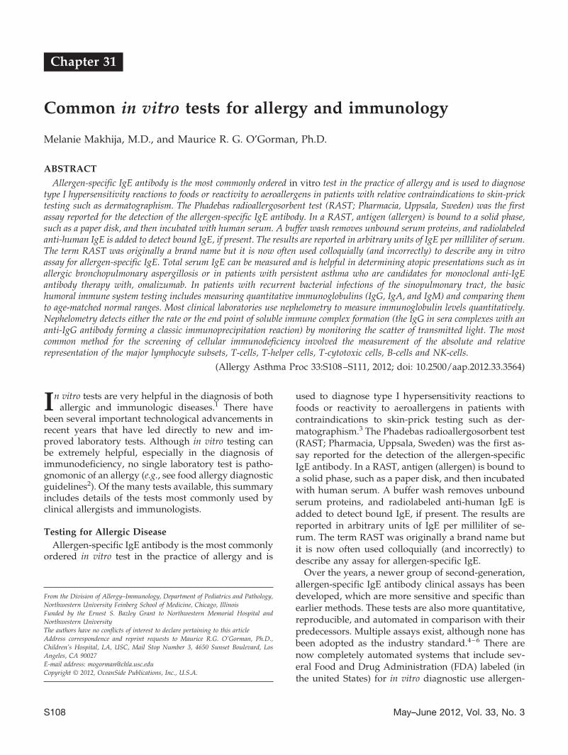
Chapter 31
Common in vitro tests for allergy and immunology
Melanie Makhija, M.D., and Maurice R. G. O’Gorman, Ph.D.
ABSTRACT
Allergen-specific IgE antibody is the most commonly ordered in vitro test in the practice of allergy and is used to diagnosetype I hypersensitivity reactions to foods or reactivity to aeroallergens in patients with relative contraindications to skin-pricktesting such as dermatographism. The Phadebas radioallergosorbent test (RAST; Pharmacia, Uppsala, Sweden) was the firstassay reported for the detection of the allergen-specific IgE antibody. In a RAST, antigen (allergen) is bound to a solid phase,such as a paper disk, and then incubated with human serum. A buffer wash removes unbound serum proteins, and radiolabeledanti-human IgE is added to detect bound IgE, if present. The results are reported in arbitrary units of IgE per milliliter of serum.The term RAST was originally a brand name but it is now often used colloquially (and incorrectly) to describe any in vitroassay for allergen-specific IgE. Total serum IgE can be measured and is helpful in determining atopic presentations such as inallergic bronchopulmonary aspergillosis or in patients with persistent asthma who are candidates for monoclonal anti-IgEantibody therapy with, omalizumab. In patients with recurrent bacterial infections of the sinopulmonary tract, the basichumoral immune system testing includes measuring quantitative immunoglobulins (IgG, IgA, and IgM) and comparing themto age-matched normal ranges. Most clinical laboratories use nephelometry to measure immunoglobulin levels quantitatively.Nephelometry detects either the rate or the end point of soluble immune complex formation (the IgG in sera complexes with ananti-IgG antibody forming a classic immunoprecipitation reaction) by monitoring the scatter of transmitted light. The mostcommon method for the screening of cellular immunodeficiency involved the measurement of the absolute and relativerepresentation of the major lymphocyte subsets, T-cells, T-helper cells, T-cytotoxic cells, B-cells and NK-cells.
(Allergy Asthma Proc 33:S108–S111, 2012; doi: 10.2500/aap.2012.33.3564)
In vitro tests are very helpful in the diagnosis of bothallergic and immunologic diseases.1 There have
been several important technological advancements inrecent years that have led directly to new and im-proved laboratory tests. Although in vitro testing canbe extremely helpful, especially in the diagnosis ofimmunodeficiency, no single laboratory test is patho-gnomonic of an allergy (e.g., see food allergy diagnosticguidelines2). Of the many tests available, this summaryincludes details of the tests most commonly used byclinical allergists and immunologists.
Testing for Allergic DiseaseAllergen-specific IgE antibody is the most commonly
ordered in vitro test in the practice of allergy and is
used to diagnose type I hypersensitivity reactions tofoods or reactivity to aeroallergens in patients withcontraindications to skin-prick testing such as der-matographism.3 The Phadebas radioallergosorbent test(RAST; Pharmacia, Uppsala, Sweden) was the first as-say reported for the detection of the allergen-specificIgE antibody. In a RAST, antigen (allergen) is bound toa solid phase, such as a paper disk, and then incubatedwith human serum. A buffer wash removes unboundserum proteins, and radiolabeled anti-human IgE isadded to detect bound IgE, if present. The results arereported in arbitrary units of IgE per milliliter of se-rum. The term RAST was originally a brand name butit is now often used colloquially (and incorrectly) todescribe any assay for allergen-specific IgE.
Over the years, a newer group of second-generation,allergen-specific IgE antibody clinical assays has beendeveloped, which are more sensitive and specific thanearlier methods. These tests are also more quantitative,reproducible, and automated in comparison with theirpredecessors. Multiple assays exist, although none hasbeen adopted as the industry standard.4–6 There arenow completely automated systems that include sev-eral Food and Drug Administration (FDA) labeled (inthe united States) for in vitro diagnostic use allergen-
From the Division of Allergy–Immunology, Department of Pediatrics and Pathology,Northwestern University Feinberg School of Medicine, Chicago, IllinoisFunded by the Ernest S. Bazley Grant to Northwestern Memorial Hospital andNorthwestern UniversityThe authors have no conflicts of interest to declare pertaining to this articleAddress correspondence and reprint requests to Maurice R.G. O’Gorman, Ph.D.,Children’s Hospital, LA, USC, Mail Stop Number 3, 4650 Sunset Boulevard, LosAngeles, CA 90027E-mail address: [email protected] © 2012, OceanSide Publications, Inc., U.S.A.
S108 May–June 2012, Vol. 33, No. 3

specific IgE tests. Currently, the ImmunoCap System™(Therma Fisher, Upsala, Sweden) is regarded by manyallergists and laboratories as the method of choice. Ituses a cellulose sponge (instead of a paper disk) toenhance the binding capacity to allergens and usesfluorescent anti-IgE in a quantitative fluorescence en-zyme immunoassay. Results are reported quantita-tively (�0.10 to �100 kU/L) as well as by classes(0-VI), although the class system is rarely used in al-lergy practice. For assessing children with food allergy,95% cutoff levels for allergen-specific IgE have beenestablished for predicting clinical reactivity to eggs,milk, peanuts, and fish using the Immunocap system.7
Of note, there is a definite trend for allergen-specificIgE assays that are currently labeled as “research useonly” to be cleared by the FDA as in vitro diagnosticuse for the diagnosis (at least a component of thediagnosis) of allergies. Recently, the FDA has approvedallergen component testing using the Immunocap sys-tem (Phadia Immunology Reference Library PiRL, Ph-adia US, Inc., Portage, MI). This testing can be used forboth food and aeroallergen protein components.Knowledge of allergen component sensitizations mayhelp differentiate between severe/anaphylactic allergyand other allergic diseases such as oral allergy syn-drome.
Pitfalls of the in vitro allergen-specific IgE testinginclude false positives in patients with a high total IgElevel (because of nonspecific binding of allergen tosome immunosorbents) and falsely low results in pa-tients with high levels of IgG antibody. Moreover, thequantity of allergen-specific IgE may not directly re-flect the biologically relevant mast cell–fixed antibodyand severity of clinical reactions.
Total serum IgE can be measured and is helpful indetermining atopic presentations. This test is useful incertain clinical scenarios including diagnosing diseasessuch as allergic bronchopulmonary aspergillosis or al-lergic bronchopulmonary mycosis. It is also tested inpatients in whom the use of the humanized monoclo-nal anti-IgE drug omalizumab (Xolair; Genentech, Inc.[San Francisco, CA] Novartis Pharmaceuticals Corp.[East Hanover, NJ]) is being considered. The clinicalusefulness is limited by age-dependent concentrationand the variation between atopic and nonatopic indi-viduals. Total serum IgE can be measured using theImmunocap™ (Therma Fisher) technology but is alsomeasured with other technologies including enzyme-linked immunosorbent assay (ELISA). Briefly, theELISA procedure includes, first, fixing anti-human IgEto a solid surface, such as a microtiter plate or plasticbead, and then incubating the latter with human se-rum. IgE from human serum binds to the anti-humanIgE fixed on the plate; unbound serum components arewashed away and a secondary enzyme-conjugated (of-ten horseradish peroxidase or alkaline phosphatase)
anti-human IgE antibody is added. The excess conju-gate is washed out, an enzymatic substrate is added,and the colorimetric change produced by the enzy-matic reaction is detected spectrophotometrically. Re-sults are quantitated by extrapolation from a standardcurve of known IgE concentration included in the samemicrotiter plate. Results for total IgE are usually re-ported in international unit per milliliter, kilounit perliter, or nanogram per milliliter (1 IU/mL � 1 kU/L �2.4 ng/mL of IgE).
Epicutaneous skin testing remains the preferredmethod for detection of allergen-specific IgE antibodybecause it is more specific, sensitive, and rapid than itsin vitro counterpart.3 Skin testing involves placing adrop of allergen solution on the skin and introducing itinto the epidermis with a testing device. A reaction(wheal and erythema) is read 15–20 minutes afterplacement. Positive (histamine) and negative (saline)controls are applied together with the allergens to en-sure validity and to control for problems such as pre-medication with antihistamines or dermatographism.The stability and concentration of allergen and tech-nique used may limit the quality of skin testing.
Intradermal skin testing involves using a tuberculinsyringe to administer allergen intracutaneously; 0.02mL of allergen is injected and the test is read 15–20minutes later. This is useful for drug and venom test-ing and is also used for aeroallergen testing if epicuta-neous testing is negative.
Precipitating IgG antibodies (precipitins) are used totest patients who have hypersensitivity pneumonitis in-duced by chronic exposure to organic dust antigens suchas molds (thermophilic actinomyces, Aspergillus species,etc.) and feces/droppings from pet birds.8 This test is onlyavailable in more specialized laboratories.
Mast cells have preformed mediators including tryp-tase, which are used by allergists and emergency depart-ment physicians to measure systemic mast cell activation.Concentrations of �-tryptase correlate with mast cellnumber while �-tryptase concentrations are associatedwith current (acute) mast cell activation. Total serumtryptase can be used to confirm a diagnosis of anaphy-laxis, although samples need to be collected within 4hours of a suspected anaphylactic reaction.9 �-Tryptaselevels are thought to peak 30–60 minutes after a reactionwith a half-life of 2 hours. Normal total tryptase rangesfrom 1 to 10 ng/mL of serum. If baseline tryptase is �20ng/mL in a patient without acute symptoms of anaphy-laxis, indolent systemic mastocytosis should be suspectedand further evaluation sought.
The availability of skin testing as well as the in-creased availability of in vitro testing10 are extremelyhelpful in the diagnosis of allergic disease. The clini-cian must, however, perform a detailed clinical historyand patient presentation to avoid unnecessary testing.
Allergy and Asthma Proceedings S109

Testing for Primary ImmunodeficiencyPatients with primary immune deficiencies present
with recurrent or severe infections, autoimmunity, fail-ure to thrive, diarrhea or any combination of thesesymptoms.11 Appropriate diagnostic testing is essen-tial to making an early diagnosis. Immunologic testingis guided by the clinical presentation of the patientincluding age, sex, type and severity of infections, au-toimmunity, and family history.
Several laboratory tests have been developed to as-sess the major components of the humoral and cellulararms of the immune system.12,13 Patients who presentwith recurrent bacterial infections of the sinopulmo-nary tract should be tested for humoral immune defi-ciencies. The basic humoral immune system testingincludes measuring quantitative immunoglobulins(IgG, IgA, and IgM) and comparing them with age-matched normal ranges. Most clinical laboratories usenephelometry to measure immunoglobulin levelsquantitatively. Nephelometry detects either the rate orthe end point of soluble immune complex formation(the IgG in sera complexes with an anti-IgG antibodyforming a classic immunoprecipitation reaction) bymonitoring the scatter of transmitted light. Accuratemeasurement of antigens (in this case the antigen ac-tually is an immunoglobulin molecule) can be madeonly with conditions of antibody excess. Fluorescentlight sources can be used to enhance the sensitivity ofthis test. As a general rule, in patients with recurrentinfections, an IgG concentration in serum of �300mg/dL in adolescents or adults or values below theage-matched reference value in a child warrants fur-ther workup for humoral immune deficiency.
Further evaluation of humoral immune deficiencies in-clude measurement of specific antibody responses to pre-vious immunizations and isohemagglutinins (usuallyIgM antiblood group A or B antibodies). A patient withlow levels of specific antibody can be immunized withprotein (i.e., tetanus toxoid) or polysaccharide antigen(i.e., pneumococcal polysaccharide) and have antibodiesmeasured 4–6 weeks after vaccination to confirm theability of the patient’s B cells to make specific antibodies.
Combined immune deficiencies and T-cell immune de-ficiencies are evaluated initially with a white blood cellcount and differential. Marked lymphopenia (�3000/mm3) in an infant warrants immediate evaluation.Human immunodeficiency virus infection should beruled out by appropriate antibody/antigen testing. Themost common screening test for cellular immunodefi-ciency involves lymphocyte immunophenotyping. Flowcytometry is used to measure the relative representationand absolute numbers of the major lymphocyte subsets,i.e., B cells, T cells, NK cells, T-helper cells, and cytotoxicT cells. Lymphocyte subset numbers can be comparedwith age-matched reference ranges. Briefly, laser(s)
within the flow cytometer is focused on a fluid streamcontaining cells as they pass single file. The cytometercontains photodiodes and photomultipliers that capturethe scattered laser light in both a forward (roughly cor-relating to size) and at a right angle (roughly correlatedwith the internal complexity of the cell) as well as lightemitted by the fluorochromes that are conjugated to an-tibody (usually monoclonal antibody [mAb]). The mAbare directed against specific targets (e.g., clusters of dif-ferentiation [CD4]) on specific cells (e.g., CD3� T cells).Based on their innate physical properties lymphocytesgenerate specific light scatter signals that allows them tobe differentiated from monocytes and granulocytes. Thelymphocytes will also emit a specific fluorescence sig-nal(s) depending on the specific mAb bound to theirsurface. Various panels of mAb are designed to measurespecific subsets of lymphocytes based primarily on theexpression of different CDs. Abnormalities in the relativerepresentation of lymphocyte subsets are readily identi-fied and known to be associated with specific primaryimmunodeficiency diseases. It is also possible to measurethe expression of known antigens associated with specificprimary immunodeficiency diseases (e.g., CD40 ligand,major histocompatibility complex class I, IL-2R� chain,and CD3 chains); however, because of the rare incidenceof many of the primary immunodeficiency diseases andthe relative complex and expensive nature of these morespecific flow cytometry–based tests, they are only per-formed in highly specialized laboratories.
Flow cytometry is extremely useful in the classificationof hematopoietic malignancies. Assessment of T-cellfunction can be performed by stimulating T cells withmitogens (e.g., phytohemagglutinin, concanavalin A, andpokeweed mitogen) and antigens (Candida, tetanus, andothers). If 22q11 deletion syndrome (DiGeorge syn-drome) is suspected, fluorescence in situ hybridization forthe microdeletion can be performed. Measuring the num-ber of T-cell receptor excision circles (TRECs) in driedblood spots has recently been adopted by a few states inthe United States as a newborn screen for severe com-bined immune deficiency. TRECs numbers also havebeen measured for evaluating the severity of T-cell de-pletion in DiGeorge patients and evaluating immune re-constitution after bone marrow transplantation althoughthis has not been widely adopted. TRECs are circularpieces of DNA formed as T cells rearrange their T-cellreceptor genes during T-cell maturation in the thymusand do not replicate; therefore, the number of TRECscorrelates with the production of new T cells from thethymus. TRECs are measured using appropriate DNAprimers and quantitative DNA amplification techniques.
Another more specialized test involves the assessmentof the breadth of the T-cell repertoire. Certain primaryimmunodeficiency diseases, e.g., Omenn syndrome pa-tients, develop only a few mature T-cell clones in theperiphery, so-called oligoclonal expansion. A relatively
S110 May–June 2012, Vol. 33, No. 3

easy method to measure the T-cell repertoire can be per-formed using a panel of monoclonal antibodies directedagainst 24 V �-family–specific targets on the T-cell recep-tor. Each of the 24 T-cell receptor V�-chain families arefound in healthy subjects. In patients with Omenn syn-drome or atypical DiGeorge syndrome, only a few V�-families will be represented.
Testing for other immune deficiencies are available andinclude a variety of procedures designed to detect cellulartargets known to be associated with specific immune abnor-malities. Screening for chronic granulomatous disease isnow commonly performed using a flow cytometry assayand an oxygen-sensitive dye, dihydrorhodamine 123 tomeasure the ability of granulocytes to exhibit an oxidativeburst in vitro or can be done using the older nitroblue tetra-zolium test. Leukocyte adhesion deficiency testing is alsoperformed by flow cytometry assessment of CD11 andCD18 or CD15, adhesion molecules on neutrophils, whichare absent or decreased in this disorder.
Complement deficiency screening is performed in pa-tients with recurrent sinopulmonary infections (C2 defi-ciency), pyogenic infections (C3, factor I, and H), systemiclupus erythematosus-like symptoms of autoimmunity (C1q,r, s, C2, or C4 deficiencies) or susceptibility to neisserialinfections (terminal complement defects C5-C9, factor D, orproperdin). Testing involves complement function screen-ing of both the classic (CH50) and the alternative (AH50)pathways and assessing specific complement componentlevels when function is markedly decreased. A decreasedAH50 test suggests problems with factor B, D, or properdin.Decreased CH50 and AH50 suggests a defect in a sharedcomplement component (C3–C9).
Other testing for immune deficiencies is disease spe-cific. A few examples include ALPS (increased numberCD4 and CD8 [double negative] T-cells expression the�/�-form of the T-cell receptor); IPEX (diminished orabsent FOXP3 protein in the nucleus of CD4 T cells),APECED/chronic mucocutaneous candidiasis (AIRE orother genetic sequencing), or X-linked lymphoprolifera-tive disorder presenting with hemophagocytic lympho-histiocytosis (NK-cell testing and genetic sequencing) andToll-like receptor pathway testing for disorders such asIRAK4, MYD88, and NEMO.
As with allergic diseases, laboratory-based testingfor immune deficiencies is imperative in guiding cor-rect diagnosis and management; however, testingshould be used judiciously in patients in whom a highlevel of suspicion is present.
CLINICAL PEARLS
• In vitro tests for anti-allergen IgE can not be used assole evidence in the diagnosis of allergy. (high sen-sitivity but not high enough specificity).
• The ImmunoCAP™ System is widely regarded asthe method of choice for detection of allergen-spe-cific IgE antibody. However, the in vivo test is pref-erable to all in vitro methods.
• New component allergy testing is starting to be usedto differentiate patients with reactivity that is asso-ciated with severe anaphylaxis.
• Total IgE is can be measured by ImmunoCAP, neph-elometry, and ELISA.
• Quantitative immunoglobulins are measured bynephelometry and used to screen for humoral im-mune deficiencies.
• Flow cytometry is used to detect abnormalities inthe major lymphocyte subsets (such as T and B cells).
• Flow cytometry also is used to identify the abnormalrepresentation of rare lymphocyte subsets, abnormalsurface marker expression, the T-cell repertoire, andspecific neutrophil functions associated with knownprimary immunodeficiency diseases.
REFERENCES1. Bernstein IL, Li JT, Bernstein DI, et al. Allergy diagnostic test-
ing: An updated practice parameter. Ann Allergy. 100(Supp3):S1–S148, 2008.
2. Boyce JA, Assa’ad A, Burks W, et al. Guidelines for the diag-nosis and management of food allergy in the United States:Summary of the NIAID-Sponsored expert panel report. J Al-lergy Clin Immunol 126:1105–1118, 2010.
3. Carr TF, and Saltoun CA. Skin testing in allergy. AllergyAsthma Proc 33:S6–S8, 2012.
4. Jiang XD, Li GY, Dong Z, and Zhu DD. Correlation analysis oftwo serum-specific immunoglobulin E test systems and skin-prick test in allergic rhinitis patients from northeast China. Am JRhinol Allergy 25:116–119, 2011.
5. Jung YG, Cho HJ, Park GY, et al. Comparison of the skin-pricktest and Phadia ImmunoCAP as tools to diagnose house-dustmite allergy. Am J Rhinol Allergy 24:226–229, 2010.
6. Sarratud T, Donnanno S, Terracciano L, et al. Accuracy of apoint-of-care testing device in children with suspected respira-tory allergy. Allergy Asthma Proc 31:e11–e17, 2010.
7. Sampson HA. Utility of food specific IgE concentrations inpredicting symptomatic food allergy. J Allergy Clin Immunol107:891–896, 2001.
8. Blatman KH, and Grammer LC. Hypersensitivity pneumonitis.Allergy Asthma Proc. 33:S64–S66, 2012.
9. Greenberger PA, and Ditto AM. Anaphylaxis. Allergy AsthmaProc 33:S80–S83, 2012.
10. Hamilton RG. Clinical laboratory assessment of immediate-typehypersensitivity. J Allergy Clin Immunol 125:S284–S296, 2010.
11. UzzamannA, and Fuleihan RL. Approach to primary immuno-deficiency. Allergy Asthma Proc 33:S91–S95, 2012.
12. Oliveira JB, and Fleischer TA. Laboratory evaluation of primaryimmunodeficiencies. J Allergy Clin Immunol 125:S297–S305,2010.
13. O’Gorman MRG. Role of flow cytometry in the diagnosis andmonitoring of primary immunodeficiency disease. In Hand-book of Human Immunology, 2nd ed. O’Gorman MRG, andDonnengerg AD (Eds). Boca Raton, FL: CRC Press, Taylor andFrances Group, 267–311, 2008. e
Allergy and Asthma Proceedings S111