Embed Size (px)
Citation preview
7/21/2013 1
Allergy and Hypersensitivity

7/21/2013 2
Introduction
Allergy is an altered reactivity to antigenic stimulation.
von Pirquet intended that the term refers to all forms of
changed reactivity to antigenic stimulation (immunity or
hypersensitivity).
By contrast, the term allergy now is almost exclusively
used, particularly in the clinical setting, to refer to a subset
of potentially harmful immune responses.
7/21/2013 3
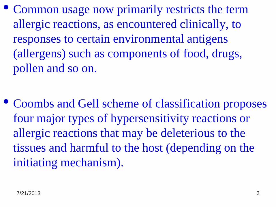
• Common usage now primarily restricts the term
allergic reactions, as encountered clinically, to
responses to certain environmental antigens
(allergens) such as components of food, drugs,
pollen and so on.
• Coombs and Gell scheme of classification proposes
four major types of hypersensitivity reactions or
allergic reactions that may be deleterious to the
tissues and harmful to the host (depending on the
initiating mechanism).

7/21/2013 4

7/21/2013 5
Type 1 Hypersensitivity – Allergy
Immediate type (IgE – Mediated): Asthma, Hay fever, Atopic dermatitis, and Allergic gastroenteritis
Atopy : affects up to 40% of the population.
Hygienic hypothesis.
General Features
• genetically determined
• continued production of IgE.
• target organ hyper-responsiveness.

7/21/2013 6
Type 1 Hypersensitivity – AllergyPathology is related to mast cell degranulation, and the
reaction is driven by mast cell mediators
General features of IgE – associated allergies:
• They are often associated with high levels of IgE production.
• They are now known to be promoted by antigen – specific Th2 cells.
• The tissue responses may be affected by genetic and diverse nongenetic factors
• They have two phases, sensitization and effector phases.
• Clinically they have acute, late and chronic phases.

7/21/2013 7
AllergensAllergens are antigens that can elicit specific IgE
responses.
The identification of a simple set of “ general rules for
allergenicity” has remained elusive.
Certain generalizations about the properties of allergens ( all
of which have exceptions) have emerged:
• Allergens typically are proteins (often glycoproteins) or
chemicals (haptens) that can become bound to proteins.
• Analysis of common allergens so far have not revealed why
these substances induce a Th2 cell driven IgE associated
response.
7/21/2013 8
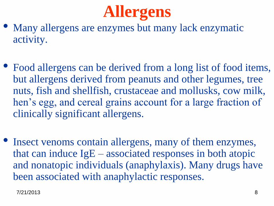
Allergens• Many allergens are enzymes but many lack enzymatic
activity.
• Food allergens can be derived from a long list of food items, but allergens derived from peanuts and other legumes, tree nuts, fish and shellfish, crustaceae and mollusks, cow milk, hen’s egg, and cereal grains account for a large fraction of clinically significant allergens.
• Insect venoms contain allergens, many of them enzymes, that can induce IgE – associated responses in both atopic and nonatopic individuals (anaphylaxis). Many drugs have been associated with anaphylactic responses.
7/21/2013 9
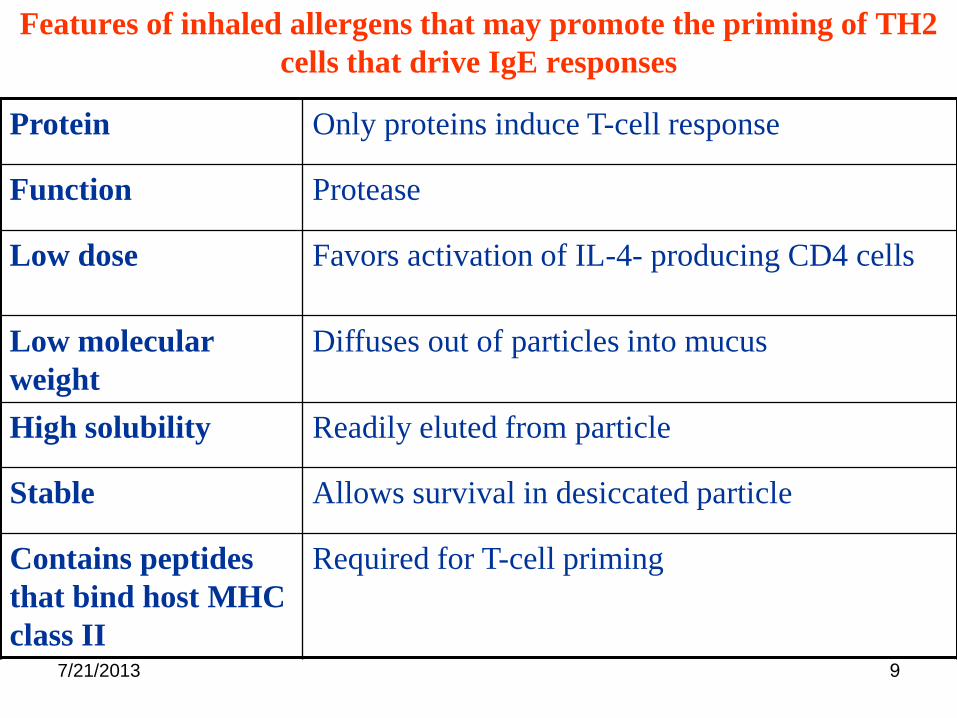
Features of inhaled allergens that may promote the priming of TH2
cells that drive IgE responses
Protein Only proteins induce T-cell response
Function Protease
Low dose Favors activation of IL-4- producing CD4 cells
Low molecular
weight
Diffuses out of particles into mucus
High solubility Readily eluted from particle
Stable Allows survival in desiccated particle
Contains peptides
that bind host MHC
class II
Required for T-cell priming
7/21/2013 10
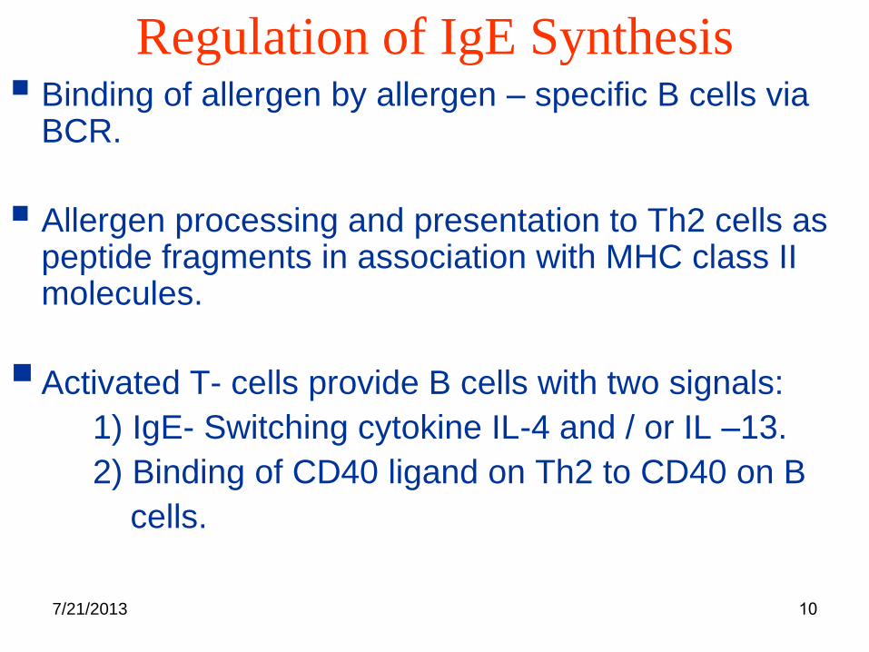
Regulation of IgE Synthesis Binding of allergen by allergen – specific B cells via
BCR.
Allergen processing and presentation to Th2 cells as peptide fragments in association with MHC class II molecules.
Activated T- cells provide B cells with two signals:
1) IgE- Switching cytokine IL-4 and / or IL –13.
2) Binding of CD40 ligand on Th2 to CD40 on B
cells.

7/21/2013 11
Y
T cell help to B cells
B
Antigen
ThTh2
IL-4 and IL-13
CD40 Ligand
CD40

7/21/2013 12
Receptors For IgEThe high affinity receptor Fcε R1
• primarily expressed on mast cells and basophils
• The activation of mast cells or basophils by FCεR1
aggregation initiates a coordinated sequence of
biochemical and morphological events that result in:
1) Exocytosis of secretary granules.
2) Synthesis and secretion of newly formed mediators.
3) Synthesis and secretion of cytokines

7/21/2013 13
Mast Cells and the Allergic Response

7/21/2013 14
Receptors For IgE• Mediators are responsible for symptoms.
• Serum IgE concentration and FcεR1 expression correlate.
• Levels of FcεR1 expression can be regulated by IgE.
• IgE - dependent upregulation of FcεR1 expression may be
part of a positive feedback mechanism for inducing further
production of IgE ( mast cells and basophils produce IL-4).
Fc εR II / CD23( binds IgE with a relatively low affinity)

7/21/2013 15
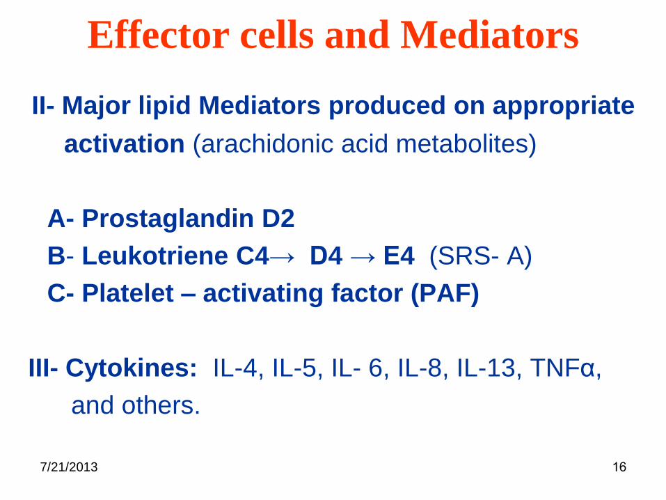
Effector cells and Mediators
Mast Cells and Basophils
• Distributed strategically throughout normal connective
tissues.
• Contain or elaborate on appropriate stimulation, a diverse
array of potent biologically active mediators.
I- Major mediators stored in cytoplasmic granules:
A- Histamine
B- Others ( heparin, proteases, acid hydrolases,
cathepsin G, and Carboxypeptidases).
7/21/2013 16
Effector cells and Mediators
II- Major lipid Mediators produced on appropriate
activation (arachidonic acid metabolites)
A- Prostaglandin D2
B- Leukotriene C4→ D4 → E4 (SRS- A)
C- Platelet – activating factor (PAF)
III- Cytokines: IL-4, IL-5, IL- 6, IL-8, IL-13, TNFα,
and others.

7/21/2013 17
Compounds Released from Mast Cells
Pre-formed and
in granules
Synthesized
upon mast cell
activation

7/21/2013 18
Effector cells and MediatorsEosinophils
• Granules contain Lysosomal hydrolases and cationic proteins.
• Crystalloid core of the granule is composed of the major basic protein ( MBP).
• The non core matrix contains eosinophil cationic protein (ECP), eosinophil- derived neurotoxin (EDN), eosinophil peroxidase, lysosomal hydrolases and lysophospholipase (Charcot – Lyden crystals)

7/21/2013 19
Eosinophils
• They also produce lipid mediators upon
activation (Lt- C4, Lipoxins) and Cytokines
(IL-1α, IL-2, IL-3, IL-4, IL-5, IL-6, IL-8, IL-10,
IL–16, GM-CSF, TNF-α ….)
• The potential for eosinophils to cause tissue
injury is illustrated by the rare hypereosinophilic
syndromes.

7/21/2013 20
Effector cells and Mediators
Eosinophils
• The clinical manifestations are damage to the
endocardium and nerves leading to heart failure
and neuropathy.
• Eosinophils accumulate in large numbers in
local allergic reactions
• Their continued presence is characteristic of
chronic allergic inflammation
• Eosinophils are thought to be the chief
contributor to tissue damage that occurs.

7/21/2013 21
Compounds Released from Eosinophils
7/21/2013 22
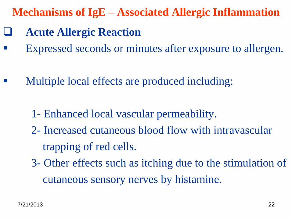
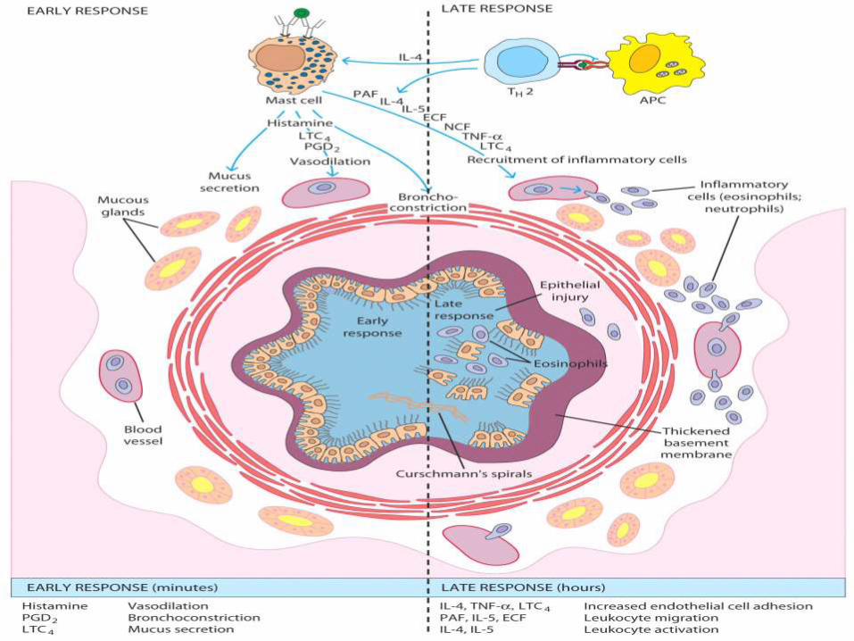
Mechanisms of IgE – Associated Allergic Inflammation
Acute Allergic Reaction
Expressed seconds or minutes after exposure to allergen.
Multiple local effects are produced including:
1- Enhanced local vascular permeability.
2- Increased cutaneous blood flow with intravascular
trapping of red cells.
3- Other effects such as itching due to the stimulation of
cutaneous sensory nerves by histamine.

7/21/2013 23
Mechanisms of IgE – Associated Allergic Inflammation
Late – Phase Reaction
It characteristically does not develop until several hours
after initial allergen challenge, in many cases after the
signs and symptoms related to the acute allergic reaction
have greatly diminished or even disappeared.
It is now clear that a large fraction of patients with
allergic asthma (50% of adults and < 70% of children)
express late phase reaction to inhaled allergens (second
phase of bronchoconstriction which usually is maximal at
6-12 hours and resolves by 24 hours).
7/21/2013 24

7/21/2013 25
Mechanisms of IgE – Associated Allergic Inflammation
Several points about human LPR appear to be well
established:
• The response can be elicited by appropriate allergen
challenge.
• The reaction is almost always preceded by an acute allergic
reaction.
• The signs and symptoms characteristic of LPR are
associated with the recruitment of circulating leukocytes to
the site of the reaction.

7/21/2013 26
Mechanisms of IgE – Associated Allergic Inflammation
Chronic Allergic Inflammation
• It typically occurs at anatomic sites that have been repeatedly challenged with allergens over prolonged periods.
• Sites contain effector cells that have been recruited from the circulation and also can be associated with striking, chronic changes in underlying tissues.
• The inflammatory infiltrate at such sites typically include eosinophils and T – cells (esp. Th2) and the affected tissue exhibit significant alterations in their function.

7/21/2013 27
• Eosinophil & mononuclear cells infiltrate the bronchi of
asthmatics
• Activated T cells elevated in the peripheral blood of
severe acute asthmatics
• Activated T cells in peripheral blood correlated with
airway narrowing
• Bronchial CD4 lymphocyte numbers correlated with
eosinophil numbers
• Elevated IL-5 expressing T cells in asthmatic bronchial
mucosa and BAL
Cellular culprits of allergy: T cells

7/21/2013 28
• T cells that release IL-5 co-localise with eosinophils
• Eosinophils cause airway hyperresponsiveness, inflammation, desquamative bronchitis, mucous hypersecretion and smooth muscle contraction
• IL-5 promotes differentiation and regulates the survival of eosinophils
• Steroid treatment is associated with a decrease in IL-5 producing cells
7/21/2013 29
Anaphylaxis
• Refers to the occurrence of IgE - mediated reaction simultaneously in multiple organs.
• The usual causative allergen is a drug, insect venom or food. It can be evoked by minute amounts of an antigen.
• It lacks the genetic propensity of atopy and it has no predilection for the atopic individual.
• Urticaria (skin) and angioedema (subcutaneous tissue) are mild localized forms of anaphylaxis.

7/21/2013 30
IgE-Mediated Allergic ReactionsSyndrome Common
allergens
Route of
entry
Response
Systemic
anaphylaxis
Drugs, serum,
venoms, peanuts
Intravenous
(either directly
or following
rapid
absorption)
Edema, increased vascular
permeability. Tracheal
occlusion. Circulatory
collapse. Death
Wheal-and-flare Insect bites
Allergy testing
Subcutaneous Local increase in blood flow
and vascular permeability
Allergic rhinitis
(hay fever)
Pollens (ragweed,
timothy, birch)
Dust- mite feces
Inhaled Edema of nasal mucosa.
Irritation of nasal mucosa
Bronchial
asthma
Pollens
Dust- mite feces
Inhaled Bronchial constriction
Increased mucus production
Airway inflammation
Food allergy Shellfish, Milk,
Eggs, Fish, Wheat
Oral Vomiting, Diarrhea, Pruritis
(itching), Urticaria (hives),
Anaphylaxis (rarely)

7/21/2013 31

7/21/2013 32

7/21/2013 33Normal larynx Laryngeal oedema

7/21/2013 34
Diagnosis and Management of IgE – Mediated Allergy
Diagnosis
RIST, RAST, Skin testing
Management
- Drug treatment
- Desensitization
- Vaccination by allergen peptides
- Anti IgE receptor
- Inhibitors of cytokines
- Blocking of mediator actions.

7/21/2013 35
Allergy treatments
make IgG response
to compete with IgE
7/21/2013 36
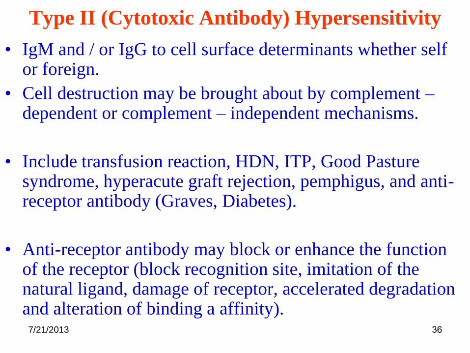
Type II (Cytotoxic Antibody) Hypersensitivity
• IgM and / or IgG to cell surface determinants whether self or foreign.
• Cell destruction may be brought about by complement –dependent or complement – independent mechanisms.
• Include transfusion reaction, HDN, ITP, Good Pasture syndrome, hyperacute graft rejection, pemphigus, and anti-receptor antibody (Graves, Diabetes).
• Anti-receptor antibody may block or enhance the function of the receptor (block recognition site, imitation of the natural ligand, damage of receptor, accelerated degradation and alteration of binding a affinity).

7/21/2013 37
Type III (Immune complex) Hypersensitivity
• Soluble immune complexes at slight antigen excess.
• Deposition is related to hemodynamics.
• The prototype of the reaction is Arthus Reaction in rabbits.
• Serum sickness in humans.
• The pathology is marked by a neutrophil - rich infiltrate
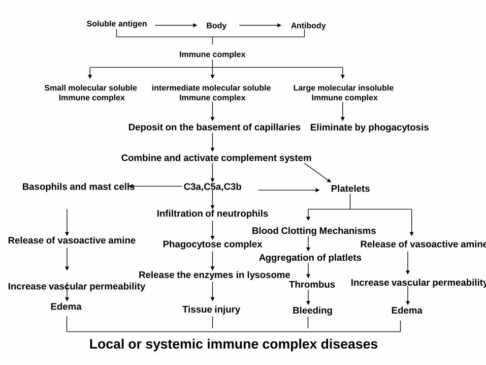
Soluble antigen Body Antibody
Immune complex
Small molecular soluble
Immune complex
intermediate molecular soluble
Immune complex
Large molecular insoluble
Immune complex
Deposit on the basement of capillaries
Combine and activate complement system
C3a,C5a,C3b
Infiltration of neutrophils
Phagocytose complex
Release the enzymes in lysosome
Tissue injury
Eliminate by phogacytosis
Platelets
Thrombus
Aggregation of platlets
Blood Clotting Mechanisms
Release of vasoactive amine
Increase vascular permeability
Bleeding Edema
Basophils and mast cells
Release of vasoactive amine
Increase vascular permeability
Edema
Local or systemic immune complex diseases

7/21/2013 39
Serum Sickness
• A systemic immune- complex complement
dependent reaction to extrinsic antigens
• Severity is antigen-dose dependent
• Fever, skin rash, lymphadenopathy, and
arthralgia
• C5 activates neutrophils to secrete protease
that produce tissue damage

7/21/2013 40
Serum Sickness (immune complexes in the blood)
.
7/21/2013 41
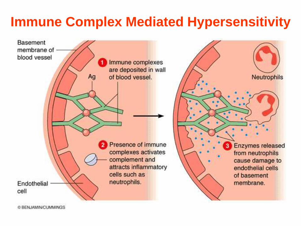
Immune Complex Mediated Hypersensitivity
7/21/2013 42
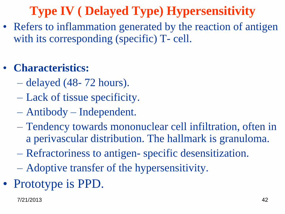
Type IV ( Delayed Type) Hypersensitivity
• Refers to inflammation generated by the reaction of antigen with its corresponding (specific) T- cell.
• Characteristics:
– delayed (48- 72 hours).
– Lack of tissue specificity.
– Antibody – Independent.
– Tendency towards mononuclear cell infiltration, often in a perivascular distribution. The hallmark is granuloma.
– Refractoriness to antigen- specific desensitization.
– Adoptive transfer of the hypersensitivity.
• Prototype is PPD.

7/21/2013 43
Delayed-type hypersensitivity (DTH)
(e.g., tuberculin skin test)
TH1 from a previous
immunization (memory)

7/21/2013 44

7/21/2013 45
7/21/2013 46
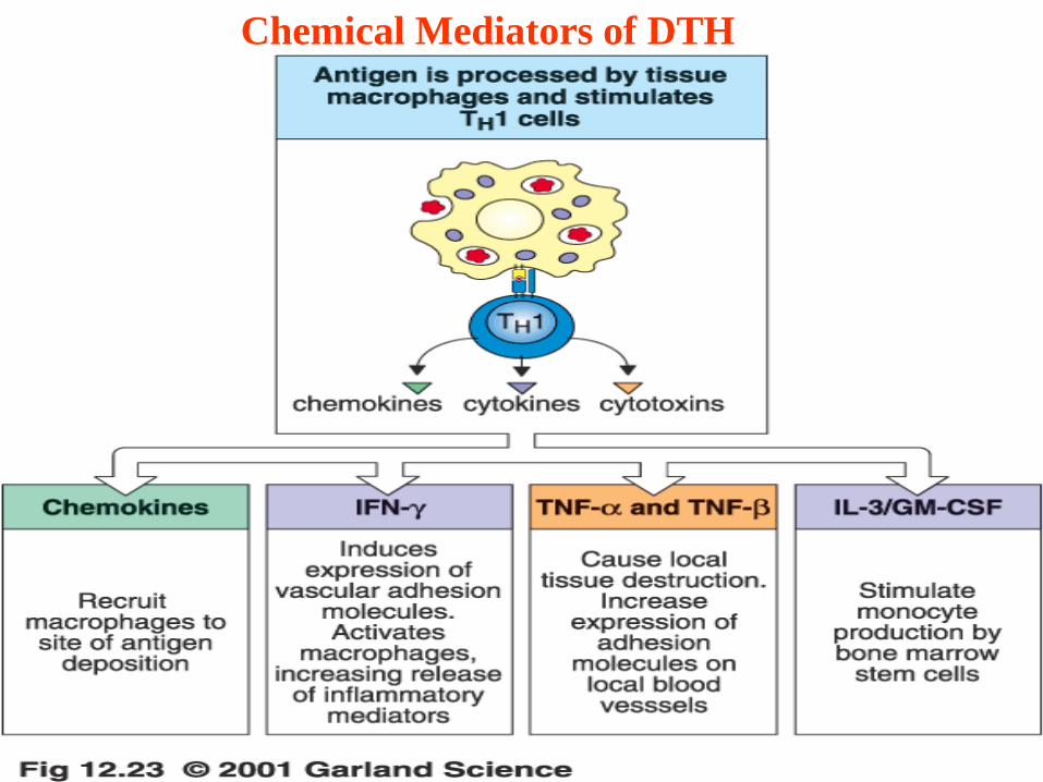
Chemical Mediators of DTH

7/21/2013 47
Delayed-type hypersensitivity

7/21/2013 48
Type IV ( Delayed Type) Hypersensitivity
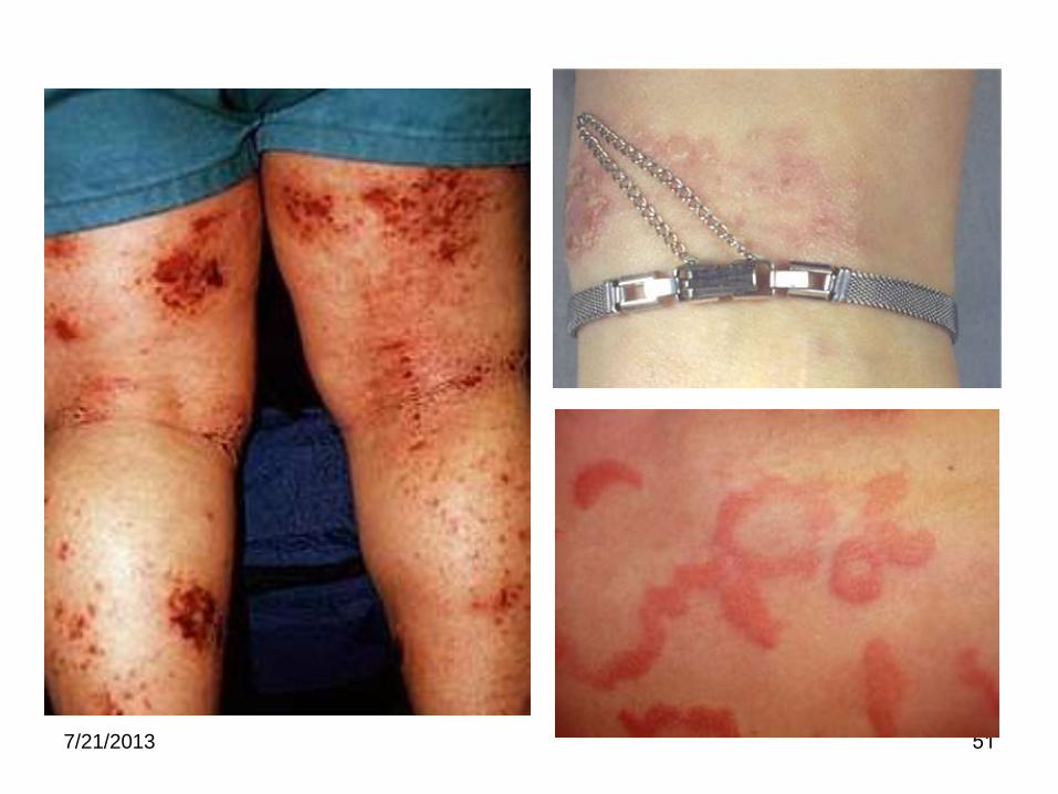
Allergic contact dermatitis
• Pentadecyl catechol (poison ivy and poison oak).
• Organic chemicals (cosmetic, insecticides, disinfectants).
• Metals (nickel, mercury, chromium, cupper).
• Synthetic products (rubber, leather).
Photoallergic contact Dermatitis
• Antigens require activation by UV light.
• Affects sun exposed areas.
• Associated with drugs or chemical constituents of topical products such as soap, cosmetics, and topical drugs.

7/21/2013 49
Allergic Contact Dermatitis Response
to Poison Ivy Hapten
7/21/2013 50
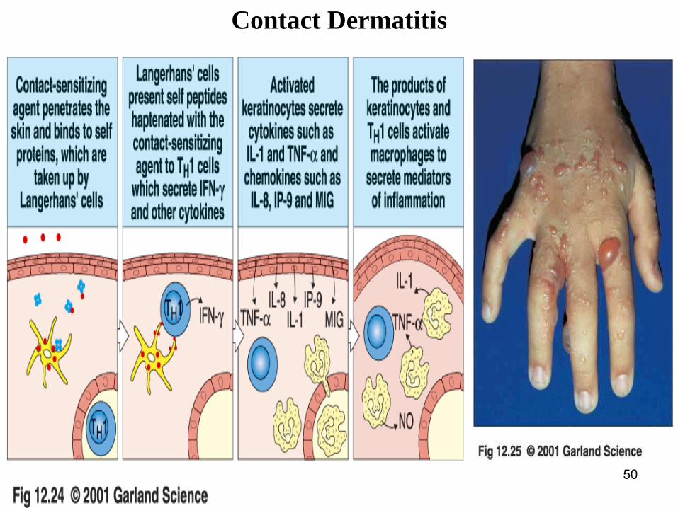
Contact Dermatitis
7/21/2013 51

7/21/2013 52
Allergic Bronchopulmonary Aspergillosis
• Biphasic: IgE to spore allergens (atopy) and IgG to
mycelial antigen (serum sickness).
Hypersensitivity pneumonitis (extrinsic allergic alveolitis)
• It involoves antibodies, T cells or both but T – cell
mechanisms predominate.
• Allergens are frequently components of biologic organisms
or their products.
• It is an occupational disease ( Bird handlers disease, birds
fanciers lungs, pigeon breeder’s disease …)

7/21/2013 53
Tuberculoid leprosy
Low infectivity
Localised infection
Normal serum Ig
Normal T cell response
Poor growth of mycobacteria in
macrophages
Th2Th1
Lepromatous leprosy
High infectivity
Disseminated infection
Hypergammaglobulinaemia
Unresponsive
Florid growth of mycobacteria in
macrophages
Relevance of Th subsets in humans
Lepromatous and tuberculoid leprosy
Infection with Mycobacterium leprae shows two main
clinical forms associated with Th1 and Th2 responses

7/21/2013 54
Tuberculoid
leprosy

7/21/2013 55Lepromatous Leprosy

7/21/2013 56