Embed Size (px)
Citation preview
DCIS = ductal carcinoma in situ; IDC = invasive ductal carcinoma; LOH = loss of heterozygosity; PCR = polymerase chain reaction.
Available online http://breast-cancer-research.com/content/3/3/192
Primary researchAllelic loss on chromosome band 18p11.3 occurs early andreveals heterogeneity in breast cancer progressionKanokwan Kittiniyom*, Karen M Gorse†, Fabienne Dalbegue‡, Jack H Lichy‡, Jeffery K Taubenberger‡ and Irene F Newsham*†
*Department of Pathology and †Department of Anatomy, Virginia Commonwealth University, Richmond, Virginia, USA‡Department of Cellular Pathology, Armed Forces Institute of Pathology, Washington, District of Columbia, USA
Correspondence: Irene Newsham, PhD, Department of Neurosurgery, Henry Ford Hospital, E&R Buillding, Room 3096, 2799 West Grand Boulevard,Detroit, MI 48202, USA. Tel: +1 313 916 8640; fax: +1 413 487 9415; e-mail: [email protected]
Abstract
We examined the stage specificity and heterogeneity of 18p11 alterations in a series of tumorsrepresenting 96 microdissected samples. Significant loss of heterozygosity (LOH) (63%) was found,with 56% occurring early in ductal carcinoma in situ. Although most cases indicated LOH was clonallyinherited, heterogeneity for 18p LOH occurred in 27% of tumors. When compared with other LOHdata, 18p LOH was found in conjunction with allelic deletion on 3p, 9p, 17p and 17q, while 13q, 16q,and 11p were less frequently associated. These analyses suggest chromosome 18p11 alteration is acommon and early event in breast disease.
Keywords: 18p, breast cancer, heterogeneity, loss of heterozygosity, progression
Synopsis
Introduction: A complete understanding of the genetic eventsinvolved in breast tumorigenesis and their heterogeneity inrelation to stage specificity will be critical to the development ofsuccessful treatments. This laboratory and other workersrecently reported the presence of a novel LOH region in band18p11.3 in non-small cell lung carcinomas, glioblastomas andinvasive breast carcinomas [1,2]. This current study usesinformative loci in 18p11.3 to investigate the stage specificityand extent of molecular heterogeneity exhibited by this locus inbreast cancer progression. These results are analyzed inconjunction with other LOH data available for markers on 3p,11p, 13q, 16p, 17p, and 17q in an effort to place chromosome18p alterations on the breast cancer progression pathway.Materials and methods: The material used in this study,consisting of formalin-fixed, paraffin-embedded tissue from thearchives of the Armed Forces Institute of Pathology, has beenpreviously described [3]. Tumors were selected at random forthose with the presence of lesions representing multiple stages of
breast disease within individual patient samples. Ninety-six focirepresenting normal, intraductal and infiltrating tumor weremicrodissected from 30 tumors. Matching normal and tumor DNAwere subjected to polymerase chain reaction (PCR)-based LOHanalysis using two 18p11.3 markers (D18S59 and D18S481)and one 18p11.2 marker (D18S452). Forward primers weresynthesized with either a fluorescent FAM tag (D18S481 andD18S452) or a fluorescent TET tag (D18S59) on the 5′ end.PCR reactions were performed in 10 µl with aliquots of normaland tumor DNA using a PCR protocol consisting of 94°C for30 s, 55°C for 30 s, and 72°C for 1 min for a total of 40 cycles.Horizontal ultrathin, high throughput fluorescence-based DNAfragment gel electrophoresis (GTI-9600; Genesys Technologies,Inc, Sauk City, WI, USA) was applied to the separation andanalysis of PCR-generated alleles as previously described [4].Allelic ratios were calculated and expressed as a percentage ofloss of intensity for the tumor allele compared with thecorresponding normal allele (D-value) after normalization.
Received: 18 September 2000
Revisions requested: 31 October 2000
Revisions received: 5 December 2000
Accepted: 19 January 2001
Published: 12 February 2001
Breast Cancer Res 2001, 3:192–198
This article may contain supplementary data which can only be foundonline at http://breast-cancer-research.com/content/3/3/192
© 2001 Kittiniyom et al, licensee BioMed Central Ltd(Print ISSN 1465-5411; Online ISSN 1465-542X)
Available online http://breast-cancer-research.com/content/3/3/192
comm
entaryreview
reportsprim
ary research
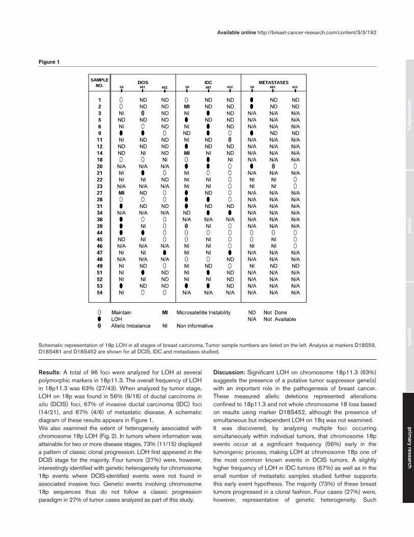
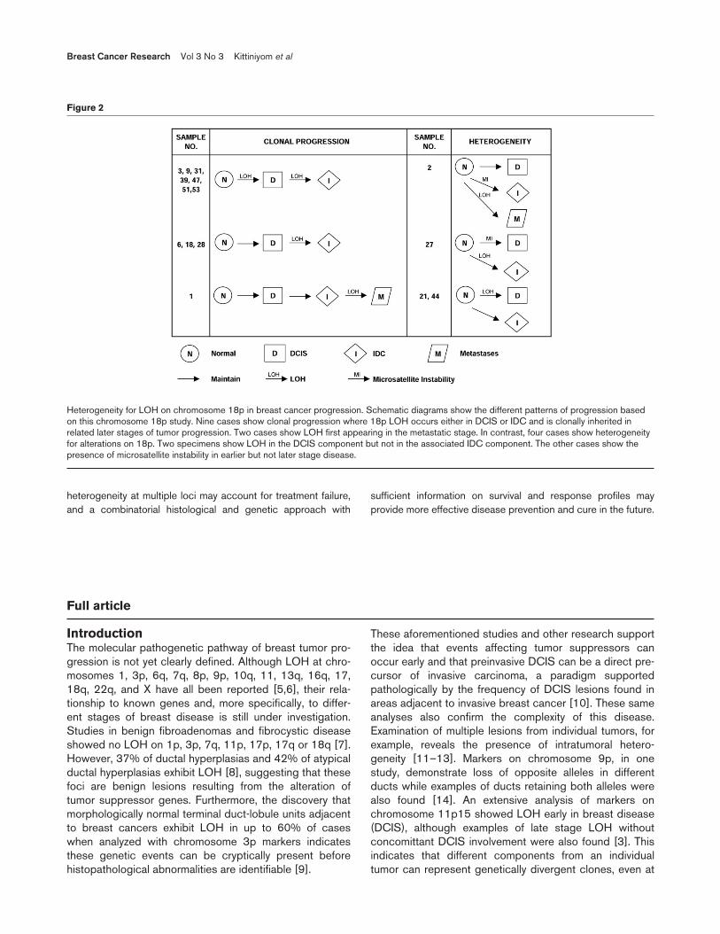
Results: A total of 96 foci were analyzed for LOH at severalpolymorphic markers in 18p11.3. The overall frequency of LOHin 18p11.3 was 63% (27/43). When analyzed by tumor stage,LOH on 18p was found in 56% (9/16) of ductal carcinoma insitu (DCIS) foci, 67% of invasive ductal carcinoma (IDC) foci(14/21), and 67% (4/6) of metastatic disease. A schematicdiagram of these results appears in Figure 1.We also examined the extent of heterogeneity associated withchromosome 18p LOH (Fig. 2). In tumors where information wasattainable for two or more disease stages, 73% (11/15) displayeda pattern of classic clonal progression. LOH first appeared in theDCIS stage for the majority. Four tumors (27%) were, however,interestingly identified with genetic heterogeneity for chromosome18p events where DCIS-identified events were not found inassociated invasive foci. Genetic events involving chromosome18p sequences thus do not follow a classic progressionparadigm in 27% of tumor cases analyzed as part of this study.
Discussion: Significant LOH on chromosome 18p11.3 (63%)suggests the presence of a putative tumor suppressor gene(s)with an important role in the pathogenesis of breast cancer.These measured allelic deletions represented alterationsconfined to 18p11.3 and not whole chromosome 18 loss basedon results using marker D18S452, although the presence ofsimultaneous but independent LOH on 18q was not examined.It was discovered, by analyzing multiple foci occurringsimultaneously within individual tumors, that chromosome 18pevents occur at a significant frequency (56%) early in thetumorigenic process, making LOH at chromosome 18p one ofthe most common known events in DCIS tumors. A slightlyhigher frequency of LOH in IDC tumors (67%) as well as in thesmall number of metastatic samples studied further supportsthis early event hypothesis. The majority (73%) of these breasttumors progressed in a clonal fashion. Four cases (27%) were,however, representative of genetic heterogeneity. Such
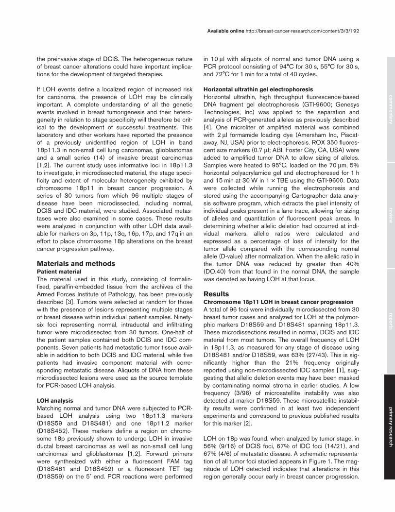
Figure 1
Schematic representation of 18p LOH in all stages of breast carcinoma. Tumor sample numbers are listed on the left. Analysis at markers D18S59,D18S481 and D18S452 are shown for all DCIS, IDC and metastases studied.
Breast Cancer Research Vol 3 No 3 Kittiniyom et al
IntroductionThe molecular pathogenetic pathway of breast tumor pro-gression is not yet clearly defined. Although LOH at chro-mosomes 1, 3p, 6q, 7q, 8p, 9p, 10q, 11, 13q, 16q, 17,18q, 22q, and X have all been reported [5,6], their rela-tionship to known genes and, more specifically, to differ-ent stages of breast disease is still under investigation.Studies in benign fibroadenomas and fibrocystic diseaseshowed no LOH on 1p, 3p, 7q, 11p, 17p, 17q or 18q [7].However, 37% of ductal hyperplasias and 42% of atypicalductal hyperplasias exhibit LOH [8], suggesting that thesefoci are benign lesions resulting from the alteration oftumor suppressor genes. Furthermore, the discovery thatmorphologically normal terminal duct-lobule units adjacentto breast cancers exhibit LOH in up to 60% of caseswhen analyzed with chromosome 3p markers indicatesthese genetic events can be cryptically present beforehistopathological abnormalities are identifiable [9].
These aforementioned studies and other research supportthe idea that events affecting tumor suppressors canoccur early and that preinvasive DCIS can be a direct pre-cursor of invasive carcinoma, a paradigm supportedpathologically by the frequency of DCIS lesions found inareas adjacent to invasive breast cancer [10]. These sameanalyses also confirm the complexity of this disease.Examination of multiple lesions from individual tumors, forexample, reveals the presence of intratumoral hetero-geneity [11–13]. Markers on chromosome 9p, in onestudy, demonstrate loss of opposite alleles in differentducts while examples of ducts retaining both alleles werealso found [14]. An extensive analysis of markers onchromosome 11p15 showed LOH early in breast disease(DCIS), although examples of late stage LOH withoutconcomittant DCIS involvement were also found [3]. Thisindicates that different components from an individualtumor can represent genetically divergent clones, even at
Full article
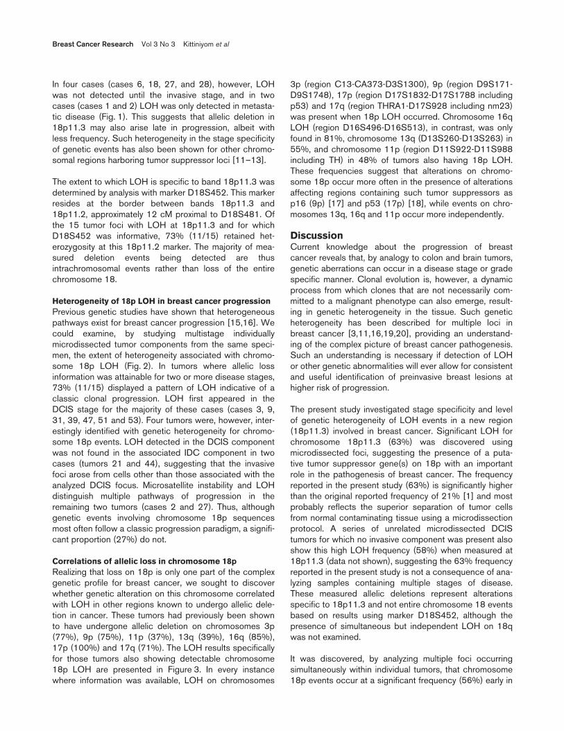
Figure 2
Heterogeneity for LOH on chromosome 18p in breast cancer progression. Schematic diagrams show the different patterns of progression basedon this chromosome 18p study. Nine cases show clonal progression where 18p LOH occurs either in DCIS or IDC and is clonally inherited inrelated later stages of tumor progression. Two cases show LOH first appearing in the metastatic stage. In contrast, four cases show heterogeneityfor alterations on 18p. Two specimens show LOH in the DCIS component but not in the associated IDC component. The other cases show thepresence of microsatellite instability in earlier but not later stage disease.
heterogeneity at multiple loci may account for treatment failure,and a combinatorial histological and genetic approach with
sufficient information on survival and response profiles mayprovide more effective disease prevention and cure in the future.
the preinvasive stage of DCIS. The heterogeneous natureof breast cancer alterations could have important implica-tions for the development of targeted therapies.
If LOH events define a localized region of increased riskfor carcinoma, the presence of LOH may be clinicallyimportant. A complete understanding of all the geneticevents involved in breast tumorigenesis and their hetero-geneity in relation to stage specificity will therefore be crit-ical to the development of successful treatments. Thislaboratory and other workers have reported the presenceof a previously unidentified region of LOH in band18p11.3 in non-small cell lung carcinomas, glioblastomasand a small series (14) of invasive breast carcinomas[1,2]. The current study uses informative loci in 18p11.3to investigate, in microdissected material, the stage speci-ficity and extent of molecular heterogeneity exhibited bychromosome 18p11 in breast cancer progression. Aseries of 30 tumors from which 96 multiple stages ofdisease have been microdissected, including normal,DCIS and IDC material, were studied. Associated metas-tases were also examined in some cases. These resultswere analyzed in conjunction with other LOH data avail-able for markers on 3p, 11p, 13q, 16p, 17p, and 17q in aneffort to place chromosome 18p alterations on the breastcancer progression pathway.
Materials and methodsPatient materialThe material used in this study, consisting of formalin-fixed, paraffin-embedded tissue from the archives of theArmed Forces Institute of Pathology, has been previouslydescribed [3]. Tumors were selected at random for thosewith the presence of lesions representing multiple stagesof breast disease within individual patient samples. Ninety-six foci representing normal, intraductal and infiltratingtumor were microdissected from 30 tumors. One-half ofthe patient samples contained both DCIS and IDC com-ponents. Seven patients had metastatic tumor tissue avail-able in addition to both DCIS and IDC material, while fivepatients had invasive component material with corre-sponding metastatic disease. Aliquots of DNA from thesemicrodissected lesions were used as the source templatefor PCR-based LOH analysis.
LOH analysisMatching normal and tumor DNA were subjected to PCR-based LOH analysis using two 18p11.3 markers(D18S59 and D18S481) and one 18p11.2 marker(D18S452). These markers define a region on chromo-some 18p previously shown to undergo LOH in invasiveductal breast carcinomas as well as non-small cell lungcarcinomas and glioblastomas [1,2]. Forward primerswere synthesized with either a fluorescent FAM tag(D18S481 and D18S452) or a fluorescent TET tag(D18S59) on the 5′ end. PCR reactions were performed
in 10 µl with aliquots of normal and tumor DNA using aPCR protocol consisting of 94°C for 30 s, 55°C for 30 s,and 72°C for 1 min for a total of 40 cycles.
Horizontal ultrathin gel electrophoresisHorizontal ultrathin, high throughput fluorescence-basedDNA fragment gel electrophoresis (GTI-9600; GenesysTechnologies, Inc) was applied to the separation andanalysis of PCR-generated alleles as previously described[4]. One microliter of amplified material was combinedwith 2 µl formamide loading dye (Amersham Inc, Piscat-away, NJ, USA) prior to electrophoresis. ROX 350 fluores-cent size markers (0.7 µl; ABI, Foster City, CA, USA) wereadded to amplified tumor DNA to allow sizing of alleles.Samples were heated to 95°C, loaded on the 70 µm, 5%horizontal polyacrylamide gel and electrophoresed for 1 hand 15 min at 30 W in 1 × TBE using the GTI-9600. Datawere collected while running the electrophoresis andstored using the accompanying Cartographer data analy-sis software program, which extracts the pixel intensity ofindividual peaks present in a lane trace, allowing for sizingof alleles and quantitation of fluorescent peak areas. Indetermining whether allelic deletion had occurred at indi-vidual markers, allelic ratios were calculated andexpressed as a percentage of loss of intensity for thetumor allele compared with the corresponding normalallele (D-value) after normalization. When the allelic ratio inthe tumor DNA was reduced by greater than 40%(DO.40) from that found in the normal DNA, the samplewas denoted as having LOH at that locus.
ResultsChromosome 18p11 LOH in breast cancer progressionA total of 96 foci were individually microdissected from 30breast tumor cases and analyzed for LOH at the polymor-phic markers D18S59 and D18S481 spanning 18p11.3.These microdissections resulted in normal, DCIS and IDCmaterial from most tumors. The overall frequency of LOHin 18p11.3, as measured for any stage of disease usingD18S481 and/or D18S59, was 63% (27/43). This is sig-nificantly higher than the 21% frequency originallyreported using non-microdissected IDC samples [1], sug-gesting that allelic deletion events may have been maskedby contaminating normal stroma in earlier studies. A lowfrequency (3/96) of microsatellite instability was alsodetected at marker D18S59. These microsatellite instabil-ity results were confirmed in at least two independentexperiments and correspond to previous published resultsfor this marker [2].
LOH on 18p was found, when analyzed by tumor stage, in56% (9/16) of DCIS foci, 67% of IDC foci (14/21), and67% (4/6) of metastatic disease. A schematic representa-tion of all tumor foci studied appears in Figure 1. The mag-nitude of LOH detected indicates that alterations in thisregion generally occur early in breast cancer progression.
Available online http://breast-cancer-research.com/content/3/3/192
comm
entaryreview
reportsprim
ary research
In four cases (cases 6, 18, 27, and 28), however, LOHwas not detected until the invasive stage, and in twocases (cases 1 and 2) LOH was only detected in metasta-tic disease (Fig. 1). This suggests that allelic deletion in18p11.3 may also arise late in progression, albeit withless frequency. Such heterogeneity in the stage specificityof genetic events has also been shown for other chromo-somal regions harboring tumor suppressor loci [11–13].
The extent to which LOH is specific to band 18p11.3 wasdetermined by analysis with marker D18S452. This markerresides at the border between bands 18p11.3 and18p11.2, approximately 12 cM proximal to D18S481. Ofthe 15 tumor foci with LOH at 18p11.3 and for whichD18S452 was informative, 73% (11/15) retained het-erozygosity at this 18p11.2 marker. The majority of mea-sured deletion events being detected are thusintrachromosomal events rather than loss of the entirechromosome 18.
Heterogeneity of 18p LOH in breast cancer progressionPrevious genetic studies have shown that heterogeneouspathways exist for breast cancer progression [15,16]. Wecould examine, by studying multistage individuallymicrodissected tumor components from the same speci-men, the extent of heterogeneity associated with chromo-some 18p LOH (Fig. 2). In tumors where allelic lossinformation was attainable for two or more disease stages,73% (11/15) displayed a pattern of LOH indicative of aclassic clonal progression. LOH first appeared in theDCIS stage for the majority of these cases (cases 3, 9,31, 39, 47, 51 and 53). Four tumors were, however, inter-estingly identified with genetic heterogeneity for chromo-some 18p events. LOH detected in the DCIS componentwas not found in the associated IDC component in twocases (tumors 21 and 44), suggesting that the invasivefoci arose from cells other than those associated with theanalyzed DCIS focus. Microsatellite instability and LOHdistinguish multiple pathways of progression in theremaining two tumors (cases 2 and 27). Thus, althoughgenetic events involving chromosome 18p sequencesmost often follow a classic progression paradigm, a signifi-cant proportion (27%) do not.
Correlations of allelic loss in chromosome 18pRealizing that loss on 18p is only one part of the complexgenetic profile for breast cancer, we sought to discoverwhether genetic alteration on this chromosome correlatedwith LOH in other regions known to undergo allelic dele-tion in cancer. These tumors had previously been shownto have undergone allelic deletion on chromosomes 3p(77%), 9p (75%), 11p (37%), 13q (39%), 16q (85%),17p (100%) and 17q (71%). The LOH results specificallyfor those tumors also showing detectable chromosome18p LOH are presented in Figure 3. In every instancewhere information was available, LOH on chromosomes
3p (region C13-CA373-D3S1300), 9p (region D9S171-D9S1748), 17p (region D17S1832-D17S1788 includingp53) and 17q (region THRA1-D17S928 including nm23)was present when 18p LOH occurred. Chromosome 16qLOH (region D16S496-D16S513), in contrast, was onlyfound in 81%, chromosome 13q (D13S260-D13S263) in55%, and chromosome 11p (region D11S922-D11S988including TH) in 48% of tumors also having 18p LOH.These frequencies suggest that alterations on chromo-some 18p occur more often in the presence of alterationsaffecting regions containing such tumor suppressors asp16 (9p) [17] and p53 (17p) [18], while events on chro-mosomes 13q, 16q and 11p occur more independently.
DiscussionCurrent knowledge about the progression of breastcancer reveals that, by analogy to colon and brain tumors,genetic aberrations can occur in a disease stage or gradespecific manner. Clonal evolution is, however, a dynamicprocess from which clones that are not necessarily com-mitted to a malignant phenotype can also emerge, result-ing in genetic heterogeneity in the tissue. Such geneticheterogeneity has been described for multiple loci inbreast cancer [3,11,16,19,20], providing an understand-ing of the complex picture of breast cancer pathogenesis.Such an understanding is necessary if detection of LOHor other genetic abnormalities will ever allow for consistentand useful identification of preinvasive breast lesions athigher risk of progression.
The present study investigated stage specificity and levelof genetic heterogeneity of LOH events in a new region(18p11.3) involved in breast cancer. Significant LOH forchromosome 18p11.3 (63%) was discovered usingmicrodissected foci, suggesting the presence of a puta-tive tumor suppressor gene(s) on 18p with an importantrole in the pathogenesis of breast cancer. The frequencyreported in the present study (63%) is significantly higherthan the original reported frequency of 21% [1] and mostprobably reflects the superior separation of tumor cellsfrom normal contaminating tissue using a microdissectionprotocol. A series of unrelated microdissected DCIStumors for which no invasive component was present alsoshow this high LOH frequency (58%) when measured at18p11.3 (data not shown), suggesting the 63% frequencyreported in the present study is not a consequence of ana-lyzing samples containing multiple stages of disease.These measured allelic deletions represent alterationsspecific to 18p11.3 and not entire chromosome 18 eventsbased on results using marker D18S452, although thepresence of simultaneous but independent LOH on 18qwas not examined.
It was discovered, by analyzing multiple foci occurringsimultaneously within individual tumors, that chromosome18p events occur at a significant frequency (56%) early in
Breast Cancer Research Vol 3 No 3 Kittiniyom et al

the tumorigenic process, making LOH at chromosome18p one of the most common known events in DCIStumors. For 73% of the cases in which multiple stages ofdisease arose simultaneously and for which information onat least one marker was available, 18p11.3 LOH in theDCIS component could also be found in the invasive com-ponent isolated for analysis (Fig. 2). This suggests that themajority of breast tumors progress in a clonal fashion. Fourcases (27%) were, however, consistent with genetic het-erogeneity at this chromosomal region. These latter obser-vations argue that invasive foci of an individual tumor canrepresent genetically divergent clones rather than progres-sive stages of the disease. Such conclusions are furthersupported by flow cytometric and comparative genomichybridization analyses on primary and metastatic diseasecomponents [19,20]. In the comparative genomichybridization study [20], 69% of metastatic lesionsshowed a high degree of clonal progression from theprimary tumor, whereas 31% did not. These frequenciesparallel the 70% clonal/30% heterogeneous progressionfrequencies reported for chromosome 18p events in thepresent study.
We are not yet at a stage in our understanding of breastdisease such that genetic alterations can substitute for thehistopathology of the lesion. A combinatorial histologicaland genetic approach with sufficient information on sur-vival and response profiles may, however, make for moreeffective disease prevention and cure in the future. Thepower of genetic profiles in prognostic relevance hasrecently been shown by Emi et al [21]. Studying 15 loci in264 women, Emi et al found that LOH at markers 1p34,13q12, 17p13.3 and 17q21.1, as well as two pairs ofmarkers (1p34/17p13.3 and 13q12/17p13.3), had signifi-cant prognostic value and carried significant relative risk ofdeath. It would be interesting, given the high frequency ofLOH discovered for chromosomal region 18p11.3 in thisreport, to include this marker along with these regions todetermine its usefulness as a predictive risk marker inbreast cancer.
To that extent, it was interesting to view a partial geneticprofile for other regions of LOH in these tumors whenchromosome 18 LOH was present. Chromosome 18pLOH always occurred in conjunction with LOH on 3p, 9p,
Available online http://breast-cancer-research.com/content/3/3/192
comm
entaryreview
reportsprim
ary research
Figure 3
Other LOH in tumors with chromosome 18p allelic deletion.

17p and 17q independent of tumor stage, suggesting thisgroup of loci were critical to the development of breastcancer. Allelic deletion on chromosomes 16q, 13q and11p occurred less consistently (81, 55 and 46%, respec-tively), suggesting these regional events are more varieddepending on the stage and/or type of tumor foci ana-lyzed. Of further interest is the putative tumor suppressorgene, DAL-1, which has recently been mapped to chromo-some band 18p11.3 [22]. It will be important to learnmore about the function of this gene and its potentialmutational profile in breast cancer. Future studies willdetermine whether this or another tumor suppressor geneis the target of the high frequency of allelic deletion mea-sured in this study for breast cancer.
AcknowledgementsThe authors would like to thank Dr Oliver Bogler for hiscritical reading of this manuscript. This work was sup-ported in part by R29 CA77330 (IN). KK is the recipient ofa Royal Thailand Scholar Fellowship.
References1. Tran Y, Benbatoul K, Gorse K, Rempel, S, Futreal A, Green M,
Newsham I: Novel regions of allelic deletion on chromo-some18p in tumors of the lung, brain and breast. Oncogene1998, 17:3499–3505.
2. Osborne RJ, Hamshere MG: A gemone-wide map showingcommon regions of loss of heterozygosity/allelic imbalancein breast cancer. Cancer Res 2000, 60:3706–3712.
3. Lichy JH, Zavar M, Tsai MM, O’Leary TJ, Taubenberger JK: Lossof heterozygosity on chromosome 11p15 during histologicalprogression in microdissected ductal carcinoma of the breast.Am J Pathol 1998, 153:271–278.
4. Newsham IF, Gorse KM, Rempel SA, Luckey J, Golden JB, BoglerO: Use of horizontal ultrathin gel electrophoresis to analyzeallelic deletions in chromsome band 11p15 in gliomas. Neuro-Oncol 2000, 2:1–5.
5. Ingvarsson S: Molecular genetics of breast cancer progression.Semin Cancer Biol 1999, 9:277–288.
6. Beckmann MW, Niederacher D, Schnurch H-G, Gusterson BA,Bender HG: Multistep carcinogenesis of breast cancer andtumour heterogeneity. J Mol Med 1997, 75:429–439.
7. Lizard-Nacol S, Lidereau R, Collin F, Arnal M, Hahnel L, Roignot P,Cuisenier J, Guerrin J: Benign breast disease: absence ofgenetic alterations at several loci implicated in breast cancermalignancy. Cancer Res 1995, 55:4416–4419.
8. O’Connell P, Pekkel V, Fuqua SAW, Osborne CK, Clark GM,Allred DC: Analysis of loss of heterozygosity in 399 premalig-nant breast lesions at 15 genetic loci. J Natl Cancer Inst 1998,90:697–703.
9. Deng G, Lu Y, Zlotnikov G, Thor AD, Smith HS: Loss of het-erozygosity in normal tissue adjacent to breast carcinomas.Science 1996, 274:2057–2059.
10. Ottesen GL, Graversen HP, Blichert TM, Zedeler K, Andersen JA:Ductal carcinoma in situ of the female breast: short-termresults of a prospective nationwide study. Am J Surg Pathol1992, 16:1183–1196.
11. Lichy JH, Dalbegue F, Zavar M, Washington C, Tsai MM, ShengZM, Taubenberger JK: Genetic heterogeneity in ductal carci-noma of the breast. Lab Invest 2000, 80:291–301.
12. Bonsing BA, Devilee P, Cleton-Jansen A-M, Kuipers-Dijkshoorn N,Fleuren GJ, Cornelisse CJ: Evidence for limited moleculargenetic heterogeneity as defined by allelotyping and clonalanalysis in nine metastatic breast carcinomas. Cancer Res1993, 53:3804–3811.
13. Anbazhagan R, Hiroaki F, Gabrielson E: Allelic loss of chromo-somal arm 8p in breast cancer progression. Am J Pathol 1998,152:815–819.
14. Marsh KL, Varley JM: Loss of heterozygosity at chromosome9p in ductal carcinoma in situ and invasive carcinoma of thebreast. Br J Cancer 1998, 77:1439–1447.
15. Newsham IF: The long and short of chromosome 11 in breastcancer. Am J Pathol 1998, 153:5–9.
16. Fujii H, Szumel R, Marsh C, Zhou W, Gabrielson E: Genetic pro-gression, histological grade and allelic loss in ductal carci-noma in situ of the breast. Cancer Res 1996, 56:5260–5265.
17. Nobori T, Miura K, Wu DJ, Lois A, Takabayashi K, Carson DA:Deletions of the cyclin-dependent kinase-4 inhibitor gene inmultiple human cancers. Nature (London) 1994, 368:753–756.
18. Vos CB, ter Haar NT, Rosenberg C, Peterse JL, Cleton-JansenAM, Cornelisse CJ, van de Vijver MJ: Genetic alterations onchromosome 16 and 17 are important features of ductal car-cinoma in situ of the breast and are associated with histologictype. Br J Cancer 1999, 81:1410–1418.
19. Bonsing BA, Corver WE, Fleuren GJ, Cleton-Jansen AM, DevileeP, Cornelisse CJ: Allelotype analysis of flow-sorted breastcancer cells demonstrates genetically related diploid andaneuploid subpopulations in primary tumors and lymph nodemetastases. Genes Chrom Cancer 2000 28:173–183.
20. Kuukasjärvi T, Karhu R, Tanner M, Kähkönen M, Schäffer A, Nup-ponen N, Pennanen S, Kallioniemi A, Kallioniemi OP, Isola J:Genetic heterogeneity and clonal evolution underlying devel-opment of asynchronous metastasis in human breast cancer.Cancer Res 1997, 57:1597–1604.
21. Emi M, Yoshimoto M, Sato T, Matsumoto S, Utada Y, Ito I, MinobeK, Iwase T, Katagiri T, Bando K, Akiyama F, Harada Y, Fukino K,Sakamoto G, Matsushima M, Iida A, Tada T, Saito H, Miki Y,Kasumi F, Nakamura, Y: Allelic loss at 1p34, 13q12, 17p13.3and 17q21.1 correlated with poor postoperative prognosis inbreast cancer. Genes Chrom Cancer 1999, 26:134–141.
22. Tran YK, Bogler O, Gorse K, Wieland I, Green MR, Newsham IF:A novel member of the NF2.ERM/4.1 superfamily with growthsuppressing properties in lung cancer. Cancer Res 1999, 59:35–43.
Breast Cancer Research Vol 3 No 3 Kittiniyom et al