Embed Size (px)
Citation preview

Page 1
About Thermography…

Page 2
About Beacon Thermography, Inc. Based in Surf City, NC
Started March 2016
3 sisters
Mostly mobile Home office
Wilmington Elite Chiropractic
Shaver Chiropractic
McKay Healing Arts
Raleigh-Durham Health Touch NC LLC
Website www.beaconthermography.com

Page 3
Shelly LaineQualifications, Certifications and Accreditations Clinical Thermographer – MediTherm and The American College of Clinical
Thermography
Certified Health Coach – The Institute for Integrative Nutrition, New York City Board Certified and Accredited Member of the American Association of Drugless
Practitioners (AADP)
New Healers Master Coaching Program
Certified BioEnergetic Synchronization Technique (B.E.S.T.) Practitioner – MorterHealth Systems
Active Isolated Stretching Practitioner – Aaron Mattes AIS and Strengthening, Basic and Advanced
Trapped Emotion Release
Aromatherapy – Raindrop Technique, Applied VitaFlex, Essential Oil Chemistry and Emotional Release with Essential Oils – Center for Aromatherapy Research and Education
Continuing Education – Master Herbalist studies through School of Natural Healing
BS Computer Science with 30 years of experience in Information Technology Leadership

Page 4
Cecilia Laine-Meinhold
Qualifications, Certifications and Accreditations Clinical Thermographer – MediTherm and member of The American
College of Clinical Thermography Energy Work via Dowsing – mentored by internationally-known
dowser Trapped Emotion Release Develops personalized organic skin care products Current studies: Master Herbalist through School of Natural Healing
and Crystal Healing Certification 25+ years of experience in Information Technology leadership

Page 5
Tanis Clark
Qualifications, Certifications and Accreditations Clinical Thermographer – MediTherm and member of The American
College of Clinical Thermography
Licensed Paralegal – heavy background in real estate and family law
The mother of 3 of my favorite people in the world Works part-time at Sugar Island Bakery and is a large part of the magic
there!

Page 6
MediTherm and The American College of Clinical Thermology (ACCT) ACCT members operate under a code of ethics and have access to Quality
Control and Continuing Education Our equipment is medical grade and regulatory compliant EMI, our telemedicine software, complies with all telemedicine and HIPAA
regulations Centralized client database, accessible by practitioners world wide All scan interpreters are Medical Doctors who are board certified in Thermology Fast turn-around of reports, with option for over-read and second opinion
service Consults for PCPs with audited Quality Control Largest network of accredited thermography practitioners worldwide, with
standardization throughout the network Dr. Mercola used MediTherm equipment in his clinic; Duke University in research
Page 7
Are you looking for a 100% safe screening test that is…
Non-invasive / no contactNo radiationPainlessPrivateFDA RegisteredOne that can reveal a roadmap to
improve your health, wellness and life longevity?

Page 8Page 8
Clinical Thermography –What is It?
It is widely acknowledged that cancers, even in their earliest stages, need nutrients to maintain or accelerate their growth. In order to facilitate this process, blood vessels are caused to remain open, inactive blood vessels are activated, and new ones are formed through a process known as neoangiogenesis. This vascular process causes an increase in surface temperature in the affected regions, which can be viewed with infrared imaging cameras. Additionally, the newly formed or activated blood vessels have a distinct appearance, which thermography can detect.Heat is an indication that inflammation exists, and typically inflammation is present in precancerous and cancerous cells, too. (It’s also present in torn muscles and ligaments as well as arthritic joints, which thermography can also detect)

Page 9
Why Thermography? What Can It Be Used For? It is part of the International Classification of Procedures in Medicine
published by the World Health Organization (Geneva, 1990) Breast health screening
It is FDA registered and approved as an adjunct to mammography
Can increase your chances of detecting breast cancer in its earliest stages by locating irregular patterns in the breast -- conditions that occur before a noticeable lump is formed and detectable by mammography, ultrasound, x-rays and MRIs.
Indications and Injuries It has been established that much additional diagnostic value is provided by
thermography when full body images are performed.
Many people choose to have full-body screening performed on a routine basis due to thermography's ability to detect conditions and diseases before they become apparent/detectable on standard diagnostic tests.
Thermal changes are often the earliest sign of vascular disease, immune dysfunction, diabetes, thyroid dysfunction. It is also the earliest indication of systemic inflammation, a precursor to many diseases—including cancer.

Page 10
Scan Types – Head & Neck, Breast

Page 11
Scan Types – Abdomen, Trunk, Legs

Page 12
Scan Types – Arms, Hands, Thyroid/Digastric/Carotid, Feet, Knees

Page 13
Let’s Talk About Breast Screening First…

Page 14
Breast Thermography….
How Does it Work? Thermography is based on the scientific premise that changes in
breast health cause an increased blood supply to the growth area (angiogenesis). Breast thermography measures the heat generated by the circulation of blood in the breast during this process. Breast thermography can identify women with changes so they can be monitored more closely and work with their health care professional to set proactive strategies in place, through diet, environmental and lifestyle changes.
Anatomical tests, such as mammograms, ultrasound and MRIs, rely on finding physical lesions (lumps). Breast thermography is a physiological test that can identify abnormal blood vessel circulation within the breast. Physiological changes are known to precede anatomical changes, therefore both anatomical and physiological information is valuable in fully assessing breast health.

Page 15
Breast Thermography….
Is Thermography a Standalone Test? Breast thermography is not a diagnostic or stand-alone
tool in the screening and diagnosis of breast cancer. It is an FDA (US Food & Drug Administration) approved
adjunctive to mammography screening.When a thermogram is positive, a closer look at the
patient's hormonal metabolism, diet, exposure to environmental pollution, toxins and lifestyle factors is in order.
Clinical blood work in addition to anatomical testing is essential. When additional tests and blood work are negative or equivocal, thermographic monitoring could continue on a quarterly to semi-annual basis.

Page 16
Average Growth Rate of Breast Cancer TumorCancer Cells Double in Number On Average Every 80-90 Days
Still undetectable with mammography
90 days1 year2 years3 years4 years5 years6 years7 years8 years
2 cells16 cells256 cells4,096 cells65,536 cells1,048,576 cells16,777,216 cells268,435,456 cells4,294,967,296 cells Normally detectable by mammogram at this
stage, approx. 1cm size (32 doublings)
40 Doublings (approx 10 years) is generally consideredlethal
The vascular changes associated with atypical pre-cancerous cells detectable with thermography
Buchanan JB, et al. Tumor growth, doubling times, and inability of the radiologist to diagnose certain cancers. Radiol Clin N Am.1983;21:115-26
Source:
This early detection of change can lead to earlier diagnosis and better treatment options as well as the opportunity for patients and their healthcare
practitioners to intervene at an early stage with preventative treatment.

Page 17
What Doctors Are Saying About Breast Thermography
Breast thermography needs to be embraced more widely by the medical community and awareness increased among women. Not only has it demonstrated a higher degree of success in identifying women with breast cancer under the age of 55 in comparison to other technologies, but it is also an effective adjunct to clinical breast exams and mammography for women over 55.
Len Saputo, M.D. – Duke University Medical School Founder of Health Medicine Forum and Health Medicine Center
With the addition of infrared imaging, our sensitivity of image detection has increased from 83% to 93%.
John Keyserlingk, M.D., Ph.D. Surgical OncologistVille Marie Breast and Oncology CenterDepartment of Oncology - St. Mary's Hospital, Montreal, Quebec

Page 18
What Doctors Are Saying About Breast Thermography
The use of computerized medical infrared imaging for breast cancer detection, diagnosis, and as a high risk and prognostic indicator leads to both earlier detection of breast cancer and increases the overall survival of breast cancer patients.
Robert Elliot, M.D., Ph.D.Comprehensive Breast Care Specialist Founder and DirectorEEH Breast Cancer Research and Treatment CenterPresident American Mastology Association
Every woman should include breast thermography as part of their regular breast health care. In fact, studies have shown that an abnormal infrared image is the single most important indicator of high risk for developing breast cancer.
Susan M. Lark, M.D.Distinguished Author and Leading Expert in Women’s Health

Page 19
What Doctors Are Saying About Breast Thermography
For years, I’ve been looking for a test to offer my patients who refuse mammograms. Some are concerned about cumulative radiation and complications from compression, and others have personal reasons for refusing this test. Yet, to do nothing is to put their heads in the sand, and this doesn’t save lives… I found in my research, and personally, that thermography can help a woman take a possible pre-cancerous condition and turn it around. It supports mammography and provides additional information. Prevention is the gold standard of health care. In my opinion, breast thermography should be part of every woman’s yearly exam to help prevent breast cancer, and for early detection.
Nan Kathryn Fuchs, Ph.DEditor, Women’s Health Letter
The option for breast screening that I most highly recommend is called thermographic breast screening. More men’s lives could also be spared from the disease as mammography is not frequently used on men, which leads to most men with breast cancer being diagnosed at a very late stage.
Dr. Joseph Mercola, DOBoard-certified family medicine doctorFounder of Mercola.com, the World’s #1 Natural Health WebsiteFellow at American College of NutritionNew York Times Best-Selling Author

Page 20
What Research Is Saying About Breast Thermography
The use of computerized medical infrared imaging for breast cancer detection, diagnosis, and as a high risk and prognostic indicator leads to both earlier detection of breast cancer and increases the overall survival of breast cancer patients.
Robert Elliot, M.D., Ph.D. Comprehensive Breast Care SpecialistFounder, Director EEH Breast Cancer Research and Treatment CtrPresident - American Mastology Association
Infrared imaging of the breast should be an integral part of any breast cancer screening program due to its value as an independent risk factor for breast cancer and its value as a prognostic indicator.
Jonathan Head, Ph.D.Tumor Cell Biologist and Pioneer in Breast Cancer VaccinesDirector of Research EEH Breast Cancer Research and Treatment Center Associate
Professor of Biochemistry - Tulane University

Page 21
What Research Is Saying About Breast Thermography
In this prospective clinical trial of 92 women undergoing DITI with suspicious breast lesions identified on prior mammogram or ultrasound, we have shown that a modernized DITI system can detect breast pathology with sensitivity up to 97%. DITI is a valuable adjunct to mammography and ultrasound, especially in women with dense breast parenchyma.
American Society of Breast SurgeonsThe American Journal of Surgery (2008) 196, 523-526
John Gofman, M.D., Ph.D. – a nuclear physicist and a medical doctor, and one of the leading experts in the world on the dangers of radiation – presents compelling evidence in his book, Radiation from Medical Procedures in the Pathogenesis of Cancer and Ischemic Heart Disease, that over 50 percent of the death-rate from cancer is in fact induced by x-rays. Now consider the fact that the routine practice of taking four films of each breast annually results in approximately 1 rad (radiation absorbed dose) exposure, which is about 1000 times greater than that from a chest x-ray.
Dr Joseph Mercola, D.O.Founder of Mercola.com, the World’s #1 Natural Health WebsiteFellow at American College of Nutrition

Page 22
A Few Case Studies…

Page 23
Baseline
3 months 6 months 9 months 12 monthsThis patient was also age 37 when her first baseline thermogram showed a slight hyperthermic asymmetry in the upper right breast. The follow-up study showed the pattern had become more well defined and although clinical correlation did not find anything remarkable it was decided to repeat the exam again in a further 3 months, when again significant changes were seen. Mammography was performed at this stage with the thermographic guidance of the locally suspicious area at 1 o’clock to the right nipple. The mammographic findings were inconclusive and the patient was referred for a repeat mammogram in 12 months. Thermographic monitoring was continued and at the fifth comparative study at 12 months significant changes were still evident and the hyperthermic asymmetry (temperature differentials) had increased. Immediate further investigation was strongly recommended despite a scheduled mammogram in 6 months, and at the patient’s insistence, a repeat mammogram was performed which clearly showed a small calcification (1 mm) at 1 o’clock. Within one week a lumpectomy had been performed with good margins and the pathology confirmed as a malignant carcinoma (DCIS).This patient has now had stable thermograms for the last 2 years and is expected to remain healthy.

Page 24
This is the specific area of the small DCIS. We can see the vascular feed and the discreet area of hypothermia that is displacing the surrounding hyperthermia.
(DCIS is Ductal Carcinoma In Situ, and refers to the abnormal growth of cells within the milk ducts of the breast forming a lesion commonly between 1-1.5 cm in diameter, and is considered non-invasive or "stage zero breast cancer," with some experts arguing for its complete re-classification as a non-cancerous condition.)

Page 25
10 Year evolving change with diagnosis of malignancy at year 7
No significant history or symptoms. Mammogram at year 1. Within normal limits.
Thermography established a baseline at year 1 but classified left breast at risk for developing pathology and recommended clinical correlation.
Thermography reported suspicious change between year 2 and year 3 in left breast. Clinically no findings.
Continued thermal changes reported year 4, 5, and 6.
Mammogram Ultrasound and MRI at year 7 diagnosed small suspicious pea-sized lump likely malignant in left breast. Patient refused biopsy. Thermography reported significant change and vascular activity consistent with angiogenesis and active pathology, suspicious for malignancy.
Year 9 MRI and Ultrasound diagnosed carcinoma 8cm x 3cm in left breast. Thermography findings correlate with aggressive malignant pathology. Later diagnosed as invasive ductal carcinoma.

Page 26
17 Years of Annual Screening
Case history Year of birth 1949. No significant history or symptoms. Mammogram at year 3. Within
normal limits. Mammogram at year 17: No suspicious findings. Within normal limits. Thermography shows no clinically significant changes over time and
no indication of developing pathology. Report: Stable and Within Normal Limits.

Page 27
Breast cancers tend to grow significantly faster in younger women under 50
Source: Cancer 71:3547-3551, 1993
Age Average Tumor Doubling TimeUnder Age 50 80 daysAge 50 – 70 157 daysOver Age 70 188 days
There is an abundance of scientific evidence supporting that breast thermography is the most sensitive and accurate way to identify women with breast cancer, especially in women under the age of 55 and those with dense breast tissue. For women over 55, breast thermography is an important adjunct to clinical breast examination and other anatomical testing, such as mammography, ultrasound and MRIs, as this combination has been documented to increase the identification of breast cancers to 98 percent.
Because of its low cost and high degree of sensitivity and accuracy, all women who want to be screened for breast cancer should begin having breast thermograms beginning at age 25. (Dr. Len Saputo)

Page 28
Breast cancers tend to grow significantly faster in younger women under 50
DITI is especially appropriate for younger women between 25 & 50 whose denser breast tissue makes it more difficult for mammography to pick up suspicious lesions. This test can provide a 'clinical marker' to the doctor or mammographer that a specific area of the breast needs particularly close examination.
This 37 year old client presented for routine thermographic breast screening. She was not in a high risk category and had no family history. No breast exams had been
performed previously. The vascular asymmetry in the upper left breast and the local hypothermia at 11 o’clock was
particularly suspicious and subsequent clinical investigation indicated a palpable mass at the position indicated. A biopsy was performed and a DCIS of 2 cm was diagnosed.
Unfortunately this patient only survived for 12 months after diagnosis.

Page 29
Inflammatory breast disease cannot be detected by mammography and is most commonly seen in younger women, the prognosis is always poor. Early detection provides the best hope of survival.
Breast cancers tend to grow significantly faster in younger women under 50
The results of this routine study led to the diagnosis of inflammatory carcinoma in the right breast. There were no clinical indications at
this stage. Thermography can show significant
indicators several months before any of the clinical signs of inflammatory breast disease, skin discoloration, swelling and pain.

Page 30
This significant thermal asymmetry is caused by an early stage DCIS and was only just dense enough to be seen with thermography-guided mammography.
There was nothing palpable and this patient was 10 years away from her first routine mammogram.
Breast cancers tend to grow significantly faster in younger women under 50

Page 31
DCIS
Gray scale
Thermography can show suspicious findings that are outside the range of mammography…… this tumor was missed by a mammogram as it was just outside the border of the medial left breast.
Thermography Scan Areas

Page 32
The very significant vascular activity in the left breast justified clinical correlation and close monitoring which returned an opinion of fibrocystic changes taking place.
These changes can be monitored thermographically at regular intervals until a stable baseline is established and is reliable enough for annual comparison.

Page 33
The increased vascular activity in both breasts is consistent with the hormonal changes in pregnancy. This patient was unaware that she was 4 weeks pregnant at the time of her scan.The thermography tech was an experienced operator who asked the patient….. “you’re not pregnant are you ?” The patient was amused and said that she wasn’t but phoned the clinic a few days later to tell them the good news.

Page 34Progressive development of invasive carcinoma

Page 35
1% of breast cancers are found in men. The survival rate is much lower than in women as most breast cancers in men are only detected in advanced stages.
This tumor was palpable at the time of imaging There is a well-established vascular feed which had caused increased blood
flow at the left brachial plexus There is drainage toward the sternum that extends to below the left breast. Metastasis were later found in other organs and this patient did not survive.

Page 36
Serial thermography can be used to monitor the response to treatments like chemo and radiation as well as angiogenesis-inhibiting drugs. This is the first study pre-diagnosis and shows vascular activity relating to a palpable mass.
A mammogram correlated with the mass and FNB confirmed malignancy. The decision was to reduce activity before surgery and thermography was used to monitor the response to chemo before surgery. The tumor is above the left nipple.
Monitoring the Response to Treatment Before Surgery

Page 37
Statistical analysis is performed by doctors who evaluate temperature findings to a sensitivity of 1/100th degree C.
Proprietary interpretation software incorporates pattern recognition with artificial intelligence which continually improves the central database grading system.
We Don’t Just Rely on Visual Interpretation…

Page 38
We Don’t Just Rely on Visual Interpretation…
Features like ‘Vascular Enhancement’ assist doctors to identify vascular abnormality and changes over time.

Page 39
We Don’t Just Rely on Visual Interpretation…
A spatial derivative processing of high-definition findings enables a virtual 3D rendering of the functional vascular activity. This assists in the identification of abnormal vascular activity and changes.

Page 40
We Don’t Just Rely on Visual Interpretation…
3D temperature modeling provides perspective between statistical thermal analysis and clinical significance as well as contributing visual comparison for change over time significance.

Page 41
Color grading delta T for reporting clinical significance
We Don’t Just Rely on Visual Interpretation…
Automatic application of contralateral comparative analysis provides more objective findings that are color coded for instant grading.

Page 42
We Don’t Just Rely on Visual Interpretation…
3D modeling of suspicious area at 12 o’clock to right nipple. 3D model can be dynamically manipulated by interpreting doctor.

Page 43
Without realignment
We Don’t Just Rely on Visual Interpretation…
Pattern recognition software realignment for accurate comparative analysis over time.
With realignment

Page 44
Self Exam + Thermography + Mammography

Page 45
The Breast Scanning Process Preparation Health HistoryQuestionnaires Thermal Scans Sample Report Follow Up

Page 46
Preparation
Understanding of Preparation Guidelines
Sign and date the Preparation Check List. Bring it with you to your appointment

Page 47
Health History
Fill out the Health History form to the best of your ability prior to your appointment.
A template with explanations is included with the form on our website.
We’ll go over this together prior to your scans.

Page 48
Breast Exam Questionnaires
Fill out the Breast Questionnaires to the best of your ability prior to your appointment.
We’ll go over this together prior to your scans.

Page 49
Thermal Scans
Upon arriving, you’ll be asked to disrobe from the waist up, and possibly only underwear, depending on clothing worn
We’ll go over your forms while you’re acclimating to the room (this ensures proper imaging)
• Time on this varies, depending on complexity and completeness of health history and questionnaires prior to arrival
5 – 6 views are taken (we usually take a posterior view as well)
Total time is approximately 15 minutes for the scans themselves
We’ll review the scans quickly to ensure we have everything we need
Scans and histories are sent to our group of MDs who are board-certified in thermal scan analysis
Your report will be sent via your preferred method usually within 2-3 days
Page 50
Patient:Date of Birth: # Referring Doctor:
Page 1 of 1
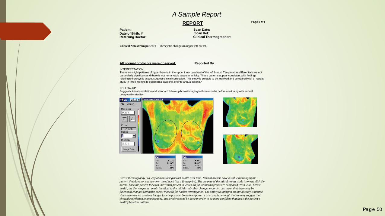
A Sample ReportREPORT
Scan Date: Scan Ref:
Clinical Thermographer:
Clinical Notes from patient : Fibrocystic changes in upper left breast.
All normal protocols were observed. Reported By :INTERPRETATION:There are slight patterns of hyperthermia in the upper inner quadrant of the left breast. Temperature differentials are not particularly significant and there is not remarkable vascular activity. These patterns appear consistent with findings relating to fibrocystic tissue, suggest clinical correlation. This study is suitable to be archived and compared with a repeat study in three months to establish a baseline, prior to annual testing.*
FOLLOW-UP:Suggest clinical correlation and standard follow-up breast imaging in three months before continuing with annual comparativestudies.
Breast thermography is a way of monitoring breast health over time. Normal breasts have a stable thermographic pattern that does not change over time (much like a fingerprint). The purpose of the initial breast study is to establish thenormal baseline pattern for each individual patient to which all future thermograms are compared. With usual breast health, the thermograms remain identical to the initial study. Any changes recorded can mean that there may be functional changes within the breast that call for further investigation. The ability to interpret an initial study is limited since there are no previous images for comparison. Sometimes patterns are complex enough that we may suggest that clinical correlation, mammography, and/or ultrasound be done in order to be more confident that this is the patient’s healthy baseline pattern.

Page 51
Report
All normal protocols wereobserved Reported By: Monte Elgarten MD.
INTERPRETATION:There are no significant thermal asymmetries seen in the breasts. There is no indication of any neovas-cularity. The slight areas of hyperthermia in the upper quadrants of both breasts do not appear suspi-cious but should be monitored for change. This study is suitable to be archived and compared with a repeat study in three months to establish a baseline, prior to annual testing.
FOLLOW-UP:Suggest standard follow-up breast imaging in three months before continuing with annual comparative studies.
PROCEDURE:This patient was examined with digital infrared thermal imaging to identify thermal findings which may suggest abnormal physiology.
Thermography is a physiologic test, which demonstrates thermal patterns in skin temperature that may be normal or which may indicate pain, injury, disease or other abnormality. If abnormal heat patterns are identified relating to a specific region of interest or function, clinical correlation and further investi-gation may be necessary to assist your health care provider in diagnosis and treatment.
Thermal imaging is an adjunctive test, which contributes to the process of differential diagnosis, and is not independently diagnostic of pathology.
Breast thermography is a way of monitoring breast health over time. Every woman has a unique thermal pattern that should not change over time, like a fingerprint. The purpose of the two initial breast studies (usually obtained three months apart) is to establish the baseline pattern for each patient to which all future thermograms are compared to monitor stability. With continued breast health, the thermograms remain identical to the initial study. Changes may be identified on follow up studies that could represent physiological differences within the breast that warrant further investigation.
The ability to interpret the first breast study is limited since there are no previous images for comparison.
This exam is an adjunctive diagnostic procedure and all interpretive findings must be clinically correlated. DITI is not a substitute for mammography.
PROTOCOLS:The thermographer certifies that this exam was conducted under standard and clinically acceptable protocols.
Scan Date: 7/11/2003 Report Ref: 11051 Report Type: Breast
Patient: xxxxxxxxDate of Birth: 09/18/1953Patient ID: 2191
Electronic Medical Interpretation Inc. 4404 S. Florida Ave. Suite 3.Lakeland FL 33813 USA Ph. 1-888-281-8700
Ronald L Blum M.D. FACOEM. FAAFP.
Linda Hegstrand M.D. Ph.D
Peter Leando Ph.D D.Sc.D.Ac.
Lauren SwerdloffM.D.
Jim Blum Ph.D.
Monte Elgarten M.D.
Arlette PharoD.O.
Daniel Farrier M.D.
Mario Soteriou M.D.
Sample EMI Report
Page 1 of3

Page 52
This Report is intended for use by trained health providers to assist in evaluation, diagnosis,and treatment. It is not intended for use by individuals for self–evaluation or self-diagnosis.This Report does not provide a diagnosis of illness, disease or other condition.
PATIENT HISTORY:The interpretation represents objective descriptions of thermal patterns. Clinical significance of such patterns is interpreted in relation to and limited by the patient data and history provided.
REPORTING:Results are reported by certified thermologists. Results are determined by studying the varying patterns and temperature differentials as recorded in the thermal images.
NORMAL FINDINGS:Normal findings are diffuse thermal patterns with good symmetry between similar regions on both sides of thebody. Comparative imaging may identify specific asymmetries that have remained stable and unchanged overtime and therefore regarded as normal.
ABNORMAL FINDINGS:Abnormal findings may be localized areas of hyperthermia or hypothermia, or thermal asymmetry between similar regions on both sides of the body with temperature differentials of more than 1° C. There may be vascular patterns that suggest pathology. Comparative imaging may identify specific changes or new asymme-tries that warrant further investigation.
COLD STRESS:Routine breast thermography monitoring for changes over time precludes the necessity for cold stressing under these protocols. A cold stress test can be conducted when appropriate or when ordered by a referring physician.
The referring health care provider should contact the EMI administrator with any questions relating to this interpretive report.
Page 2 of3

Page 53
Page 3 of3

Page 54
Now, Let’s Talk About the Other Indications for a Thermagraphic Exam…

Page 55
Other Indications (Partial List) Arteriosclerosis
Brachial Plexus Injury
Biomechanical Impropriety
Breast Disease
Bursitis
Carpal Tunnel Syndrome
Cord Pain/Injury
Deep Vascular Disease
Disc Disease
Disc Syndromes Dystrophy
External Carotid Insufficiency
Headache Evaluation
Herniated Disc
Inflammatory Disease
Infectious Disease (Shingles, Leprosy)
Lumbosacral Plexus Injury Ligament Tear
Lupus
Morton's Neuroma
Nerve Impingement
Neurovascular Compression
Neuralgia (melanoma, squamous cell, basal)
Nutritional Disease (Alcoholism, Diabetes)
Peripheral Nerve Injury
Raynaud's
Referred Pain Syndrome
Reflex Sympathetic Dystrophy
Ruptured Disc
Soft Tissue Injury Sprain/Strain
Stroke Screening
Sensory Nerve Abnormality
Somatic Abnormality
Skin Abnormalities
Thoracic Outlet Syndrome
Temporal Arteritis
Trigeminal Neuralgia Trigger Points
TMJ Dysfunction Tendonitis

Page 56
Sample Conditions & Injuries

Autonomic pattern relating to Coronary Artery Disease
Local dentalinfection
Muscular inflammation infraspinatus and deltoid

RSD/CRPS (Reflex Sympathetic
Dystrophy/Complex Regional
Pain Syndrome) of the left
hand. Glove-like hypothermia.
The return of normal sympathetic
function after treatment was
short term.

This elderly lady had undergone a left hip
replacement surgery 3 months previously and her
continued leg pain raised a suspicion for DVT. The
thermographic findings were not consistent with DVT
(Deep Vein Thrombosis), but showed a focal area of
inflammation that guided a sonographer to a deep
abscess near the bone. This was lanced and
successfully treated with antibiotics.
Small metastatic tumors indicated by focal areas of hypothermia over the lumbar spine

This patient presented with low back pain, there were no thermal findings in the back but the abdomen showed a well defined area of inflammation over the right kidney which could refer pain to the back. Subsequent tests confirmed a kidney infection.
The primary finding here is the local area of hyperthermia over the hepatic flexure of the colon. Diverticulitis was diagnosed after clinical correlation with thermographic findings.

Muscular inflammation over trapezius, hypothermia over T2 consistent with fibromyalgia.
Local inflammation from osteoarthritis

Patient with elevated C-Reactive Protein, an early risk indication for heart disease

Patient with an advanced infection in mitral valve, detected with Thermal Imaging

Increased lymph node activity in the right digastric triangle

TMJ
Digastric inflammation

Frozen shoulder
Torticollis (wry neck)

Focal hypothermia relating to a malignant cyst
One per cent of breast cancers are found in men. The survival rate is much lower than in women as most breast cancers in men are only detected in advanced stages.This tumor was palpable at the time of imaging and there is a well established vascular feed which has even caused increased blood flow at the left brachial plexus. There is also drainage toward the sternum that extends to below the left breast.Metastasis were later found in other organs and this patient did not survive.

C7 / 8 Radiculopathy
Carpal Tunnel
Epicondylitis (painful inflammation of the muscle and surrounding
tissues of the elbow)

Three stress fractures of the transverse processes of the lumbar spine.
This patient fell from a ladder. X- raywas inconclusive, Scintigraphy showedthe fractures.
Mid lumbar disc bulge to the right

Scoliosis
Fibromyalgia

Right Lung Cancer
Fibromyalgia

Sprained ankle
Osteoarthritis of the left knee
Varicosities

We use examples of animal studies to emphasize that thermography can provide useful information when a patient cannot tell us where it hurts……..
Senile dementia Alzheimer's unconscious patients language difficulties young patients mental disabilities and any other reason that
a patient cannot give coherent description of symptoms.

Page 74
There are several strategies out there for addressing lifestyle changes that may greatly decrease your chances of getting cancer…
Workshops on both Breast Health and General Healthy Lifestyle Strategies forthcoming…
Focus on Prevention via early detection with Thermography and Lifestyle Changes
to Support a Healthy Body

Page 75
Add Thermal Scans to Your Annual Checkup!
General Physical Exam and Blood Work For women,
Pap Smears to check for cellular changes
Mammograms, depending on age and breast tissue density
Thermal Scans to detect changes in physiology Inflammation
Heart – elevated CRP
Pain – OA, fibromyalgia, etc.
Breast disease
Angiogenesis (potential tumor development)
(see list of indications!)

Page 76
Current Scanning Locations
4421 Junction Park Dr, #100Wilmington, NC 28412
4916 Wrightsville AveWilmington, NC 28403
1319 Military Cutoff, Suite LLWilmington, NC 28405
3500 Westgate Dr, Suite 504 Durham, NC 27707
406 Adelaide DrSurf City, NC 28445Home Office

Page 77
Scan Types Price Intro Price
Initial Breast Scan $165 $115.50
3-Month Follow-Up Breast Scan $145 $101.50
Baseline Package(includes Initial Breast Scan & 3-Month Follow-Up) $245 $171.50
Annual Breast Scan $145 $101.50
Single Region of Interest ($60 with breast scan) $110 $77.00
Half Body Scan – 3 ROIs (including Breast Scan $325) $285 $199.50
Full Body Scan – 4+ ROIs (including Breast Scan $400) $375 $262.50
Introductory Special: 30% discount on your first set of scansExpires 11/30/2016
www.beaconthermography.com

Referral Appreciation ProgramFor every referral that results in a new client for
Beacon Thermography, the referrer will receive a 5% discount off the regular price of his/her next scan
session (one-time usage).
Referral Appreciation Program is subject to change and/or terminate as determined by Beacon Thermography.

Page 80
Wrap-Up
Be sure to check out our website for new events and research information! (This ppt will be available soon!)www.beaconthermography.com
To schedule your scan and/or educational talk, please call us at (910) 803-2159, (727) 470-1694
Take advantage of our introductory discount – 30% off your first set of scans! (expires 11/30/2016)
Refer a friend!

Page 81
The Gifts of ThermographyNo PainNon-Invasive (no touch) Earliest Indicators100% Safe - No RadiationFDA Registered & ApprovedMDs Board-Certified in Thermal Scan
Analysis Breast Screening Sees Surrounding Areas