Embed Size (px)
Citation preview

i
ALK1 SIGNALING IS REQUIRED FOR DIRECTED ENDOTHELIAL CELL
MIGRATION IN THE PREVENTION OF ARTERIOVENOUS MALFORMATIONS
by
Elizabeth R Rochon
B.A. Biology, Rhode Island College, 2007
Submitted to the Graduate Faculty of
The Kenneth P. Dietrich School of Arts and Sciences in partial fulfillment
of the requirements for the degree of
Doctor of Philosophy
University of Pittsburgh
2015

ii
UNIVERSITY OF PITTSBURGH
Kenneth P. Dietrich School of Arts and Sciences
This thesis was presented
by
Elizabeth R Rochon
It was defended on
March 27, 2015
and approved by
Jon Boyle, Ph.D., Associate Professor, Biological Sciences
Jeffery Hildebrand, Ph.D., Associate Professor, Biological Sciences
Neil Hukriede, Ph.D., Associate Professor and Vice Chair, Developmental Biology
Beth L. Roman, Ph.D., Associate Professor, Human Genetics
Committee Chair: Deborah Chapman, Ph.D., Associate Professor, Biological Sciences

iii
Copyright © by Elizabeth R. Rochon
2015
iv
ALK1, a TGF-β type I receptor serine/threonine kinase, is critical for proper vascular
development. Heterozygous loss of ALK1 results in the vascular disorder, hereditary
hemorrhagic telangiectasia type 2 (HHT2), which is characterized by the development of
arteriovenous malformations (AVMs) and affects 1:8000 people worldwide. alk1-/- zebrafish
develop embryonic lethal AVMs which form via a two-step mechanism. First, loss of alk1
results in an increase in endothelial cell number in cranial arteries, which results in increased
vessel caliber. In the second step, normally transient connections between arteries and veins are
maintained as an adaptive mechanism to cope with an increased hemodynamic load. Using
zebrafish as a tool to study the AVM formation due to loss of Alk1 signaling, I have found that
Alk1 is required for directed arterial endothelial cell migration in opposition to blood flow.
Embryos lacking alk1 experience a redistribution of cells, with endothelial cells failing to
efficiently migrate against the direction of blood flow and accumulating in more distal regions of
alk1-dependent arteries. This altered cellular distribution causes an increase in arterial caliber
and consequent retention of downstream arteriovenous connections, resulting in fatal AVMs.
ALK1 SIGNALING IS REQUIRED FOR DIRECTED ENDOTHELIAL CELL
MIGRATION IN THE PREVENTION OF ARTERIOVENOUS MALFORMATIONS
Elizabeth R. Rochon, PhD
University of Pittsburgh, 2015
v
Notch and ALK1 have been implicated in arterial specification and loss of function of
either pathway causes AVMs. Furthermore, ALK1 can cooperate with Notch to upregulate
expression of Notch target genes in cultured endothelial cells. These findings have led to the
hypothesis that Notch and ALK1 collaboratively program arterial identity and prevent AVMs. I
modulated Notch and Alk1 activities in zebrafish embryos and examined effects on Notch target
gene expression and vascular morphology. Results demonstrate that control of Notch targets is
context-dependent, with gene-specific and region-specific requirements for Notch and Alk1.
Although loss of alk1 increases expression of dll4, which encodes a Notch ligand, and enhanced
Notch signaling causes AVMs, AVMs in alk1 mutants could neither be phenocopied by Notch
activation nor rescued by Notch inhibition. In conclusion, Alk1 is dispensable for acquisition
and maintenance of arterial identity, and perturbations in Notch signaling cannot account or
AVM development in alk1 mutants.
vi
TABLE OF CONTENTS
PREFACE ................................................................................................................................. XIV
1.0 INTRODUCTION ........................................................................................................ 1
1.1 VERTEBRATE VASCULAR DEVELOPMENT ............................................ 1
1.1.1 Origins of the primitive vasculature ........................................................... 1
1.1.2 Lumen Formation ......................................................................................... 2
1.1.3 Elaboration and remodeling of the primitive vasculature ........................ 3
1.1.4 Vessel stabilization ........................................................................................ 7
1.1.5 Arterial and Venous Specification ............................................................... 7
1.2 BLOOD FLOW AND MECHANOTRANSDUCTION ................................. 10
1.2.1 Hemodynamic forces in blood vessels ....................................................... 10
1.2.2 Mechanosensation of shear stress .............................................................. 11
1.2.2.1 Primary cilia ........................................................................................ 11
1.2.2.2 Glycocalyx ............................................................................................ 12
1.2.2.3 Adherens complex ............................................................................... 12
1.2.2.4 Hemodynamic drag ............................................................................. 13
1.3 ARTERIOVENOUS MALFORMATIONS .................................................... 15
vii
1.3.1 Anatomical and functional differences between different types of blood
vessels...........................................................................................................................15
1.3.2 Anatomy of AVMS ...................................................................................... 15
1.3.3 Clinical consequences of AVMs ................................................................. 16
1.3.4 Genetic basis for AVMs .............................................................................. 16
1.3.5 Hereditary Hemorrhagic Telangiectasia .................................................. 17
1.3.6 Genotype/phenotype correlations in HHT................................................ 17
1.3.6.1 ALK1 signaling .................................................................................... 18
1.4 ZEBRAFISH AS A MODEL SYSTEM FOR STUDYING HHT-
ASSOCIATED AVMS AND ALK1 SIGNALING .......................................................... 24
1.4.1 General attributes of the zebrafish model ................................................ 24
1.4.2 Zebrafish alk1 mutants develop cranial AVMs ....................................... 25
1.4.2.1 Zebrafish cranial blood vessel development:.................................... 25
1.4.2.2 alk1-/- AVMs arise via a two-step process ......................................... 26
1.4.3 alk1 expression is regulated by blood flow ............................................... 26
1.4.3.1 Alk1 lies downstream of blood flow in phosphorylation of
Smad1/5/9 ........................................................................................................... 27
1.4.3.2 Alk1 lies downstream of blood flow in expression of some shear
stress responsive genes ...................................................................................... 28
1.5 NOTCH SIGNALING AND AVMS ................................................................ 29
1.5.1 Notch pathway summary ........................................................................... 30
1.5.2 Perturbation of Notch signaling results in AVMs .................................... 31
1.5.3 Evidence for Notch/Alk1 interactions ....................................................... 32
viii
1.6 SUMMARY AND DISSERTATION AIMS .................................................... 32
2.0 ALK1 ALLOWS ARTERIAL ENDOTHELIAL CELLS TO RESIST
MIGRATION IN THE DIRECTION OF BLOOD FLOW .................................................... 34
2.1 INTRODUCTION ............................................................................................. 35
2.2 ORIGIN AND PATTERNING OF ALK1-POSITIVE ZEBRAFISH
CRANIAL ARTERIES ...................................................................................................... 38
2.3 PROXIMAL AND DISTAL ARTERIAL ENDOTHELIAL CELL
NUMBERS ARE DIFFERENTIALLY AFFECTED IN ALK1 MUTANTS ............... 40
2.4 ARTERIAL ENDOTHELIAL CELL NUMBER CHANGES IN ALK1
MUTANTS DO NOT RESULT FROM CHANGES IN PROLIFERATION OR
APOPTOSIS ....................................................................................................................... 43
2.5 ARTERIAL ENDOTHELIAL CELLS PROXIMAL TO THE OUTFLOW
TRACT MIGRATE TOWARD THE HEART, AGAINST THE DIRECTION OF
BLOOD FLOW .................................................................................................................. 45
2.6 ARTERIAL ENDOTHELIAL CELLS REPRESENT A NOVEL SOURCE
OF ENDOCARDIAL CELLS ........................................................................................... 52
2.7 DISCUSSION ..................................................................................................... 53
3.0 CONTEXT-SPECIFIC INTERACTIONS BETWEEN NOTCH AND ALK1
CANNOT EXPLAIN ALK1-ASSOCIATED ARTERIOVENOUS MALFORMATIONS . 55
3.1 INTRODUCTION ............................................................................................. 56
3.2 NOTCH IS ACTIVE CONCOMITANT WITH ALK1 IN CRANIAL
ARTERIAL ENDOTHELIUM ......................................................................................... 59
ix
3.3 NOTCH AND ALK1 COOPERATIVELY REGULATE HEY2 AND
EFNB2A BUT OPPOSITELY REGULATE DLL4 IN THE DORSAL AORTA ........ 62
3.4 NOTCH AND ALK1 EXHIBIT GENE-SPECIFIC ANTAGONISTIC
INTERACTIONS IN REGULATION OF CRANIAL ARTERIAL ENDOTHELIAL
GENE EXPRESSION ........................................................................................................ 66
3.5 NOTCHGOF AND ALK1LOF GENERATE VASCULAR MORPHOLOGI ES
WITH SOME PHENOTYPIC OVERLAP BUT WITH INDEPENDENT
ETIOLOGIES ..................................................................................................................... 70
3.6 NOTCH ACTIVITY IS NOT REQUIRED FOR AVM DEVELOPMENT IN
ALK1 MUTANTS ............................................................................................................... 76
3.7 DISCUSSION ..................................................................................................... 81
4.0 CONCLUSIONS AND FUTURE DIRECTIONS ................................................... 85
4.1 CURRENT HHT TREATMENTS ................................................................... 87
5.0 MATERIAL AND METHODS ................................................................................. 89
5.1 ZEBRAFISH LINES AND MAINTENANCE ................................................ 89
5.2 MORPHOLINOS AND MORPHOLINO VALIDATION ............................ 91
5.3 CONFOCAL AND TWO-PHOTON IMAGING ........................................... 91
5.4 ENDOTHELIAL CELL TRACKING ............................................................. 92
5.5 KAEDE PHOTOCONVERSION..................................................................... 94
5.6 CRYOSECTIONS AND IMMUNOFLUORESCENCE ................................ 95
5.7 IN SITU HYBRIDIZATION ............................................................................ 95
5.8 CENTRAL ARTERY SPROUT QUANTIFICATION .................................. 97
5.9 BCA AREA QUANTIFICATION.................................................................... 97

x
5.10 FLUORESCENCE INTENSITY MEASUREMENTS OF NOTCH
REPORTER EMBRYOS ................................................................................................... 98

xi
LIST OF TABLES
Table 1: Qualitative changes in arterial gene expression in response to altered Notch and/or Alk1
signaling ........................................................................................................................................ 67
Table 2: Transgenic zebrafish used in this work .......................................................................... 90
Table 3: Morpholino sequences used in this study ....................................................................... 91
Table 4: Primers and Assayed used for genotyping and morpholino validation .......................... 93
xii
LIST OF FIGURES
Figure 1: Arterial and venous differentiation and the formation of AVMs .................................... 6
Figure 2: Angiogenic sprouts are composed of tip and stalk cells. ................................................ 8
Figure 3: Mechanosensation of shear stress, four possible mechanisms. ..................................... 14
Figure 4: BMP/Alk1 signaling ...................................................................................................... 20
Figure 5: Blood Flow/BMP10/Alk1 Schematic. ........................................................................... 28
Figure 6: Zebrafish cranial blood vessel development ................................................................. 39
Figure 7: Proximal arteries have altered endothelial cell distribution and vessel morphology in
alk1 mutants .................................................................................................................................. 42
Figure 8: Distal cranial arteries have increased endothelial cell number in alk1 mutants. ........... 44
Figure 9: AA1 EC migration towards the heart is impaired in alk1 morphants. .......................... 46
Figure 10: Photoconverted cells in AA1 enter the heart and this migration is altered in alk1
mutants and embryos lacking blood flow. .................................................................................... 47
Figure 11: Endothelial cell migration in the distal cranial vasculature. ....................................... 51
Figure 12: Endothelial cells migrating into the heart appear to become a part of the endocardium.
....................................................................................................................................................... 52
Figure 13: Notch is active concomitant with Alk1 in cranial arterial endothelium ...................... 61
xiii
Figure 14: Notch- and Alk1-mediated control of Notch target gene expression is gene-specific
and context-dependent. ................................................................................................................. 64
Figure 15: dll4 is negatively regulated by Alk1 signaling. ........................................................... 65
Figure 16: Notch targets are differentially regulated by Alk1 and Notch signaling in cranial
arterial endothelium. ..................................................................................................................... 68
Figure 17: Opposing roles of Alk1 and Notch signaling in cranial arterial hey2 expression. ...... 69
Figure 18: notchgof and alk1lof cranial AVMs have independent etiologies.................................. 72
Figure 19: dll4 expression is not required for AVM development in alk1 mutants. .................... 74
Figure 20: dll4 morpholino validation. ......................................................................................... 75
Figure 21: Notch activity is not required for AVM development in alk1 mutants. ...................... 78
Figure 22: The Notchlof hypersprouting phenotype in midbrain and forebrain central arteries is
partially rescued by alk1 mutation. ............................................................................................... 79
Figure 23: Notch signaling output is unaffected by alk1 knockdown. ......................................... 80
Figure 24: Cranial vessel architecture resulting from Alk1 and/or Notch manipulation. ............. 81

xiv
PREFACE
I would like to begin by thanking Beth Roman for her outstanding mentorship throughout my
graduate school career. She has been a model of integrity, hard work, honesty and perseverance.
With her assistance, I have developed a solid base of knowledge and gained the tools I need to be
successful in where ever my science career may take me. May she be rewarded for the quality of
her efforts, the creativity of her ideas, the depth of her knowledge and the intensity of her need to
understand, no one is more deserving.
I would also like to thank each of my committee members, Dr. Jon Boyle, Dr. Jeff
Hildebrand, Dr. Neil Hukriede and especially Dr. Debbie Chapman, who provided my with
much support and humor throughout this process. Each of them has been instrumental to my
growth as a scientist, and the aid that they have provided to me has made this research possible.
I will always remember their kindness and support.
A very special thanks to Dr. Suzanne Conklin. Her belief in my abilities and her
persistence in urging me to apply to graduate school is the sole reason I am here today. Her
presence in my life has changed the course of my life and for that I will never be able to repay
her.

xv
I have had the honor of working with many talented people in the lab who have touch my
heart in many ways. A special thanks to Sarah Young (the best co-worker, mentor and friend),
Paola Corti (who always has a balanced perspective on the world), Zak Kupchinski
(Kupchinsky? Kupcinski? Fish whisperer), Derek Laux (my partner in crime and friend for life),
Shane Wright (fastest learner and most supportive lab mate), Teresa Capasso (the sunshine in my
life) and all of our other lab technicians, undergraduates and supportive help I’ve worked with
throughout my years here at Pitt.
To my friends, thank you for your patience and love. I would like to thank each one of
them for providing me with nothing but love and happiness over the past several years. You
have been my family here in Pittsburgh and for that I am thankful.
To my family, your support has been the only reason I have been able to complete this
journey. This accomplishment is ours, not just mine. Thank you.
xvi
LIST OF COMMONLY USED ABBREVIATIONS
AA1: first aortic arch
Alk1: activin receptor-like kinase
ALPM: anterior lateral plate mesoderm
AVM: arteriovenous malformation
BA: basilar artery
BCA: basal communicating artery
BMP: bone morphogenetic protein
CaDI: caudal division of the internal carotid artery
CCV: common cardinal vein
Coup-TFII: chicken ovalbumin upstream promoter transcription factor II
CrDI: cranial division of the internal carotid artery
CtA: central arteries
CV: caudal vein
DA: dorsal aorta
DLL4: delta-like ligand 4
dpf: days post fertilization
ECM: extracellular matrix
xvii
Edn1: endothelin-1
Efnb2: ephrinb2
ENG: endoglin
Fli1a: friend leukemia integration 1a
Gof: gain of function
HES1: hairy and enhancer of split
HHT: hereditary hemorrhagic telangiectasia
HIF-1α: hypoxia-inducible factor-1α
Hpf: hours post fertilization
HUVECS: human umbilical vein endothelial cells
ICA: internal carotid artery
KLF: krüppel-like factor
LDA: lateral dorsal aorta
Lof: loss-of-function
MO: morpholino
MOC: midbrain organizing center
MtA: metencephalic artery
MTOC: microtubule organizing center
NF-κB: nuclear factor kappa B
NICD: notch intracellular domain
NO: nitric oxide
OA: optic artery
PCS: posterior communicating segments
xviii
PECAM-1: platelet endothelial cell adhesion molecule-1
PHBC: primordial hindbrain channel
PLPM: posterior lateral plate mesoderm
PMBC: primordial midbrain channel
RBPJ: recombination signal binding protein for immunoglobulin kappa J
ROC: rostral organizing center
Shh: sonic hedgehog
Sih: silent heart
Tg: transgenic
TGFβ: transforming growth factor β
VECAD: vascular endothelial cadherin
VEGF: vascular endothelial growth factor
vSMC: vascular smooth muscle cell.
1
1.0 INTRODUCTION
1.1 VERTEBRATE VASCULAR DEVELOPMENT
1.1.1 Origins of the primitive vasculature
The circulatory system is one of the first organ systems to form in development and is
responsible for the transport of gases, nutrients, hormones and metabolites throughout the
embryo. In all vertebrate embryos, the first blood vessels arise via the process of vasculogenesis,
the de novo formation of blood vessels from endothelial precursor cells known as angioblasts.
The molecular mechanisms involved in angioblast differentiation are highly conserved among
vertebrate species: members of the ETS and FOX transcription factor families are important for
specifying endothelial cell lineages within mesoderm in fish, mice and humans [1].
Extraembryonically, blood islands arise within the yolk sac mesoderm of developing
mammals and avians: the outer cells differentiate into endothelial cells, cells that line the
vascular lumen, whereas the inner cells differentiate into red blood cells [2, 3].
In the embryo proper, cells within the posterior lateral plate mesoderm (PLPM) give rise
to angioblasts that arrange into clusters by embryonic day (E)7.0 in mice [2], by the 10-somite
stage in zebrafish [4] , and by the 1-somite stage in quail [5]. These cells migrate individually or
as groups towards the midline, where they coalesce into cords and lumenize to form the trunk
axial blood vessels [2, 4, 5].
2
Cranial vessels develop from anterior lateral plate mesoderm (ALPM) via a hybrid
vasculogenic and angiogenic process. Instead of migrating to the midline, angioblasts migrate as
discrete groups of cells to form large clusters; cells within these clusters proliferate and migrate
via an angiogenesis-like sprouting mechanism to shape the cranial vessels. Specifically, within
the developing zebrafish embryo, two sets of bilateral angioblast clusters form adjacent to the
midbrain and the most rostral point of the ALPM. Sprouting from these clusters gives rise to the
majority of cranial vessels [6]. A similar hybrid vasculogenic/angiogenic mechanism is involved
in the formation of the pharyngeal arch arteries of mice and zebrafish: individual angiogenic
clusters (one per arch artery) differentiate in the pharyngeal mesoderm and sprout dorsally to
connect to the already patent dorsal aorta(e), and ventrally to give rise to the ventral aorta [7-9].
The ventral aorta connects directly to the outflow tract of the heart, thereby completing the initial
primitive vascular loop.
1.1.2 Lumen Formation
Once a cord of endothelial cells has formed, it must hollow out to carry blood flow. While there
is some evidence suggesting that cell hollowing (intracellular vacuole formation and fusion) may
be involved in the formation of vessel lumens in vivo [10], a majority of the evidence now
indicates that cord hollowing is the predominant mechanism by which vessels lumenize [11-13].
In the cord hollowing mechanism, apicobasal polarity is established via a Par3-mediated
redistribution of junctional complexes away from the future apical surface and towards the
periphery of endothelial cells within a cord [14]. Once polarity has been established,
podocalyxin (PODXL) and CD34 are recruited to the apical surface [13]. These proteins, which
3
are members of the CD34-sialomucin family of proteins, are cell surface transmembrane proteins
that mediate de-adhesion [15]. Additionally, PODXL/CD34 recruit moesin, which binds to F-
actin, creating a network of actin filaments along the apical surface of the vessel and allowing
further separation of apical surfaces via myosin-mediated contraction. Fluid passively enters the
luminal space through paracellular gaps that are later resolved, resulting in an enclosed lumen
[13].
1.1.3 Elaboration and remodeling of the primitive vasculature
Angiogenesis is the process by which the basic vessel architecture laid down during
vasculogenesis is remodeled and expanded. Angiogenesis includes remodeling of capillary
plexuses into hierarchical structures (Figure 1); sprouting of new vessels from existing vessels;
and splitting of single vessels into multiple vessels (intussusception) [16].
During the activation phase of angiogenesis, basal lamina is degraded, mural cells detach
and there is an increase in endothelial cell proliferation and migration. Vascular endothelial
growth factor (VEGF) signaling is a critical regulator of angiogenesis and is required for nearly
all aspects of vascular development. Signaling induces endothelial cell proliferation, migration
and supports endothelial cell survival [17, 18]. There are five ligands in the VEGF family,
VEGFA, VEGFB, VEGFC, VEGFD and placenta growth factor (PlGF), and three VEGF
receptors, VEGFR1 (also known as FLT1), VEGFR2 (also known as FLK1 and KDRL), and
VEGFR3 (FLT4). VEGFR2 and VEGFR3 are tyrosine kinases and ligand binding results in
receptor dimerization and phosphorylation. VEGFA binding to VEGFR2 acts as the primary
positive regulator of angiogenesis. Phosphorylation of VEGFR2 activates a complex network of
4
intracellular signal transduction pathways including phospholipase C gamma (PLCγ), mitogen-
activated protein kinase (MAPK), protein kinase B (PKB or AKT), focal adhesion kinase (FAK),
and nitric oxide (NO) signaling [17, 19, 20]. In mice, heterozygous loss of either Vegfa or
Vegfr2 is embryonic lethal due to decreased vascular density [21]. VEGFC and D interact with
VEGFR3, and signaling through this receptor is critically important for lymphatic development
and is involved in early venous specification [22, 23]. VEGFR1 has limited kinase activity [24]
and acts primarily as a competitive inhibitor of VEGFR2 signaling by binding to VEGFA and
VEGFB and acting as a ligand sink [25]. Vegfr1-/- mice die embryonically due to a
hypervascular phenotype [26].
VEGF ligands and receptors are regulated by a wide variety of mechanisms. Hypoxia-
inducible factor-1α (HIF-1α) upregulates VEGF signaling in response to tissue hypoxia, resulting
in increased angiogenesis during tissue growth, development and wound repair [27]. ETS
transcription factors (involved in angioblast specification as discussed above) can induce the
expression of VEGF ligands and receptors [28]. Also Notch signaling is critical for regulating
and restricting expression of specific VEGF receptors during sprouting angiogenesis [29-34].
An angiogenic sprout is composed of two different cell types, the tip cell and the stalk
cell (Figure 2). Tip cells extend their filopodia towards an angiogenic stimulus and lead the
sprout, while the stalk cells trail the tip cell and maintain a connection to the parent vessel and
establish a vessel lumen [35, 36]. Microarray analysis of individual tip and stalk cells isolated by
laser capture microdissection has revealed cohorts of genes that are differentially expressed in
the two cell types [37]. VEGFR2 is highly expressed in tip cells and permits cells to migrate
towards VEGFA, which is expressed in nearby tissues in a morphogenetic gradient. Delta-like
ligand 4 (DLL4), a membrane-bound ligand of the Notch pathway, is also preferentially
5
expressed in the tip cell and binds to Notch receptors in the neighboring stalk cell to active Notch
signaling. Notch signaling represses expression of VEGFR2 in stalk cells, thereby decreasing
responsiveness to VEGFA [34, 38-41]. Cells with higher levels of VEGF signaling and lower
levels of Notch signaling are found in the tip position [29]. However, tip and stalk cells do not
have a fixed identity in an angiogenic sprout. Additional genetic interactions between Notch and
VEGF signaling result in a transient induction of DLL4 mRNA by VEGF [29, 42]. This tight
regulation of DLL4 results in fluctuating levels of VEGFR2 expression. This results in a
dynamic competition between overlapping cells within sprouts for the tip cell position [29, 43].
6
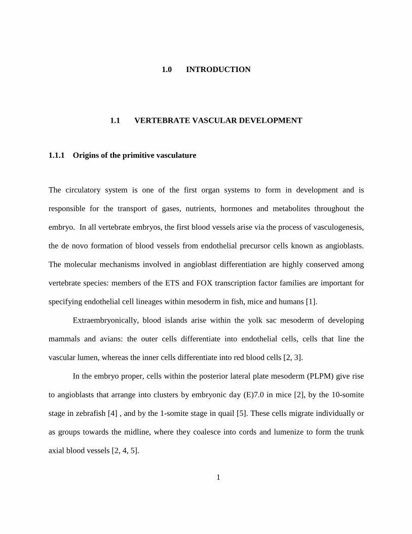
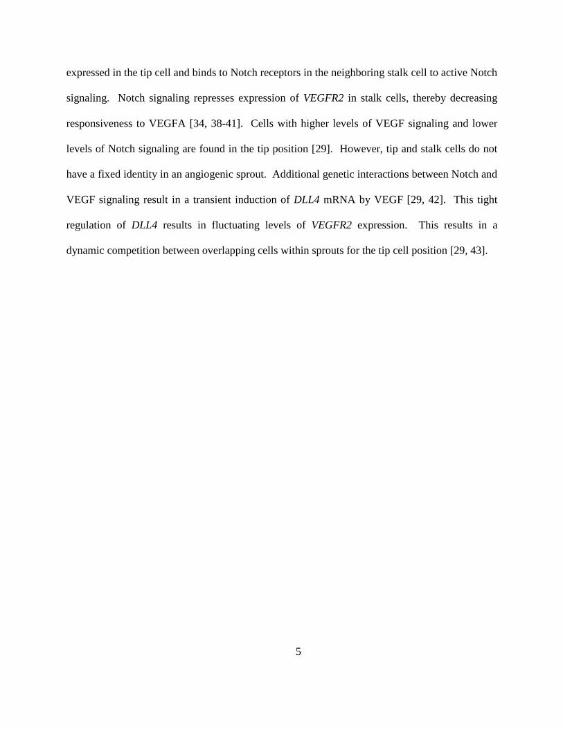
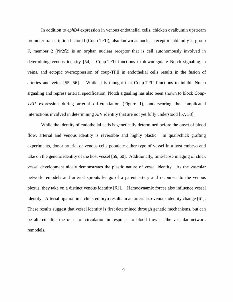
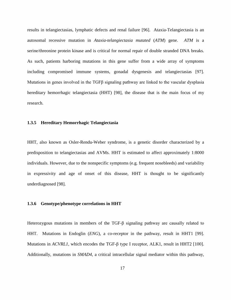
Figure 1: Arterial and venous differentiation and the formation of AVMs
Arteries (in red) are specified by Vegf-induced activation of Notch signaling, resulting in the expression of hey1,
hey2, hes1 and ephrinb2. Notch/Dll4 signaling also represses the expression of ephb4 (a venous marker) in the
arterial endothelium. Veins (in blue) express Coup-TFII, which represses Notch signaling in the venous endothelium
and vice versa. High flow arteries have thick walls lined with vascular smooth muscle cells (brown cells) and flow
into smaller caliber arterioles and finally into capillaries (purple), the location of oxygen and nutrient exchange (blue
dots). Venules and then veins receive low magnitude flow and have valves to prevent backflow. AVMs are direct
connections between arteries and veins, lacking an intervening capillary bed. These high flow shunts decrease
oxygen and nutrient exchange. Also, veins that are downstream of these high flow shunts are enlarged. Impaired
arterial venous specification has been implicated in AVM formation.
7
1.1.4 Vessel stabilization
During the resolution phase of angiogenesis, endothelial cells cease to migrate and proliferate
and vessels are stabilized through the deposition of basal lamina and the recruitment of vascular
smooth muscle cells (vSMCs) and pericytes, a type of vascular support cell [44]. Vascular mural
cells function to support the endothelial cell layer and are able to contract and regulate vascular
tone in response to environmental cues [45]. A number of signaling pathways are important for
the resolution phase of angiogenesis. Platelet derived growth factor (PDGF) produced by
endothelial cells attracts PDGFR-expressing pericytes and induces mural cell proliferation [46].
Additionally, sphingosine-1 phosphate (S1P)/S1P receptor 1 (S1PR1), angiopoetin1/Tie2 and
Transforming growth factor β (TGFβ) signaling all contribute to vascular maturation through
pericyte recruitment and/or extracellular matrix deposition [45].
1.1.5 Arterial and Venous Specification
Arteries and veins are genetically distinct prior to the onset of blood flow. Shortly after
angioblasts are specified as endothelial cells they take on an arterial or venous (A/V) identity. In
the trunk, sonic hedgehog (shh) expressed in the notochord induces the expression of vegf in the
somites [47-49]. Vegf signaling induces dll4 expression and Notch activation in the endothelium
(Figure 1) [49]. It is thought that venous identity is the default identity, while Notch signaling
confers an arterial identity through the regulation of hey1, hey2 and hes genes [30, 50].
Additionally, Notch has been shown to regulate ephrinB2 [30]. EphrinB2 is a ligand for
the EphB4 receptor. EphrinB2/EphB4 are transmembrane proteins that signal through cell-cell
8
contact and label arterial and venous cells, respectively [51-53]. While Notch signaling induces
ephrinB2 expression in arterial endothelial cells, loss of Notch signaling results in the ectopic
expression of ephB4 in these same cells [30].
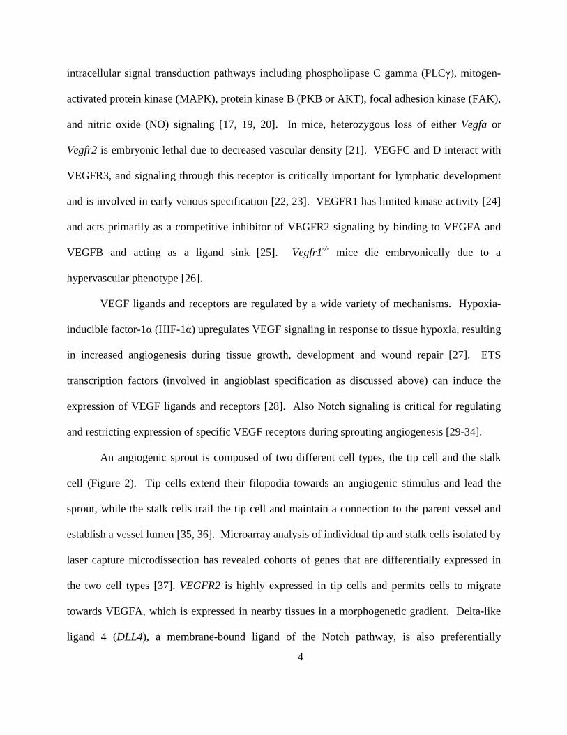
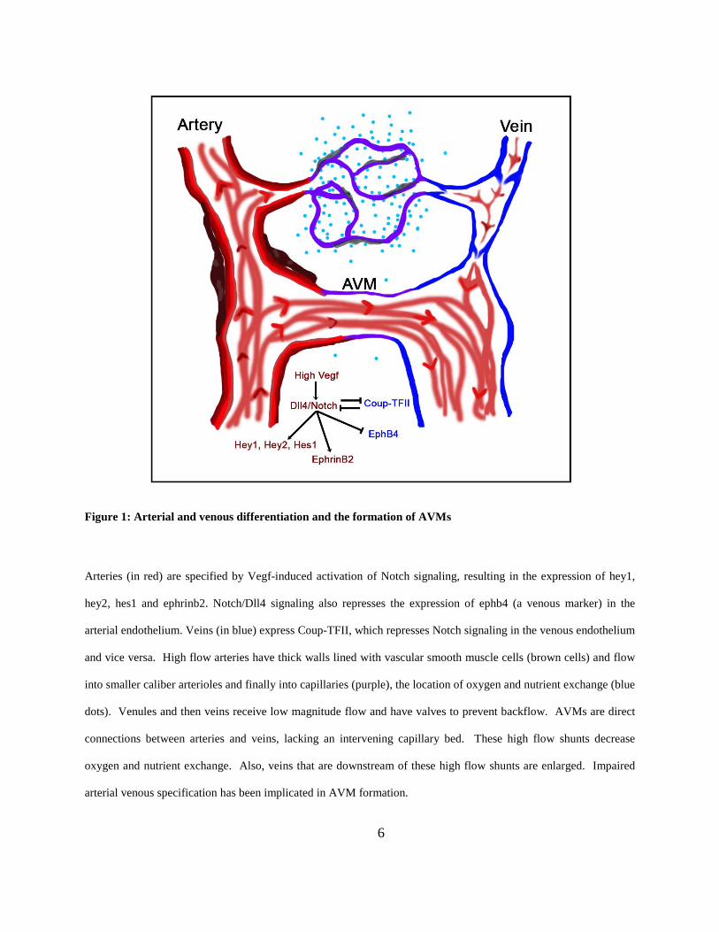
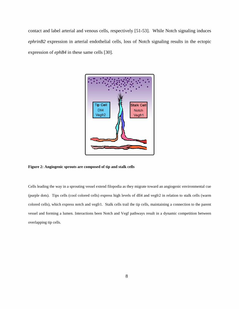
Figure 2: Angiogenic sprouts are composed of tip and stalk cells
Cells leading the way in a sprouting vessel extend filopodia as they migrate toward an angiogenic environmental cue
(purple dots). Tips cells (cool colored cells) express high levels of dll4 and vegfr2 in relation to stalk cells (warm
colored cells), which express notch and vegfr1. Stalk cells trail the tip cells, maintaining a connection to the parent
vessel and forming a lumen. Interactions been Notch and Vegf pathways result in a dynamic competition between
overlapping tip cells.
9
In addition to ephB4 expression in venous endothelial cells, chicken ovalbumin upstream
promoter transcription factor II (Coup-TFII), also known as nuclear receptor subfamily 2, group
F, member 2 (Nr2f2) is an orphan nuclear receptor that is cell autonomously involved in
determining venous identity [54]. Coup-TFII functions to downregulate Notch signaling in
veins, and ectopic overexpression of coup-TFII in endothelial cells results in the fusion of
arteries and veins [55, 56]. While it is thought that Coup-TFII functions to inhibit Notch
signaling and repress arterial specification, Notch signaling has also been shown to block Coup-
TFII expression during arterial differentiation (Figure 1), underscoring the complicated
interactions involved in determining A/V identity that are not yet fully understood [57, 58].
While the identity of endothelial cells is genetically determined before the onset of blood
flow, arterial and venous identity is reversible and highly plastic. In quail/chick grafting
experiments, donor arterial or venous cells populate either type of vessel in a host embryo and
take on the genetic identity of the host vessel [59, 60]. Additionally, time-lapse imaging of chick
vessel development nicely demonstrates the plastic nature of vessel identity. As the vascular
network remodels and arterial sprouts let go of a parent artery and reconnect to the venous
plexus, they take on a distinct venous identity [61]. Hemodynamic forces also influence vessel
identity. Arterial ligation in a chick embryo results in an arterial-to-venous identity change [61].
These results suggest that vessel identity is first determined through genetic mechanisms, but can
be altered after the onset of circulation in response to blood flow as the vascular network
remodels.
10
1.2 BLOOD FLOW AND MECHANOTRANSDUCTION
1.2.1 Hemodynamic forces in blood vessels
Prior to the onset of blood flow, vessel development is governed by local paracrine factors. Upon
the onset of circulation, mechanical forces and endocrine factors influence vessel morphology,
endothelial cell behavior and maintenance of arterial-venous identity.
There are three mechanical forces sensed by vessels: shear stress, cyclic strain, and
hydrostatic pressure. Shear stress is a frictional force that acts directly on luminal surface of
endothelial cells, parallel to the surface of the vessel. It is proportional to the flow rate and
viscosity of the blood and inversely proportional to the vessel radius. Cyclic strain results in
circumferential stretching of the vessel wall, whereas hydrostatic pressure acts perpendicular to
and pushes outward on the vessel wall. Whereas shear stress is directly sensed only by
endothelial cells, cyclic strain and hydrostatic pressure can be sensed by all cells in the vessel
[62]. Of these forces, shear stress is the best studied. Vessels attempt to maintain normalized
forces. In the face of increased blood flow rate (increased cardiac output) or viscosity, vessels
will increase their radius to decrease the total shear forces experienced by the endothelial cells.
Likewise, decreased shear forces result in decreased vessel caliber and in some cases, vessel
regression [63-67].
Different types of shear stress have been shown to result in different biochemical
responses. Straight vessels experience pulsatile laminar shear stress associated with high
magnitude shear forces. Branched and curved vessels experience disturbed flow and low shear
stress [62]. These different types of flow have different effects on gene expression and
11
endothelial cell behavior [68]. High pulsatile laminar shear stress activates Krüppel-like factor 2
(KLF2), a flow regulated transcription factor that favors vessel quiescence [69-71], while
disturbed flow activates nuclear factor kappa B (NF-κB), resulting in an activated inflammatory
response [72]. KLF2 inhibits the expression of NF-κB responsive genes, suggesting a
mechanism by which an inflammatory response is silenced as the endothelium establishes a
laminar shear flow pattern and the vessels become quiescent [73]. To accomplish these changes,
the vessels need to be able to sense these different flow patterns and mechanical forces and then
translate them into a biochemical response.
1.2.2 Mechanosensation of shear stress
It is not yet fully understood how shear stress is sensed nor how sensation is transduced to
changes in gene expression and cell behavior. However, the primary cilium, cell-cell adhesion
dynamics, the glycocalyx and nuclear hydrodynamic drag have all been implicated in vascular
mechanosensation of shear stress (Figure 3) [62, 74].
1.2.2.1 Primary cilia
Primary cilia (Figure 3) are composed of microtubules arranged in a 9+0 pattern. They are
nonmotile and reside on the apical surface of a cell. In endothelial cells, cilial bending results in
a transient calcium influx that ultimately results in the production of NO, a potent vasodilator
[75]. However it is unlikely that this is the primary means of mechanosensation. In regions of
disturbed flow, some endothelial cells have a primary cilium [76, 77], whereas cells experiencing
high physiological levels of shear stress dismantle their primary cilium [78-80]. Additionally,
12
while this mechanism would relay flow magnitude to the cell, it is unclear how the calcium
influx could translate directional information [74].
1.2.2.2 Glycocalyx
The endothelial glycocalyx (Figure 3) consists of sulfated proteoglycans, hyaluronan and
glycoproteins creating a gel-like layer that covers the apical membrane of endothelial cells [81].
The glycocalyx serves many functions including regulating vascular permeability and the
formation of docking sites for plasma-derived molecules, creating microenvironments of growth
factors and atheroprotective proteins [82]. In relation to mechanotransduction, it is thought that
the glycocalyx is displaced in the direction of flow and transduces mechanical forces to the actin
cytoskeleton via adherens junctions [83].
1.2.2.3 Adherens complex
Platelet endothelial cell adhesion molecule-1 (PECAM-1) and vascular endothelial cadherin
(VECAD) are endothelial-specific proteins that localize to adherens junctions. Along with
VEGFR2, these cadherins have been implicated in a flow-sensing complex that is critical in
transducing shear stress into a biochemical response (Figure 3). PECAM-1 is thought to act as a
direct mechanosensor because cultured endothelial cells incubated with PECAM-1 antibody-
coated magnetic beads exhibit rapid PECAM-1 phosphorylation upon application of magnetic
force, similar to that seen upon application of fluid shear stress. [84]. Through a mechanism that
is not yet fully understood, shear stress leads to an accumulation of VEGFR2 at adherens
junctions, and shear stress-induced phosphorylation of PECAM-1 results in ligand-independent
phosphorylation of VEGFR2. With VECAD acting as a scaffolding protein, VEGFR2 initiates a
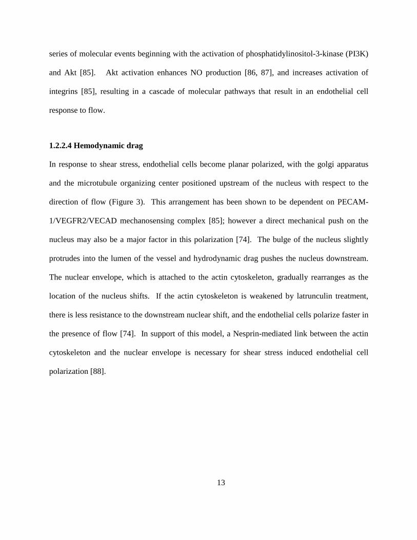
13
series of molecular events beginning with the activation of phosphatidylinositol-3-kinase (PI3K)
and Akt [85]. Akt activation enhances NO production [86, 87], and increases activation of
integrins [85], resulting in a cascade of molecular pathways that result in an endothelial cell
response to flow.
1.2.2.4 Hemodynamic drag
In response to shear stress, endothelial cells become planar polarized, with the golgi apparatus
and the microtubule organizing center positioned upstream of the nucleus with respect to the
direction of flow (Figure 3). This arrangement has been shown to be dependent on PECAM-
1/VEGFR2/VECAD mechanosensing complex [85]; however a direct mechanical push on the
nucleus may also be a major factor in this polarization [74]. The bulge of the nucleus slightly
protrudes into the lumen of the vessel and hydrodynamic drag pushes the nucleus downstream.
The nuclear envelope, which is attached to the actin cytoskeleton, gradually rearranges as the
location of the nucleus shifts. If the actin cytoskeleton is weakened by latrunculin treatment,
there is less resistance to the downstream nuclear shift, and the endothelial cells polarize faster in
the presence of flow [74]. In support of this model, a Nesprin-mediated link between the actin
cytoskeleton and the nuclear envelope is necessary for shear stress induced endothelial cell
polarization [88].
14
Figure 3: Mechanosensation of shear stress, four possible mechanisms
1: Primary cilia are displaced by blood flow resulting in a calcium influx, resulting in the production of NO. 2: The glycocalyx on the surface of endothelial cells
bends in the direction of flow, transducing mechanical forces to the actin cytoskeleton via adherens junctions. 3: Shear stress activated PECAM-1
phosphorylates VEGFR2 and together with VECAD initiates signaling cascades that alter gene expression and cell behavior. 4: The nucleus bulges into the
lumen of the vessel, experiencing hemodynamic drag, and orients itself downstream of the golgi apparatus and the microtubule organizing center, relaying both
direction and strength of force to the actin cytoskeleton
15
1.3 ARTERIOVENOUS MALFORMATIONS
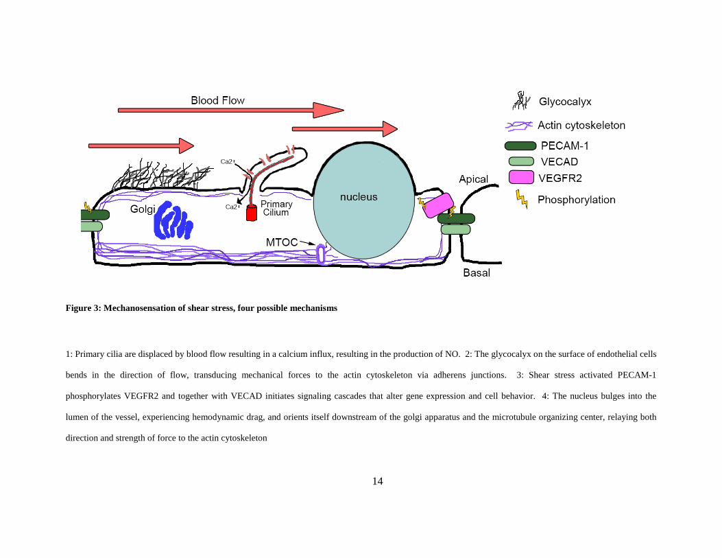
1.3.1 Anatomical and functional differences between different types of blood vessels
Arteries and veins (Figure 1) have developed structural features that reflect differences in the
hemodynamic factors that each vessel type encounters. Arteries carry blood away from the heart
and are designed to cope with high hemodynamic forces (pressure, shear stress, stretch): they
have thick walls composed of several layers of smooth muscle and elastic fibers [16, 62].
Arteries lead to smaller caliber arterioles, which then ramify into a complex network of thin-
walled capillaries [16]. Capillaries are sparsely supported by pericytes and serve as the site of
nutrient and oxygen exchange [45]. As blood flows though the highly branched capillary bed,
velocity decreases dramatically, and thus veins experience much lower magnitudes of
hemodynamic force [89]. Accordingly, veins have thin walls and valves, which function to
prevent back flow of blood.
1.3.2 Anatomy of AVMS
Arteriovenous malformations (AVMs) are direct, high flow connections between thick walled
arteries and thin walled veins, lacking an intervening capillary bed. Over time, these connections
become increasingly complex and tortuous, forming a tangled web of enlarged vessels, or nidus,
which leads to a grossly enlarged draining vein. These malformations acquire a thick smooth
muscle coat, thereby barring gas exchange (Figure 1) [90, 91]. Although the etiology of AVMs is
16
unclear, there is evidence to suggest that in some cases they arise due to failed regression of
normally transient arterial/venous connections, or failed repulsion between arteries and veins due
to improper arterial/venous (A/V) specification [53, 92].
1.3.3 Clinical consequences of AVMs
The clinical consequences of an AVM will depend on the location and size of the lesion.
Generally, AVMs decrease gas exchange and cause localized ischemia, and these malformations
may rupture due to the inability of veins to handle high magnitudes of mechanical forces [91].
Specifically, cerebral AVMs may cause localized ischemia or hemorrhagic stroke; pulmonary
AVMs rarely rupture but can lead to cyanosis, brain abscess, transient ischemic attacks, and
embolic stroke; and very high flow hepatic AVMs can lead to high output cardiac failure [93].
AVMs connecting small, mucocutaneous vessels are known as telangiectasias. Dermal
telangiectasias may bleed but are primarily a cosmetic issue, whereas bleeding from GI and nasal
telangiectasias can cause anemia and severe hemorrhage [94].
1.3.4 Genetic basis for AVMs
A majority of AVMs are sporadic, however, a subset of these vascular lesions is caused by
genetic mutations. Capillary malformation-arteriovenous malformation (CM-AVM) is caused by
heterozygous mutations in Rasa1, which encodes RAS p21 protein activator 1, and is
characterized by multiple small capillary malformations [95]. Hypotrichosis-lymphedema-
telangiectasia syndrome (HLTS) results from mutations in Sox18, a known regulator of Dll4, and
17
results in telangiectasias, lymphatic defects and renal failure [96]. Ataxia-Telangiectasia is an
autosomal recessive mutation in Ataxia-telangiectasia mutated (ATM) gene. ATM is a
serine/threonine protein kinase and is critical for normal repair of double stranded DNA breaks.
As such, patients harboring mutations in this gene suffer from a wide array of symptoms
including compromised immune systems, gonadal dysgenesis and telangiectasias [97].
Mutations in genes involved in the TGFβ signaling pathway are linked to the vascular dysplasia
hereditary hemorrhagic telangiectasia (HHT) [98], the disease that is the main focus of my
research.
1.3.5 Hereditary Hemorrhagic Telangiectasia
HHT, also known as Osler-Rendu-Weber syndrome, is a genetic disorder characterized by a
predisposition to telangiectasias and AVMs. HHT is estimated to affect approximately 1:8000
individuals. However, due to the nonspecific symptoms (e.g. frequent nosebleeds) and variability
in expressivity and age of onset of this disease, HHT is thought to be significantly
underdiagnosed [98].
1.3.6 Genotype/phenotype correlations in HHT
Heterozygous mutations in members of the TGF-β signaling pathway are causally related to
HHT. Mutations in Endoglin (ENG), a co-receptor in the pathway, result in HHT1 [99].
Mutations in ACVRL1, which encodes the TGF-β type I receptor, ALK1, result in HHT2 [100].
Additionally, mutations in SMAD4, a critical intracellular signal mediator within this pathway,
18
result in a combined juvenile polyposis-HHT syndrome [101]. Two additional loci on
chromosomes 5q31.3-32 and 7p14 have been linked to HHT; however, the responsible genes
within these loci have not yet been identified [102, 103]. HHT1 and HHT2 present with
different phenotypic severity and location. HHT1 patients tend to experience more severe
symptoms with an earlier age of onset. 49-75% of HHT1 patients present with pulmonary
AVMs (PAVMs), 15-20% with cerebral AVMs (CAVMs) and 2-8% with hepatic AVMs
(HAVMs). Between 60-72% of HHT1 patients have GI telangiectasias with approximately 18%
of these patients experiencing bleeding [104-106]. HHT2 patients are typically diagnosed
around the age of 40 and, compared to HHT1 patients, present with similar incidences of GI
(gastrointestinal) telangiectasias/bleeding and lower incidences of PAVMS and CAVMs (5-44%
and 0-2%). However, the incidence of HAVMs is between 28-84% in these patients [104-106].
The reason for the differences in the phenotypic severity and presentation between the two HHT
sub-groups is unknown but may reflect differential tissue distribution or function of Endoglin
and Alk1 [107].
1.3.6.1 ALK1 signaling
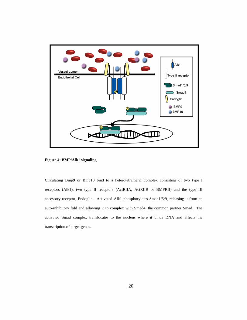
Overview of TGF-β family signaling
In TGF-β signaling, dimeric ligand binds to a heterotetrameric complex of type I and type II
receptors. Upon ligand binding, the type II receptor phosphorylates the type I receptor. The type
I receptor then phosphorylates Smad transcription factors. Once activated, Smad proteins bind to
the common partner Smad, Smad4, and translocate into the nucleus to regulate the transcription
of target genes (Figure 4). TGF-β superfamily signaling involves seven type I receptors (ALK1-
19
ALK7) and five type II receptors (ActRIIA, ActRIIB, BMPRII, TGFβII and AMHRII), all of
which are serine/threonine kinases. Ligand binding to a heterotetrameric complex of type I and
type II receptors can be facilitated by a type III receptor, ENG, which does not have enzymatic
activity [108]. The family of TGF-β ligands is large and can be divided into multiple sub-
families. Ligands in the TGF-β and activin subfamilies bind to receptor complexes that
phosphorylate Smad2 and Smad3. Bone morphogenetic protein (BMP) ligands bind to different
receptor complexes that phosphorylate Smad1, Smad5 and Smad9 [109].
Alk1 in vascular development
ALK1 is a transmembrane protein containing an extracellular N-terminal domain that binds
ligand, a short, single pass transmembrane domain and a large intracellular domain. The
intracellular domain contains three main motifs: a GS domain, a serine/threonine kinase domain
and a cytoplasmic tail. The GS domain is phosphorylated by the type II receptor and contains a
highly conserved TTSGSGSG motif [110, 111]. To date, 434 mutants in Alk1 have been
identified, of which 50% have been found to be pathogenic
(http://www.arup.utah.edu/database/hht/). Of these, 46% are missense mutations. Limited in
vitro analysis of HHT2-associated ALK1 mutations suggests that the majority of mutant proteins
are localized to the cell surface and are able to bind to BMP9 (except for mutants in the
extracellular domain), and that mutant protein does not affect activity of wild type protein [112,
113]. These data indicate that these mutations do not act as a dominant negative and suggest
instead that phenotypes result from a haploinsufficiency [112].
20
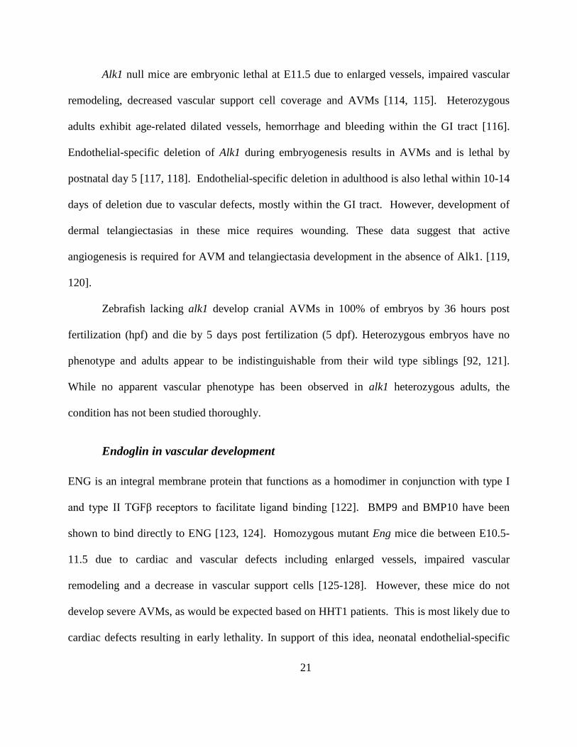
Figure 4: BMP/Alk1 signaling
Circulating Bmp9 or Bmp10 bind to a heterotetrameric complex consisting of two type I
receptors (Alk1), two type II receptors (ActRIIA, ActRIIB or BMPRII) and the type III
accessory receptor, Endoglin. Activated Alk1 phosphorylates Smad1/5/9, releasing it from an
auto-inhibitory fold and allowing it to complex with Smad4, the common partner Smad. The
activated Smad complex translocates to the nucleus where it binds DNA and affects the
transcription of target genes.
21
Alk1 null mice are embryonic lethal at E11.5 due to enlarged vessels, impaired vascular
remodeling, decreased vascular support cell coverage and AVMs [114, 115]. Heterozygous
adults exhibit age-related dilated vessels, hemorrhage and bleeding within the GI tract [116].
Endothelial-specific deletion of Alk1 during embryogenesis results in AVMs and is lethal by
postnatal day 5 [117, 118]. Endothelial-specific deletion in adulthood is also lethal within 10-14
days of deletion due to vascular defects, mostly within the GI tract. However, development of
dermal telangiectasias in these mice requires wounding. These data suggest that active
angiogenesis is required for AVM and telangiectasia development in the absence of Alk1. [119,
120].
Zebrafish lacking alk1 develop cranial AVMs in 100% of embryos by 36 hours post
fertilization (hpf) and die by 5 days post fertilization (5 dpf). Heterozygous embryos have no
phenotype and adults appear to be indistinguishable from their wild type siblings [92, 121].
While no apparent vascular phenotype has been observed in alk1 heterozygous adults, the
condition has not been studied thoroughly.
Endoglin in vascular development
ENG is an integral membrane protein that functions as a homodimer in conjunction with type I
and type II TGFβ receptors to facilitate ligand binding [122]. BMP9 and BMP10 have been
shown to bind directly to ENG [123, 124]. Homozygous mutant Eng mice die between E10.5-
11.5 due to cardiac and vascular defects including enlarged vessels, impaired vascular
remodeling and a decrease in vascular support cells [125-128]. However, these mice do not
develop severe AVMs, as would be expected based on HHT1 patients. This is most likely due to
cardiac defects resulting in early lethality. In support of this idea, neonatal endothelial-specific
22
deletion of Eng results in AVMs in a high percentage of mice [129], and adult mice harboring a
single Eng mutation tend to develop age-related HHT-like phenotypes including nosebleeds,
enlarged vessels and telangiectasias [126, 130, 131].
ALK1 Ligands, BMP9 and BMP10
BMP9 and BMP10 have recently been identified as the physiologically relevant ligands for
ALK1 signaling and vascular development [112, 132-135]. BMP9 and BMP10 are highly
related proteins, sharing 65% sequence identity at the protein level. Both proteins undergo very
similar biosynthesis [112, 136]. Pre-pro-proteins are cleaved by convertase enzymes such as
furin into a prodomain and mature peptide. After secretion, the prodomain remains associated
with the mature peptide through non-covalent interactions. BMP9 is active when associated with
the prodomain and able to bind to ALK1 and induce Smad1/5 phosphorylation [136]. However,
BMP10 is latent until the prodomain is removed [137]. Although the metalloproteinase BMP-1
(unrelated to BMP ligands) can cleave the BMP10 prodomain in vitro [137], the mechanism by
which BMP10 is activated in vivo is not understood. Perhaps mechanical forces, interactions
with the extracellular matrix or accessory receptors are required to dissociate the prodomain
from the BMP10 mature peptide in a physiologic setting.
In humans, BMP9 is expressed in the liver (hepatocytes, biliary epithelial cells) and
circulates in its biologically active form at 110 pg/ml in serum [133, 138, 139]. In mice, Bmp9 is
also expressed in liver as early as E9.75 and is simultaneously detectable in serum. Bmp9 null
mice have no vascular defect and are viable [140]. BMP10 is produced by the heart [141],
specifically within the ventricular cardiomyocytes as early as E8.5 then restricted to the atrial
cardiomyocytes by E16.5 in mice [140], and is also detectable in mouse and human serum [112,
23
142], though in an inactive prodomain-bound complex [112]. BMP10 null mice die at E10.0
from failed trabeculation and AVMs. Interestingly, vascular but not cardiac defects in these
mice could be rescued by the insertion of Bmp9 into the Bmp10 locus [140]. Together, these
data suggest that BMP9 and BMP10 are endocrine ALK1 ligands, and that BMP9 can
functionally compensate for BMP10 in the vasculature if expressed in a BMP10-like
spatiotemporal pattern.
In zebrafish, morpholino oligonucleotide mediated knockdown of bmp9 is not lethal but
results in a failure of the caudal vein to properly remodel [143]. These embryos do not develop
enlarged cranial shunts similar to those observed in alk1 mutants [92, 121]. Like in mice, bmp10
is expressed earlier than bmp9 and concomitant knockdown of bmp10 and bmp10-like (a
zebrafish bmp10 paralog) results in large cranial shunts and is embryonic lethal [135]. Together
with the data from the mouse models, these results suggest that in early development, BMP10 is
necessary for embryonic vascular development and BMP9 and BMP10 ultimately function
redundantly to maintain normal vasculature.
Type II receptors that complex with ALK1
Ligand binds to a heterotetrameric complex of type I and type II receptors. Type II receptors are
thought to be constitutively active and phosphorylate the type I receptors when they are brought
into a complex together by ligand binding. BMPs have been shown to preferentially interact
with ActRIIA, ActRIIB or BMPRII [109].
24
1.4 ZEBRAFISH AS A MODEL SYSTEM FOR STUDYING HHT-ASSOCIATED
AVMS AND ALK1 SIGNALING
1.4.1 General attributes of the zebrafish model
Zebrafish are an excellent model system for the study of vertebrate development.
External fertilization, small size, and optical clarity allow for the observation of development
from the 1-cell stage. Development occurs rapidly, with gastrulation occurring at 6 hpf,
heartbeat beginning at 24 hpf and circulation through the head and tail of the embryo by 27 hpf
[144]. Furthermore, using confocal or two-photon microscopy, development or particular organ
systems can be monitored with high spatiotemporal resolution in live transgenic embryos
expressing fluorescent proteins under the control of cell type-specific promoters.
Zebrafish are also amenable to genetic manipulation. DNA and RNA can be injected
easily into 1-cell stage zebrafish embryos to ectopically express genes in a tissue-specific manner
or globally, respectively. In addition, genes of interest can be knocked down transiently using
morpholino-modified antisense oligonucleotides. These short (~25 bases) oligos are highly
stable and contain a morpholine ring in place of the deoxyribose ring and non-ionic
phosphorodiamidate group in place of the anionic phosphodiester linkages between bases [145].
They are designed to target specific mRNA sequences and through steric inhibition block either
translation of the message or proper mRNA splicing [146, 147].
Forward genetic screens using N-ethyl-N-nitrosourea (ENU) mutagenesis, viral insertion
mutagenesis or transposon-mediated gene disruption have generated thousands of genetic
mutants that are used to study gene functions and signaling cascades important for embryonic
25
and larval development [148]. More recent advances in reverse genetics have allowed for the
targeted disruption of genes using TALEN and CRISPR/Cas9 technology [149, 150].
In addition to genetic approaches, zebrafish are easily manipulated using
pharmacological approaches. Addition of soluble small molecules to the water in which the
embryos are reared allows researchers to perturb specific biochemical pathways using previously
characterized drugs, and to perform large scale, medium throughput chemical screens to identify
novel small molecules that perturb particular signaling pathways or developmental processes.
[148]. Together, these attributes make zebrafish an extremely powerful system for the study of
vertebrate vascular development in a physiologically relevant in vivo setting.
1.4.2 Zebrafish alk1 mutants develop cranial AVMs
1.4.2.1 Zebrafish cranial blood vessel development:
Embryonic zebrafish vascular morphology has been studied in great detail, first through the use
of confocal microangiography and then through live time-lapse imaging of transgenic embryos
expressing fluorescent proteins under the control of endothelial-specific promoters [6, 144].
Microangiography specifically highlights vessels that are lumenized and carrying plasma/blood
flow, while imaging of transgenics allows assessment of development both prior to and
subsequent to lumen formation. By utilizing both techniques, a complete understanding of
vessel formation and maturation can be gained. .
26
1.4.2.2 alk1-/- AVMs arise via a two-step process
The spatial and temporal predictability of shunt formation in alk1-/- zebrafish embryos make
them an ideal model for studying the molecular and cellular missteps that lead to AVM
formation. In the absence of Alk1 signaling, zebrafish embryos develop grossly enlarged cranial
arteries that contain supernumerary endothelial cells: alk1-/- embryos have a 1.2-fold increase in
the number of endothelial cells in the BCA/PCS beginning at 32 hpf and a 1.8-fold increase by
48 hpf [92, 121, 135]. By 40 hpf, AVMs form downstream of these arteries, between the BCA
and PMBC (anterior shunt) and/or the BA and PHBC (posterior shunt) [92]. Thus, arterial
enlargement precedes shunt formation. Time-lapse confocal microscopy has revealed that AVMs
are the result of failed regression of one or more transient BCA/PMBC or BA/PHBC
connections. These connections are retained in alk1 mutants only in the presence of blood flow,
suggesting that the increased shear stress caused by cranial arterial enlargement triggers an
adaptive response aimed at normalizing hemodynamic force [92]. In sum, these data support the
idea that AVM development in alk1 mutants is not genetically determined but instead represents
a two-step process that involves genetically programmed arterial enlargement followed by an
Alk1-independent response to changes in the hemodynamic environment [92].
1.4.3 alk1 expression is regulated by blood flow
Alk1 is expressed in arteries that are proximal to the heart in zebrafish embryos. Beginning at 26
hpf, alk1 expression can be observed in the AA1 and the LDA, vessels that are a part of the
initial circulatory loop. Shortly thereafter, expression within the ICA can be detected followed
by CaDI and BCA expression at 30 hpf. It is interesting to note that alk1 is not expressed in the
27
PCS or BA, and is restricted in the cranial arterial endothelium to arteries that are proximal to the
heart and experience the highest magnitude of hemodynamic forces [92, 121]. In silent heart
(sih) embryos, which lack heartbeat and therefore blood flow, overall vascular patterning is
normal; however, alk1 is not expressed, indicating that flow is required for alk1 expression.
Additionally, pharmacological inhibition of heartbeat also inhibits alk1 expression [92].
Maintenance of alk1 expression is also highly dependent on flow. In experiments where flow
was stopped after alk1 expression had been initiated, expression quickly faded in a distal-to-
proximal pattern. Upon restoration of heartbeat, alk1 expression was detectable within an hour in
the AA1 and completely restored by 8 hours (Jim Donovan, unpublished data). In gata1 mutant
embryos, which lack erythrocytes, alk1 expression is unaffected, indicating that expression is not
dependent on endothelial-cell/red blood cell interaction [92].
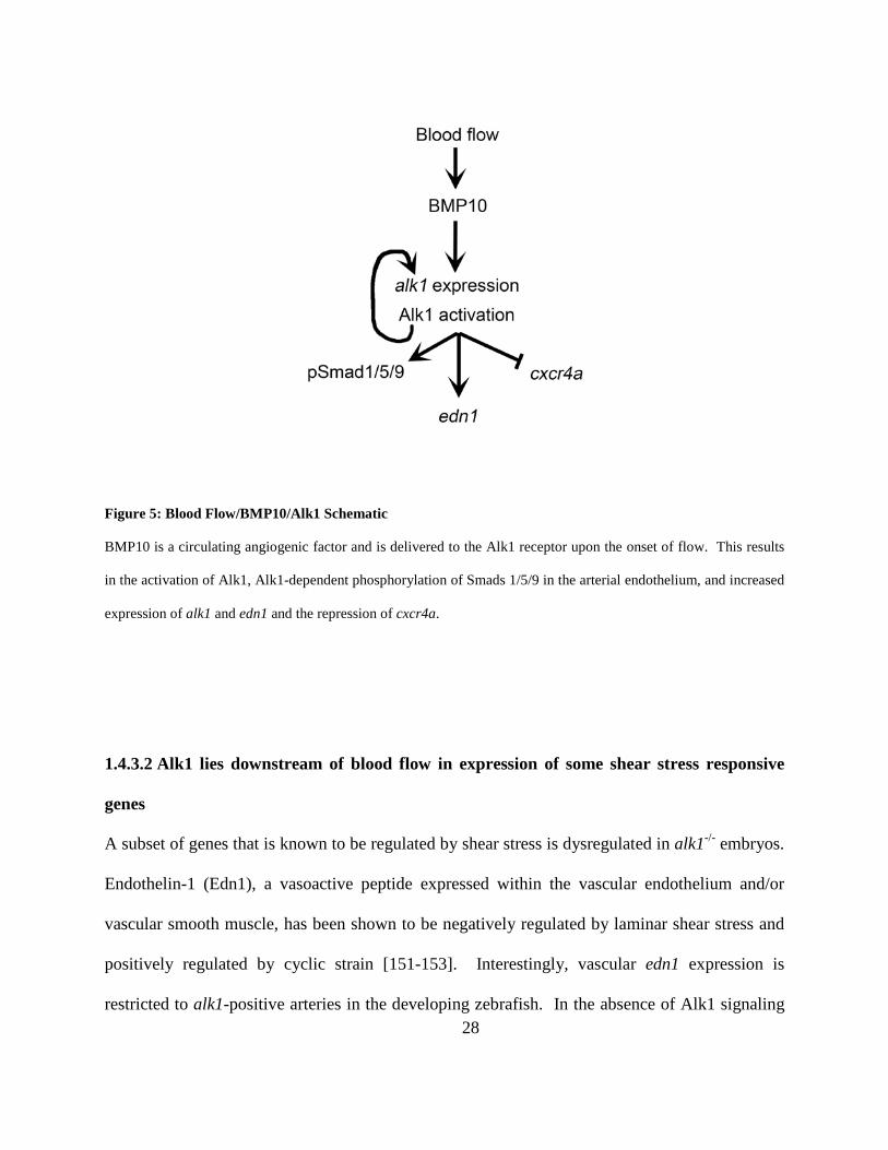
1.4.3.1 Alk1 lies downstream of blood flow in phosphorylation of Smad1/5/9
Alk1 activity within the vascular endothelium can be assayed by assessing pSmad1/5 [110, 133,
134], which is absent in alk1 mutants and in sih morphant embryos lacking blood flow (and
therefore also lacking alk1 expression) [135]. Stable transgenic expression of alk1 driven by a
flow-independent vascular endothelial promoter is able to rescue pSmad1/5 in alk1 mutants but
not sih morphants, suggesting that flow is necessary for Alk1 activity as well as expression
[135]. Accordingly, injection of recombinant human BMP10 protein directly into the base of the
CaDI of flow-deprived embryos ectopically expressing alk1 was able to restore pSmad1/5,
suggesting that blood flow is required to activate Alk1 signaling by circulating Bmp10, which is
produced in the heart [135] (Figure 5).
28
Figure 5: Blood Flow/BMP10/Alk1 Schematic
BMP10 is a circulating angiogenic factor and is delivered to the Alk1 receptor upon the onset of flow. This results
in the activation of Alk1, Alk1-dependent phosphorylation of Smads 1/5/9 in the arterial endothelium, and increased
expression of alk1 and edn1 and the repression of cxcr4a.
1.4.3.2 Alk1 lies downstream of blood flow in expression of some shear stress responsive
genes
A subset of genes that is known to be regulated by shear stress is dysregulated in alk1-/- embryos.
Endothelin-1 (Edn1), a vasoactive peptide expressed within the vascular endothelium and/or
vascular smooth muscle, has been shown to be negatively regulated by laminar shear stress and
positively regulated by cyclic strain [151-153]. Interestingly, vascular edn1 expression is
restricted to alk1-positive arteries in the developing zebrafish. In the absence of Alk1 signaling
29
or blood flow, edn1 expression is lost, indicating that alk1 is either directly or indirectly required
for edn1 expression. Cxcr4a is a promigratory chemokine receptor and has been shown to be
negatively regulated by laminar shear stress [154]. cxcr4a expression is increased in alk1-
postive arteries in the absence of Alk1 or blood flow (Figure 5).
Not all flow responsive genes are dysregulated in alk1-/- embryos. klf2a, a transcription
factor known to integrate shear stress-responsive pathways, is positively regulated by shear stress
[70, 151] and expression is completely lost in zebrafish embryos lacking blood flow [92].
However, klf2a expression is unaffected in alk1-/- embryos [92]. These results suggest that Alk1
signaling may act upstream of edn1 and cxcr4a in a klf2a-independent flow responsive pathway
required to limit endothelial caliber upon the onset of flow. In support of this hypothesis,
BMP10 protein injection coupled with transgenic, flow-independent alk1 expression restored
cxcr4a and edn1 expression and endothelial cell number to wild type levels in the absence of
blood flow [135] (Figure 5).
1.5 NOTCH SIGNALING AND AVMS
With its membrane bound ligand and receptors, Notch signaling allows communication between
neighboring cells. It is a fundamentally conserved signaling pathway that is used reiteratively in
the development of many different tissues and has been shown to regulate proliferation,
apoptosis, self-renewal and differentiation. Notch signaling is highly context specific and
therefore it is unwise to make generalized predictions on how Notch signaling will influence the
development of one cell type based on results of another [155]. Throughout much of the

30
introduction, I have touched on Notch signaling and its key roles in angiogenesis, specifically in
tip and stalk cell determination, regulation of VEGF signaling and arterial-venous identity. Notch
signaling is implicated in the formation of AVMs, and there is also a large amount of evidence
suggesting that a Notch and Alk1 interaction is important for proper vascular development.
1.5.1 Notch pathway summary
Notch receptors are single-pass transmembrane proteins that contain an extracellular ligand
binding domain and an intracellular domain critical for Notch signal transduction. Notch ligands
are transmembrane proteins from the Delta-like ligand (Dll) and Jagged families. Of these, Dll4
has the most prominent role in vascular development. The notch intracellular domain (NICD) is
processed upon ligand binding by ADAM metalloproteases [156], which activate the receptor,
and then further cleaved by γ-secretase enzymes that release the activated NICD into the cell
[157]. The intracellular domain of the Notch receptor binds to CSL (Cp-binding factor 1 (CBF-
1), Suppressor of hairless [Su(h)], and Lag-1), also known as recombination signal sequence-
binding protein-J kappa (RBPJκ) [158]. In the absence of NICD, RBPJκ acts as a transcriptional
repressor. Binding of NICD to RBPJκ converts the complex to a co-activator and results in the
transcription of Notch target genes, including the hairy and enhancer of split-1 (HES1) family
[155, 159].
31
1.5.2 Perturbation of Notch signaling results in AVMs
Arterial/venous specification and its role in AVM prevention are evidenced by Notch
perturbations resulting in these vascular malformations in zebrafish and mice. In zebrafish,
mutations in the mindbomb (mib), a regulator of Notch ligand endocytosis, results in the
formation of AVMs as well as impaired arterial/venous specification [30, 160]. While mutations
in hey2 results in defects in dorsal aorta due to altered arterial/venous identity [160, 161].
Similarly in mice, decreased expression of the Notch ligand, dll4 or endothelial specific deletion
of RBPJκ results in loss of arterial specification and AVMs that are small in caliber and appear
atretic [162, 163]. Ectopic activation of Notch signaling also results in improper A/V
specification and vascular abnormalities. Zebrafish ectopically overexpressing the NICD in the
vascular endothelium have enlarged vessels and decreased expression of venous ephB4 [164,
165], and expression of a constitutively active Notch4 (Notch4CA) in the mouse endothelium
results in venous expression of arterial markers and AVMs in the liver, uterus, skin, brain and
lung of adult mice [166-169]. Brain AVMs that result from inducible transient expression of
Notch4CA are reversible: when the transgene is turned off, vessels shrink back to normal size
and A/V marker expression is restored [170]. While AVMs resulting from decreased Notch
signaling are small in caliber, Notch overexpression AVMs are large and contain an increased
number of endothelial cells [31, 166, 168, 171, 172].
32
1.5.3 Evidence for Notch/Alk1 interactions
Evidence from cultured endothelial cells demonstrates that Smad1/5 can bind to the promoters of
the canonical notch target genes, hey1, hey2 and hes1 and enhance expression [173], and Alk1
activation increases expression of some Notch targets [32, 174, 175], In addition, Notch target
gene expression is synergistically increased by simultaneous Notch and Alk1 pathway activation
[175]. BMP9/ALK1 effects were found to be independent of RBPJκ suggesting that Alk1
signaling through Smad1/5 may function to reinforce arterial identity independent of NICD/
RBPJκ [175]. Phenotypic evidence of an interaction between Notch and Alk1 signaling is less
convincing. While inhibition of both pathways separately has been shown to enhance VEGF-
stimulated angiogenesis both in cell culture and in vivo, combined inhibition does not appear to
have an additive effect [175]. Similarly, constitutively active Notch or Alk1 has been shown to
inhibit sprouting with no increased effects of combined pathway activation [174]. And lastly, the
inhibition of either pathway does not alter phenotypic effects of activation of the other [174]. A
better understanding of how each of these pathways function and interact is required to
understand why an apparent genetic synergy does not translate into a phenotypic synergy.
1.6 SUMMARY AND DISSERTATION AIMS
Patients with HHT2 develop AVMs due to a mutation in ALK1 [91, 112, 113], but the function
of ALK1 in endothelial cells and the natural history of AVMs is unknown. Zebrafish alk1 mutant
embryos develop enlarged arteries containing supernumerary endothelial cells, causing a change
33
in hemodynamic environment that leads to AVMs [92, 121, 135]. The source of the additional
endothelial cells has been thought to be a result of increased proliferation and/or migration,
supporting the hypothesis that Alk1 signaling is antiangiogenic and contributes to vessel
stabilization [132, 176]. Here, I show that the increase in endothelial cell number in the cranial
arterial endothelium is due to an improper distribution of cells. Arteries proximal to the heart
experience a decrease in cell number due to an accumulation of cells in the more distal arteries.
These data suggest that Alk1 is required for directed endothelial cell migration towards the heart
and in opposition to blood flow.
In addition, an interaction between Alk1 and Notch signaling pathways has been thought
to be important for proper vascular development and AVM prevention [175, 177]. Here I
demonstrate that Alk1 and Notch signaling have context specific interactions in the regulation of
the expression of some Notch target genes, but there are only weak phenotypic interactions
between the two pathways in vivo.
34
2.0 ALK1 ALLOWS ARTERIAL ENDOTHELIAL CELLS TO RESIST MIGRATION
IN THE DIRECTION OF BLOOD FLOW
ALK1, a TGF-β type I receptor serine/threonine kinase, is critical for proper vascular
development. Heterozygous loss of ALK1 results in the vascular disorder, hereditary
hemorrhagic telangiectasia type 2 (HHT2), which is characterized by the development of
arteriovenous malformations (AVMs) and affects 1:8000 people worldwide. alk1-/- zebrafish
develop embryonic lethal AVMs which form via a two-step mechanism. First, loss of alk1
results in an increase in endothelial cell number in cranial arteries, which results in increased
vessel caliber. In the second step, normally transient connections between arteries and veins are
maintained as an adaptive mechanism to cope with an increased hemodynamic load. Using
zebrafish as a tool to study the AVM formation due to loss of Alk1 signaling, I have found that
Alk1 is required for directed arterial endothelial cell migration in opposition to blood flow.
Embryos lacking alk1 experience a redistribution of cells, with endothelial cells failing to
efficiently migrate against the direction of blood flow and accumulating in more distal regions of
alk1-dependent arteries. This altered cellular distribution causes an increase in arterial caliber
and consequent retention of downstream arteriovenous connections, resulting in fatal AVMs.
35
2.1 INTRODUCTION
Hereditary hemorrhagic telangiectasia (HHT) is a haploinsufficiency characterized by a
predisposition to development of arteriovenous malformations (AVMs). These fragile, direct
connections between arteries and veins can lead to hemorrhage or stroke. HHT is caused by
defects in transforming growth factor-beta (TGF-β) superfamily signaling. Specifically,
mutations in the type III accessory receptor, endoglin (ENG), cause HHT1; mutations in the type
I receptor serine threonine kinase, activin receptor-like kinase 1 (ACVRL1, or ALK1), cause
HHT2; and mutations in the signaling mediator, SMAD4, cause a combined syndrome of juvenile
polyposis with HHT [99-101]. Together, mutations in these three genes account for
approximately 85% of HHT. Despite the fact that these gene products all participate in TGF-β
signaling, whether mutations affect one or more discrete pathways and how these pathways
function to prevent AVMs remain poorly understood.
Based on histological observation of cutaneous AVMs (telangiectasias) from HHT
patients, it has been postulated that the first step in AVM development is focal dilation of a
postcapillary venule, followed by arteriole dilation and subsequent loss of intervening capillaries
[90]. However, these conclusions were reached from static observations of independent lesions
and not from longitudinal analysis. In Alk1- and Eng-deleted adult mice, wound-induced
subdermal AVMs develop via angiogenic elongation of both arteries and veins, with de novo
arterial-venous connections developing prior to vessel dilation [117, 120]. Although these
findings represent a longitudinal analysis, imaging of vascular growth was performed only once
per day and was not at cellular resolution. Therefore, the aberrant cell behaviors that lead to
AVMs could not be elucidated.
36
Zebrafish are an excellent model for the study of both normal and pathological vascular
development because signaling pathways that control endothelial cell differentiation and vessel
patterning are conserved from fish to mammals, and because optically transparent transgenic
zebrafish embryos allow real-time imaging of vessel development at cellular resolution.
Zebrafish alk1 mutants develop AVMs at a predictable time (approximately 40 hours post-
fertilization, hpf) in a predictable location (beneath the midbrain or hindbrain) and therefore
serve as an excellent model for exploring the cellular basis of HHT-associated AVM
development [92, 121, 135].
In zebrafish, alk1 is expressed after the onset of blood flow in cranial arterial endothelial
cells closest to the heart, including (in ordered series) the outflow tract and first aortic arch
(AA1), internal carotid artery (ICA), caudal division of the internal carotid artery (CaDI), and
basal communicating artery (BCA). We previously reported increases in arterial endothelial cell
number in and diameter of the contiguous CaDI, BCA, and posterior communicating segments
(PCS) in alk1 loss-of-function mutants as early as 32 hpf [92, 135]. Between 32-40 hpf,
BCA/PCS endothelial cell number increases similarly in the absence of alk1 function or in the
absence of blood flow, and blood flow is required for alk1 expression [92]. These data suggest
that Alk1 transmits a flow-based signal that limits arterial caliber. In alk1 mutants, high-flow
shunts develop by 40 hpf downstream of enlarged arteries, connecting either the BCA to the
primordial midbrain channel (PMBC) or the downstream alk1-negative basilar artery (BA) to the
primordial hindbrain channel (PHBC). These shunts represent aberrant retention of normally
transient arteriovenous connections that initiate development of and serve as early drainage for
the nascent arterial system [92, 121]. In alk1 mutants, late increases in endothelial cell number
(40-48 hpf) and AVM development require blood flow [92], suggesting that these effects are
37
secondary to enlargement of alk1-positive cranial arteries closest to the heart and represent an
adaptive response of downstream vessels to altered hemodynamics. Therefore, I focused this
study on defining the primary role of Alk1 in arterial endothelium in limiting arterial caliber.
Results demonstrate a defect in arterial endothelial cell distribution within lumenized
vessels as the primary effect of loss of Alk1 function. With the onset of blood flow, wild type
cranial arterial endothelial cells in the lumenized AA1, ICA, and CaDI migrate in a distal-to-
proximal fashion towards the heart, against the direction of blood flow. Some cells originally
located in AA1 or the ICA enter the heart and incorporate into ventricular endocardium. In
contrast, cells distal to the ICA/CaDI junction generally remain in place after the onset of blood
flow, and there is little to no mixing of arterial cells derived from different sprouts or angioblast
pools. In alk1 mutants, endothelial cell distribution is altered, with decreased cranial arterial
endothelial cell contribution to endocardium, increased distal migration of endothelial cells, and
increased mixing of arterial endothelial cells derived from different sprouts or angioblast pools.
Together, these data suggest that loss of alk1 results in enhanced movement of arterial
endothelial cells in the direction of blood flow, resulting in accumulation of cells in and
enlargement of cranial arteries distal to the heart.
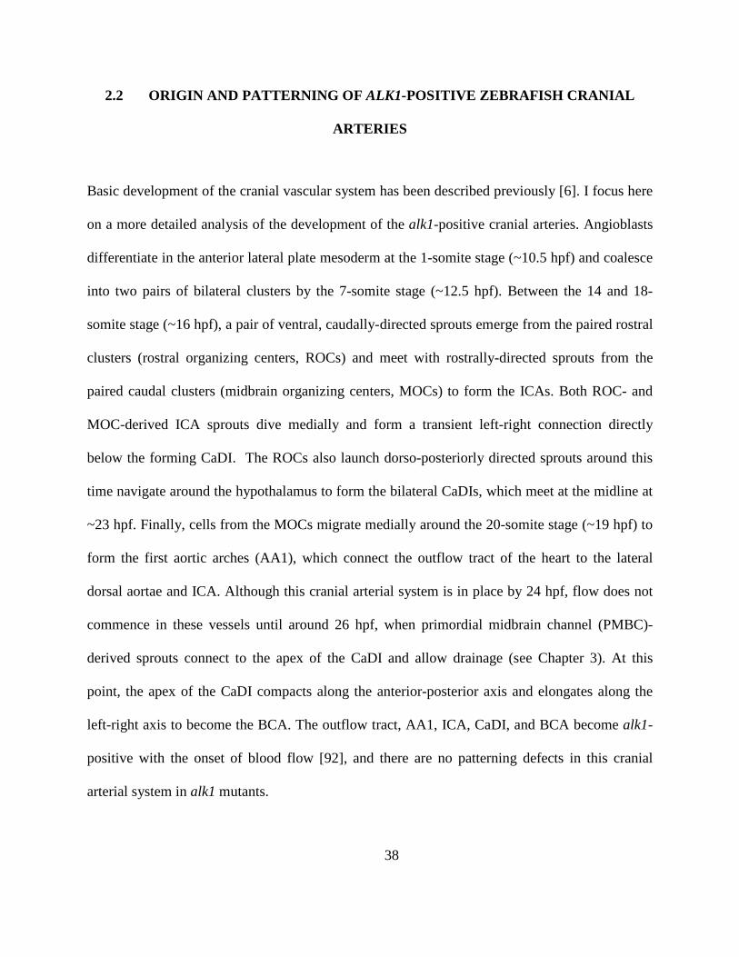
38
2.2 ORIGIN AND PATTERNING OF ALK1-POSITIVE ZEBRAFISH CRANIAL
ARTERIES
Basic development of the cranial vascular system has been described previously [6]. I focus here
on a more detailed analysis of the development of the alk1-positive cranial arteries. Angioblasts
differentiate in the anterior lateral plate mesoderm at the 1-somite stage (~10.5 hpf) and coalesce
into two pairs of bilateral clusters by the 7-somite stage (~12.5 hpf). Between the 14 and 18-
somite stage (~16 hpf), a pair of ventral, caudally-directed sprouts emerge from the paired rostral
clusters (rostral organizing centers, ROCs) and meet with rostrally-directed sprouts from the
paired caudal clusters (midbrain organizing centers, MOCs) to form the ICAs. Both ROC- and
MOC-derived ICA sprouts dive medially and form a transient left-right connection directly
below the forming CaDI. The ROCs also launch dorso-posteriorly directed sprouts around this
time navigate around the hypothalamus to form the bilateral CaDIs, which meet at the midline at
~23 hpf. Finally, cells from the MOCs migrate medially around the 20-somite stage (~19 hpf) to
form the first aortic arches (AA1), which connect the outflow tract of the heart to the lateral
dorsal aortae and ICA. Although this cranial arterial system is in place by 24 hpf, flow does not
commence in these vessels until around 26 hpf, when primordial midbrain channel (PMBC)-
derived sprouts connect to the apex of the CaDI and allow drainage (see Chapter 3). At this
point, the apex of the CaDI compacts along the anterior-posterior axis and elongates along the
left-right axis to become the BCA. The outflow tract, AA1, ICA, CaDI, and BCA become alk1-
positive with the onset of blood flow [92], and there are no patterning defects in this cranial
arterial system in alk1 mutants.
39
Figure 6: Zebrafish cranial blood vessel development
By 12 somites, 2 bilateral clusters of angioblasts referred to as the midbrain organizing center (MOC) and rostral organizing center (ROC) have formed. By 18
somites, the MOC has begun to sprout anteriorly and posteriorly, with the more dorsal cells giving rise to the cranial veins and the more ventral cells contributing
to the first aortic arch (AA1), lateral dorsal aortae (LDA) and the internal carotid arteries (ICA). The ROC has also begun to sprout and form the cranial division
of the internal carotid artery (CrDI), the optic artery (OA), the caudal division of the internal carotid artery (CaDI) and the ICA. By 24 hpf, the heart has begun
to beat and the final cranial connections are being made in anticipation of circulation. At 30 hpf, transient connections between the basal communicating artery
(BCA) and the primordial midbrain channel (PMBC) are carrying flow and providing the only circulatory outlet until the connections between the metencephalic
arteries (MtA), posterior communicating segments (PCS) and the basilar artery (BA) become patent at ~36 hpf
40
2.3 PROXIMAL AND DISTAL ARTERIAL ENDOTHELIAL CELL NUMBERS ARE
DIFFERENTIALLY AFFECTED IN ALK1 MUTANTS
Our laboratory previously reported an increase in endothelial cell number in alk1 mutant
embryos compared to wild type embryos in the combined BCA/PCS [92, 121, 135] and CaDI
[135]. Increases in BCA/PCS cell number were significant from 32-48 hpf, whereas CaDI cell
number was examined only at 36 hpf. To better understand the origin of these increases in cell
number, I investigated the development of and endothelial cell number in (from proximal to
distal, with respect to the heart) AA1, ICA, CaDI, and BCA between 24 and 36 hpf. Because
these vessels form a contiguous arterial system, I defined the boundaries of each based on their
parent vessels, according to the diagrams in Figure 7 and Figure 11.
In wild type embryos, the number of endothelial cells in the proximal regions of AA1
(shaded in gray in Figure 6) decreased steadily between 24 hpf and 36 hpf (15.3± .6 to 12.0± 1.2
cells, mean ± SEM, Students T test, p<0.05 Figure 7A, C). Over this same period of time, AA1
diameters increased slightly, by approximately 5 m (Figure 7B). Endothelial cell number in
the ICA also decreased over this time period in wild type embryos (35.8± 1.3 to 22± .91 cells,
mean ± SEM, Students T test, p<0.05 Figure 7A, C)
In alk1 mutant embryos, AA1 endothelial cell number also decreased over time and was
indistinguishable from wild type siblings at 24-28 hpf, but was decreased compared to wild type
by 30-36 hpf (16± .46 to 9± .55 cells, mean ± SEM, Students T test, p<0.05 Figure 7A,C). The
morphology of AA1 was dynamic and variable in alk1 mutant embryos: the paired vessels often
41
developed asymmetrically, with one side dramatically decreasing in diameter (Figure 7A) and in
rare cases seemingly disconnecting from the heart outflow tract. In contrast to wild type
embryos, in which mean AA1 diameters increased slightly over time, the mean AA1 diameter
did not change between 24 and 36 hpf in alk1 mutants, though variability was very high (Figure
7B). In the ICA, endothelial cell number was not different from wild type at 24-26 hpf but failed
to decrease, as in wild type, at later times, resulting in a significant increase in cell number
compared to wild type between 28 and 36 hpf (31.9± 1.7 to 34± 1.4 cells, mean ± SEM, Students
T test, p<0.05 Figure 7A,C).
In contrast to the steady decrease in endothelial cell number in AA1 and the ICA,
endothelial cell number in the more distal CaDI and BCA increased steadily over time in wild
type embryos (CaDI: 14.5± .5 to 25.4± .6 cells, PCS: 0± 0 to 7.9± .7 cells mean ± SEM, Students
T test, p<0.05 Figure 8). Endothelial cell number in the CaDI and BCA was significantly
increased in alk1 mutants compared to wild type siblings from 30-36 hpf (CaDI: 15.3± .7 to
36.6± .5 cells, PCS: 0± 0 to 7± .5 cells mean ± SEM, Students T test, p<0.05 Figure 8). These
data demonstrate that endothelial cell number in alk1 mutants is decreased in AA1, the artery
most proximal to the heart, but increased in more distal arteries, including ICA, CaDI and BCA.
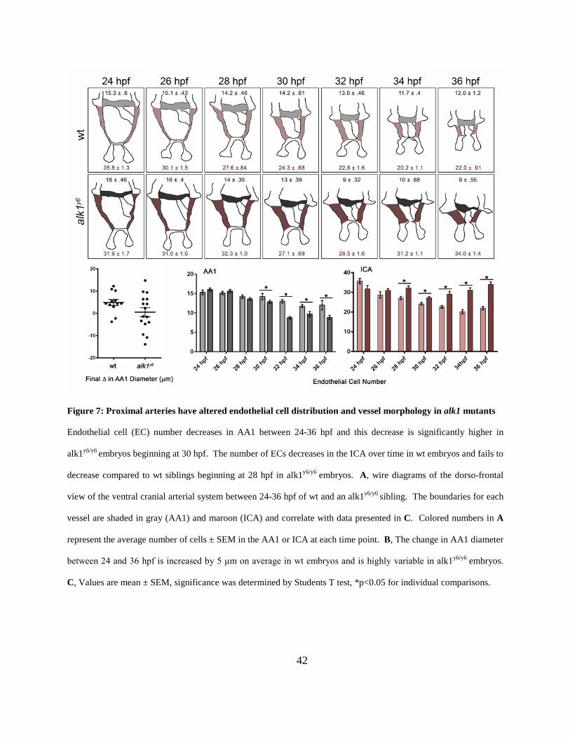
42
Figure 7: Proximal arteries have altered endothelial cell distribution and vessel morphology in alk1 mutants
Endothelial cell (EC) number decreases in AA1 between 24-36 hpf and this decrease is significantly higher in
alk1y6/y6 embryos beginning at 30 hpf. The number of ECs decreases in the ICA over time in wt embryos and fails to
decrease compared to wt siblings beginning at 28 hpf in alk1y6/y6 embryos. A, wire diagrams of the dorso-frontal
view of the ventral cranial arterial system between 24-36 hpf of wt and an alk1y6/y6 sibling. The boundaries for each
vessel are shaded in gray (AA1) and maroon (ICA) and correlate with data presented in C. Colored numbers in A
represent the average number of cells ± SEM in the AA1 or ICA at each time point. B, The change in AA1 diameter
between 24 and 36 hpf is increased by 5 μm on average in wt embryos and is highly variable in alk1y6/y6 embryos.
C, Values are mean ± SEM, significance was determined by Students T test, *p<0.05 for individual comparisons.
43
2.4 ARTERIAL ENDOTHELIAL CELL NUMBER CHANGES IN ALK1 MUTANTS
DO NOT RESULT FROM CHANGES IN PROLIFERATION OR APOPTOSIS
alk1 mutants have fewer arterial endothelial cells in AA1 but more in the ICA and CaDI.
Because all of these endothelial cells are alk1 positive, it seems unlikely that these changes could
be caused by enhanced apoptosis in AA1 and increased proliferation or decreased apoptosis in
the ICA and CaDI. In support of this reasoning, no differences in apoptosis or proliferation were
identified in these vessels in alk1 mutants versus wild type siblings by time-lapse (having
analyzed 8 wt/control morpholino time-lapse movies and 12 alk1y6/alk1 morpholino time-lapse
movies). In fact, we detected almost no proliferation or apoptosis in this vessel system regardless
of genotype. These data suggest that a fixed number of differentiated endothelial cells distribute
themselves over time in a stereotypical way within these contiguous arteries, and that this
distribution is altered in alk1 mutants.
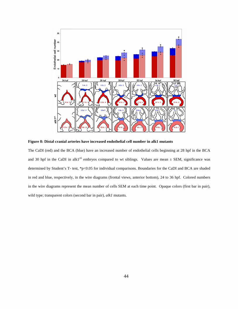
44
Figure 8: Distal cranial arteries have increased endothelial cell number in alk1 mutants
The CaDI (red) and the BCA (blue) have an increased number of endothelial cells beginning at 28 hpf in the BCA
and 30 hpf in the CaDI in alk1y6 embryos compared to wt siblings. Values are mean ± SEM, significance was
determined by Student’s T- test, *p<0.05 for individual comparisons. Boundaries for the CaDI and BCA are shaded
in red and blue, respectively, in the wire diagrams (frontal views, anterior bottom), 24 to 36 hpf. Colored numbers
in the wire diagrams represent the mean number of cells SEM at each time point. Opaque colors (first bar in pair),
wild type; transparent colors (second bar in pair), alk1 mutants.
45
2.5 ARTERIAL ENDOTHELIAL CELLS PROXIMAL TO THE OUTFLOW TRACT
MIGRATE TOWARD THE HEART, AGAINST THE DIRECTION OF BLOOD FLOW
The lack of proliferation or apoptosis in the developing cranial arterial system suggested that cell
number changes in wild type cranial arteries from 24-36 hpf (decreased cell number in AA1 and
ICA, increased cell number in CaDI) resulted from a redistribution of endothelial cells. To better
appreciate the cell movements that generate the cranial arterial system, I performed time-lapse
two-photon microscopy of Tg(fli1a:negfp)y7;Tg(fli1a.ep:mRFP-CAAX)pt504 embryos, which
express GFP in endothelial cell nuclei and mRFP in endothelial cell membranes. Imaging
between 23 and 33 hpf revealed a striking net movement of AA1 endothelial cells toward the
heart beginning at approximately 24-25 hpf, just after the onset of heartbeat and blood flow
(Figure 9A-B). Tracking of individual endothelial cells (Figure 9B-B’) demonstrated that on
average 6-10 cells entered the heart from AA1 (red arrows), 3-6 cells entered AA1 from the
ICA/LDA (blue arrows) and the rest of the cells remained in AA1 while migrating towards the
heart (black arrows) during this window of development.
To confirm this observation, I performed fate mapping using
Tg(fli1a:GAL4FF;UAS:kaede) embryos which expresses a photoconvertible fluorescent protein
in the vascular endothelium. All cells in either the left or right AA1 were photoconverted from
green to red at 24 hpf using a 405 nm laser, and photoconverted cell locations were recorded at
48 hpf (Figure 10). Nearly all embryos showed photoconverted cells in the outflow tract and
heart, with approximately 60% of embryos having photoconverted cells within the proximal
portion of the ventricle, adjacent to the atrioventricular canal. Approximately 20% of embryos
had photoconverted cells remaining in AA1 but no embryos had photoconverted cells more distal
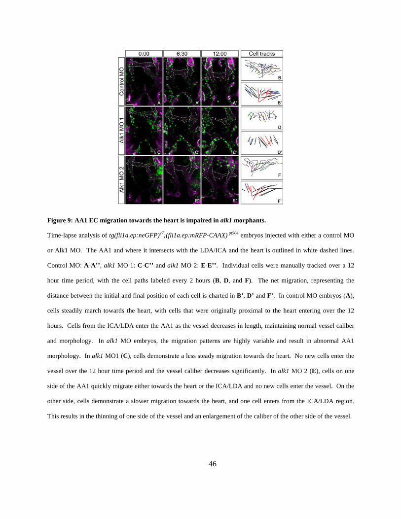
46
Figure 9: AA1 EC migration towards the heart is impaired in alk1 morphants.
Time-lapse analysis of tg(fli1a.ep:neGFP)y7;(fli1a.ep:mRFP-CAAX) pt504 embryos injected with either a control MO
or Alk1 MO. The AA1 and where it intersects with the LDA/ICA and the heart is outlined in white dashed lines.
Control MO: A-A’’, alk1 MO 1: C-C’’ and alk1 MO 2: E-E’’. Individual cells were manually tracked over a 12
hour time period, with the cell paths labeled every 2 hours (B, D, and F). The net migration, representing the
distance between the initial and final position of each cell is charted in B’, D’ and F’. In control MO embryos (A),
cells steadily march towards the heart, with cells that were originally proximal to the heart entering over the 12
hours. Cells from the ICA/LDA enter the AA1 as the vessel decreases in length, maintaining normal vessel caliber
and morphology. In alk1 MO embryos, the migration patterns are highly variable and result in abnormal AA1
morphology. In alk1 MO1 (C), cells demonstrate a less steady migration towards the heart. No new cells enter the
vessel over the 12 hour time period and the vessel caliber decreases significantly. In alk1 MO 2 (E), cells on one
side of the AA1 quickly migrate either towards the heart or the ICA/LDA and no new cells enter the vessel. On the
other side, cells demonstrate a slower migration towards the heart, and one cell enters from the ICA/LDA region.
This results in the thinning of one side of the vessel and an enlargement of the caliber of the other side of the vessel.
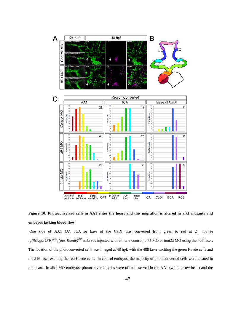
47
Figure 10: Photoconverted cells in AA1 enter the heart and this migration is altered in alk1 mutants and
embryos lacking blood flow
One side of AA1 (A), ICA or base of the CaDI was converted from green to red at 24 hpf in
tg(fli1:gal4FF)ubs4;(uas:Kaede)rk8 embryos injected with either a control, alk1 MO or tnnt2a MO using the 405 laser.
The location of the photoconverted cells was imaged at 48 hpf, with the 488 laser exciting the green Kaede cells and
the 516 laser exciting the red Kaede cells. In control embryos, the majority of photoconverted cells were located in
the heart. In alk1 MO embryos, photoconverted cells were often observed in the AA1 (white arrow head) and the
48
heart. Images at 24 hpf are single z-stacks, dorsal, anterior down. 48 hpf are two-dimensional confocal
projections, frontal with dorsal up. B. The cranial vasculature was divided into regions demarcated on the wire
diagram. Cells were converted on one side of AA1, ICA or CaDI at 24 hpf and the location of the converted cells
was scored at 48 hpf and the percentage of embryos with cells in each vessel region was calculated and graphed (C).
The distribution of cells is shifted away from the heart in alk1 and tnnt2a morphants. Numbers in the top right hand
corner of each graph represents the number of embryos assayed for each condition.
than the middle (loop) region of AA1 (Figure 10). Additional photoconversion experiments
revealed that cells in the ICA at 24 hpf either remain in the ICA or have moved toward the heart,
against the direction of blood flow, by 48 hpf. However, ICA cells reach only distal regions of
the ventricle (22% of embryos). Endothelial cells at the base of the CaDI also move toward the
heart and contribute to the ICA in nearly all cases (90% of embryos) but only rarely to more
proximal arteries (10-18% of embryos) and never to the heart. Together, these data demonstrate
that endothelial cells initially residing in lumenized arteries most proximal to the heart migrate
towards the heart, against the direction of blood flow, in wild type embryos.
In tnnt2a morphants, which lack a heartbeat and blood flow, converted cells within one
half of AA1 at 24 hpf did not migrate efficiently into the heart by 48 hpf (Figure 10). The
location of these converted cells were shifted more distally when compared to the control
morpholino embryos, with only 30% of embryos having cells located in the most distal region of
the ventricle (versus 90%), ~90% in the outflow tract and 10% of embryos with cells located as
distally as the CaDI. Photoconversion of the ICA and base of the CaDI revealed similar trends,
with embryos having photoconverted cells present in more distal vessels at 48 hpf when
49
compared to control morpholino siblings. These results indicate that blood flow triggers the
migration of endothelial cells towards the heart between 24 and 48 hpf.
In alk1 morphants, which completely phenocopy alk1 mutants, there was high variability
in the migratory behavior of AA1 endothelial cells. In the Alk1 MO1 example (Figure 9C-D), 3
cells entered the heart from AA1, only 1 cell entered AA1 from distal vessels, and in general the
migration was less directed. In the Alk1 MO2 example (Figure 9E-F), 6 cells entered the heart, 2
cells exited AA1 toward distal vessels, (migrating in the direction of blood flow) and no new
cells entered AA1 from the LDA/ICA. These trends are also observed in additional experiments
(n=6 for control morpholino and 8 for alk1 morpholino). Additionally, in kaede conversion
experiments, the distribution of arterial endothelial cells was shifted distally compared to control
siblings: a lower percentage of embryos showed AA1-derived photoconverted cells in the
proximal ventricle (10% versus 60%), and a higher percentage showed photoconverted cells
remaining in proximal (62% versus 21%) and distal (30% versus 0%) regions of AA1. Arterial
endothelial cells residing at 24 hpf in the ICA and CaDI were also shifted distally in alk1
morphants compared to control siblings at 48 hpf (Figure 10). Together, with proliferation and
apoptosis data, these data demonstrate that 1) the loss of arterial endothelial cells from wild type
AA1 and ICA over time is due to proximal migration of these cells into the heart; and 2) the
changes in arterial endothelial cell number in alk1 mutants (decreased in AA1, increased in ICA
and CaDI) are likely due to decreased proximal migration (against the direction of blood flow)
and/or increased distal migration (with the direction of blood flow).
To directly determine whether increased distal migration contributes to increased
endothelial cell number in the CaDI in alk1 morphants, I performed time-lapse two-photon
microscopy to image endothelial cell contributions to this vessel in Tg(fli1a:negfp) embryos, 24-
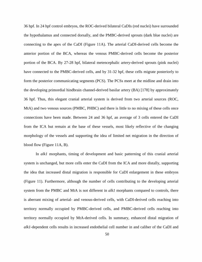
50
36 hpf. In 24 hpf control embryos, the ROC-derived bilateral CaDIs (red nuclei) have surrounded
the hypothalamus and connected dorsally, and the PMBC-derived sprouts (dark blue nuclei) are
connecting to the apex of the CaDI (Figure 11A). The arterial CaDI-derived cells become the
anterior portion of the BCA, whereas the venous PMBC-derived cells become the posterior
portion of the BCA. By 27-28 hpf, bilateral metencephalic artery-derived sprouts (pink nuclei)
have connected to the PMBC-derived cells, and by 31-32 hpf, these cells migrate posteriorly to
form the posterior communicating segments (PCS). The PCSs meet at the midline and drain into
the developing primordial hindbrain channel-derived basilar artery (BA) [178] by approximately
36 hpf. Thus, this elegant cranial arterial system is derived from two arterial sources (ROC,
MtA) and two venous sources (PMBC, PHBC) and there is little to no mixing of these cells once
connections have been made. Between 24 and 36 hpf, an average of 3 cells entered the CaDI
from the ICA but remain at the base of these vessels, most likely reflective of the changing
morphology of the vessels and supporting the idea of limited net migration in the direction of
blood flow (Figure 11A, B).
In alk1 morphants, timing of development and basic patterning of this cranial arterial
system is unchanged, but more cells enter the CaDI from the ICA and more distally, supporting
the idea that increased distal migration is responsible for CaDI enlargement in these embryos
(Figure 11). Furthermore, although the number of cells contributing to the developing arterial
system from the PMBC and MtA is not different in alk1 morphants compared to controls, there
is aberrant mixing of arterial- and venous-derived cells, with CaDI-derived cells reaching into
territory normally occupied by PMBC-derived cells, and PMBC-derived cells reaching into
territory normally occupied by MtA-derived cells. In summary, enhanced distal migration of
alk1-dependent cells results in increased endothelial cell number in and caliber of the CaDI and
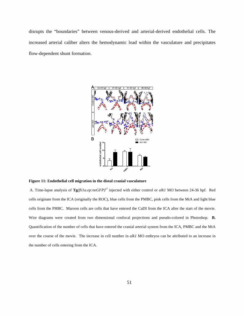
51
disrupts the “boundaries” between venous-derived and arterial-derived endothelial cells. The
increased arterial caliber alters the hemodynamic load within the vasculature and precipitates
flow-dependent shunt formation.
Figure 11: Endothelial cell migration in the distal cranial vasculature
A. Time-lapse analysis of Tg(fli1a.ep:neGFP)y7 injected with either control or alk1 MO between 24-36 hpf. Red
cells originate from the ICA (originally the ROC), blue cells from the PMBC, pink cells from the MtA and light blue
cells from the PHBC. Maroon cells are cells that have entered the CaDI from the ICA after the start of the movie.
Wire diagrams were created from two dimensional confocal projections and pseudo-colored in Photoshop. B.
Quantification of the number of cells that have entered the cranial arterial system from the ICA, PMBC and the MtA
over the course of the movie. The increase in cell number in alk1 MO embryos can be attributed to an increase in
the number of cells entering from the ICA.
52
2.6 ARTERIAL ENDOTHELIAL CELLS REPRESENT A NOVEL SOURCE OF
ENDOCARDIAL CELLS
The contribution of arterial endothelial cells to the heart has not previously been reported. To
determine which cardiac cell type these AA1-derived endothelial cells become, I performed
double immunofluorescence on Tg(alk1e5:egfp) embryos for EGFP and MF20. EGFP
expression in Tg(alk1e5:egfp) embryos marks alk1-positive endothelial cells in AA1 at 24 hpf
but is not detectable in the heart at 48 hpf. MF20 marks sarcomeric myosin heavy chain and
only labels the myocardium [179]. Preliminary analysis indicates that these cells do not
contribute to myocardium and are likely therefore endocardial, as would be expected by their
endothelial origin (Figure 12). Future work is required to clearly identify the fate of these cells.
Figure 12: Endothelial cells migrating into the heart appear to become a part of the endocardium
Tg(alk1e5:egfp)pt517 marks alk1-positive endothelial cells with gfp, but is to faint to visualize without antibody
staining before ~32 hpf. MF20 labels sarcomeric myosin heavy chain and will specifically label myocardium.
EGFP expressing (green) cells do not colocalize with MF20 (red) but are present inside the myocardium, indicating
these cells are in the endocardium. Images are 2D confocal projections of 30 µM cryosections, dorsal view, anterior
downward. Scale bar, 50 µm.
53
2.7 DISCUSSION
Alk1 signaling functions to transmit a flow-based signal that is required to limit arterial
endothelial cell number and vessel caliber [92]. Here I demonstrate that upon the onset of flow,
endothelial cells within the first aortic arch (AA1) migrate in opposition to flow and enter the
heart, likely contributing to the endocardium. The cranial arterial system is derived from cells of
both arterial and venous origins. Due to the carefully coordinated timing of the migration of
these cells to their final destination, there is rarely any cell mixing and these cells generally cease
to migrate after the onset of flow. In the absence of alk1, endothelial cell distribution is altered,
with fewer cells in AA1 and more cells in the more distal ICA, CaDI and BCA. Because there
was no observed endothelial cell apoptosis or differences in proliferation, these data suggest that
the primary defect in alk1 mutants is due to aberrant migration and allocation of a fixed number
of arterial endothelial cells. Arterial endothelial cells proximal to the heart fail to migrate in
opposition to flow and/or migrate with flow, resulting in an accumulation of cells in distal cranial
arteries.
We have previously reported that alk1 mutant zebrafish embryos develop AVMs via a
two-step mechanism. The first step occurs independent of blood flow and results in an increased
number of endothelial cells in the CaDI, resulting in an increased vessel caliber. In the second
flow-dependent step, transient connections between arteries and veins are maintained as an
adaptive response to the increased flow through the system due to the increased CaDI caliber
[92, 135]. In this work, I have demonstrated that the increase in endothelial cell number that is
central to the first step in AVM development occurs due to aberrant endothelial cell migration in
what are normally alk1-positive arteries. Furthermore, I show through time-lapse analysis that

54
Alk1 signaling does not influence endothelial cell proliferation and increased proliferation does
not account for the increase in arterial cell number in alk1 mutants (data not shown), as has been
previously speculated [176, 180]. However, EDU and TUNEL staining are required to confirm
proliferation and apoptosis time-lapse observations.
55
3.0 CONTEXT-SPECIFIC INTERACTIONS BETWEEN NOTCH AND ALK1
CANNOT EXPLAIN ALK1-ASSOCIATED ARTERIOVENOUS MALFORMATIONS
Notch and activin receptor-like kinase 1 (ALK1) have been implicated in arterial specification,
angiogenic tip/stalk cell differentiation, and development of arteriovenous malformations
(AVMs), and ALK1 can cooperate with Notch to upregulate expression of Notch target genes in
cultured endothelial cells. These findings suggest that Notch and ALK1 might collaboratively
program arterial identity and prevent AVMs. I therefore sought to investigate the interaction
between Notch and Alk1 signaling in the developing vertebrate vasculature. I modulated Notch
and Alk1 activities in zebrafish embryos and examined effects on Notch target gene expression
and vascular morphology. Although Alk1 is not necessary for expression of Notch target genes
in arterial endothelium, loss of Notch signaling unmasks a role for Alk1 in supporting hey2 and
ephrinb2a expression in the dorsal aorta. In contrast, Notch and Alk1 play opposing roles in hey2
expression in cranial arteries and dll4 expression in all arterial endothelium, with Notch inducing
and Alk1 repressing these genes. Although alk1 loss increases expression of dll4, AVMs in alk1
mutants could neither be phenocopied by Notch activation nor rescued by Dll4/Notch inhibition.
Control of Notch targets in arterial endothelium is context-dependent, with gene-specific and
region-specific requirements for Notch and Alk1. Alk1 is not required for arterial identity, and
perturbations in Notch signaling cannot account for alk1 mutant-associated AVMs. These data
56
suggest that AVMs in HHT patients are not caused by defective arterial specification or altered
Notch signaling.
3.1 INTRODUCTION
Notch signaling is critical for cell fate determination in many tissues. When activated by
transmembrane ligands of the Delta and Jagged families, the Notch intracellular domain (NICD)
is cleaved, translocates to the nucleus, binds to the DNA binding protein, recombination signal
binding protein for immunoglobulin kappa J (RBPJ), and induces target gene expression [181].
In the vasculature, delta-like ligand 4 (Dll4)/Notch signaling controls arterial specification and
angiogenic tip/stalk cell selection [30, 33, 38, 182], and in mouse and zebrafish models,
decreased Dll4/Notch function leads to direct connections between arteries and veins, or
arteriovenous malformations (AVMs) [30, 162, 166, 172, 183]. Because Dll4/Notch signaling
transcriptionally upregulates the arterial endothelial marker, ephrinb2 (Efnb2) [184], and
decreased Dll4/Notch signaling results in loss of Efnb2 and ectopic arterial expression of the
venous marker, Ephb4 [30, 31, 162, 166, 171, 174, 183, 184], AVMs resulting from decreased
Dll4/Notch signaling are generally attributed to disruption of arterial-venous identity. Notch loss-
of-function (Notchlof) generates small caliber AVMs that are associated with thin, nearly atretic
arteries [162, 183, 185]. Notch gain-of-function (Notchgof), which enhances Efnb2 and represses
Ephb4, also results in AVMs in mice [30, 31, 162, 166, 171, 174, 183, 184], and human brain
AVMs exhibit increased Notch signaling [168, 186]. AVMs associated with Notchgof involve
enlarged arteries containing supernumerary endothelial cells [31, 166, 167, 171, 172], suggesting
57
that failed repulsion mediated by EfnB2/EphB4, which is required for segregation of venous and
arterial cells in developing vessels [187, 188], may be responsible for these AVMs. Thus, both
Notchlof and Notchgof result in AVMs associated with disrupted arterial-venous identity, but the
morphological characteristics of these AVMs are distinct, with low flow, small caliber shunts
associated with Notchlof and high flow, large caliber shunts associated with Notchgof.
Similar to Notch signaling, bone morphogenetic protein (BMP) signaling has also been
implicated in AVM prevention. BMP ligands bind to a heterotetrameric complex of type I and
type II serine/threonine kinase receptors; non-signaling type III receptors facilitate ligand
binding. Upon complex formation, type II receptors phosphorylate type I receptors, which in turn
phosphorylate Smad1, Smad5, and/or Smad9. Phosphorylated Smads bind to Smad4, translocate
to the nucleus, and bind to DNA to regulate gene expression [189]. In humans, heterozygous loss
of endoglin (ENG, encoding a type III receptor), activin receptor-like kinase 1 (ACVRL1 or
ALK1, encoding a type I receptor), or SMAD4 results in hereditary hemorrhagic telangiectasia
(HHT), a disease characterized by a predisposition to development of telangiectasias and AVMs
[98, 101, 190, 191]. Alk1 mutant mice exhibit decreased Efnb2 expression in the dorsal aorta
(DA) [192], and BMP9/ALK1 transcriptionally induces EFNB2 in cultured human umbilical
artery endothelial cells [174], suggesting that ALK1, similar to Notch, is required for arterial
identity. Also like Notch, ALK1 has been implicated in maintenance of stalk cell identity [175].
Because both Notch and ALK1 are required for arterial differentiation, stalk cell fate
determination, and prevention of AVMs, ALK1 and Notch might function in a common pathway
to control arterial and/or stalk cell identity and prevent AVMs.
Several lines of evidence suggest that Notch and ALK1 interact in endothelial cells.
Activation of either DLL4/Notch or BMP9/ALK1 in cultured endothelial cells enhances
58
expression of canonical Notch targets HEY1, HEY2, and HES1,[32, 174, 175] and simultaneous
activation of these pathways synergistically increases expression of HEY1 and HEY2 [175]. A
less dramatic effect on HEY2 expression is observed in response to combined stimulation by
constitutively active forms of Notch (NICD) and ALK1 (ALK1CA) [184]. Induction of HEY1 and
HEY2 by BMP9/ALK1 requires SMAD4 but not RBPJ [175], and BMP9 induces SMAD1/5
binding to HEY1, HEY2, and HES1 promoters in cultured human umbilical vein endothelial cells
(HUVECs) [193]. These findings suggest that BMP9/ALK1 directly stimulates canonical Notch
targets by a SMAD-dependent and NICD/RBPJ-independent pathway. Notch and ALK1
additively induce VEGFR1, a stalk cell marker, and inhibit APELIN, a tip cell marker, and
mosaic sprouting assays demonstrate that endothelial cells lacking ALK1, SMAD4, or HEY2 are
more likely to be in the tip position [175]. Together, these results suggest that
BMP9/ALK1/SMAD signaling induces canonical Notch targets independently of NICD/RBPJ
and reinforces Notch-mediated acquisition of arterial identity and maintenance of stalk cell fate.
Although Notch and ALK1 exhibit synergistic interactions with respect to Notch target
gene expression in cultured endothelial cells, evidence for synergy is less compelling for
phenotypic endpoints. Both NICD and ALK1CA dampen endothelial cell sprouting, but
simultaneous pathway activation shows no further effects, and inhibition of one pathway does
not inhibit effects of activation of the other pathway [174]. Similarly, both ALK1 and Notch
inhibition enhance VEGF-stimulated tube formation in HUVECs and increase vascular area in
the postnatal retina; however, combined inhibition of these pathways shows less than additive
effects [175]. Thus, the synergy between Notch and ALK1 in controlling gene expression may
not directly translate to synergistic effects on endothelial cell behavior.
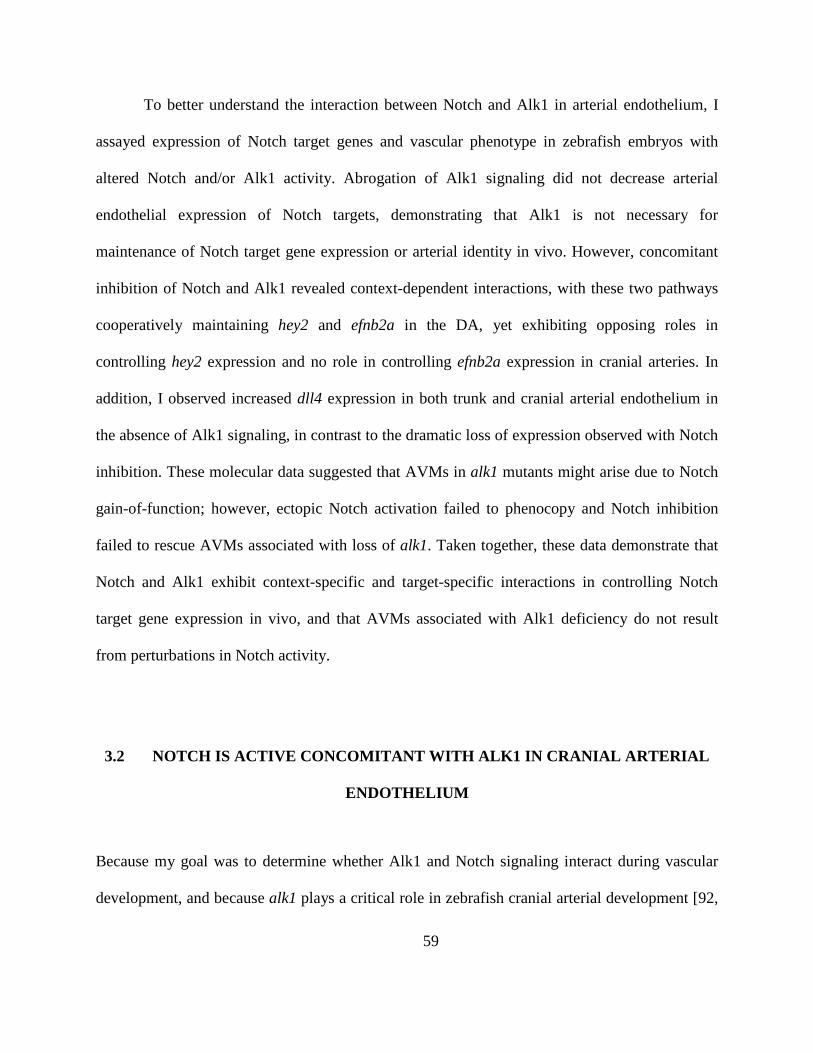
59
To better understand the interaction between Notch and Alk1 in arterial endothelium, I
assayed expression of Notch target genes and vascular phenotype in zebrafish embryos with
altered Notch and/or Alk1 activity. Abrogation of Alk1 signaling did not decrease arterial
endothelial expression of Notch targets, demonstrating that Alk1 is not necessary for
maintenance of Notch target gene expression or arterial identity in vivo. However, concomitant
inhibition of Notch and Alk1 revealed context-dependent interactions, with these two pathways
cooperatively maintaining hey2 and efnb2a in the DA, yet exhibiting opposing roles in
controlling hey2 expression and no role in controlling efnb2a expression in cranial arteries. In
addition, I observed increased dll4 expression in both trunk and cranial arterial endothelium in
the absence of Alk1 signaling, in contrast to the dramatic loss of expression observed with Notch
inhibition. These molecular data suggested that AVMs in alk1 mutants might arise due to Notch
gain-of-function; however, ectopic Notch activation failed to phenocopy and Notch inhibition
failed to rescue AVMs associated with loss of alk1. Taken together, these data demonstrate that
Notch and Alk1 exhibit context-specific and target-specific interactions in controlling Notch
target gene expression in vivo, and that AVMs associated with Alk1 deficiency do not result
from perturbations in Notch activity.
3.2 NOTCH IS ACTIVE CONCOMITANT WITH ALK1 IN CRANIAL ARTERIAL
ENDOTHELIUM
Because my goal was to determine whether Alk1 and Notch signaling interact during vascular
development, and because alk1 plays a critical role in zebrafish cranial arterial development [92,
60
121, 135], I first assessed Notch activity in cranial endothelium using a double transgenic line,
Tg(tp1:egfp)um14;Tg(fli1a.ep:mRFP-CAAX)pt504. These fish report Notch activity, as visualized by
EGFP expression [194], on a background of mRFP-labeled endothelial cells. Embryos were
imaged at 36 hpf, a time point when Alk1 is active in cranial arterial endothelium [135]. Notch
activity was weak to moderate in cranial arteries, including the first aortic arch (AA1), internal
carotid artery (ICA), caudal division of the internal carotid artery (CaDI), optic artery (OA), and
basal communicating artery (BCA) [Figure 13A]. All of these arteries are alk1-positive at 36 hpf
[92]. Notch activity was also detected in the alk1-negative posterior communicating segments
(PCS) and metencephalic arteries (MtA), but was absent in cranial veins (Figure 13A). These
data demonstrate that cranial vascular Notch activity is arterial-specific, and that all alk1-positive
arteries have active Notch signaling.
Next, I assayed cranial vessel expression of endogenous Notch targets. Hairy and
enhancer of split (HES)-related proteins are transcriptional repressors that are induced by
NICD/RBPJ [195], and the HES-related genes HES1, HEY1, and HEY2 are upregulated by
BMP9/ALK1 in cultured endothelial cells [175, 184, 193]. The zebrafish genome contains two
hes1 paralogs, her6 and her9 [196]. However, neither these genes nor hey1 were detectable in
endothelium at 24-36 hpf (data not shown). In contrast, hey2 was expressed in all alk1-positive
cranial arteries at 36 hpf (Figure 13B). efnb2a, another Notch target [184], as well as dll4, which
encodes a Notch ligand that is positively regulated by Notch signaling [42, 197, 198], were also
expressed in all alk1-positive cranial arteries at 36 hpf (Figure 13B). dll4 was additionally
expressed in the alk1-negative PCS and MtA. These data demonstrate that Notch targets hey2,
efnb2a, and dll4 are expressed in cranial arterial endothelium concomitant with active Alk1
signaling and are good candidates for cooperative regulation by Notch and Alk1.
61
Figure 13: Notch is active concomitant with Alk1 in cranial arterial endothelium
A, Notch activity is detectable in cranial arterial but not venous endothelium (“v”) at 36 hpf. Two-dimensional
confocal projections of Tg(tp1:egfp)um14 (green in merge) and Tg(fli1a.ep:mRFP-CAAX) pt504 (magenta in merge).
alk1-positive arteries: AA1, aortic arch 1; ICA, internal carotid artery; LDA, lateral dorsal aorta; CaDI, caudal
division of internal carotid artery; OA, optic artery; BCA, basal communicating artery. alk1-negative arteries: PCS,
posterior communicating segment; MtA, metencephalic artery. Images represent N = 20 embryos. Lateral and dorsal
views, anterior leftward; frontal view, anterior up. Scale bar, 50 μm. B, Wiring diagrams (arteries black, veins gray)
and representative whole mount in situ hybridization for alk1, hey2, efnb2a, and dll4 at 36 hpf. Expression of Notch
62
targets is detected in the alk1-positive AA1 (white arrow), ICA (blue arrowhead), LDA (white asterisk), CaDI
(white arrowhead), OA (blue asterisk), and BCA (blue arrow). dll4 is also expressed in the alk1-negative PCS
(black asterisk) and MtA (black arrowhead). Plane of focus of dll4, frontal view, is deeper than other frontal images
because of interfering dll4 brain expression. Images represent N > 63 embryos. Lateral and dorsal views, anterior
leftward; frontal view, anterior up. Scale bar, 100 μm.
3.3 NOTCH AND ALK1 COOPERATIVELY REGULATE HEY2 AND EFNB2A BUT
OPPOSITELY REGULATE DLL4 IN THE DORSAL AORTA
To investigate the interaction between Notch and Alk1 in the regulation of Notch targets in vivo,
I assayed tp1:egfp, hey2, efnb2a, and dll4 in embryos with impaired Notch and/or Alk1
signaling. To inhibit Notch signaling, I treated embryos with 10 µmol/L LY411575, a gamma-
secretase inhibitor, between 23 and 36 hpf. This time period brackets the critical time period of
Alk1 function: alk1 is first detectable around 26 hpf, and alk1 mutation results in enlargement of
cranial arteries by 32 hpf [92]. LY411575 treatment had no effect on heartbeat or blood flow but
resulted in severe trunk curvature (data not shown) as expected in Notch-inhibited embryos [30,
33, 199].
Notch signaling is active in the zebrafish DA throughout embryonic development [30,
161, 182], and despite no obvious requirement for Alk1, alk1 is indeed expressed in the DA as
early as 26 hpf [92]. LY411575 treatment resulted in complete loss of DA tp1:egfp expression
(Figure 14A), demonstrating effective abrogation of NICD/RBPJ-mediated transcription.
However, the effect of Notch inhibition on expression of endogenous Notch targets was variable.
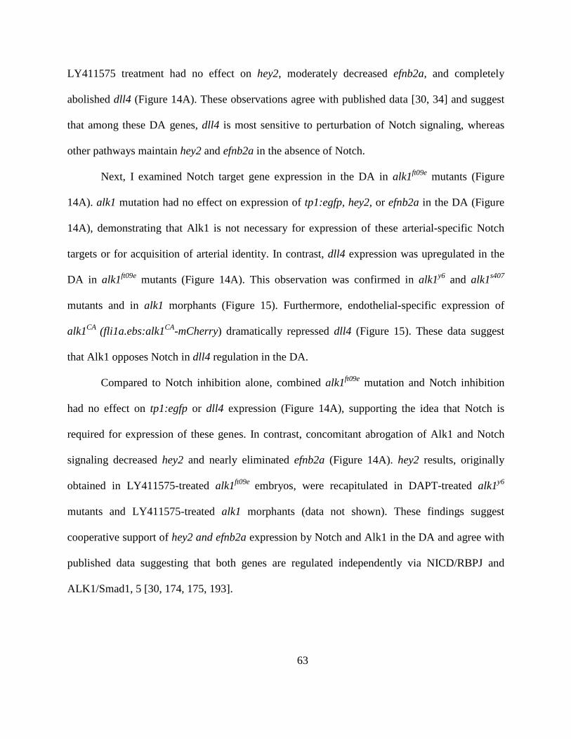
63
LY411575 treatment had no effect on hey2, moderately decreased efnb2a, and completely
abolished dll4 (Figure 14A). These observations agree with published data [30, 34] and suggest
that among these DA genes, dll4 is most sensitive to perturbation of Notch signaling, whereas
other pathways maintain hey2 and efnb2a in the absence of Notch.
Next, I examined Notch target gene expression in the DA in alk1ft09e mutants (Figure
14A). alk1 mutation had no effect on expression of tp1:egfp, hey2, or efnb2a in the DA (Figure
14A), demonstrating that Alk1 is not necessary for expression of these arterial-specific Notch
targets or for acquisition of arterial identity. In contrast, dll4 expression was upregulated in the
DA in alk1ft09e mutants (Figure 14A). This observation was confirmed in alk1y6 and alk1s407
mutants and in alk1 morphants (Figure 15). Furthermore, endothelial-specific expression of
alk1CA (fli1a.ebs:alk1CA-mCherry) dramatically repressed dll4 (Figure 15). These data suggest
that Alk1 opposes Notch in dll4 regulation in the DA.
Compared to Notch inhibition alone, combined alk1ft09e mutation and Notch inhibition
had no effect on tp1:egfp or dll4 expression (Figure 14A), supporting the idea that Notch is
required for expression of these genes. In contrast, concomitant abrogation of Alk1 and Notch
signaling decreased hey2 and nearly eliminated efnb2a (Figure 14A). hey2 results, originally
obtained in LY411575-treated alk1ft09e embryos, were recapitulated in DAPT-treated alk1y6
mutants and LY411575-treated alk1 morphants (data not shown). These findings suggest
cooperative support of hey2 and efnb2a expression by Notch and Alk1 in the DA and agree with
published data suggesting that both genes are regulated independently via NICD/RBPJ and
ALK1/Smad1, 5 [30, 174, 175, 193].
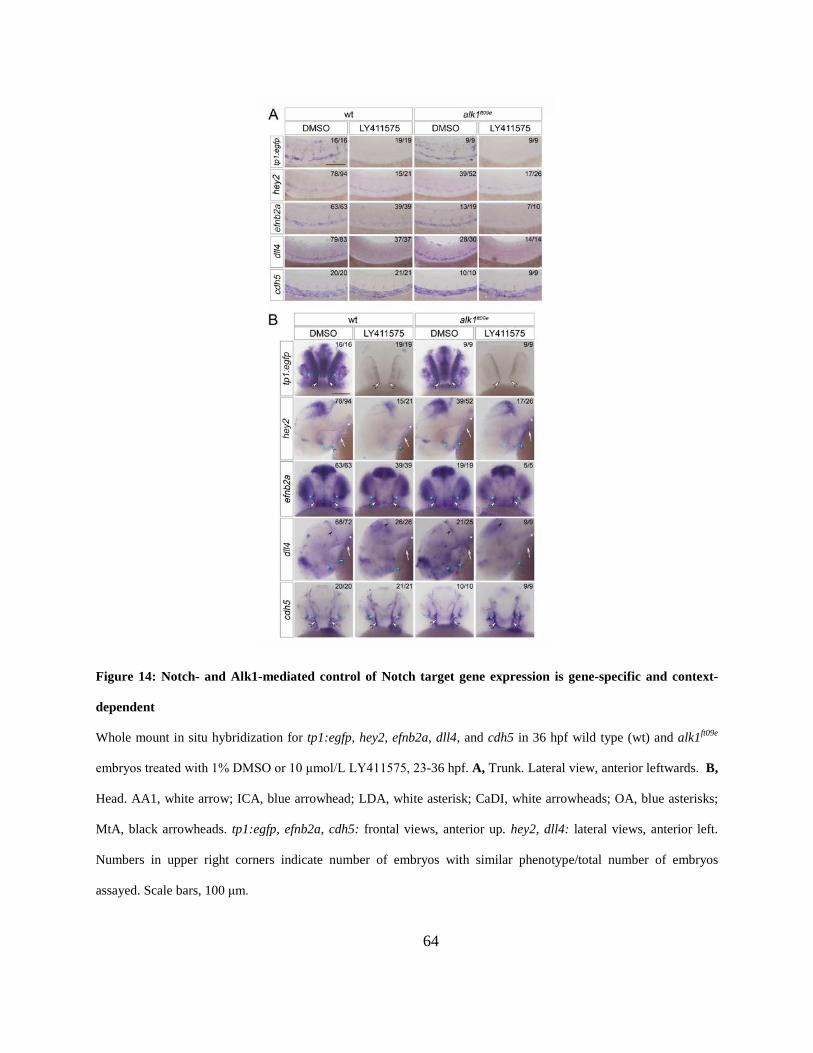
64
Figure 14: Notch- and Alk1-mediated control of Notch target gene expression is gene-specific and context-
dependent
Whole mount in situ hybridization for tp1:egfp, hey2, efnb2a, dll4, and cdh5 in 36 hpf wild type (wt) and alk1ft09e
embryos treated with 1% DMSO or 10 μmol/L LY411575, 23-36 hpf. A, Trunk. Lateral view, anterior leftwards. B,
Head. AA1, white arrow; ICA, blue arrowhead; LDA, white asterisk; CaDI, white arrowheads; OA, blue asterisks;
MtA, black arrowheads. tp1:egfp, efnb2a, cdh5: frontal views, anterior up. hey2, dll4: lateral views, anterior left.
Numbers in upper right corners indicate number of embryos with similar phenotype/total number of embryos
assayed. Scale bars, 100 μm.
65
Figure 15: dll4 is negatively regulated by Alk1 signaling
Whole mount in situ hybridization for dll4 in alk1y6/y6 (36 hpf), alk1s407/s407 (32 hpf) and alk1 morphants (MO) (36
hpf) in the trunk (A) and head (C). In the absence of alk1, dll4 expression is increased in the dorsal aorta (trunk),
aortic arch 1 (white arrow), internal carotid artery (blue arrowhead), lateral dorsal aorta (white asterisks), and
metencephalic artery (black arrowhead) when compared to wild type (wt) or control MO siblings.
Tg(fli1a.ebs:alk1CA-mcherry) embryos show no vascular dll4 expression at 36 hpf in the trunk (B) or head (D).
Expression of collagen type IV alpha1 (col4a1), a pan-endothelial marker, demonstrates the presence of vessels in
Tg(fli1a.ebs:alk1CA-mcherry) embryos. Numbers in lower right corners indicate number of embryos with similar
phenotype/total number of embryos assayed. Lateral views, anterior left. Scale bar, 200 μm.
66
3.4 NOTCH AND ALK1 EXHIBIT GENE-SPECIFIC ANTAGONISTIC
INTERACTIONS IN REGULATION OF CRANIAL ARTERIAL ENDOTHELIAL
GENE EXPRESSION
I next examined regulation of Notch target genes in cranial arteries, which enlarge upstream of
AVMs in alk1 mutants. LY411575 treatment abrogated tp1:egfp expression in cranial arterial
endothelium and neural domains (Figure 14B and Figure 16), as expected. However, whereas
Notch inhibition dramatically decreased hey2 in cranial arterial endothelium, efnb2a was
refractory to this treatment (Figure 14B and Figure 16). These effects were different from those
observed in the DA (Figure 14A), suggesting context-specific gene regulation. LY411575
treatment eliminated arterial dll4 expression in cranial arteries, similar to the DA, but increased
dll4 in neural tissues (Figure 14B and Figure 16). These observations support the idea of a
positive feedback loop specific to arterial endothelial cells in which Notch signaling directly
regulates DLL4 expression [34, 42, 197, 198]. As in the DA, in cranial arteries tp1:egfp, hey2,
and efnb2a were unaffected by alk1 mutation or knockdown, whereas dll4 was markedly
upregulated, and a fli1a.ebs:alk1CA-mCherry transgene dramatically repressed dll4 (Figure 14B,
Figure 15C-D, and Figure 16). Also similar to the DA, loss of alk1 failed to rescue abrogation of
tp1:egfp or dll4 expression induced by LY411575 treatment (Figure 14B and Figure 16). In
contrast, hey2 and efnb2a behaved differently in the absence of both Notch and Alk1 signaling in
trunk versus cranial arterial endothelial domains. In cranial arteries, efnb2a expression proved
refractory to combined loss of Notch and Alk1 signaling, whereas this same treatment increased
hey2 expression compared to Notch inhibition alone (Figure 14B and Figure 16). hey2 results,
originally obtained in LY411575-treated alk1ft09e embryos, were recapitulated in DAPT-treated
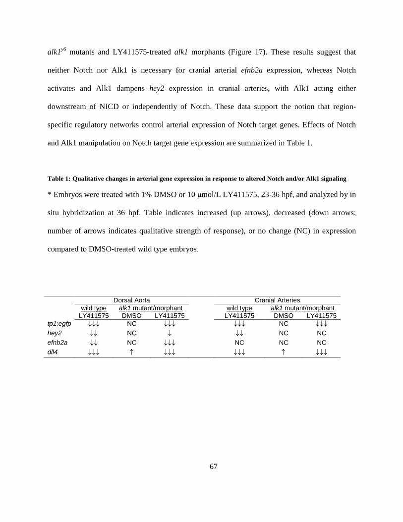
67
alk1y6 mutants and LY411575-treated alk1 morphants (Figure 17). These results suggest that
neither Notch nor Alk1 is necessary for cranial arterial efnb2a expression, whereas Notch
activates and Alk1 dampens hey2 expression in cranial arteries, with Alk1 acting either
downstream of NICD or independently of Notch. These data support the notion that region-
specific regulatory networks control arterial expression of Notch target genes. Effects of Notch
and Alk1 manipulation on Notch target gene expression are summarized in Table 1.
Table 1: Qualitative changes in arterial gene expression in response to altered Notch and/or Alk1 signaling
* Embryos were treated with 1% DMSO or 10 μmol/L LY411575, 23-36 hpf, and analyzed by in
situ hybridization at 36 hpf. Table indicates increased (up arrows), decreased (down arrows;
number of arrows indicates qualitative strength of response), or no change (NC) in expression
compared to DMSO-treated wild type embryos.
Dorsal Aorta Cranial Arteries wild type alk1 mutant/morphant wild type alk1 mutant/morphant
LY411575 DMSO LY411575 LY411575 DMSO LY411575 tp1:egfp ↓↓↓ NC ↓↓↓ ↓↓↓ NC ↓↓↓ hey2 ↓↓ NC ↓ ↓↓ NC NC efnb2a ↓↓ NC ↓↓↓ NC NC NC dll4 ↓↓↓ ↑ ↓↓↓ ↓↓↓ ↑ ↓↓↓
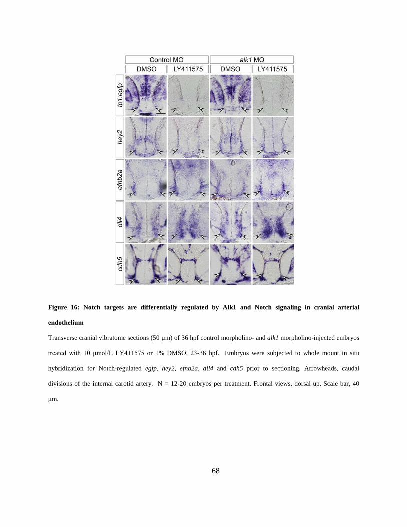
68
Figure 16: Notch targets are differentially regulated by Alk1 and Notch signaling in cranial arterial
endothelium
Transverse cranial vibratome sections (50 µm) of 36 hpf control morpholino- and alk1 morpholino-injected embryos
treated with 10 μmol/L LY411575 or 1% DMSO, 23-36 hpf. Embryos were subjected to whole mount in situ
hybridization for Notch-regulated egfp, hey2, efnb2a, dll4 and cdh5 prior to sectioning. Arrowheads, caudal
divisions of the internal carotid artery. N = 12-20 embryos per treatment. Frontal views, dorsal up. Scale bar, 40
μm.
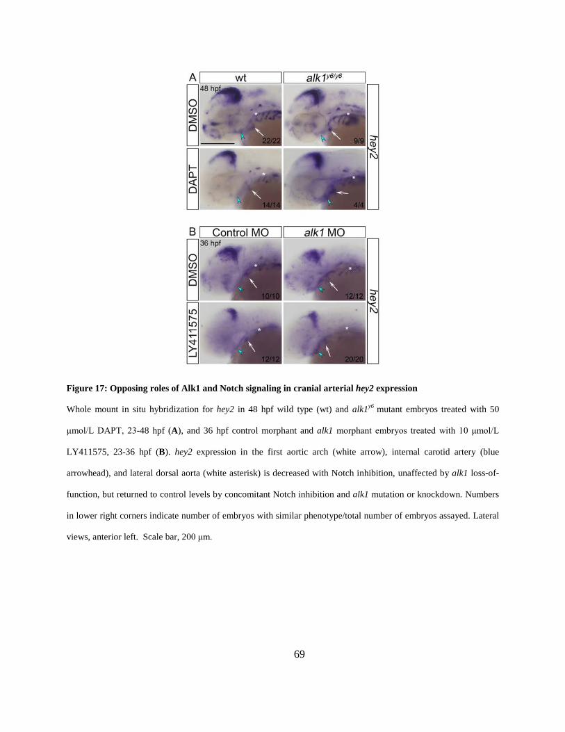
69
Figure 17: Opposing roles of Alk1 and Notch signaling in cranial arterial hey2 expression
Whole mount in situ hybridization for hey2 in 48 hpf wild type (wt) and alk1y6 mutant embryos treated with 50
μmol/L DAPT, 23-48 hpf (A), and 36 hpf control morphant and alk1 morphant embryos treated with 10 μmol/L
LY411575, 23-36 hpf (B). hey2 expression in the first aortic arch (white arrow), internal carotid artery (blue
arrowhead), and lateral dorsal aorta (white asterisk) is decreased with Notch inhibition, unaffected by alk1 loss-of-
function, but returned to control levels by concomitant Notch inhibition and alk1 mutation or knockdown. Numbers
in lower right corners indicate number of embryos with similar phenotype/total number of embryos assayed. Lateral
views, anterior left. Scale bar, 200 μm.
70
3.5 NOTCHGOF AND ALK1LOF GENERATE VASCULAR MORPHOLOGI ES WITH
SOME PHENOTYPIC OVERLAP BUT WITH INDEPENDENT ETIOLOGIES
My in vivo gene expression studies demonstrated that loss of alk1 is associated with increased
endothelial dll4 expression. Therefore, I reasoned that enhanced Dll4/Notch signaling might
phenocopy cranial AVMs in alk1 mutants. To investigate this possibility, I compared cranial
vascular development in wild type embryos, Tg(fli1a:GAL4FF)ubs3;Tg(5xUAS-E1b:6xMYC-
notch1a)kca3 embryos [which ectopically express Notch1a ICD in all endothelial cells; hereafter
referred to as Tg(endo:N1ICD)], and alk1 morphant embryos.
In wild type embryos, single sprouts emerged from the most posterior aspect of each
bilateral venous primordial midbrain channel (PMBC) ~22 hpf and migrated medially to connect
to a sprout emanating from the apex of the paired CaDIs by ~ 26 hpf, forming the BCA. These
transient BCA/PMBC connections serve as the primary drainage for the CaDI/BCA between 26-
36 hpf but regress thereafter as downstream arteries develop [92].
In Tg(endo:N1ICD) embryos, the CaDIs developed and lumenized normally, but
sprouting from the PMBC was impaired, with BCA/PMBC connections delayed up to 8 hours
compared to wild type [Figure 18, compare A-E, WT to F-J, Tg(endo:N1ICD)]. Vascular
morphology was variable in 36 hpf Tg(endo:N1ICD) embryos, with establishment of early
alternative drainage connections (for example, BCA to PCS/MtA, Figure 18G-H) associated with
relatively normal caliber vessels, and establishment of late connections associated with
CaDI/BCA engorgement and enlargement (Figure 18P,Q). Regardless of 36 hpf phenotype,
BCA area was not significantly increased compared to control at 48 hpf (Figure 18S), but all
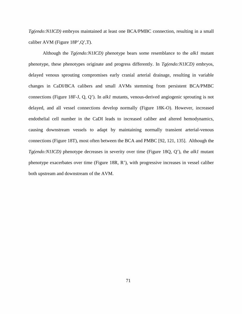
71
Tg(endo:N1ICD) embryos maintained at least one BCA/PMBC connection, resulting in a small
caliber AVM (Figure 18P’,Q’,T).
Although the Tg(endo:N1ICD) phenotype bears some resemblance to the alk1 mutant
phenotype, these phenotypes originate and progress differently. In Tg(endo:N1ICD) embryos,
delayed venous sprouting compromises early cranial arterial drainage, resulting in variable
changes in CaDI/BCA calibers and small AVMs stemming from persistent BCA/PMBC
connections (Figure 18F-J, Q, Q’). In alk1 mutants, venous-derived angiogenic sprouting is not
delayed, and all vessel connections develop normally (Figure 18K-O). However, increased
endothelial cell number in the CaDI leads to increased caliber and altered hemodynamics,
causing downstream vessels to adapt by maintaining normally transient arterial-venous
connections (Figure 18T), most often between the BCA and PMBC [92, 121, 135]. Although the
Tg(endo:N1ICD) phenotype decreases in severity over time (Figure 18Q, Q’), the alk1 mutant
phenotype exacerbates over time (Figure 18R, R’), with progressive increases in vessel caliber
both upstream and downstream of the AVM.
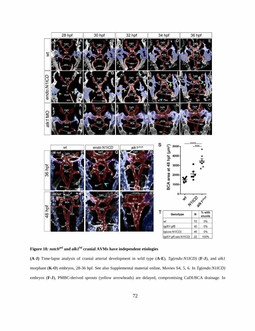
72
Figure 18: notchgof and alk1lof cranial AVMs have independent etiologies
(A-J) Time-lapse analysis of cranial arterial development in wild type (A-E), Tg(endo:N1ICD) (F-J), and alk1
morphant (K-O) embryos, 28-36 hpf. See also Supplemental material online, Movies S4, 5, 6. In Tg(endo:N1ICD)
embryos (F-J), PMBC-derived sprouts (yellow arrowheads) are delayed, compromising CaDI/BCA drainage. In
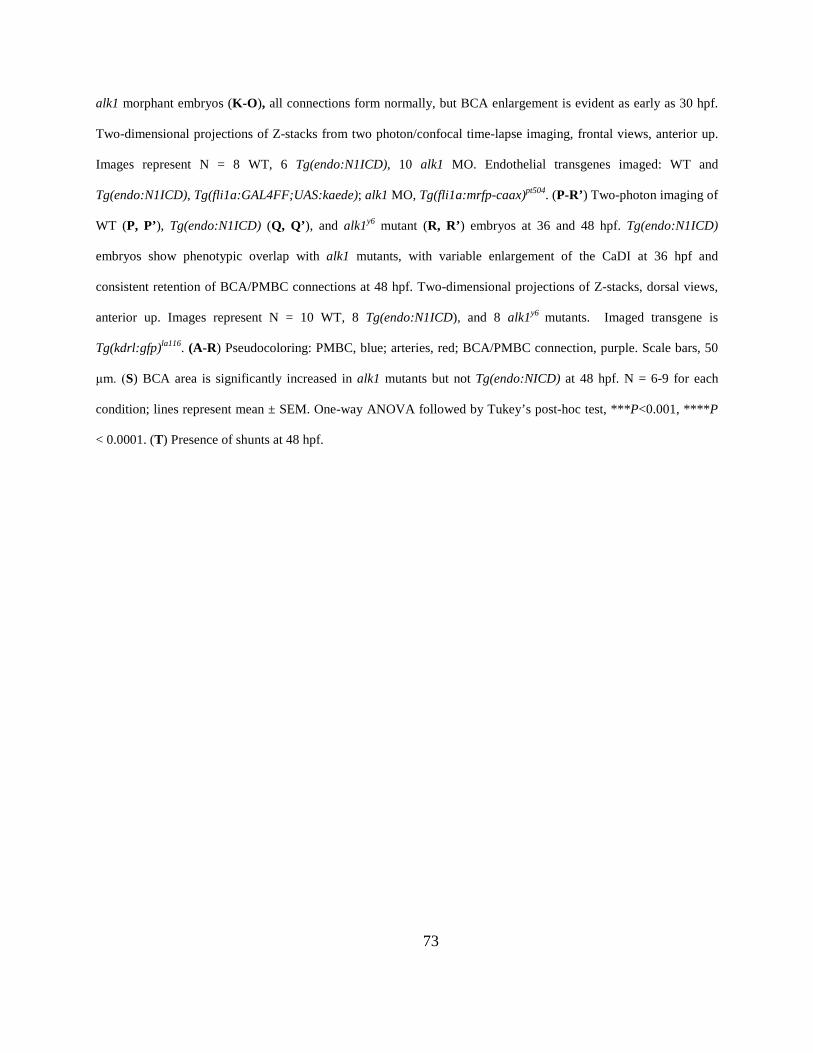
73
alk1 morphant embryos (K-O), all connections form normally, but BCA enlargement is evident as early as 30 hpf.
Two-dimensional projections of Z-stacks from two photon/confocal time-lapse imaging, frontal views, anterior up.
Images represent N = 8 WT, 6 Tg(endo:N1ICD), 10 alk1 MO. Endothelial transgenes imaged: WT and
Tg(endo:N1ICD), Tg(fli1a:GAL4FF;UAS:kaede); alk1 MO, Tg(fli1a:mrfp-caax)pt504. (P-R’) Two-photon imaging of
WT (P, P’), Tg(endo:N1ICD) (Q, Q’), and alk1y6 mutant (R, R’) embryos at 36 and 48 hpf. Tg(endo:N1ICD)
embryos show phenotypic overlap with alk1 mutants, with variable enlargement of the CaDI at 36 hpf and
consistent retention of BCA/PMBC connections at 48 hpf. Two-dimensional projections of Z-stacks, dorsal views,
anterior up. Images represent N = 10 WT, 8 Tg(endo:N1ICD), and 8 alk1y6 mutants. Imaged transgene is
Tg(kdrl:gfp)la116. (A-R) Pseudocoloring: PMBC, blue; arteries, red; BCA/PMBC connection, purple. Scale bars, 50
μm. (S) BCA area is significantly increased in alk1 mutants but not Tg(endo:NICD) at 48 hpf. N = 6-9 for each
condition; lines represent mean ± SEM. One-way ANOVA followed by Tukey’s post-hoc test, ***P<0.001, ****P
< 0.0001. (T) Presence of shunts at 48 hpf.
74
Figure 19: dll4 expression is not required for AVM development in alk1 mutants
Embryos from an alk1y6/+;Tg(kdrl:gfp)la116 incross were injected at the 1 to 4-cell stage with 15 ng dll4 MO or 5-bp
mismatch control MO, imaged at 36 (A-D) or 48 (E-H) hpf, and genotyped. Two-dimensional projections of two-
photon Z-stacks. Pseudo-coloring: PMBC (blue), arteries (red), and BCA/PMBC connection (purple). Dorsal views,
anterior up. Images represent N = 7-41 embryos per condition. Scale bars, 50 μm. (I) dll4 knockdown failed to
rescue increased BCA area in alk1y6 mutants at 36 hpf. Lines represent mean ± SEM, N = 6-21 embryos per
condition. One-way ANOVA followed by Tukey’s post-hoc test, * P<0.05; ****P<0.0001. (J) dll4 knockdown
failed to rescue cranial shunt formation in alk1y6 mutants at 48 hpf.

75
Figure 20: dll4 morpholino validation
(A) RT-PCR following injection of dll4 MO or 5-bp mismatch control MO (15 ng) using primers flanking the MO
binding site. In 3 dpf control morphant cDNA, a single 345 bp band was detected. In 3 dpf dll4 MO cDNA, dll4
wild type product was decreased to 37% of control and an additional exon 3-deleted product (287 bp) was detected.
Band intensities were normalized to -actin and identities were confirmed by sequencing of gel-extracted bands.
(B) Injection of dll4 MO but not 5-bp mismatch control MO (15 ng) caused hypersprouting (arrowheads) in trunk
intersegmental vessels, 3 dpf, in wild type and alk1y6 mutant embryos. Two-dimensional confocal projections,
Tg(fli1a:mrfp-caax)pt504, anterior left. Scale bar, 50 µm (C) Hypersprouting was present in 63% of dll4 MO-injected
embryos, with similar representation in wild type and alk1 mutant embryos.
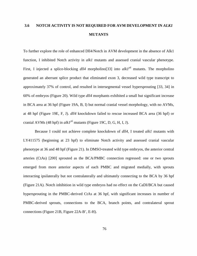
76
3.6 NOTCH ACTIVITY IS NOT REQUIRED FOR AVM DEVELOPMENT IN ALK1
MUTANTS
To further explore the role of enhanced Dll4/Notch in AVM development in the absence of Alk1
function, I inhibited Notch activity in alk1 mutants and assessed cranial vascular phenotype.
First, I injected a splice-blocking dll4 morpholino[33] into alk1y6 mutants. The morpholino
generated an aberrant splice product that eliminated exon 3, decreased wild type transcript to
approximately 37% of control, and resulted in intersegmental vessel hypersprouting [33, 34] in
60% of embryos (Figure 20). Wild type dll4 morphants exhibited a small but significant increase
in BCA area at 36 hpf (Figure 19A, B, I) but normal cranial vessel morphology, with no AVMs,
at 48 hpf (Figure 19E, F, J). dll4 knockdown failed to rescue increased BCA area (36 hpf) or
cranial AVMs (48 hpf) in alk1y6 mutants (Figure 19C, D, G, H, I, J).
Because I could not achieve complete knockdown of dll4, I treated alk1 mutants with
LY411575 (beginning at 23 hpf) to eliminate Notch activity and assessed cranial vascular
phenotype at 36 and 48 hpf (Figure 21). In DMSO-treated wild type embryos, the anterior central
arteries (CtAs) [200] sprouted as the BCA/PMBC connection regressed: one or two sprouts
emerged from more anterior aspects of each PMBC and migrated medially, with sprouts
interacting ipsilaterally but not contralaterally and ultimately connecting to the BCA by 36 hpf
(Figure 21A). Notch inhibition in wild type embryos had no effect on the CaDI/BCA but caused
hypersprouting in the PMBC-derived CtAs at 36 hpf, with significant increases in number of
PMBC-derived sprouts, connections to the BCA, branch points, and contralateral sprout
connections (Figure 21B, Figure 22A-B’, E-H).
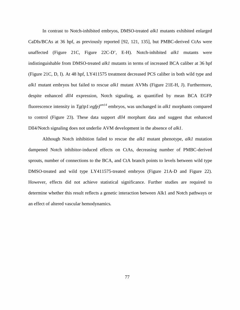
77
In contrast to Notch-inhibited embryos, DMSO-treated alk1 mutants exhibited enlarged
CaDIs/BCAs at 36 hpf, as previously reported [92, 121, 135], but PMBC-derived CtAs were
unaffected (Figure 21C, Figure 22C-D’, E-H). Notch-inhibited alk1 mutants were
indistinguishable from DMSO-treated alk1 mutants in terms of increased BCA caliber at 36 hpf
(Figure 21C, D, I). At 48 hpf, LY411575 treatment decreased PCS caliber in both wild type and
alk1 mutant embryos but failed to rescue alk1 mutant AVMs (Figure 21E-H, J). Furthermore,
despite enhanced dll4 expression, Notch signaling, as quantified by mean BCA EGFP
fluorescence intensity in Tg(tp1:egfp)um14 embryos, was unchanged in alk1 morphants compared
to control (Figure 23). These data support dll4 morphant data and suggest that enhanced
Dll4/Notch signaling does not underlie AVM development in the absence of alk1.
Although Notch inhibition failed to rescue the alk1 mutant phenotype, alk1 mutation
dampened Notch inhibitor-induced effects on CtAs, decreasing number of PMBC-derived
sprouts, number of connections to the BCA, and CtA branch points to levels between wild type
DMSO-treated and wild type LY411575-treated embryos (Figure 21A-D and Figure 22).
However, effects did not achieve statistical significance. Further studies are required to
determine whether this result reflects a genetic interaction between Alk1 and Notch pathways or
an effect of altered vascular hemodynamics.
78
Figure 21: Notch activity is not required for AVM development in alk1 mutants
Embryos from an alk1y6/+;Tg(kdrl:gfp)la116 incross were treated with 1% DMSO or 10 μmol/L LY411575 at 23 hpf
and cranial vasculature imaged at 36 (A-D) and 48 (E-H) hpf. Two-dimensional projections of two-photon Z-stacks;
in (E-H), dorsal planes were removed to highlight BCA/PCS. Pseudo-coloring: PMBC (blue), CtA (cyan);
CaDI/BCA/PCS/BA (red), and BCA/PMBC shunt (purple). Dorsal views, anterior up. Scale bars, 50 μm (36 hpf)
and 100 μm (48 hpf). (I) Notch inhibition failed to rescue increased BCA area in alk1y6 mutants at 36 hpf. Lines
represent mean ± SEM, N = 4-7 embryos per condition. One-way ANOVA followed by Tukey’s post-hoc test,
*P<0.05; ***P<0.001. (J) Notch inhibition failed to rescue cranial shunt formation in alk1y6 mutants at 48 hpf.
79
Figure 22: The Notchlof hypersprouting phenotype in midbrain and forebrain central arteries is partially
rescued by alk1 mutation
Embryos from an alk1y6/+; Tg(kdrl:gfp)la116 incross were treated with 1% DMSO or 10 μmol/L LY411575, 23-36
hpf. Two-dimensional two-photon projections of the 36 hpf vasculature (A-D) were traced to generate wiring
diagrams (A’-D’) of the midbrain and forebrain central arteries (CtA). Blue dots: connections to the primordial
midbrain channel (PMBC); red dots: connections to the basal communicating artery (BCA); green dots: branch
points. Frontal views, dorso-anterior up. Scale bar, 50 µm. Graphs quantify the number of PMBC-derived sprouts
(E), CtA/BCA connections (F), CtA branch points (G), and CtA contralateral connections (H). N= 4-7 for each
condition; lines represent mean ± SEM. One-way ANOVA followed by Tukey’s post-hoc test, *P<0.05,
***P<0.001.
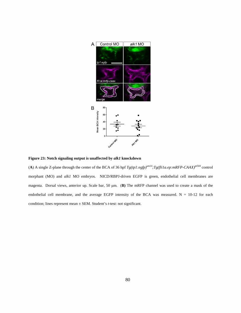
80
Figure 23: Notch signaling output is unaffected by alk1 knockdown
(A) A single Z-plane through the center of the BCA of 36 hpf Tg(tp1:egfp)um14;Tg(fli1a.ep:mRFP-CAAX)pt504 control
morphant (MO) and alk1 MO embryos. NICD/RBPJ-driven EGFP is green, endothelial cell membranes are
magenta. Dorsal views, anterior up. Scale bar, 50 μm. (B) The mRFP channel was used to create a mask of the
endothelial cell membrane, and the average EGFP intensity of the BCA was measured. N = 10-12 for each
condition; lines represent mean ± SEM. Student’s t-test: not significant.
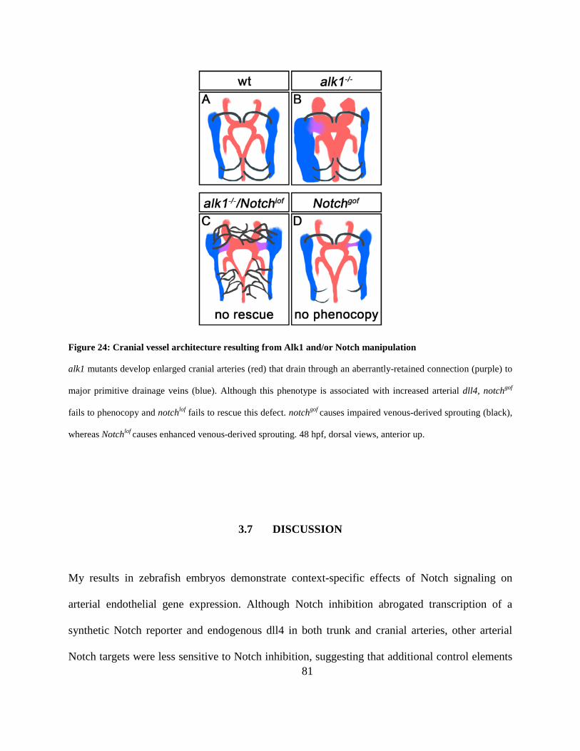
81
Figure 24: Cranial vessel architecture resulting from Alk1 and/or Notch manipulation
alk1 mutants develop enlarged cranial arteries (red) that drain through an aberrantly-retained connection (purple) to
major primitive drainage veins (blue). Although this phenotype is associated with increased arterial dll4, notchgof
fails to phenocopy and notchlof fails to rescue this defect. notchgof causes impaired venous-derived sprouting (black),
whereas Notchlof causes enhanced venous-derived sprouting. 48 hpf, dorsal views, anterior up.
3.7 DISCUSSION
My results in zebrafish embryos demonstrate context-specific effects of Notch signaling on
arterial endothelial gene expression. Although Notch inhibition abrogated transcription of a
synthetic Notch reporter and endogenous dll4 in both trunk and cranial arteries, other arterial
Notch targets were less sensitive to Notch inhibition, suggesting that additional control elements
82
sustain expression of these genes in the absence of Notch. Furthermore, Notch sensitivity of
particular genes showed regional variation, suggesting unique regulatory mechanisms in
different vascular beds. These data suggest that context-specific regulation, which may be lost or
dampened in cultured endothelial cells, plays an important role in the control of Notch target
gene expression in vivo.
Based on published work demonstrating cooperative interactions between DLL4/Notch
and BMP9/ALK1 in enhancing arterial Notch target gene expression in cultured endothelial cells
[175], I had anticipated that combined Notch and Alk1 inhibition might additively if not
synergistically decrease Notch target gene expression and impair arterial specification. However,
abrogation of Alk1 signaling failed to decrease expression of Notch targets or disrupt arterial
(hey2, efnb2a, dll4) or venous (data not shown) identity, and I uncovered only minor cooperative
interactions between Alk1 and Notch, with both contributing to maintenance of efnb2a and hey2
expression in trunk but not cranial arteries. Furthermore, I uncovered opposing roles of Notch
and Alk1 in expression of the Notch target and arterial marker, dll4. Although dll4 is upregulated
in the zebrafish DA in the absence of blood flow [201], lack of blood flow cannot account for
increased dll4 in alk1 mutants: blood flow remains strong in alk1 mutants at 36 hpf, with only a
subtle redistribution of flow towards cranial vessels [92]. Together with previous data
demonstrating increased cxcr4a and decreased edn1 in the absence of blood flow or alk1
expression [92], these data support the idea that Alk1 mediates a flow-based signal that controls
expression of a subset of arterial genes. It is also possible that dll4 upregulation in the absence of
alk1 might be attributed to the loss of the Smad1/5 target, id1. In the DA, id1 is downregulated in
the absence of alk1 or blood flow (data not shown), and ID1 stabilizes HES1 (Her6 in zebrafish),
which in turn represses DLL4 [177, 202]. Therefore, decreased Id1 might decrease Her6, thereby
83
resulting in increased dll4. However, I was unable to detect her6 in zebrafish arterial endothelial
cells by in situ hybridization.
In addition to the unanticipated opposing effects of Notch and Alk1 on dll4 expression, I
demonstrated a paradoxical interaction with respect to cranial arterial hey2 regulation, with loss
of alk1 restoring hey2 to near control levels in Notch-inhibited embryos. This finding suggests
that Alk1 may repress hey2 in cranial arterial endothelial cells either downstream of NICD
cleavage or via an independent mechanism. Although it is possible that enhanced blood flow
through enlarged, alk1-dependent vessels might increase hey2 expression, this seems unlikely
given that hey2 expression is unchanged in alk1 mutants with intact Notch signaling (Table 1) or
in the absence of blood flow (data not shown). Further studies are required to untangle the
relationship between Notch and Alk1 in cranial arterial hey2 regulation.
Dll4 is a critical arterial endothelial Notch ligand [182, 184] and Notchgof and in
particular Dll4 overexpression results in large caliber arteries and AVMs similar in morphology
to AVMs in Alk1 knockout mice [31, 166, 167, 171, 172]. Therefore, my finding that Alk1
inhibited dll4 expression initially suggested to us that increased Notch signaling might contribute
to AVMs in alk1 mutants. However, multiple lines of evidence fail to support this hypothesis.
First, although ectopic endothelial expression of N1ICD results in small cranial AVMs involving
the same vessels as in alk1 mutants, the origin and progression of these AVMs differs
dramatically. The primary defect leading to AVMs in N1ICD-expressing embryos is delayed
venous-derived sprouting, whereas the primary defect in alk1 mutants is increased endothelial
cell number in and caliber of upstream arteries [92]. Differences in the spatiotemporal
expression of fli1a-driven N1ICD versus alk1 limit the utility of my approach; however, I would
expect that earlier N1ICD expression would cause an even more pronounced arterial phenotype
84
if enhanced arterial Notch signaling could serve as a proxy for arterial alk1 loss. Second, neither
dll4 knockdown nor Notch inhibition rescues alk1 mutant AVMs. Third, despite increased dll4
expression, I failed to detect increased canonical Notch activity in alk1 mutants. Together, these
results suggest that AVMs arise independently in Notchgof and Alk1lof embryos.
Although AVMs initiate via independent mechanisms in Notchgof and Alk1lof embryos,
both represent retention of normally transient BCA/PMBC connections. In alk1 mutants,
maintenance of BCA/PMBC connections occurs as an adaptive response to increased shear stress
caused by enlargement of upstream arteries [92]. In Notchgof embryos, impaired venous-derived
sprouting delays CaDI/BCA drainage, which likely alters cranial vascular hemodynamics and
affects remodeling. Thus, an adaptive response to altered hemodynamics, downstream of
independent primary molecular and cellular defects, may be a unifying factor in development of
Notchgof and Alk1lof AVMs.
Although I failed to rescue vascular defects in alk1 mutants via Notch inhibition, I
partially rescued CtA hypersprouting defects in Notch-inhibited embryos via alk1 mutation,
suggesting some interaction between Notch and Alk1 in the cranial vasculature. It is possible that
restored hey2 expression in the absence of both Notch and Alk1 signaling might contribute to
this phenomenon, as knockdown of HEY2 enhances sprouting in cultured HUVECs [175].
However, whether these observations represent a true genetic interaction or a response to
changes in vascular hemodynamics remains to be determined.
In summary, my in vivo analysis of Notch and Alk1 signaling demonstrates gene-specific
and context-specific interactions, with examples of both cooperative and antagonistic control of
gene expression. However, results fail to support the idea that these pathways interact
synergistically to control Notch target genes, program arterial identity, and prevent AVMs.
85
4.0 CONCLUSIONS AND FUTURE DIRECTIONS
The work presented in this thesis provides insight into the role of Alk1 signaling in endothelial
cell migration and presents a model for AVM formation caused by pathway disruption. Previous
work in the lab had demonstrated that AVMs form via a two-step mechanism upon loss of alk1.
In the first step, endothelial cell number increases in cranial arteries and this increase is
independent of blood flow [92, 121, 135]. As an adaptive response to increased flow, transient
connections are maintained forming flow dependent arteriovenous shunts [92]. My work has
focused on furthering the understanding of how loss of alk1 results in an increase in endothelial
cell number in cranial arteries in the zebrafish embryo. I show that Alk1 is necessary for
directed endothelial cell migration upon the onset of blood flow and that alk1 mutant AVMs do
not form as a result of defects in arterial/venous identity. I have also contributed to the general
knowledge of vascular development by describing in detail the formation of the cranial vascular
system between 24 and 36 hpf, a key developmental period when cranial circulation begins. To
this end, I have found that endothelial cells migrate in opposition to flow and contribute to the
endocardium, described the sources of the endothelial cells that contribute to the cranial arterial
system, and described how the timing of sprouting is key to forming a normal mature vascular
system.
86
There are many unresolved questions. Whether Alk1 functions as a
mechanosensor/transducer is not clear. Alk1-positive arteries do not appear to have primary cilia
(B. Roman, unpublished), and the expression of vegfr and vecad is unaffected in alk1 mutants
(data not shown). However, Alk1 may regulate vegf or vecad at the translational or post-
translational level and experiments testing the role of Alk1 signaling on the localization of
Vegfr2 and Vecad protein in the presence of flow would be informative. Based on the
observation that endothelial cells are defective in directed endothelial cell migration, it is
possible that Alk1 is involved in endothelial cell polarization. In the presence of high magnitude
flow endothelial cells polarize with the nucleus downstream of the microtubule organizing center
and the golgi apparatus [74]. In preliminary experiments, I was able to inject a GFP tagged
centrin construct (centrin is a key protein of the centrosomes) into zebrafish embryos and
visualize endothelial cell centrin in AA1 (data not shown). I hope to determine if endothelial cell
polarization is affected in alk1 mutant embryos and in ALK1-depleted cultured endothelial cells
exposed to shear stress.
Angiogenesis is often viewed as a binary process. A vessel is either actively undergoing
angiogenesis (endothelial cell proliferation, migration and degradation of the ECM) or is
quiescent (no proliferation or migration and vessels are stabilized through ECM deposition and
smooth muscle recruitment). A long standing model in the field suggested that TGFβ1 activation
of Alk1 and Alk5 induced activation or stabilization, respectively, and that the relative activation
of the two pathways determined the activation state of the endothelium [176, 180, 203, 204].
AVMs were thought to arise due to a disruption in this balance causing aberrant angiogenesis
[203, 205]. This model was shown to be inaccurate because in mice and zebrafish, Alk1 and
Alk5 are not coexpressed in the vascular endothelium and Alk5 is not required in endothelial
87
cells for normal vascular development [118, 206, 207]. Subsequent work has postulated that
AVMs form in HHT models due to defects in arterial venous identity (as evidenced by decreased
ephrinB2 expression in the DA of mouse alk1 mutants [114, 121]) or due to focal capillary
regression [90].
It is my hope that the work I present in this document provides a more nuanced
understanding of angiogenesis and AVM development. ALK1 signaling is neither pro- nor anti-
angiogenic, and AVM formation is not the result of “too much” or “too little” angiogenesis.
Instead, AVM development involves defects in post-angiogenic endothelial cell behaviors and
force-dependent remodeling. Alk1 is important upon the onset of flow to provide a means for
endothelial cells to migrate towards the heart in the vessels that experience the highest
hemodynamic load. Without this signal, arterial endothelial cells are not able to respond
appropriately and while the immediate result is somewhat variable, the final result is a decrease
in cells proximal to the heart and an accumulation of cells in downstream arteries. My work has
also demonstrated that while Alk1 is not necessary for determining or maintaining arterial
identity, there is still much to be understood regarding the relationship and interactions between
Alk1, Notch and blood flow.
4.1 CURRENT HHT TREATMENTS
Current HHT treatments focus on treating symptoms. Large PAVMs or BAVMs may be coil-
embolized or surgically resected, but these procedures are invasive and risky. The observation
that wounding is necessary for adult endothelial-targeted Eng (Eng-iKOe) [120] and inducible
88
knockout Alk1 (Alk1-iKOe) mice [117] to develop AVMs has led to the hypothesis that sites of
active angiogenesis are more susceptible to AVM formation in HHT patients. To this end, anti-
angiogenic treatments have been shown to greatly improve Eng-iKOe and Alk1-iKOe mouse
vascular phenotypes. Specifically treatment with antibodies designed to block VEGF have
reversed vascular abnormalities in these mice and restored levels of key angiogenic factors that
had been dysregulated in affected tissues [208, 209]. In addition, HHT patients treated with
similar therapies have experience significant improvement in nose bleeds, GI bleeding and liver
complications [210-213]. Given that VEGF does not appear to be involved in Eng-iKOe and
Alk1-iKOe mouse disease pathology, the mechanism by which anti-VEGF treatment improves
HHT symptoms is not understood [107]. In light of my research, I suggest that AVMs develop
in immature vessels because of aberrant distribution of arterial endothelial cells that causes
arterial enlargement, which results in an altered hemodynamic load and consequent downstream
AVM formation to normalize hemodynamic forces. Inhibiting angiogenesis prevents the
initiation of a sequence of events that would lead to improper endothelial cell migration and
distribution.
89
5.0 MATERIAL AND METHODS
5.1 ZEBRAFISH LINES AND MAINTENANCE
Adult zebrafish (Danio rerio) were maintained according to standard protocols [214]. Embryos
were grown at 28.5°C in 30% Danieau [17 mM NaCl, 2 mM KCl, 0.12 mM MgSO4, 1.8 mM
Ca(NO3)2, 1.5 mM HEPES]. For imaging, embryo medium was supplemented with 0.003%
phenylthiourea (Sigma, St. Louis, MO, USA) at ~8 hpf hours post-fertilization (hpf) to prevent
melanin synthesis. Mutant lines alk1ft09e (p.Y88X), alk1y6 (p.L240F), alk1s407 (g.IVS8-2A>T),
alk1 splice-blocking morpholino, and alk1y6 genotyping assay have been described [92, 121,
215]. Line alk1ft09e was genotyped by dCAPs assay [216] using primers 5’-
GTGCTACGTACCTGCTATTCCTGGAGTCTA-3’ and 5’-CGAACAACCCAGAAACGAG-
3’. The forward primer contains a single mismatch (underlined) that creates an XbaI site in the
mutant allele. Line alk1s407 was genotyped using PCR primers 5’-
GACAATTTCCAGTCATCCTC-3’ and 5’-CTGGGCCTGTGCTGGTC-3’ followed by
restriction digest with DdeI (cuts wild type only). Transgenic lines Tg(fli1a:GAL4FF)ubs3,
Tg(UAS:Kaede)rk8 and Tg(kdrl:GFP)la116 have been described [217-219]. Two new transgenic
lines were created by Gateway cloning (Invitrogen/Life Technologies, Carlsbad, CA, USA) into
tol2 transposon arm-flanked vectors followed by injection into one-cell stage embryos [220-222].
Tg(fli1a.ep:mRFP-CAAX)pt504 has mRFP-labeled endothelial cell membranes.
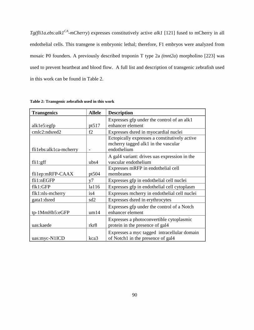
90
Tg(fli1a.ebs:alk1CA-mCherry) expresses constitutively active alk1 [121] fused to mCherry in all
endothelial cells. This transgene is embryonic lethal; therefore, F1 embryos were analyzed from
mosaic P0 founders. A previously described troponin T type 2a (tnnt2a) morpholino [223] was
used to prevent heartbeat and blood flow. A full list and description of transgenic zebrafish used
in this work can be found in Table 2.
Table 2: Transgenic zebrafish used in this work
Transgenics Allele Description
alk1e5:egfp pt517 Expresses gfp under the control of an alk1 enhancer element
cmlc2:ndsred2 f2 Expresses dsred in myocardial nuclei
fli1ebs:alk1ca-mcherry -
Ectopically expresses a constitutively active mcherry tagged alk1 in the vascular endothelium
fli1:gff ubs4 A gal4 variant: drives uas expression in the vascular endothelium
fli1ep:mRFP-CAAX pt504 Expresses mRFP in endothelial cell membranes
fli1:nEGFP y7 Expresses gfp in endothelial cell nuclei flk1:GFP la116 Expresses gfp in endothelial cell cytoplasm flk1:nls-mcherry is4 Expresses mcherry in endothelial cell nuclei gata1:dsred sd2 Expresses dsred in erythrocytes
tp-1MmHb5:eGFP um14 Expresses gfp under the control of a Notch enhancer element
uas:kaede rkr8 Expresses a photoconvertible cytoplasmic protein in the presence of gal4
uas:myc-N1ICD kca3 Expresses a myc tagged intracellular domain of Notch1 in the presence of gal4
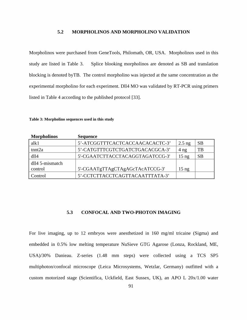
91
5.2 MORPHOLINOS AND MORPHOLINO VALIDATION
Morpholinos were purchased from GeneTools, Philomath, OR, USA. Morpholinos used in this
study are listed in Table 3. Splice blooking morpholinos are denoted as SB and translation
blocking is denoted byTB. The control morpholino was injected at the same concentration as the
experimental morpholino for each experiment. Dll4 MO was validated by RT-PCR using primers
listed in Table 4 according to the published protocol [33].
Table 3: Morpholino sequences used in this study
Morpholinos Sequence alk1 5’-ATCGGTTTCACTCACCAACACACTC-3’ 2.5 ng SB tnnt2a 5’-CATGTTTCGTCTGATCTGACACGCA-3’ 4 ng TB dll4 5'-CGAATCTTACCTACAGGTAGATCCG-3' 15 ng SB dll4 5-mismatch control 5'-CGAATgTTAgCTAgAGcTAcATCCG-3' 15 ng Control 5’-CCTCTTACCTCAGTTACAATTTATA-3’
5.3 CONFOCAL AND TWO-PHOTON IMAGING
For live imaging, up to 12 embryos were anesthetized in 160 mg/ml tricaine (Sigma) and
embedded in 0.5% low melting temperature NuSieve GTG Agarose (Lonza, Rockland, ME,
USA)/30% Danieau. Z-series (1.48 mm steps) were collected using a TCS SP5
multiphoton/confocal microscope (Leica Microsystems, Wetzlar, Germany) outfitted with a
custom motorized stage (Scientifica, Uckfield, East Sussex, UK), an APO L 20x/1.00 water
92
immersion objective or an HCX IRAPO L 25x/0.95 water immersion objective, non-descanned
detectors, and spectral detectors, with a 1.7X zoom. EGFP was excited with a Mai Tai DeepSee
Ti:Sapphire laser (Newport/Spectra Physics, Santa Clara, CA, USA) at 900 nm, whereas
mCherry and dsRed were excited with a 561 nm diode. Sequential frame scanning was
performed using a resonant scanner with unidirectional (8000 Hz) or bidirectional (1600 Hz)
scanning and 16x or 32x line averaging. For time lapse experiments, (X,Y) coordinates were set
using the LAS AF “Mark and Find” function, and images were collected every 18-23 minutes,
with z-stack parameters redefined for each (X,Y) coordinate. Images were analyzed using LAS
AF (version 3.0.0 build 834) and Adobe Photoshop CS6. Confocal time series were converted to
QuickTime (.mov) files using LAS AF and annotated using Final Cut Pro and iMovie.
5.4 ENDOTHELIAL CELL TRACKING
Maximum projection z-stacks were compiled using the Leica ASF-AF software and each time
point was saved as an individual tiff. Tiffs were imported into an Adobe Photoshop file, with
each time point occupying a single layer. Individual cells were labeled in Photoshop using the
paint tool and tracked over each layer. 3D projections were referenced in the Leica ASF-AF
software when it became difficult to track an individual cell with certainty.
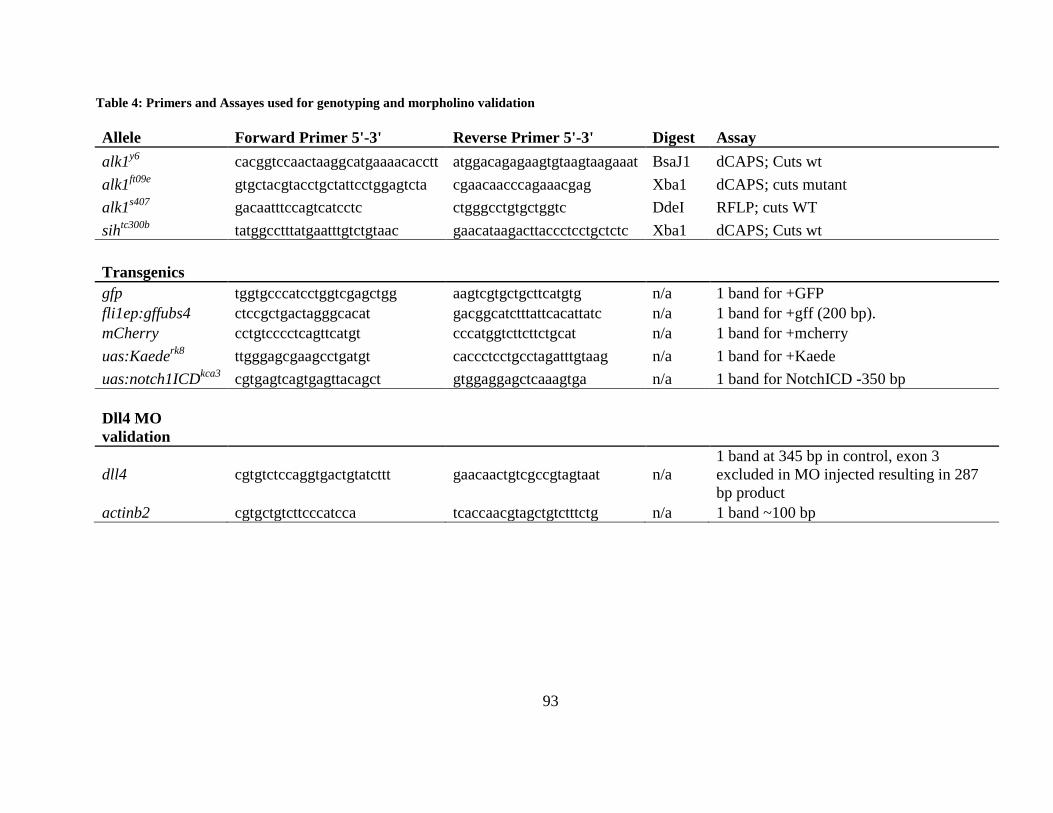
93
Table 4: Primers and Assayes used for genotyping and morpholino validation
Allele Forward Primer 5'-3' Reverse Primer 5'-3' Digest Assay alk1y6 cacggtccaactaaggcatgaaaacacctt atggacagagaagtgtaagtaagaaat BsaJ1 dCAPS; Cuts wt alk1ft09e gtgctacgtacctgctattcctggagtcta cgaacaacccagaaacgag Xba1 dCAPS; cuts mutant alk1s407 gacaatttccagtcatcctc ctgggcctgtgctggtc DdeI RFLP; cuts WT sihtc300b tatggcctttatgaatttgtctgtaac gaacataagacttaccctcctgctctc Xba1 dCAPS; Cuts wt Transgenics gfp tggtgcccatcctggtcgagctgg aagtcgtgctgcttcatgtg n/a 1 band for +GFP fli1ep:gffubs4 ctccgctgactagggcacat gacggcatctttattcacattatc n/a 1 band for +gff (200 bp). mCherry cctgtcccctcagttcatgt cccatggtcttcttctgcat n/a 1 band for +mcherry uas:Kaederk8 ttgggagcgaagcctgatgt caccctcctgcctagatttgtaag n/a 1 band for +Kaede uas:notch1ICDkca3 cgtgagtcagtgagttacagct gtggaggagctcaaagtga n/a 1 band for NotchICD -350 bp Dll4 MO validation
dll4 cgtgtctccaggtgactgtatcttt gaacaactgtcgccgtagtaat n/a 1 band at 345 bp in control, exon 3 excluded in MO injected resulting in 287 bp product
actinb2 cgtgctgtcttcccatcca tcaccaacgtagctgtctttctg n/a 1 band ~100 bp
94
5.5 KAEDE PHOTOCONVERSION
Up to 12 embryos were embedded in 0.5% low melt agarose at 24 hpf. The Leica “Frap Wizard”
software was used for imaging and bleaching. An initial scan was captured with the argon laser
set to 28% power, with the 488 nm line at 25%; the 561 nm diode at 25% and the 405 nm laser at
0%, 600Hz laser speed (3x line average) and a 2x zoom. Using this image, a region of interest
(ROI) was drawn around the section of vessel that was to be photoconverted. For bleaching all
parameters remained the same, with the exception of the 405 laser, which was set to 20%. The
“set background to zero” and “use laser settings for all ROIs” options were selected. The
bleaching time course consisted of 2 pre-bleach scans, 5 bleach scans, and 2 post-bleach scans.
Embryos were removed from the agarose and placed in a 12-well plate and incubated at 28.5°C
in the dark until 48 hpf. Embryos were then embedded again and imaged using the 488 nm and
516 nm wavelengths to determine the location of the converted cells.
To quantify the location of photoconverted cells, I divided the vessels into anatomical
sections and scored whether or not an individual embryo had a converted cell in that specific
region. The data represents the percentage of embryos that had at least one photoconverted cell
in each vessel region.
95
5.6 CRYOSECTIONS AND IMMUNOFLUORESCENCE
Embryos were fixed overnight in 4% paraformaldehyde at 4°C. Embryos were then washed into
a 15% and 30% sucrose/PBS solution before being embedded in Tissue Freezing Medium (TFM,
Triangle Biomedical Sciences) and stored at -80°C. Cryosectioning was performed on a Leica
CM 1850 and 30 µM sections were immediately placed on a Shandon Superfrost Plus positively
charged slide (Thermo Scientific), dried at 37°C for 30 minutes and stored at -20°C overnight.
Immunohistochemistry was performed using primary antibodies mouse anti-MF20 at 1:500
(sarcomeric myosin, Developmental Studies Hybridoma Bank, Iowa City, IA, USA) or rabbit
anti-GFP at 1:500 (Invitrogen, A-11122) and secondary antibodies goat-anti-rabbit Alexa Fluor
488 at 1:1000, and goat-anti-mouse Alexa Fluor 568 at 1:1000. Embryos were washed in a
PBS/0.1% triton X-100/0.1% DMSO (PBDT) solution and blocked in 5% goat serum in PBDT.
Sections were mounted with Vectashield Fluorescent mounting medium (Vector) and imaged
with an Olympus Fluoview 1000 confocal microscope outfitted with a UPFLN 20x oil
immersion objective. Two-dimensional projections were generated from Z-series (1 µm steps)
using ImageJ 1.45s (National Institutes of Health, USA).
5.7 IN SITU HYBRIDIZATION
All embryos were collected at 36 hpf, fixed in 4% paraformaldehyde/PBS for approximately 36
hours at 4°C, dehydrated in methanol, and stored at -20°C for in situ hybridization. Digoxigenin-
labeled riboprobes were generated according to the manufacturer’s protocol (Roche,
96
Indianapolis, IN, USA). cdh5 plasmid [92], dll4 plasmid [33], and efnb2a plasmid [30] have
been described. hey2 was amplified from zebrafish cDNA and cloned into PCRII-TOPO
(Invitrogen/Life Technologies). egfp was amplified from plasmid DNA and cloned into pCRII-
TOPO. collagen type IV alpha 1 (col4a1) was amplified from zebrafish cDNA using primers
appended with T3 (sense) and T7 (antisense) polymerase sites, and the PCR product was column
purified (Qiaquick PCR Purification Kit) and used for riboprobe synthesis. Whole mount in situ
hybridization was performed in an InSituPro VSi liquid handler (Intavis Inc, Chicago, IL, USA).
Briefly, 36 hpf embryos were rehydrated to PBS/0.1% tween-20 (PBT), permeabilized with 50
mg/ml proteinase K for 15 minutes, and the permeabilization terminated with 0.2% glycine/PBT,
5 minutes. Embryos were then post-fixed for 20 minutes with 4% paraformaldehyde/PBS and
hybridized at 65°C for 12 hr with riboprobe diluted 1:500 in hybridization buffer (50%
formamide, 5x SSC, 5 mg/mL yeast tRNA, 50 mg/ml heparin, 0.1% tween-20). Embryos were
then washed with 50% formamide/2x SSC/0.1% tween-20 (2 x 30 minutes, 65°C), 2x SSC/0.1%
tween-20 (15 minutes, 65°C), and 0.2x SSC/0.1% tween-20 (2 x 30 mintues, room temperature).
Specimens were then blocked with 5% sheep serum (Sigma)/PBT and incubated with embryo-
adsorbed anti-digoxigenin-AP, Fab fragments (Roche), 1:5000 in 5% sheep serum/PBT.
Embryos were then washed 6 x 15 minutes followed by overnight in PBT, transferred to 6-well
plates containing NTMT (100 mmol/L NaCl, 100 mmol/L Tris-HCl, pH 9.5, 50 mmol/L MgCl2,
1% tween-20), and incubated in NBT/BCIP (340 mg/ml nitro blue tetrazolium; 350 µg/ml 5-
bromo-4-chloro-3-indolyly phosphate in NTMT) for color development. Embryos were
photographed using an MVX-10 MacroView microscope and DP71 camera (Olympus America,
Center Valley, PA, USA), scored for expression pattern and relative staining intensity, and then
processed for genotyping (Table 4). Stained embryos were embedded in 4% NuSieve GTG
97
agarose (Lonza, Rockland, ME, USA), sectioned at 50 mm with a VT1000S vibratome (Leica
Microsystems, Buffalo Grove, IL, USA), and photographed using a BX51 compound microscope
with UPLFLN 20x/0.5 objective and DP71 camera (Olympus). All figures represent embryos
that were simultaneously processed for fixation and staining.
5.8 CENTRAL ARTERY SPROUT QUANTIFICATION
Manual tracing (Adobe Photoshop CS6) of maximum projections generated from confocal Z-
stacks was used to generate simplified wiring diagrams of the forebrain and midbrain central
arteries, which originate from the primordial midbrain channels. From these wiring diagrams, I
counted: sprouts emerging from the primordial midbrain channel, sprout connections to the basal
communicating artery, branch points, and contralateral connections (midline crossings). Each
parameter was averaged within treatment groups and values are presented as mean ± SEM.
5.9 BCA AREA QUANTIFICATION
Approximate basal communicating area measurements were achieved by creating a region of
interest on a two dimensional maximal projection around the basal communicating artery. Using
the analyze tool in the LAS AF (version 3.0.0 build 8134) software, the area within the ROI was
calculated. Areas were averaged within treatment groups and values are presented as mean ±
SEM.
98
5.10 FLUORESCENCE INTENSITY MEASUREMENTS OF NOTCH REPORTER
EMBRYOS
Fluorescence intensity of cranial arterial EGFP in Tg(tp1:egfp)um14;Tg(fli1a.ep:mRFP-CAAX)pt504
embryos was quantified using the LAS AF Version 3.0.0 build 8134 software. An ROI was
created based on the mRFP channel (threshold, 55; background, 30), and GFP intensity within
the masked ROI was measured and averaged over a single z plane, yielding a mean intensity of
GFP fluorescence within the ROI. Mean intensities were averaged across samples (n=10 control
morphants, 12 alk1 morphants), and values expressed as mean ± SEM. Results were verified by
manually drawing ROIs over the basal communicating artery, using the mRFP channel as a
guide, and averaging the colocalized GFP intensity across an entire stack for two embryos per
treatment. This method gave similar results to the automated method.
99
BIBLIOGRAPHY
1. De Val, S., Key transcriptional regulators of early vascular development. Arterioscler Thromb Vasc Biol, 2011. 31(7): p. 1469-75.
2. Risau, W. and I. Flamme, Vasculogenesis. Annu Rev Cell Dev Biol, 1995. 11: p. 73-91.
3. Patan, S., Vasculogenesis and angiogenesis as mechanisms of vascular network formation, growth and remodeling. J Neurooncol, 2000. 50(1-2): p. 1-15.
4. Fouquet, B., et al., Vessel patterning in the embryo of the zebrafish: guidance by notochord. Dev Biol, 1997. 183(1): p. 37-48.
5. Coffin, J.D. and T.J. Poole, Embryonic vascular development: immunohistochemical identification of the origin and subsequent morphogenesis of the major vessel primordia in quail embryos. Development, 1988. 102(4): p. 735-48.
6. Proulx, K., A. Lu, and S. Sumanas, Cranial vasculature in zebrafish forms by angioblast cluster-derived angiogenesis. Dev Biol, 2010. 348(1): p. 34-46.
7. Anderson, M.J., et al., Loss of unc45a precipitates arteriovenous shunting in the aortic arches. Dev Biol, 2008. 318(2): p. 258-67.
8. Li, P., M. Pashmforoush, and H.M. Sucov, Mesodermal retinoic acid signaling regulates endothelial cell coalescence in caudal pharyngeal arch artery vasculogenesis. Dev Biol, 2012. 361(1): p. 116-24.
9. Paffett-Lugassy, N., et al., Heart field origin of great vessel precursors relies on nkx2.5-mediated vasculogenesis. Nat Cell Biol, 2013. 15(11): p. 1362-9.
10. Kamei, M., et al., Endothelial tubes assemble from intracellular vacuoles in vivo. Nature, 2006. 442(7101): p. 453-6.
11. Blum, Y., et al., Complex cell rearrangements during intersegmental vessel sprouting and vessel fusion in the zebrafish embryo. Dev Biol, 2008. 316(2): p. 312-22.
12. Parker, L.H., et al., The endothelial-cell-derived secreted factor Egfl7 regulates vascular tube formation. Nature, 2004. 428(6984): p. 754-8.
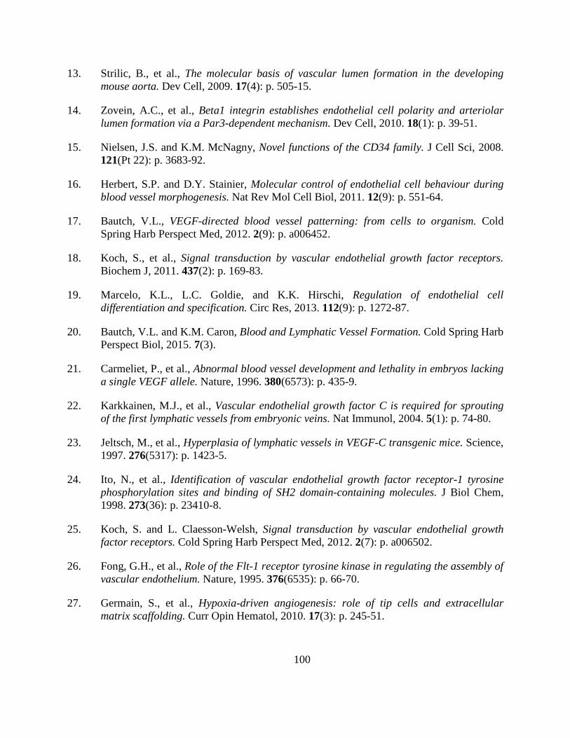
100
13. Strilic, B., et al., The molecular basis of vascular lumen formation in the developing mouse aorta. Dev Cell, 2009. 17(4): p. 505-15.
14. Zovein, A.C., et al., Beta1 integrin establishes endothelial cell polarity and arteriolar lumen formation via a Par3-dependent mechanism. Dev Cell, 2010. 18(1): p. 39-51.
15. Nielsen, J.S. and K.M. McNagny, Novel functions of the CD34 family. J Cell Sci, 2008. 121(Pt 22): p. 3683-92.
16. Herbert, S.P. and D.Y. Stainier, Molecular control of endothelial cell behaviour during blood vessel morphogenesis. Nat Rev Mol Cell Biol, 2011. 12(9): p. 551-64.
17. Bautch, V.L., VEGF-directed blood vessel patterning: from cells to organism. Cold Spring Harb Perspect Med, 2012. 2(9): p. a006452.
18. Koch, S., et al., Signal transduction by vascular endothelial growth factor receptors. Biochem J, 2011. 437(2): p. 169-83.
19. Marcelo, K.L., L.C. Goldie, and K.K. Hirschi, Regulation of endothelial cell differentiation and specification. Circ Res, 2013. 112(9): p. 1272-87.
20. Bautch, V.L. and K.M. Caron, Blood and Lymphatic Vessel Formation. Cold Spring Harb Perspect Biol, 2015. 7(3).
21. Carmeliet, P., et al., Abnormal blood vessel development and lethality in embryos lacking a single VEGF allele. Nature, 1996. 380(6573): p. 435-9.
22. Karkkainen, M.J., et al., Vascular endothelial growth factor C is required for sprouting of the first lymphatic vessels from embryonic veins. Nat Immunol, 2004. 5(1): p. 74-80.
23. Jeltsch, M., et al., Hyperplasia of lymphatic vessels in VEGF-C transgenic mice. Science, 1997. 276(5317): p. 1423-5.
24. Ito, N., et al., Identification of vascular endothelial growth factor receptor-1 tyrosine phosphorylation sites and binding of SH2 domain-containing molecules. J Biol Chem, 1998. 273(36): p. 23410-8.
25. Koch, S. and L. Claesson-Welsh, Signal transduction by vascular endothelial growth factor receptors. Cold Spring Harb Perspect Med, 2012. 2(7): p. a006502.
26. Fong, G.H., et al., Role of the Flt-1 receptor tyrosine kinase in regulating the assembly of vascular endothelium. Nature, 1995. 376(6535): p. 66-70.
27. Germain, S., et al., Hypoxia-driven angiogenesis: role of tip cells and extracellular matrix scaffolding. Curr Opin Hematol, 2010. 17(3): p. 245-51.
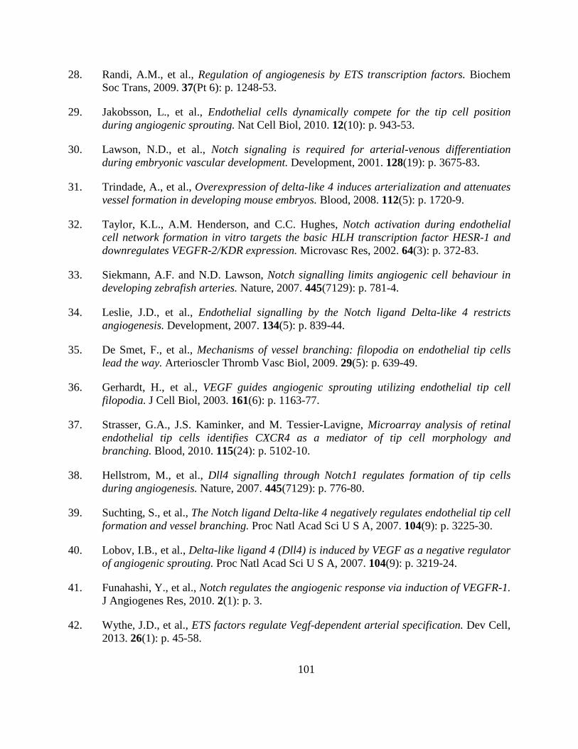
101
28. Randi, A.M., et al., Regulation of angiogenesis by ETS transcription factors. Biochem Soc Trans, 2009. 37(Pt 6): p. 1248-53.
29. Jakobsson, L., et al., Endothelial cells dynamically compete for the tip cell position during angiogenic sprouting. Nat Cell Biol, 2010. 12(10): p. 943-53.
30. Lawson, N.D., et al., Notch signaling is required for arterial-venous differentiation during embryonic vascular development. Development, 2001. 128(19): p. 3675-83.
31. Trindade, A., et al., Overexpression of delta-like 4 induces arterialization and attenuates vessel formation in developing mouse embryos. Blood, 2008. 112(5): p. 1720-9.
32. Taylor, K.L., A.M. Henderson, and C.C. Hughes, Notch activation during endothelial cell network formation in vitro targets the basic HLH transcription factor HESR-1 and downregulates VEGFR-2/KDR expression. Microvasc Res, 2002. 64(3): p. 372-83.
33. Siekmann, A.F. and N.D. Lawson, Notch signalling limits angiogenic cell behaviour in developing zebrafish arteries. Nature, 2007. 445(7129): p. 781-4.
34. Leslie, J.D., et al., Endothelial signalling by the Notch ligand Delta-like 4 restricts angiogenesis. Development, 2007. 134(5): p. 839-44.
35. De Smet, F., et al., Mechanisms of vessel branching: filopodia on endothelial tip cells lead the way. Arterioscler Thromb Vasc Biol, 2009. 29(5): p. 639-49.
36. Gerhardt, H., et al., VEGF guides angiogenic sprouting utilizing endothelial tip cell filopodia. J Cell Biol, 2003. 161(6): p. 1163-77.
37. Strasser, G.A., J.S. Kaminker, and M. Tessier-Lavigne, Microarray analysis of retinal endothelial tip cells identifies CXCR4 as a mediator of tip cell morphology and branching. Blood, 2010. 115(24): p. 5102-10.
38. Hellstrom, M., et al., Dll4 signalling through Notch1 regulates formation of tip cells during angiogenesis. Nature, 2007. 445(7129): p. 776-80.
39. Suchting, S., et al., The Notch ligand Delta-like 4 negatively regulates endothelial tip cell formation and vessel branching. Proc Natl Acad Sci U S A, 2007. 104(9): p. 3225-30.
40. Lobov, I.B., et al., Delta-like ligand 4 (Dll4) is induced by VEGF as a negative regulator of angiogenic sprouting. Proc Natl Acad Sci U S A, 2007. 104(9): p. 3219-24.
41. Funahashi, Y., et al., Notch regulates the angiogenic response via induction of VEGFR-1. J Angiogenes Res, 2010. 2(1): p. 3.
42. Wythe, J.D., et al., ETS factors regulate Vegf-dependent arterial specification. Dev Cell, 2013. 26(1): p. 45-58.
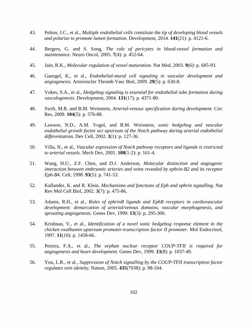
102
43. Pelton, J.C., et al., Multiple endothelial cells constitute the tip of developing blood vessels and polarize to promote lumen formation. Development, 2014. 141(21): p. 4121-6.
44. Bergers, G. and S. Song, The role of pericytes in blood-vessel formation and maintenance. Neuro Oncol, 2005. 7(4): p. 452-64.
45. Jain, R.K., Molecular regulation of vessel maturation. Nat Med, 2003. 9(6): p. 685-93.
46. Gaengel, K., et al., Endothelial-mural cell signaling in vascular development and angiogenesis. Arterioscler Thromb Vasc Biol, 2009. 29(5): p. 630-8.
47. Vokes, S.A., et al., Hedgehog signaling is essential for endothelial tube formation during vasculogenesis. Development, 2004. 131(17): p. 4371-80.
48. Swift, M.R. and B.M. Weinstein, Arterial-venous specification during development. Circ Res, 2009. 104(5): p. 576-88.
49. Lawson, N.D., A.M. Vogel, and B.M. Weinstein, sonic hedgehog and vascular endothelial growth factor act upstream of the Notch pathway during arterial endothelial differentiation. Dev Cell, 2002. 3(1): p. 127-36.
50. Villa, N., et al., Vascular expression of Notch pathway receptors and ligands is restricted to arterial vessels. Mech Dev, 2001. 108(1-2): p. 161-4.
51. Wang, H.U., Z.F. Chen, and D.J. Anderson, Molecular distinction and angiogenic interaction between embryonic arteries and veins revealed by ephrin-B2 and its receptor Eph-B4. Cell, 1998. 93(5): p. 741-53.
52. Kullander, K. and R. Klein, Mechanisms and functions of Eph and ephrin signalling. Nat Rev Mol Cell Biol, 2002. 3(7): p. 475-86.
53. Adams, R.H., et al., Roles of ephrinB ligands and EphB receptors in cardiovascular development: demarcation of arterial/venous domains, vascular morphogenesis, and sprouting angiogenesis. Genes Dev, 1999. 13(3): p. 295-306.
54. Krishnan, V., et al., Identification of a novel sonic hedgehog response element in the chicken ovalbumin upstream promoter-transcription factor II promoter. Mol Endocrinol, 1997. 11(10): p. 1458-66.
55. Pereira, F.A., et al., The orphan nuclear receptor COUP-TFII is required for angiogenesis and heart development. Genes Dev, 1999. 13(8): p. 1037-49.
56. You, L.R., et al., Suppression of Notch signalling by the COUP-TFII transcription factor regulates vein identity. Nature, 2005. 435(7038): p. 98-104.
103
57. Kang, J., et al., An exquisite cross-control mechanism among endothelial cell fate regulators directs the plasticity and heterogeneity of lymphatic endothelial cells. Blood, 2010. 116(1): p. 140-50.
58. Swift, M.R., et al., SoxF factors and Notch regulate nr2f2 gene expression during venous differentiation in zebrafish. Dev Biol, 2014. 390(2): p. 116-25.
59. Moyon, D., et al., Plasticity of endothelial cells during arterial-venous differentiation in the avian embryo. Development, 2001. 128(17): p. 3359-70.
60. Othman-Hassan, K., et al., Arterial identity of endothelial cells is controlled by local cues. Dev Biol, 2001. 237(2): p. 398-409.
61. le Noble, F., et al., Flow regulates arterial-venous differentiation in the chick embryo yolk sac. Development, 2004. 131(2): p. 361-75.
62. Roman, B.L. and K. Pekkan, Mechanotransduction in embryonic vascular development. Biomech Model Mechanobiol, 2012. 11(8): p. 1149-68.
63. Brownlee, R.D. and B.L. Langille, Arterial adaptations to altered blood flow. Can J Physiol Pharmacol, 1991. 69(7): p. 978-83.
64. Di Stefano, I., D.R. Koopmans, and B.L. Langille, Modulation of arterial growth of the rabbit carotid artery associated with experimental elevation of blood flow. J Vasc Res, 1998. 35(1): p. 1-7.
65. Kamiya, A. and T. Togawa, Adaptive regulation of wall shear stress to flow change in the canine carotid artery. Am J Physiol, 1980. 239(1): p. H14-21.
66. Langille, B.L., M.P. Bendeck, and F.W. Keeley, Adaptations of carotid arteries of young and mature rabbits to reduced carotid blood flow. Am J Physiol, 1989. 256(4 Pt 2): p. H931-9.
67. Langille, B.L. and F. O'Donnell, Reductions in arterial diameter produced by chronic decreases in blood flow are endothelium-dependent. Science, 1986. 231(4736): p. 405-7.
68. Cecchi, E., et al., Role of hemodynamic shear stress in cardiovascular disease. Atherosclerosis, 2011. 214(2): p. 249-56.
69. Dekker, R.J., et al., KLF2 provokes a gene expression pattern that establishes functional quiescent differentiation of the endothelium. Blood, 2006. 107(11): p. 4354-63.
70. Parmar, K.M., et al., Integration of flow-dependent endothelial phenotypes by Kruppel-like factor 2. J Clin Invest, 2006. 116(1): p. 49-58.
71. Fledderus, J.O., et al., KLF2 primes the antioxidant transcription factor Nrf2 for activation in endothelial cells. Arterioscler Thromb Vasc Biol, 2008. 28(7): p. 1339-46.
104
72. Hay, D.C., et al., Activation of NF-kappaB nuclear transcription factor by flow in human endothelial cells. Biochim Biophys Acta, 2003. 1642(1-2): p. 33-44.
73. SenBanerjee, S., et al., KLF2 Is a novel transcriptional regulator of endothelial proinflammatory activation. J Exp Med, 2004. 199(10): p. 1305-15.
74. Tkachenko, E., et al., The nucleus of endothelial cell as a sensor of blood flow direction. Biol Open, 2013. 2(10): p. 1007-12.
75. AbouAlaiwi, W.A., et al., Ciliary polycystin-2 is a mechanosensitive calcium channel involved in nitric oxide signaling cascades. Circ Res, 2009. 104(7): p. 860-9.
76. Van der Heiden, K., et al., Endothelial primary cilia in areas of disturbed flow are at the base of atherosclerosis. Atherosclerosis, 2008. 196(2): p. 542-50.
77. Van der Heiden, K., et al., Monocilia on chicken embryonic endocardium in low shear stress areas. Dev Dyn, 2006. 235(1): p. 19-28.
78. Hierck, B.P., et al., Primary cilia sensitize endothelial cells for fluid shear stress. Dev Dyn, 2008. 237(3): p. 725-35.
79. Iomini, C., et al., Primary cilia of human endothelial cells disassemble under laminar shear stress. J Cell Biol, 2004. 164(6): p. 811-7.
80. Nauli, S.M., et al., Endothelial cilia are fluid shear sensors that regulate calcium signaling and nitric oxide production through polycystin-1. Circulation, 2008. 117(9): p. 1161-71.
81. Weinbaum, S., J.M. Tarbell, and E.R. Damiano, The structure and function of the endothelial glycocalyx layer. Annu Rev Biomed Eng, 2007. 9: p. 121-67.
82. Reitsma, S., et al., The endothelial glycocalyx: composition, functions, and visualization. Pflugers Arch, 2007. 454(3): p. 345-59.
83. Thi, M.M., et al., The role of the glycocalyx in reorganization of the actin cytoskeleton under fluid shear stress: a "bumper-car" model. Proc Natl Acad Sci U S A, 2004. 101(47): p. 16483-8.
84. Osawa, M., et al., Evidence for a role of platelet endothelial cell adhesion molecule-1 in endothelial cell mechanosignal transduction: is it a mechanoresponsive molecule? J Cell Biol, 2002. 158(4): p. 773-85.
85. Tzima, E., et al., A mechanosensory complex that mediates the endothelial cell response to fluid shear stress. Nature, 2005. 437(7057): p. 426-31.
86. Dimmeler, S., et al., Activation of nitric oxide synthase in endothelial cells by Akt-dependent phosphorylation. Nature, 1999. 399(6736): p. 601-5.
105
87. Jin, Z.G., et al., Ligand-independent activation of vascular endothelial growth factor receptor 2 by fluid shear stress regulates activation of endothelial nitric oxide synthase. Circ Res, 2003. 93(4): p. 354-63.
88. Morgan, J.T., et al., Nesprin-3 regulates endothelial cell morphology, perinuclear cytoskeletal architecture, and flow-induced polarization. Mol Biol Cell, 2011. 22(22): p. 4324-34.
89. le Noble, F., et al., Control of arterial branching morphogenesis in embryogenesis: go with the flow. Cardiovasc Res, 2005. 65(3): p. 619-28.
90. Braverman, I.M., A. Keh, and B.S. Jacobson, Ultrastructure and three-dimensional organization of the telangiectases of hereditary hemorrhagic telangiectasia. J Invest Dermatol, 1990. 95(4): p. 422-7.
91. Guttmacher, A.E., D.A. Marchuk, and R.I. White, Jr., Hereditary hemorrhagic telangiectasia. N Engl J Med, 1995. 333(14): p. 918-24.
92. Corti, P., et al., Interaction between alk1 and blood flow in the development of arteriovenous malformations. Development, 2011. 138(8): p. 1573-82.
93. Laakso, A. and J. Hernesniemi, Arteriovenous malformations: epidemiology and clinical presentation. Neurosurg Clin N Am, 2012. 23(1): p. 1-6.
94. Ragsdale, J.A., Hereditary hemorrhagic telangiectasia: from epistaxis to life-threatening GI bleeding. Gastroenterol Nurs, 2007. 30(4): p. 293-9; quiz 300-1.
95. Hershkovitz, D., et al., RASA1 mutations may cause hereditary capillary malformations without arteriovenous malformations. Br J Dermatol, 2008. 158(5): p. 1035-40.
96. Moalem, S., et al., Hypotrichosis-lymphedema-telangiectasia-renal defect associated with a truncating mutation in the SOX18 gene. Clin Genet, 2014.
97. Chaudhary, M.W. and R.S. Al-Baradie, Ataxia-telangiectasia: future prospects. Appl Clin Genet, 2014. 7: p. 159-67.
98. McDonald, J., P. Bayrak-Toydemir, and R.E. Pyeritz, Hereditary hemorrhagic telangiectasia: an overview of diagnosis, management, and pathogenesis. Genet Med, 2011. 13(7): p. 607-16.
99. McAllister, K.A., et al., Endoglin, a TGF-beta binding protein of endothelial cells, is the gene for hereditary haemorrhagic telangiectasia type 1. Nat Genet, 1994. 8(4): p. 345-51.
100. Johnson, D.W., et al., Mutations in the activin receptor-like kinase 1 gene in hereditary haemorrhagic telangiectasia type 2. Nat Genet, 1996. 13(2): p. 189-95.
106
101. Gallione, C.J., et al., A combined syndrome of juvenile polyposis and hereditary haemorrhagic telangiectasia associated with mutations in MADH4 (SMAD4). Lancet, 2004. 363(9412): p. 852-9.
102. Cole, S.G., et al., A new locus for hereditary haemorrhagic telangiectasia (HHT3) maps to chromosome 5. J Med Genet, 2005. 42(7): p. 577-82.
103. Bayrak-Toydemir, P., et al., A fourth locus for hereditary hemorrhagic telangiectasia maps to chromosome 7. Am J Med Genet A, 2006. 140(20): p. 2155-62.
104. Bayrak-Toydemir, P., et al., Genotype-phenotype correlation in hereditary hemorrhagic telangiectasia: mutations and manifestations. Am J Med Genet A, 2006. 140(5): p. 463-70.
105. Sabba, C., et al., Hereditary hemorrhagic telangiectasia: clinical features in ENG and ALK1 mutation carriers. J Thromb Haemost, 2007. 5(6): p. 1149-57.
106. Letteboer, T.G., et al., Genotype-phenotype relationship for localization and age distribution of telangiectases in hereditary hemorrhagic telangiectasia. Am J Med Genet A, 2008. 146A(21): p. 2733-9.
107. Ardelean, D.S. and M. Letarte, Anti-angiogenic therapeutic strategies in hereditary hemorrhagic telangiectasia. Front Genet, 2015. 6: p. 35.
108. Meurer, S.K., et al., Endoglin in liver fibrogenesis: Bridging basic science and clinical practice. World J Biol Chem, 2014. 5(2): p. 180-203.
109. David, L., J.J. Feige, and S. Bailly, Emerging role of bone morphogenetic proteins in angiogenesis. Cytokine Growth Factor Rev, 2009. 20(3): p. 203-12.
110. Schmierer, B. and C.S. Hill, TGFbeta-SMAD signal transduction: molecular specificity and functional flexibility. Nat Rev Mol Cell Biol, 2007. 8(12): p. 970-82.
111. Tillet, E. and S. Bailly, Emerging roles of BMP9 and BMP10 in hereditary hemorrhagic telangiectasia. Front Genet, 2014. 5: p. 456.
112. Ricard, N., et al., Functional analysis of the BMP9 response of ALK1 mutants from HHT2 patients: a diagnostic tool for novel ACVRL1 mutations. Blood, 2010. 116(9): p. 1604-12.
113. Gu, Y., et al., Functional analysis of mutations in the kinase domain of the TGF-beta receptor ALK1 reveals different mechanisms for induction of hereditary hemorrhagic telangiectasia. Blood, 2006. 107(5): p. 1951-4.
114. Oh, S.P., et al., Activin receptor-like kinase 1 modulates transforming growth factor-beta 1 signaling in the regulation of angiogenesis. Proc Natl Acad Sci U S A, 2000. 97(6): p. 2626-31.
107
115. Urness, L.D., L.K. Sorensen, and D.Y. Li, Arteriovenous malformations in mice lacking activin receptor-like kinase-1. Nat Genet, 2000. 26(3): p. 328-31.
116. Srinivasan, S., et al., A mouse model for hereditary hemorrhagic telangiectasia (HHT) type 2. Hum Mol Genet, 2003. 12(5): p. 473-82.
117. Park, S.O., et al., Real-time imaging of de novo arteriovenous malformation in a mouse model of hereditary hemorrhagic telangiectasia. J Clin Invest, 2009. 119(11): p. 3487-96.
118. Park, S.O., et al., ALK5- and TGFBR2-independent role of ALK1 in the pathogenesis of hereditary hemorrhagic telangiectasia type 2. Blood, 2008. 111(2): p. 633-42.
119. Tual-Chalot, S., et al., Endothelial depletion of Acvrl1 in mice leads to arteriovenous malformations associated with reduced endoglin expression. PLoS One, 2014. 9(6): p. e98646.
120. Garrido-Martin, E.M., et al., Common and distinctive pathogenetic features of arteriovenous malformations in hereditary hemorrhagic telangiectasia 1 and hereditary hemorrhagic telangiectasia 2 animal models--brief report. Arterioscler Thromb Vasc Biol, 2014. 34(10): p. 2232-6.
121. Roman, B.L., et al., Disruption of acvrl1 increases endothelial cell number in zebrafish cranial vessels. Development, 2002. 129(12): p. 3009-19.
122. Barbara, N.P., J.L. Wrana, and M. Letarte, Endoglin is an accessory protein that interacts with the signaling receptor complex of multiple members of the transforming growth factor-beta superfamily. J Biol Chem, 1999. 274(2): p. 584-94.
123. Alt, A., et al., Structural and functional insights into endoglin ligand recognition and binding. PLoS One, 2012. 7(2): p. e29948.
124. Castonguay, R., et al., Soluble endoglin specifically binds bone morphogenetic proteins 9 and 10 via its orphan domain, inhibits blood vessel formation, and suppresses tumor growth. J Biol Chem, 2011. 286(34): p. 30034-46.
125. Li, D.Y., et al., Defective angiogenesis in mice lacking endoglin. Science, 1999. 284(5419): p. 1534-7.
126. Bourdeau, A., D.J. Dumont, and M. Letarte, A murine model of hereditary hemorrhagic telangiectasia. J Clin Invest, 1999. 104(10): p. 1343-51.
127. Arthur, H.M., et al., Endoglin, an ancillary TGFbeta receptor, is required for extraembryonic angiogenesis and plays a key role in heart development. Dev Biol, 2000. 217(1): p. 42-53.
128. Sorensen, L.K., et al., Loss of distinct arterial and venous boundaries in mice lacking endoglin, a vascular-specific TGFbeta coreceptor. Dev Biol, 2003. 261(1): p. 235-50.
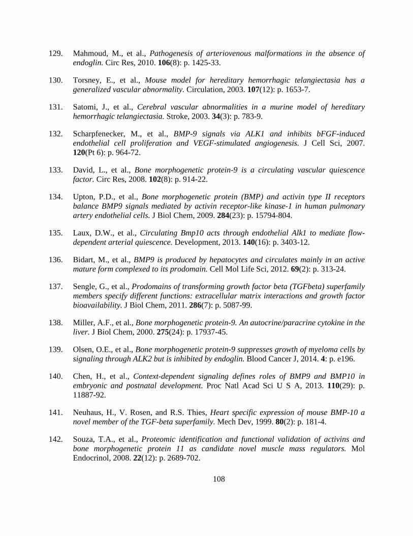
108
129. Mahmoud, M., et al., Pathogenesis of arteriovenous malformations in the absence of endoglin. Circ Res, 2010. 106(8): p. 1425-33.
130. Torsney, E., et al., Mouse model for hereditary hemorrhagic telangiectasia has a generalized vascular abnormality. Circulation, 2003. 107(12): p. 1653-7.
131. Satomi, J., et al., Cerebral vascular abnormalities in a murine model of hereditary hemorrhagic telangiectasia. Stroke, 2003. 34(3): p. 783-9.
132. Scharpfenecker, M., et al., BMP-9 signals via ALK1 and inhibits bFGF-induced endothelial cell proliferation and VEGF-stimulated angiogenesis. J Cell Sci, 2007. 120(Pt 6): p. 964-72.
133. David, L., et al., Bone morphogenetic protein-9 is a circulating vascular quiescence factor. Circ Res, 2008. 102(8): p. 914-22.
134. Upton, P.D., et al., Bone morphogenetic protein (BMP) and activin type II receptors balance BMP9 signals mediated by activin receptor-like kinase-1 in human pulmonary artery endothelial cells. J Biol Chem, 2009. 284(23): p. 15794-804.
135. Laux, D.W., et al., Circulating Bmp10 acts through endothelial Alk1 to mediate flow-dependent arterial quiescence. Development, 2013. 140(16): p. 3403-12.
136. Bidart, M., et al., BMP9 is produced by hepatocytes and circulates mainly in an active mature form complexed to its prodomain. Cell Mol Life Sci, 2012. 69(2): p. 313-24.
137. Sengle, G., et al., Prodomains of transforming growth factor beta (TGFbeta) superfamily members specify different functions: extracellular matrix interactions and growth factor bioavailability. J Biol Chem, 2011. 286(7): p. 5087-99.
138. Miller, A.F., et al., Bone morphogenetic protein-9. An autocrine/paracrine cytokine in the liver. J Biol Chem, 2000. 275(24): p. 17937-45.
139. Olsen, O.E., et al., Bone morphogenetic protein-9 suppresses growth of myeloma cells by signaling through ALK2 but is inhibited by endoglin. Blood Cancer J, 2014. 4: p. e196.
140. Chen, H., et al., Context-dependent signaling defines roles of BMP9 and BMP10 in embryonic and postnatal development. Proc Natl Acad Sci U S A, 2013. 110(29): p. 11887-92.
141. Neuhaus, H., V. Rosen, and R.S. Thies, Heart specific expression of mouse BMP-10 a novel member of the TGF-beta superfamily. Mech Dev, 1999. 80(2): p. 181-4.
142. Souza, T.A., et al., Proteomic identification and functional validation of activins and bone morphogenetic protein 11 as candidate novel muscle mass regulators. Mol Endocrinol, 2008. 22(12): p. 2689-702.
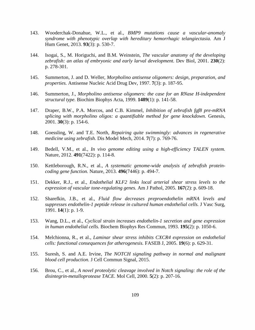
109
143. Wooderchak-Donahue, W.L., et al., BMP9 mutations cause a vascular-anomaly syndrome with phenotypic overlap with hereditary hemorrhagic telangiectasia. Am J Hum Genet, 2013. 93(3): p. 530-7.
144. Isogai, S., M. Horiguchi, and B.M. Weinstein, The vascular anatomy of the developing zebrafish: an atlas of embryonic and early larval development. Dev Biol, 2001. 230(2): p. 278-301.
145. Summerton, J. and D. Weller, Morpholino antisense oligomers: design, preparation, and properties. Antisense Nucleic Acid Drug Dev, 1997. 7(3): p. 187-95.
146. Summerton, J., Morpholino antisense oligomers: the case for an RNase H-independent structural type. Biochim Biophys Acta, 1999. 1489(1): p. 141-58.
147. Draper, B.W., P.A. Morcos, and C.B. Kimmel, Inhibition of zebrafish fgf8 pre-mRNA splicing with morpholino oligos: a quantifiable method for gene knockdown. Genesis, 2001. 30(3): p. 154-6.
148. Goessling, W. and T.E. North, Repairing quite swimmingly: advances in regenerative medicine using zebrafish. Dis Model Mech, 2014. 7(7): p. 769-76.
149. Bedell, V.M., et al., In vivo genome editing using a high-efficiency TALEN system. Nature, 2012. 491(7422): p. 114-8.
150. Kettleborough, R.N., et al., A systematic genome-wide analysis of zebrafish protein-coding gene function. Nature, 2013. 496(7446): p. 494-7.
151. Dekker, R.J., et al., Endothelial KLF2 links local arterial shear stress levels to the expression of vascular tone-regulating genes. Am J Pathol, 2005. 167(2): p. 609-18.
152. Sharefkin, J.B., et al., Fluid flow decreases preproendothelin mRNA levels and suppresses endothelin-1 peptide release in cultured human endothelial cells. J Vasc Surg, 1991. 14(1): p. 1-9.
153. Wang, D.L., et al., Cyclical strain increases endothelin-1 secretion and gene expression in human endothelial cells. Biochem Biophys Res Commun, 1993. 195(2): p. 1050-6.
154. Melchionna, R., et al., Laminar shear stress inhibits CXCR4 expression on endothelial cells: functional consequences for atherogenesis. FASEB J, 2005. 19(6): p. 629-31.
155. Suresh, S. and A.E. Irvine, The NOTCH signaling pathway in normal and malignant blood cell production. J Cell Commun Signal, 2015.
156. Brou, C., et al., A novel proteolytic cleavage involved in Notch signaling: the role of the disintegrin-metalloprotease TACE. Mol Cell, 2000. 5(2): p. 207-16.
110
157. De Strooper, B., et al., A presenilin-1-dependent gamma-secretase-like protease mediates release of Notch intracellular domain. Nature, 1999. 398(6727): p. 518-22.
158. Gordon, W.R., K.L. Arnett, and S.C. Blacklow, The molecular logic of Notch signaling--a structural and biochemical perspective. J Cell Sci, 2008. 121(Pt 19): p. 3109-19.
159. Andersson, E.R., R. Sandberg, and U. Lendahl, Notch signaling: simplicity in design, versatility in function. Development, 2011. 138(17): p. 3593-612.
160. Weinstein, B.M., et al., Gridlock, a localized heritable vascular patterning defect in the zebrafish. Nat Med, 1995. 1(11): p. 1143-7.
161. Zhong, T.P., et al., gridlock, an HLH gene required for assembly of the aorta in zebrafish. Science, 2000. 287(5459): p. 1820-4.
162. Krebs, L.T., et al., Haploinsufficient lethality and formation of arteriovenous malformations in Notch pathway mutants. Genes Dev, 2004. 18(20): p. 2469-73.
163. Nielsen, C.M., et al., Deletion of Rbpj from postnatal endothelium leads to abnormal arteriovenous shunting in mice. Development, 2014. 141(19): p. 3782-92.
164. Covassin, L., et al., Global analysis of hematopoietic and vascular endothelial gene expression by tissue specific microarray profiling in zebrafish. Dev Biol, 2006. 299(2): p. 551-62.
165. Bussmann, J., S.A. Wolfe, and A.F. Siekmann, Arterial-venous network formation during brain vascularization involves hemodynamic regulation of chemokine signaling. Development, 2011. 138(9): p. 1717-26.
166. Carlson, T.R., et al., Endothelial expression of constitutively active Notch4 elicits reversible arteriovenous malformations in adult mice. Proc Natl Acad Sci U S A, 2005. 102(28): p. 9884-9.
167. Murphy, P.A., et al., Endothelial Notch4 signaling induces hallmarks of brain arteriovenous malformations in mice. Proc Natl Acad Sci U S A, 2008. 105(31): p. 10901-6.
168. Murphy, P.A., et al., Endothelial Notch signaling is upregulated in human brain arteriovenous malformations and a mouse model of the disease. Lab Invest, 2009. 89(9): p. 971-82.
169. Miniati, D., et al., Constitutively active endothelial Notch4 causes lung arteriovenous shunts in mice. Am J Physiol Lung Cell Mol Physiol, 2010. 298(2): p. L169-77.
170. Murphy, P.A., et al., Notch4 normalization reduces blood vessel size in arteriovenous malformations. Sci Transl Med, 2012. 4(117): p. 117ra8.
111
171. Kim, Y.H., et al., Artery and vein size is balanced by Notch and ephrin B2/EphB4 during angiogenesis. Development, 2008. 135(22): p. 3755-64.
172. Krebs, L.T., et al., Notch1 activation in mice causes arteriovenous malformations phenocopied by ephrinB2 and EphB4 mutants. Genesis, 2010. 48(3): p. 146-50.
173. Morikawa, M., et al., ChIP-seq reveals cell type-specific binding patterns of BMP-specific Smads and a novel binding motif. Nucleic Acids Res, 2011. 39(20): p. 8712-27.
174. Kim, J.H., et al., BMP9 induces EphrinB2 expression in endothelial cells through an Alk1-BMPRII/ActRII-ID1/ID3-dependent pathway: implications for hereditary hemorrhagic telangiectasia type II. Angiogenesis, 2012. 15(3): p. 497-509.
175. Larrivee, B., et al., ALK1 signaling inhibits angiogenesis by cooperating with the Notch pathway. Dev Cell, 2012. 22(3): p. 489-500.
176. Goumans, M.J., et al., Activin receptor-like kinase (ALK)1 is an antagonistic mediator of lateral TGFbeta/ALK5 signaling. Mol Cell, 2003. 12(4): p. 817-28.
177. Moya, I.M., et al., Stalk cell phenotype depends on integration of Notch and Smad1/5 signaling cascades. Dev Cell, 2012. 22(3): p. 501-14.
178. Fujita, M., et al., Assembly and patterning of the vascular network of the vertebrate hindbrain. Development, 2011. 138(9): p. 1705-15.
179. Schumacher, J.A., et al., tal1 Regulates the formation of intercellular junctions and the maintenance of identity in the endocardium. Dev Biol, 2013. 383(2): p. 214-26.
180. Lebrin, F., et al., Endoglin promotes endothelial cell proliferation and TGF-beta/ALK1 signal transduction. EMBO J, 2004. 23(20): p. 4018-28.
181. Guruharsha, K.G., M.W. Kankel, and S. Artavanis-Tsakonas, The Notch signalling system: recent insights into the complexity of a conserved pathway. Nat Rev Genet, 2012. 13(9): p. 654-66.
182. Quillien, A., et al., Distinct Notch signaling outputs pattern the developing arterial system. Development, 2014.
183. Duarte, A., et al., Dosage-sensitive requirement for mouse Dll4 in artery development. Genes Dev, 2004. 18(20): p. 2474-8.
184. Iso, T., et al., Dll4-selective Notch signaling induces ephrinB2 gene expression in endothelial cells. Biochem Biophys Res Commun, 2006. 341(3): p. 708-14.
185. Benedito, R., et al., Loss of Notch signalling induced by Dll4 causes arterial calibre reduction by increasing endothelial cell response to angiogenic stimuli. BMC Dev Biol, 2008. 8: p. 117.
112
186. ZhuGe, Q., et al., Notch-1 signalling is activated in brain arteriovenous malformations in humans. Brain, 2009. 132(Pt 12): p. 3231-41.
187. Herbert, S.P., et al., Arterial-venous segregation by selective cell sprouting: an alternative mode of blood vessel formation. Science, 2009. 326(5950): p. 294-8.
188. Lindskog, H., et al., Molecular identification of venous progenitors in the dorsal aorta reveals an aortic origin for the cardinal vein in mammals. Development, 2014. 141(5): p. 1120-8.
189. Xu, P., J. Liu, and R. Derynck, Post-translational regulation of TGF-beta receptor and Smad signaling. FEBS Lett, 2012. 586(14): p. 1871-84.
190. Johnson, D.W., et al., Mutations in the activin receptor-like kinase 1 gene in hereditary haemorrhagic telangiectasia type 2. Nature Genet., 1996. 13(2): p. 189-95.
191. McAllister, K.A., et al., Endoglin, a TGF-b binding protein of endothelial cells, is the gene for hereditary haemorrhagic telangiectasia type 1. Nature Genet., 1994. 8(4): p. 345-51.
192. Urness, L.D., L.K. Sorensen, and D.Y. Li, Arteriovenous malformations in mice lacking activin receptor-like kinase-1. Nature Genet., 2000. 26(3): p. 328-31.
193. Morikawa, M., et al., ChIP-seq reveals cell type-specific binding patterns of BMP-specific Smads and a novel binding motif. Nucleic Acids Res, 2011.
194. Parsons, M.J., et al., Notch-responsive cells initiate the secondary transition in larval zebrafish pancreas. Mech Dev, 2009. 126(10): p. 898-912.
195. Borggrefe, T. and F. Oswald, The Notch signaling pathway: transcriptional regulation at Notch target genes. Cell Mol Life Sci, 2009. 66(10): p. 1631-46.
196. Zhou, M., et al., Comparative and evolutionary analysis of the HES/HEY gene family reveal exon/intron loss and teleost specific duplication events. PLoS One, 2012. 7(7): p. e40649.
197. Caolo, V., et al., Feed-forward signaling by membrane-bound ligand receptor circuit: the case of NOTCH DELTA-like 4 ligand in endothelial cells. J Biol Chem, 2010. 285(52): p. 40681-9.
198. Sacilotto, N., et al., Analysis of Dll4 regulation reveals a combinatorial role for Sox and Notch in arterial development. Proc Natl Acad Sci U S A, 2013. 110(29): p. 11893-8.
199. Jiang, Y.J., et al., Mutations affecting neurogenesis and brain morphology in the zebrafish, Danio rerio. Development, 1996. 123: p. 205-16.
113
200. Isogai, S., M. Horiguchi, and B.M. Weinstein, The vascular anatomy of the developing zebrafish: an atlas of embryonic and early larval development. Dev. Biol., 2001. 230(2): p. 278-301.
201. Watson, O., et al., Blood flow suppresses vascular Notch signalling via dll4 and is required for angiogenesis in response to hypoxic signalling. Cardiovasc Res, 2013. 100(2): p. 252-61.
202. Bai, G., et al., Id sustains Hes1 expression to inhibit precocious neurogenesis by releasing negative autoregulation of Hes1. Dev Cell, 2007. 13(2): p. 283-97.
203. Fernandez, L.A., et al., Hereditary hemorrhagic telangiectasia, a vascular dysplasia affecting the TGF-beta signaling pathway. Clin Med Res, 2006. 4(1): p. 66-78.
204. Lamouille, S., et al., Activin receptor-like kinase 1 is implicated in the maturation phase of angiogenesis. Blood, 2002. 100(13): p. 4495-501.
205. Fernandez, L.A., et al., Blood outgrowth endothelial cells from Hereditary Haemorrhagic Telangiectasia patients reveal abnormalities compatible with vascular lesions. Cardiovasc Res, 2005. 68(2): p. 235-48.
206. Seki, T., K.H. Hong, and S.P. Oh, Nonoverlapping expression patterns of ALK1 and ALK5 reveal distinct roles of each receptor in vascular development. Lab Invest, 2006. 86(2): p. 116-29.
207. Seki, T., J. Yun, and S.P. Oh, Arterial endothelium-specific activin receptor-like kinase 1 expression suggests its role in arterialization and vascular remodeling. Circ Res, 2003. 93(7): p. 682-9.
208. Han, C., et al., VEGF neutralization can prevent and normalize arteriovenous malformations in an animal model for hereditary hemorrhagic telangiectasia 2. Angiogenesis, 2014. 17(4): p. 823-30.
209. Ardelean, D.S., et al., Endoglin and activin receptor-like kinase 1 heterozygous mice have a distinct pulmonary and hepatic angiogenic profile and response to anti-VEGF treatment. Angiogenesis, 2014. 17(1): p. 129-46.
210. Mitchell, A., et al., Bevacizumab reverses need for liver transplantation in hereditary hemorrhagic telangiectasia. Liver Transpl, 2008. 14(2): p. 210-3.
211. Oosting, S., W. Nagengast, and E. de Vries, More on bevacizumab in hereditary hemorrhagic telangiectasia. N Engl J Med, 2009. 361(9): p. 931; author reply 931-2.
212. Retornaz, F., Y. Rinaldi, and C. Duvoux, More on bevacizumab in hereditary hemorrhagic telangiectasia. N Engl J Med, 2009. 361(9): p. 931; author reply 931-2.

114
213. Bose, P., J.L. Holter, and G.B. Selby, Bevacizumab in hereditary hemorrhagic telangiectasia. N Engl J Med, 2009. 360(20): p. 2143-4.
214. Westerfield, M., The zebrafish book : a guide for the laboratory use of zebrafish (Brachydanio rerio). 1993, Eugene, OR: M. Westerfield.
215. Jin, S.W., et al., Cellular and molecular analyses of vascular tube and lumen formation in zebrafish. Development, 2005. 132(23): p. 5199-209.
216. Neff, M.M., et al., dCAPS, a simple technique for the genetic analysis of single nucleotide polymorphisms: experimental applications in Arabidopsis thaliana genetics. Plant J, 1998. 14(3): p. 387-92.
217. Choi, J., et al., FoxH1 negatively modulates flk1 gene expression and vascular formation in zebrafish. Dev Biol, 2007. 304(2): p. 735-44.
218. Herwig, L., et al., Distinct cellular mechanisms of blood vessel fusion in the zebrafish embryo. Curr Biol, 2011. 21(22): p. 1942-8.
219. Hatta, K., H. Tsujii, and T. Omura, Cell tracking using a photoconvertible fluorescent protein. Nat Protoc, 2006. 1(2): p. 960-7.
220. Kawakami, K., et al., A transposon-mediated gene trap approach identifies developmentally regulated genes in zebrafish. Dev Cell, 2004. 7(1): p. 133-44.
221. Kwan, K.M., et al., The Tol2kit: a multisite gateway-based construction kit for Tol2 transposon transgenesis constructs. Dev Dyn, 2007. 236(11): p. 3088-99.
222. Villefranc, J.A., J. Amigo, and N.D. Lawson, Gateway compatible vectors for analysis of gene function in the zebrafish. Dev Dyn, 2007. 236(11): p. 3077-87.
223. Sehnert, A.J., et al., Cardiac troponin T is essential in sarcomere assembly and cardiac contractility. Nat Genet, 2002. 31(1): p. 106-10.


![Critical Reviews in OncologyHematology Volume 62 Issue 3 2007 [Doi 10.1016_j.critrevonc.2007.01.006] Robert Roskoski Jr. -- Vascular Endothelial Growth Factor (VEGF) Signaling in Tumor](https://img.dokumen.tips/doc/110x75/577cd0b41a28ab9e7892e964/critical-reviews-in-oncologyhematology-volume-62-issue-3-2007-doi-101016jcritrevonc200701006.jpg)