Embed Size (px)
Citation preview

IP Indian Journal of Neurosciences 2021;7(2):106–118
Content available at: https://www.ipinnovative.com/open-access-journals
IP Indian Journal of Neurosciences
Journal homepage: https://www.ijnonline.org/
Original Research Article
Alcohol exacerbated nitroxidative stress in brain of diabetic rats: An ameliorativerole of green tea
K Swarnalatha1, S Fareeda Begum1, M Chandra Mohan2, Ch. Venkata Ramaiah3,NCh Varadacharyulu1,*1Dept. of Biochemistry, Sri Krishnadevaraya University, Ananthapur, Andhra Pradesh, India2Dept. of Microbiology, SB Organics Pvt. Ltd., Hyderabad, Telangana, India3Dept. of Zoology, Sri Venkateshwara University, Tirupati, Andhra Pradesh, India
A R T I C L E I N F O
Article history:Received 30-11-2020Accepted 26-04-2021Available online 14-07-2021
Keywords:BrainChronic AlcoholNitroxidative stressDiabetesGreen tea
A B S T R A C T
Introduction: Alcohol abuse and diabetes exist in many populations as comorbities. Alcoholism anddiabetes can induce wide spectrum of effects on central nervous system. This study focussed on, theimpact of nitroxidative stress in alcohol treated diabetic rats and to evaluate the possible protective effectof aqueous extract of green tea (GTE).Materials and Methods: Male albino Wistar rats aged of 8 weeks were made diabetic with steptozotocin(STZ) and treated with at 5 g/kg b.w/day (20% v/v) alcohol for sixty days.Results: Alongside enhanced reactive oxygen species (ROS), reactive nitrogen species (RNS) levels andneuronal nitric oxide synthase (nNOS) expression were found to be increased and activities of antioxidantenzymes were decreased in the brain of alcohol treated diabetic rats. When the alcohol was administered tothe diabetic rats, it is difficult to know whether the observed changes are independent or overlapping effects.Increased expression of mRNAs of Bcl2, Bax, and TNF-α, IL-6, Caspase-3, and COX2 genes suggest thatthey may have functional significance about alcohol intoxication.Conclusions: This study provided information that, green tea catechin, (-)-epigallocatechin-3-O-gallate(EGCG) has a therapeutic effect and thus ameliorate the effect of alcohol in diabetic condition.
© This is an open access article distributed under the terms of the Creative Commons AttributionLicense (https://creativecommons.org/licenses/by/4.0/) which permits unrestricted use, distribution, andreproduction in any medium, provided the original author and source are credited.
1. Introduction
Diabetes mellitus (DM) is a common metabolic disorderoften results in microvascular and macrovascular diseasessuch as nephropathy, angiopathy, retinopathy, andperipheral neuropathy.1 The metabolic insults negativelyaffect the mental health status and quality life in affectedindividuals.2 Globally, 11.3% of deaths are due todiabetes.3 In 2019, a total of 463 million people areestimated to be living with diabetes, representing 9.3% ofthe global adult population (20–79 years). This number isexpected to increase to 578 million (10.2%) in 2030 and700 million (10.9%) in 20454 and currently, half (50.1%)of the people with diabetes do not know that they have
* Corresponding author.E-mail address: [email protected] (N. C. Varadacharyulu).
diabetes. Alcohol is a toxic and psychoactive substancewith dependence producing properties. Nearly 5.1% ofthe global burden of disease is attributable to alcoholconsumption, and it causes nearly 3.3 million deaths everyyear. According to a report released by the World HealthOrganization5 in 2016, more than 3 million people died asa result of the harmful effects of alcohol which represents 1in 20 deaths.
Though liver is the major organ for the oxidation ofethanol, followed by the stomach and other organs, thebrain oxidizes a significant amount of alcohol with respectto several perspectives.6 Ethanol can cross the blood-brain barrier and it can be metabolized in the brain. Theexcessive intake of alcohol is a serious public healthproblem, especially, given the severe damage provoked by
https://doi.org/10.18231/j.ijn.2021.0182581-8236/© 2021 Innovative Publication, All rights reserved. 106
Swarnalatha et al. / IP Indian Journal of Neurosciences 2021;7(2):106–118 107
chronic or prenatal exposure to alcohol that affects manyphysiological processes, such as memory, motor function,and cognitive abilities. This damage is related to theethanol oxidation in the brain. Existence of free radicalsand their roles identified in biological systems was knownin chemistry since nineteenth century,7 but the conceptof nitroxidative stress was proposed only at the end of20th century.8 Both alcoholism and diabetes affect a largepopulation worldwide.9 Excessive alcohol consumption notonly negatively impacts diabetes self-care adherence butalso affects the course of diabetes.10 Alcohol abuse11 anddiabetes can cause structural and functional deficits in thebrain.12
“Nitroxidative stress” is a term has been used to indicatethe cellular damage elicited by nitric oxide (NO) andits congener peroxynitrite (OONO-).13 Increase in NOcontent by alcohol could explain some of the pathologicalchanges occurring in diabetics. Hence, our study focusedon the association between alcohol-induced nitroxidativestress and their effect on enzyme activities in diabeticbrain. Furthermore, the role of aqueous green tea extract, apowerful antioxidant against synergistic effect of diabetesby alcohol intoxication was investigated in this study.We employed the molecular docking method for neuronalnitric oxide synthase (nNOS), as a putative target ofEGCG which will improve our understanding of thepharmacological mechanism of EGCG and identify novelpotential therapeutic targets against this double jeopardy.
2. Aim
The supplementation of green tea renders protection againstalcohol administered diabetic brain damage.
3. Materials and Methods
3.1. Chemicals
Streptozotocin (STZ), NADP, TBA, TCA and monoclonalnNOS primary antibody were purchased from Sigma-Aldrich Company (St. Louis, MO, USA). All otherchemicals were of analytical grade and obtained from localcommercial sources. Aqueous green tea leaf extract (GTE)dry powder (extract contains 75% catechins with 50%EGCG) was obtained from Guardian Biosciences, Phoenix,Arizona, USA.
3.2. Experimental animals and maintenance
Adult male albino Wistar rats, aged 10-12 weeks,weighing ~150-180g were procured from Sri VenkateswaraEnterprises, Bangalore, India) and used for all experiments.Animals were housed in clean polypropylene cages having8 rats per cage, maintained on a standard pellet diet (M/S.Hindustan Lever Ltd., Mumbai, India) and water ad libitumwith 24 h light-dark cycle throughout the experimental
period. The experiments were carried out under controlledconditions, in accordance with the guidelines and protocolapproved by Sri Krishnadevaraya Animal Ethics Committeevide Reg no: 1889/GO/Re/S/16/CPCSEA/IAEC/SKU.
3.3. Experimental design
A total of 40 rats were divided into five groups, eight rats ineach group (n=8), treated as follows.
1. Group I: Normal Control (NC) rats2. Group II: Alcoholic Control (AC) rats3. Group III: Diabetic Control (DC) rats4. Group IV: Alcohol administered diabetic (D+A) rats5. Group V: Alcohol administered diabetic rats with GTE
treatment (D+A+E).
3.4. Induction of diabetes
To the overnight fasted adult albino male Wistar rats,diabetes was induced by single intraperitoneal (i. p.)administration of a freshly prepared solution of STZ (45mg/kg b. w) dissolved in 0.1 M cold citrate buffer (pH 4.5)as described by the method.14 The STZ-induced diabeticanimals were allowed to drink 15% glucose solutionafter 6hr of STZ injection to overcome drug-inducedhypoglycaemia. Rats with fasting blood glucose (FBG) ≥250 mg/dL on the third day following STZ injection wereconfirmed as diabetic and selected for this study.
3.5. Preparation of aqueous green tea extracts (GTE)
The aqueous extract of green tea leaves was prepared bysoaking 10 g commercially available powder in 100mldistilled water in a glass jar for 72 h at room temperatureand the liquid extract separated by filtration was used fortreatment.
3.6. Alcohol administration and aqueous GTEtreatment
Ethanol was administered orally at the dose of 5 g/kg.b.w/day (20% v/v) via orogastric tube for 60 consecutivedays. Alcohol treatment was started to the STZ- diabeticrats from 3rd day, which was considered as day one for thealcohol treated diabetic rat group. GTE was administeredat a dose of 300 mg/kg b.w/day for 60 days. Clinicalmonitoring of the animals was performed for body weight,brain weight, biochemical analysis and histopathologicalchanges.
3.7. Collection of blood and tissue isolation
At the end of experimental period, animals were fastedovernight, weighed and sacrificed using anaesthetic etherfollowed by cervical dislocation. Blood samples from allthe experimental rats were withdrawn by cardiac puncture

108 Swarnalatha et al. / IP Indian Journal of Neurosciences 2021;7(2):106–118
and fasting blood glucose levels were determined. Braintissue was immediately harvested, washed with ice-coldsaline, weighed to the nearest gram levels. The tissueswere suspended in HEPES buffer (pH 7.4) in polypropylenecontainers, labelled carefully and frozen in liquid nitrogenand stored at -80◦C until further assays were carried out.A part of the tissue was stored in buffered formalin forhistological examination.
3.8. In-vitro anti-diabetic activity
3.8.1. A-Glucosidase inhibition assayThe α-glucosidase inhibitory activity of aqueous GTE wasdetermined according to the method.15 Briefly, 250 µLof different concentrations (100, 200 and 300 µg/mL) ofGTE in DMSO was incubated with α-glucosidase solution(500 µL of 1U mL−1) in 100 mmol L−1 phosphate buffer(pH 6.8) at 37 ◦C for 15 min. Thereafter, 4-Nitrophenylα-D-glucopyranoside (pNPG) solution (5 mmol L−1) in100 mmol L−1 phosphate buffer (pH 6.8) was added andthe mixture was further incubated at 37 ◦C for 20 min.Ascarbose was used as the standard drug for inhibition of α-glucosidase. The absorbance of the released p-nitrophenolwas measured at 405 nm and the inhibitory activitywas expressed as percentage of a control sample withoutinhibitors. Negative control was α-glucosidase and pNPG.
3.8.2. A-Amylase inhibition assayThe α-amylase inhibitory activity was determined byfollowing the method,16 with some modifications. 250 µLof different concentrations (100–300 µg/mL dissolved inDMSO) of GTE and porcine pancreatic amylase (500 µLof 2 UmL−1) in 100 mmol L−1 phosphate buffer (pH 6.8)were incubated at 37 ◦C for 20 min. 1% starch dissolvedin 100 mmol L−1 phosphate buffer (pH 6.8) was addedto the reaction mixture and incubated at 37 ◦C for 1hr.500 µL of DNS (3, 5-dinitrosalicylate) reagent was addedand boiled for 10 min. The absorbance of the resultingmixture was measured at 540 nm and the inhibitory activitywas expressed as percentage of a control sample withoutinhibitors. All assays were carried out in triplicate.
3.9. Evaluation of the anti-hyperglycemic activity ofaqueous GTE
Rats were divided into seven groups, each containing fourrats (n=4) for the evaluation of the anti-hyperglycemicactivity of aqueous extract of green tea., group 1: normalcontrol rats, group 2: alcoholic control rats, group 3:diabetic control rats and group 4: alcohol treated diabeticrats, where as groups 5, 6, 7: alcohol treated diabetic ratstreated with aqueous GTE with the doses of 100, 200, 300mg/kg respectively. After the oral administration of extract,blood samples were collected from the tail vein for glucoseestimation.
3.10. Evaluation of nitroxidative stress and anti-oxidantstatus
Lipid peroxidation (LPO) was determined in total braintissue homogenate by estimating the level of thiobarbuturicacid reactive substances (TBARS) and measured asmalondialdehyde (MDA) by following the previousmethod.17 Protein carbonyls (PCO) content in the abovesamples was measured using DNPH method.18 Nitric oxidelevels in the samples were estimated by using greis reagentmethod19 in terms of nitrates and nitrates and peroxynitrite(ONOO−) content was measured by using nitrophenol.20
Total reduced glutathione (GSH) content was measuredby using Ellman’s reagent.21 The activity of glutathioneperoxidase (GPx, EC 1.11.1.9) was assayed by the5, 5’- dithiobis-2-nitrobenzoic acid (DTNB) method.22
Superoxide dismutase (SOD, EC 1.15.1.1) activity wasassayed by the method.23 Catalase (CAT, EC 1.11.1.6) wasassayed by following the method.24
3.11. Tissue Glycogen assay
Brain tissues were homogenized in ice-cold 6% perchloricacid (1:5 w/v) containing 1 mM EDTA. For measuresof tissue glycogen content, glycogen was hydrolyzed toglucose in aliquots of homogenate that were removed andincubated overnight at room temperature with 0.2 M sodiumacetate, 1M KHCO3, and 20 U/mL of amyloglucosidase.Adding perchloric acid solution stopped the reactions.After centrifugation, supernatants were neutralized with aKOH solution (3M KOH, 0.3M imidazole, and 0.4M KCl),centrifuged and taken for assays of glucose content asdescribed.25
3.12. RNA isolation, cDNA synthesis and RT PCR
Total RNA from the tissue homogenate samples wasextracted using TRIzol reagent. RNA concentration andquality in the extracted samples were measured using aNano Drop ND-1000spectrophotometer (Thermo, USA). 2µg of total RNA was treated with RNase-free DNase andreverse transcribed as per the manufacturer’s instructions.Diluted cDNA (1:20 v/v) was used for RT PCR, which wasperformed in an Mx3000 P (Stratagene, USA). GAPDH,which was unaffected by the experimental factors, waschosen as the housekeeping gene. All primers used in thisstudy are listed in Table 2, and were synthesized by GenerayCompany (Shanghai, China). The2−△△Ct method was usedto analyze the PCR results, and gene mRNA levels areexpressed as the fold change relative to the mean value ofthe normal control rat group.
3.13. Western blot analysis
Western blot analysis was used to determine nNOS proteinexpression in the brain tissue of all the experimental

Swarnalatha et al. / IP Indian Journal of Neurosciences 2021;7(2):106–118 109
rat groups in comparison with normal control group andβ -tubulin was used as positive loading control. Equalamount of protein was loaded in each lane followed byseparation using sodium dodecyl sulfate-polyacrylamidegel electrophoresis (SDS-PAGE) and electroblotted onPVDF membrane (Millipore, Massachusetts, USA). Themembrane was blocked for 4.0 h at 37◦C with 5% bovineserum albumin (BSA) solution. Then the membrane wasincubated with nNOS (polyclonal) antibody (1:5000) forovernight at 4◦C, followed by incubation with alkalineconjugated anti-rabbit antibody (1:2500) for 4.0 h. Afterwashing, the membrane was developed using ECL solution.All western blots were performed under the sameexperimental conditions and the band intensities werequantified using the Image J program.
3.14. In silico studies
The structure of nNOS (PDB: 6NG1) and the referencedrug, Rosiglitazone (Pub Chem ID 77999) was downloadedfrom the RCSB protein Data Bank and Pub Chem Database.The chemical structure of title compound was preparedusing Chem Bio Draw; the energy minimization was carriedout using Argus lab and converted into Pdbqt file format.Molecular docking studies were carried against nNOSproteins with the compound, EGCG and the reference drugRosiglitazone, using the docking module implemented inPyrx2010.12. The grid dimensions were predicted as ° X:28.27, Y: 27.13, Z: 28.51 respectively. The docking wascarried out with the default parameters i.e., placement:triangle matcher, recording 1: London dG, refinement:force field and a maximum of 10 conformations ofeach compound were allowed to be saved in a separatedatabase file in a mdb format. The binding energy andbinding interactions of the protein–ligand complexes weredetermined using PyMol viewer tool by following themethod26 (www.pymol.org).
3.15. In silico ADME properties of the Ligands
The ligands (EGCG and RTG) were subjected to predictionof ADME (Absorption, Distribution, Metabolism andExcretion) properties. The various ADME propertiesincluding topological polar surface area (TPSA), molecularweight, number of rotatable bonds, molecular volume,number of hydrogen bond donors, and number of hydrogenbond acceptors, mi Log P and violations of Lipinski rulewere calculated. Pre-ADMET online server (http://preadmet.bmdrc.org/) tool has been used to determine the ADMEproperties of the EGCG and RTZ.27
3.16. Histopathological Examination
Paraffin blocks were prepared with brain tissue samples,fixed in 10% neutral buffered formalin solution after routinetissue monitorization process. From each tissue sample,
5µm thick sections were obtained, and these tissue sectionswere stained with haematoxylin and eosin.28
3.17. Statistical analysis
Student “t” test and one-way ANOVA were used todetermine the significance among parameters between thegroups. Pearson correlation coefficient and Regressionanalysis were done using Graph Pad Prism version 6.01for Windows. P ≤ 0.05 and P< 0.01were considered asstatistically significant.
4. Results
4.1. Biochemical profile in brain
4.1.1. In-vitro antidiabetic activity, fasting blood glucose,and glycogen levelsThe results of the present study (Figure 1) indicated thatthe inhibition of α-glucosidase enzyme activity analysisby different concentrations of green tea extract. Thepercentage of inhibition was increased with the increasingconcentration of the extracts of green tea. The resultswere compared with standard drug ascarbose, whichshowed highest α-glucosidase inhibitory activity comparedto the extracts of green tea tested. Effect of differentconcentrations extracts of green tea on inhibition of α-amylase is shown in Figure 2. In the present study, ascarboseand green tea extract exhibited inhibition of α-amylaseenzyme in dose-dependent manner. The hiked fasting bloodglucose levels with significant difference was reported inall experimental groups in comparison with normal controlrats during 1st week, 4th week, and 8th week. The dailyadministration of GTE to diabetic alcoholic rats for 60days caused a significant (P <0.05) reduction in fastingblood glucose (FBG) and an increase in body weights. Asignificant change in the levels of glycogen is observedbetween the AC, DC and D+A rats in comaparision with NCrats. But D+A+E rats showed a significant decrease with ACand D+A rats and no significance either with NC or DC rats.
4.1.2. Effects of alcohol and diabetes on body weight andbrain weightData demonstrated (Figure 2) that the percentage of brainweight/body weight exhibited significant decrease in DC,D+A, D+A+E and significant decrease in the percentage ofbrain weight/body weight in comparison with the normalcontrol rats. There is no significant difference between DC,D+A and D+A+E with normal healthy controls rats. Allexperimental groups except D+A+E showed diminishedbody weight and brain weight with significant difference.Supplementation of EGCG to the D+A+E showed asignificant increase in the weights of body and brain at theend of the experimentation period.

110 Swarnalatha et al. / IP Indian Journal of Neurosciences 2021;7(2):106–118
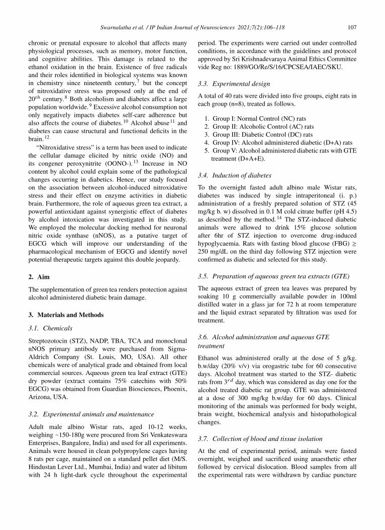
4.1.3. Status of nitroxidative stress markers, antioxidantenzymes and signalling moleculesOur results presented that an augmented level ofmalondialdehyde, protein carbonyls, peroxynitrites,and nitric oxide was observed in all experimental groups(AC, DC, D+A, D+A+E). A significant decrease wasnoticed among alcohol administered diabetic groupsstudied in comparison with normal controls and otherstudied groups (AC, DC, and D+A+E). The mean valuesof malondialdehyde showed significant change betweenexperimental groups except D+A+E rats (Figure 3).Data shown in Figure 4 that significantly diminishedconcentrations of reduced glutathione content, andantioxidant enzymes (GPx, SOD, and CAT) activities inall studied group rats and significant change was reportedamong experimental groups except levels of reducedglutathione.
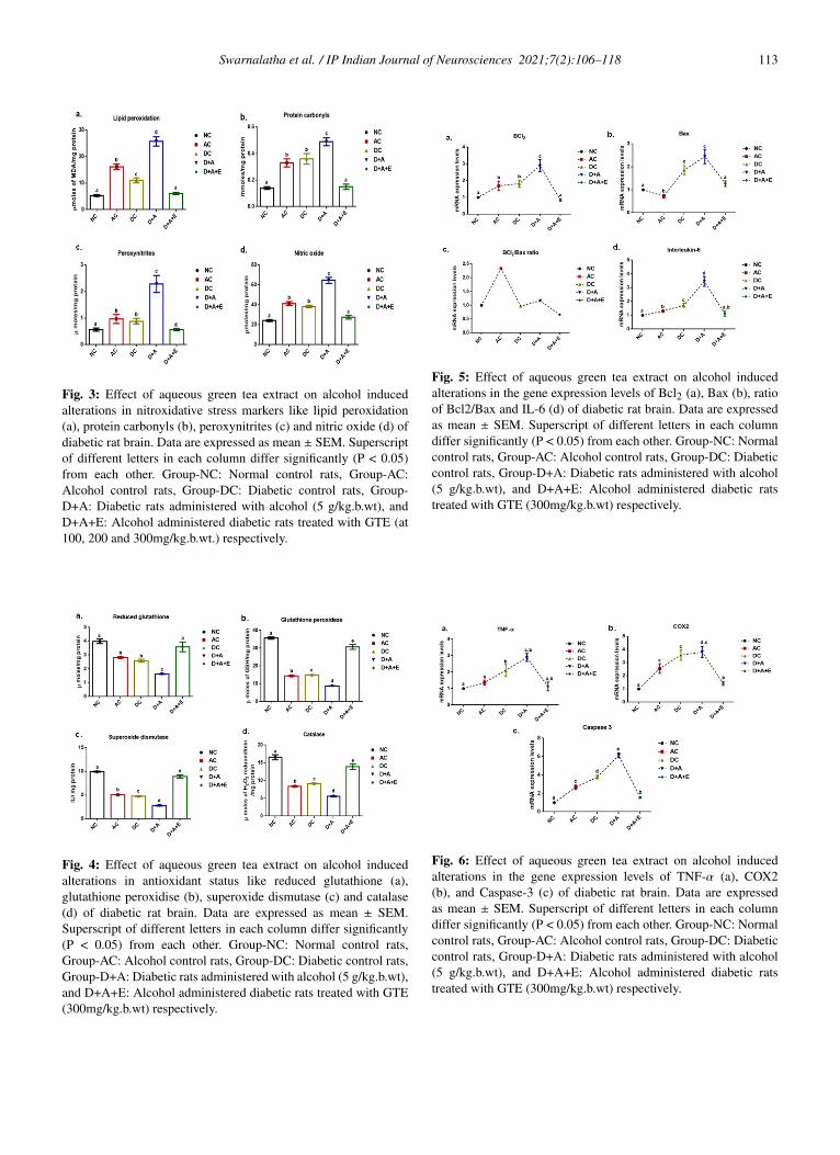
In the current study, all experimental rats demonstratedsignificant increase in the expression levels of Bcl2, Bax,caspase 3, COX2, TNF-α, and IL-6 compared to controlsrats. The mean values of COX2 and caspase 3 showedsignificant difference and remaining variables (Bcl2, Bax,IL-6, and TNF-α) did not showed significant change inD+A+E rats. There is no significant change between ACand DC group rats in the levels of Bcl2, COX2, and TNF-α,while significant difference was noticed in the mean valuesof Bax, IL-6, and caspase 3. The D+A+E rats showed asignificant difference in the expression levels Bcl2 and Baxwith NC, AC, DC and D+A rat groups but not with NCrats. And these D+A+E rats showed a DC and D+A ratsbut not with NC an AC rats. Similarly, they are significantlydifferent in the expression of TNF-α with the D+A rats butnot with the NC, AC and DC rats as well. But D+A+ Erats are statistically significant in expressing the COX2 andcaspase 3, with all the other groups viz., NC, AC, DC andD+A rats (Figures 5 and 6).
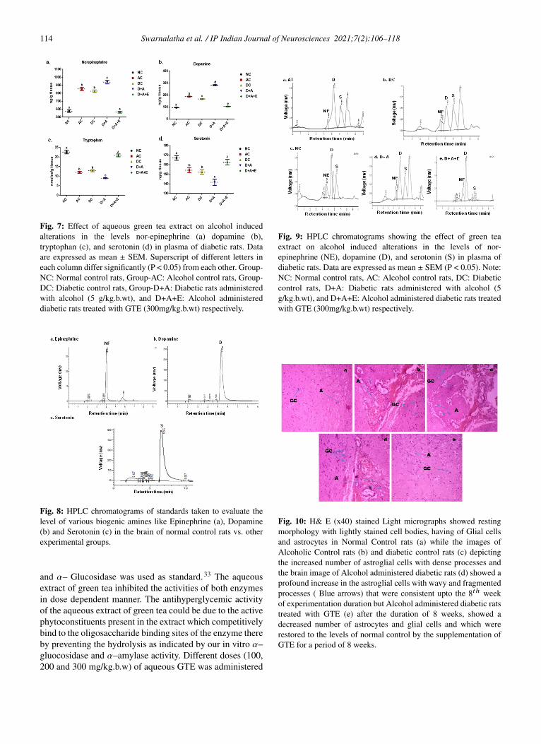
4.1.4. Levels of bioaminesOur results noticed (Figure 7) that, the levels of dopamineand norepinephrine were higher in AC, DC, and D+Acompared to control rats. The mean values of dopamineexhibited significant difference among experimental groupsand D+A groups showed statistical significant change withthe other groups in the mean values of norepinephrine.A significant reduction in the levels of serotonin andtryptophan was reported in studied groups (AC, DC, andD+A) in comparison with healthy rats. The D+A ratsshowed significant change with remaining all groups andthere is no significant difference between AC and DCgroups.
Data (Figures 8 and 9) indicated the range of retentiontime of standard nor epinephrine is 3.5-5.4 min, serotoninis 3.6-6.2 min, and 3.8-9.2 min respectively and appeareda chromatogram peak at 4.01 min of nor epinephrine, 5.34
min of dopamine, 6.57 min of serotonin respectively. Thegroup I rats reported that chromatogram peak at retentiontime at 4.99 min of nor epinephrine, at 5.40 min ofdopamine, at 6.26 min of serotonin. The chromatogram peakat 5.77 min of nor epinephrine, at 6.10 min of dopamine,at 6.90 min of serotonin was observed in group II rats.The nor epinephrine shown peak at retention time at 4.99min of nor epinephrine, at 5.40 min of dopamine, at 6.26min of serotonin in group III rats. The chromatograms ofnor epinephrine, dopamine, and serotonin at 4.39 min, 5.14min, and 5.91 min respectively in the brain tissue of groupIV rats. In the brain tissue, nor- epinephrine disclosed achromatogram peak at 4.50 min of retention time, dopamineat 5.04 min, and serotonin at 6.46 min in group V rats.
4.2. Histopathology
Photomicrographs of rat brain sections in differentexperimental groups stained with Hematoxylin and Eosin(10X), (a) Histopathological sections of brain in control ratrevealed normal cellular structure with normal astrocyte andglial cells, (b,d) Rat treated with and alcohol administereddiabetic rat respectively, showing cellular structure withreduced number of astrocytes and glial cells, (c) STZinduced diabetic control rat revealed decreased astrocytenumber and brain alterations, and (e) Brain of rats treatedwith GTE (300 mg/kg.b.w) after induction of diabetesto the alcohol treated rats , showed restoration in glialand astrocyte number to compare with normal control rats(Figure 10).
4.3. Docking studies
The binding affinities and energy profiles of both titleand reference compounds towards the active site ofthe enzyme are summarized in Table 1. The presentinvestigation demonstrated that the title compound will bethe promising next generation anti-diabetic drug, whichcan be effectively used in the treatment of manifestationof diabetic complications. The 3D bonding images of thecompounds were shown in Figure 11. Meanwhile, themolecular docking study demonstrated that EGCG is ableto strongly interact the nNOS protein with a high bindingaffinity (-6.4 kcal/mol) via strong hydrogen bindings to Asn606 and glutamic acid 661 and a hydrophobic interactionto the hydrophobic pocket of Lysine 631. The stronginteraction predicted between EGCG to nNOS may lead toinhibition of nNOS by EGCG, thus supporting the downregulation of it was observed in this study (Figure 11 a,b).
4.3.1. ADMETWe have determined the compliance of the ligands tothe Lipinski’s ‘rule of five’. According to this rule, poorabsorption or permeation is more likely when there aremore than five hydrogen bond donors, ten hydrogen

Swarnalatha et al. / IP Indian Journal of Neurosciences 2021;7(2):106–118 111
bond acceptors, the molecular weight is greater than500 and the calculated log p (logarithmic ratio of theoctanol–water partitioning coefficient) is greater than 5.Molecules violating more than one of these parameters mayhave problems with bioavailability and a high probabilityof failure to display drug-likeness. Further, the topologicalpolar surface area (TPSA) which is another key propertyin estimating drug bioavailability was also calculated.Generally, compounds with a TPSA ≥140A◦ are thought tohave low bioavailability. As shown in Table 2, the compound(EGCG) complies with these rules. Moreover, the EGCGexhibited a greater percentage of absorption (%ABS)with 98.66% whereas the RTZ is with 90.66%. Hence,theoretically, this compound should have good passive oralabsorption and drug likeness. In addition to this, differentADME predictions such as BBB penetration, percentage ofhuman intestinal absorption (HIA %), CaCO2 permeabilityand percentage of plasma protein binding (PPB %) werepredicted. Analysing the ADME predictions (Table 3), itwas observed that the compound has shown high HIA%values with 99.24 % and is well absorbed when comparedto the RTZ (94.16%). The CaCO2 cell permeability valueof the EGCG is good, that is 25.14 nm/s. Furthermore,the compound was strongly bound to plasma proteins with%PPB penetration more than 94 %. In addition, it was foundto have good penetration (0.226) to the CNS through theBBB. From all these parameters, it can be observed that,theoretically, this compound exhibited good absorption andbioavailability with good permeability through the BBBwhen compared with the reference compound, RTZ.
5. Discussion
The present study used a dose of 5g/kg. b. w/day alcohol(20% v/v) that has been proved to be neurotoxic asit selectively increased nitroxidative stress in diabeticrats. Comorbid existence of diabetes and alcoholismworsen health status, productivity and other societal costs.The current study examined the effects of GTE onnitro/oxidative stress in brain of alcohol treated diabeticrats. Diabetes causes dysregulation of various metabolicprocesses, especially the glucose homeostasis. In addition,neurobiological profiles of alcoholism are linked to theeffects of a disruption of glucose homeostasis and ofinsulin resistance, which are affected by altered appetite thatregulates the peptides and neurotrophic factors.29 Heavyalcohol consumption increases ROS production and maybe a mechanism of pancreatic β-cells dysfunction in type2 diabetes mellitus. Glucose metabolism play an importantrole related in neuroenergetics, neurotransmission, energystorage, biosynthesis and oxidative defence. Thus, tightregulation of glucose metabolism is critical for normal brainphysiology.30 The damage caused by alcohol abuse anddiabetes can become debilitating and cumulative.
Fig. 1: Effect of aqueous green tea extract on alcohol inducedalterations in percentage of α-amylase, α-glucosidase inhibition(a, b), fasting blood glucose (c) and glycogen levels (d) of diabeticbrain. Data are expressed as mean ± SEM. Superscript of differentletters in each column differ significantly (P < 0.05) from eachother. Group-NC: Normal control rats, Group-AC: Alcohol controlrats, Group-DC: Diabetic control rats, Group-D+A: Diabetic ratsadministered with alcohol (5 g/kg.b.wt), and D+A+E: Alcoholadministered diabetic rats treated with GTE (at 100, 200 and300mg/kg.b.wt.) respectively.
Fig. 2: Effect of aqueous green tea extract on alcohol inducedalterations in brain weight (a), body weight (b) and the percentagechange in the ratio of brain to body weight (c) of diabetic rat. Dataare expressed as mean ± SEM. Superscript of different letters ineach column differ significantly (P < 0.05) from each other. Group-NC: Normal control rats, Group-AC: Alcohol control rats, Group-DC: Diabetic control rats, Group-D+A: Diabetic rats administeredwith alcohol (5 g/kg.b.wt), and D+A+E: Alcohol administereddiabetic rats treatedwithGTE(300mg/kg.b.wt)respectively.

112 Swarnalatha et al. / IP Indian Journal of Neurosciences 2021;7(2):106–118
Table 1: Bonding characterization of EGCG and RTZ againstnNOS protein
nNOS-6NG1Compound Binding
energy(K calmol− )
Binding interaction Bond Length(AO ) Bond Angle (o ) Bond Type
RTZ - 5.1 Pro 343 CZ . . . .HN 1.8 120.3 H- don
EGCG - 6.4Lys 631 CA . . . .HO 2.2 96.3 H- accAsn 606 CZ . . . HO 2.3 118.7 H- donGlu 661 CZ . . . .HN 2.1 124.0 H- don
Table 2: Physicochemical properties of EGCG and RTZ
Compound Mol.Wt.a
Mol.Vol.b
n-ROTBc
n-OHNHDonor
d
n-ONacceptor
e
miLog Pf
TPSA(A◦2)g
LipinskiVio.
%ABS h
Rule ≤ 500 ≤ 5 ≤ 10 ≤ 5 ≤ 1EGCG 458.37 396.6 6 0 4 2.12 99.86 0 98.66RTZ 357.42 338.4 5 1 6 3.64 96.14 0 90.66
aMolecular weight; bMolecular volume; cNumber of rotatable bonds dNumber of hydrogen bond donors; eNumber of hydrogen bond acceptors;f Logarithmic ratio of the octanol–water partitioning coefficient; gTopological polar surface Area; hPercentage of absorption; %ABS = 109 - (0.345 9TPSA).
Table 3: Prediction of pharmacokinetic properties of EGCG and RTZ
Compound CaCO2a
permeabilityHIAb (%) PPBc (%) BBBd
(Cbrain/Cblood)EGCG 25.14 99.24 94.16 0.226RTZ 20.80 94.16 90.28 0.620
aCalcium carbonate; bHuman intestinal absorption; cPlasma protein binding; dBlood–brain barrier.
Table 4: Primer sequence of related genes
Target genes(5’→3’)forward/reverse
Primer Primer sequence Primerforward/reverse
Primer sequence
GAPDH Forward GAAGACTGTGGGACCCAGATReverse AAGGTGGATGATGTTCCAGG
Caspase-3 Forward AAATTCAAGGGACGGGTCATReverse ATTGACACAATACACGGGATCTGT
TNF-α Forward AAGCCTGTAGCCCATGTTGTReverse CAGATAGATGGGCTCATACC
IL-6 Forward TAGCCGCCCCACACAGACAGReverse GGCTGGCATTTGTGGTTGGG
Bcl-2 Forward CTGTACGGCCCCAGCATGGCGReverse GCTTTGTTTCATGGTACATC
Bax Forward GGGAATTCTGGAGCTGCAGAGGATGATTCox-2 Reverse GCGGATCCAAGTTGCCATCAGCAAACAT
Forward ACACCTTCAACATTGAAGACCReverse ATCCCTTCACTAAATGCCCTC
Hyperglycemia generates abnormally high levels offree radicals by auto-oxidation of glucose and by proteinglycation, that are also participate in the developmentof oxidative stress and the inflammatory status.31 Hence,it was found to be very difficult to manage andcure this disease effectively by using the available oralhypoglycaemic drugs or insulin. Therefore, the focus ofscientists shifted towards the development of the naturalherbal medicine with high therapeutic potential and less
or no toxic effect. In this connection, the aqueous extractof green tea leaves have been claimed to possess anti-diabetic properties by traditional healers.32 In the presentstudy, we evaluated the α-glucosidase and α-amylaseinhibitiory activity and antidiabetic activity of the GTEin alcohol treated diabetic rats. α– Gluocosidase andα–amylase are the carbohydrate hydrolyzing enzymesinvolved in the digestion of carbohydrates. Ascarbose, anoral hypoglycemic agent which inhibits both α–amylase
Swarnalatha et al. / IP Indian Journal of Neurosciences 2021;7(2):106–118 113
Fig. 3: Effect of aqueous green tea extract on alcohol inducedalterations in nitroxidative stress markers like lipid peroxidation(a), protein carbonyls (b), peroxynitrites (c) and nitric oxide (d) ofdiabetic rat brain. Data are expressed as mean ± SEM. Superscriptof different letters in each column differ significantly (P < 0.05)from each other. Group-NC: Normal control rats, Group-AC:Alcohol control rats, Group-DC: Diabetic control rats, Group-D+A: Diabetic rats administered with alcohol (5 g/kg.b.wt), andD+A+E: Alcohol administered diabetic rats treated with GTE (at100, 200 and 300mg/kg.b.wt.) respectively.
Fig. 4: Effect of aqueous green tea extract on alcohol inducedalterations in antioxidant status like reduced glutathione (a),glutathione peroxidise (b), superoxide dismutase (c) and catalase(d) of diabetic rat brain. Data are expressed as mean ± SEM.Superscript of different letters in each column differ significantly(P < 0.05) from each other. Group-NC: Normal control rats,Group-AC: Alcohol control rats, Group-DC: Diabetic control rats,Group-D+A: Diabetic rats administered with alcohol (5 g/kg.b.wt),and D+A+E: Alcohol administered diabetic rats treated with GTE(300mg/kg.b.wt) respectively.
Fig. 5: Effect of aqueous green tea extract on alcohol inducedalterations in the gene expression levels of Bcl2 (a), Bax (b), ratioof Bcl2/Bax and IL-6 (d) of diabetic rat brain. Data are expressedas mean ± SEM. Superscript of different letters in each columndiffer significantly (P < 0.05) from each other. Group-NC: Normalcontrol rats, Group-AC: Alcohol control rats, Group-DC: Diabeticcontrol rats, Group-D+A: Diabetic rats administered with alcohol(5 g/kg.b.wt), and D+A+E: Alcohol administered diabetic ratstreated with GTE (300mg/kg.b.wt) respectively.
Fig. 6: Effect of aqueous green tea extract on alcohol inducedalterations in the gene expression levels of TNF-α (a), COX2(b), and Caspase-3 (c) of diabetic rat brain. Data are expressedas mean ± SEM. Superscript of different letters in each columndiffer significantly (P < 0.05) from each other. Group-NC: Normalcontrol rats, Group-AC: Alcohol control rats, Group-DC: Diabeticcontrol rats, Group-D+A: Diabetic rats administered with alcohol(5 g/kg.b.wt), and D+A+E: Alcohol administered diabetic ratstreated with GTE (300mg/kg.b.wt) respectively.
114 Swarnalatha et al. / IP Indian Journal of Neurosciences 2021;7(2):106–118
Fig. 7: Effect of aqueous green tea extract on alcohol inducedalterations in the levels nor-epinephrine (a) dopamine (b),tryptophan (c), and serotonin (d) in plasma of diabetic rats. Dataare expressed as mean ± SEM. Superscript of different letters ineach column differ significantly (P < 0.05) from each other. Group-NC: Normal control rats, Group-AC: Alcohol control rats, Group-DC: Diabetic control rats, Group-D+A: Diabetic rats administeredwith alcohol (5 g/kg.b.wt), and D+A+E: Alcohol administereddiabetic rats treated with GTE (300mg/kg.b.wt) respectively.
Fig. 8: HPLC chromatograms of standards taken to evaluate thelevel of various biogenic amines like Epinephrine (a), Dopamine(b) and Serotonin (c) in the brain of normal control rats vs. otherexperimental groups.
and α– Glucosidase was used as standard.33 The aqueousextract of green tea inhibited the activities of both enzymesin dose dependent manner. The antihyperglycemic activityof the aqueous extract of green tea could be due to the activephytoconstituents present in the extract which competitivelybind to the oligosaccharide binding sites of the enzyme thereby preventing the hydrolysis as indicated by our in vitro α–gluocosidase and α–amylase activity. Different doses (100,200 and 300 mg/kg.b.w) of aqueous GTE was administered
Fig. 9: HPLC chromatograms showing the effect of green teaextract on alcohol induced alterations in the levels of nor-epinephrine (NE), dopamine (D), and serotonin (S) in plasma ofdiabetic rats. Data are expressed as mean ± SEM (P < 0.05). Note:NC: Normal control rats, AC: Alcohol control rats, DC: Diabeticcontrol rats, D+A: Diabetic rats administered with alcohol (5g/kg.b.wt), and D+A+E: Alcohol administered diabetic rats treatedwith GTE (300mg/kg.b.wt) respectively.
Fig. 10: H& E (x40) stained Light micrographs showed restingmorphology with lightly stained cell bodies, having of Glial cellsand astrocytes in Normal Control rats (a) while the images ofAlcoholic Control rats (b) and diabetic control rats (c) depictingthe increased number of astroglial cells with dense processes andthe brain image of Alcohol administered diabetic rats (d) showed aprofound increase in the astroglial cells with wavy and fragmentedprocesses ( Blue arrows) that were consistent upto the 8th weekof experimentation duration but Alcohol administered diabetic ratstreated with GTE (e) after the duration of 8 weeks, showed adecreased number of astrocytes and glial cells and which wererestored to the levels of normal control by the supplementation ofGTE for a period of 8 weeks.

Swarnalatha et al. / IP Indian Journal of Neurosciences 2021;7(2):106–118 115
Fig. 11: a: Molecular structures of nNOS, EGCG andRosiglitazone; b: Predicted bonding interactions between the titlecompound with ASN 606 (Yellow); GLU 661 (red) and LYS 631(orange) and standard with PRO 343 (blue) of nNOS. Positionof EGCG were showed in the hydrophobic pocket of nNOS andnNOS has deep hydrophobic grooves suitable for the binding ofEGCG.
to check its anti-diabetic activity in alcohol treated diabeticrats. After the treatment, it was found to decrease the fastingblood glucose levels significantly with the concentration of300mg/kg.b.w, at the end of 8 weeks. Highest anti-diabeticactivity at these concentrations could be due to the bioactivecompounds present in the extract that might improve theinsulin secretion or mimic the insulin action. Treatment withaqueous GTE has lowered the blood glucose concentrationsto normal levels in D+A rats as compared to the diabeticcontrols.
Glycemic equilibrium plays a very important role inthe prooxidant/antioxidant balance. A critical biomarkerof oxidative stress is lipid peroxidation and nitrosativestress is peroxynitration which are the most explored areasof research when it comes to ROS/RNS34 respectively.Involvement of oxidative/nitrosative stress in alcohol abuseddiabetic complications is the basis of the developmentof adjunct therapies with antioxidant. Alcohol anddiabetes produces disturbances in the lipid profile makingthe cells more susceptible to lipid peroxidation, andprotein nitration, critical biomarkers of nitroxidativestress, finally leading to the alterations in the levelsof membrane lipid composition. Our results showedsignificantly increased lipid peroxidation product, MDAand protein carbonyl levels of brain in chronically alcoholtreated diabetic rats. Membrane proteins are importanttargets for oxidative/nitrosative attack because ROS/RNSmight cleaves multiple bonds, oxidizes amino acid residues,augments fragmentation and aggregation of proteins andthe rate of proteolysis.35 Thus protein oxidation isconsidered one of several alcohol-related modificationsby ROS. Increased protein carbonyl levels are a majorindex of protein oxidation. There have been a relativelysmall number of reports that systematically dealt withidentification and functional characterization of nitratedproteins in various sub cellular organelles, includingmitochondria, especially under conditions with increasednitroxidative stress. Peroxynitrite, a product of superoxide
anion radical reaction with nitric oxide, is a major oxidantin pathological conditions associated with oxidative stressincluding diabetes36 and preventing peroxynitrite formationcould be an effective target for the treatment of diabeticneuropathy.37 Production and accumulation of ROS/RNScaused loss of membrane integrity and function due toincreased membrane peroxidation and nitration. Our resultsshowed significantly augmented the levels of MDA andprotein carbonyls, nitric oxide and peroxynitrite of brainof D+A+E rats in comparison with DC and AC rats.Reduced glutathione (GSH), an important endogenous non-enzymatic antioxidant was decreased in diabetic brain bychronic alcohol exposure, and this may influence the basallevels of other antioxidant enzymes in the brain of diabeticrats.
Brain is more vulnerable to nitroxidative damage thanany other organ in the body as it consumes nearly 20%of oxygen from the body, has low concentrations ofantioxidants and high content of polyunsaturated fatty acidsand catecholamines that are easily oxidised.38 Experimentalstudies show that polyunsaturated fatty acids in cellmembrane are extremely prone to attack by free radicalsdue to the presence of multiple bonds.39 Diabetes andalcoholism induces alterations in activity of enzymes GPx,SOD and CAT. Any alteration in their levels will makethe cells prone to oxidative stress and hence cell injury.Glycogen levels differ enormously from one tissue toanother. The factors which maintain the characteristic levelin a given tissue are unknown.40 Ethanol is known toperturb the carbohydrate metabolism and alter the contentof glycogen, a highly branched polysaccharide representsthe major energy store in the brain.41 At the end ofexperimental period, brain glycogen concentrations werereduced by alcohol, in alcohol treated diabetic rat groupsthat may contributed to changes in the phosphorylationrate of glycogen synthase. Altogether, the present resultsindicate that diabetes with alcohol abused condition isassociated with impaired brain glycogen metabolism. Toour knowledge, this is the first study to report glycogenlevels in an alcohol treated diabetic animal model.Furthermore, these alterations were restored to the normalby supplementation of green tea extract. Alcohol selectivelyaffects nNOS activity in different brain cells. Alterationsin the nNOS gene may constitute a genetic risk factorfor the vulnerability of high voluntary alcohol intake andsubsequent alcohol dependence.42 Western blotting showeda significant increase in the expression of nNOS in diabeticrat brain after the treatment with alcohol. These findingsindicated that an impairment of nNOS expression is mainlydue to the absence of insulin and alcohol may exert its toxiceffects via a mechanism of altered nitric oxide availabilityfrom nitric oxide synthase.43
Reversion by GTE treatment was also obtained inthe protein expression of nNOS, which could be due to
116 Swarnalatha et al. / IP Indian Journal of Neurosciences 2021;7(2):106–118
catechins that inhibits the enzyme activities by binding tothe active site nNOS, and thus decreasing the percentageof its active dimeric form, reducing the enzymaticcoupling and affecting the RNS generation during thereaction. The synthesis of a number of neurotransmittersinvolved in the regulation of various physiological functionsand behavioural patterns depends largely on precursoravailability. Bcl-2 and Bax proteins, are functionallyopposed and hence, in this study, we therefore assessedthe expression of Bcl-2 and Bax at mRNA level as wellas the Caspase-3, in order to investigate the occurrenceof apoptosis in the brain of alcohol treated diabetic rats.In the present study, we observed that, chronic alcoholadministration significantly increases the magnitude ofanti-apoptotic Bcl2 and apoptotic Bax mRNA in theD+A rat brain which was coupled with increase incaspase-3 activity, IL-6, TNF-α, and COX2, suggests therole of cytokines in alcohol-induced neuroinflammationunder diabetic condition. Increased blood-glucose level asobserved in this study and activation of p53 transcriptionfactors and caspase 3 might be the potential causes toreduce the IGF-I level and sequential apoptotic cell death inthe STZ-induced diabetic brain following alcohol treatmentas reported in this study. Results found that green teacatechins especially EGCG, a potent inhibitor of the anti-apoptotic proteins attenuated the enhanced levels of pro-inflammatory cytokines to normal, which are in accordwith earlier reports.44 Therefore, mechanism underlyingthe neuroprotective effects observed in our study mightbe due to its antioxidant, anti-inflammatory and neuro-modulatory effects of EGCG. In diabetic animals treatedwith alcohol, the basal brain concentrations of serotoninwere decreased and those of norepinephrine and dopaminewere increased compared to alcohol and diabetic controls.Since uptake of the precursor’s tryptophan and tyrosinefrom the blood is chronically reduced, it is likely that long-term adjustments of neurotransmitter metabolism occur inthe diabetic brain.45
Glial cells are susceptible to any change or disruption inthe brain. In response to adverse state, glial cells transformtheir morphology and proliferate to combat the balefulcondition.46 Following any brain insult, glial cells getactivated and exhibit morphological transformations fromresting to activated, increase in their cell population andsecrete an array of inflammatory molecules.47 In the currentstudy, hematoxylin and eosin stained alcoholic, diabetic andalcohol treated diabetic brain presented a marked incrementin activation and population of astrocytes and glial cells atthe end of 8th week. This activation might be a customizedapproach to modulate the effect of excessive cell deathfollowing diabetes under alcoholism and moreover, theseastrocytes in the healthy brain regulate neurotransmitterand neurovascular dynamics as well which has beendemonstrated to be disturbed in alcohol treated diabeticrats and in turn improved by the treatment with GTE.
Meanwhile, molecular docking analysis was carried out forthe title compound with selective pharmacological targetsuch as nNOS, which is a suitable target for anti-diabeticactivity. The docking results revealed that, the compoundEGCG has shown higher binding energy than the referencedrug, Rosiglitazone. Interestingly, raw brain-to-body weightratio is an important and useful tool for comparingencephalization within species or between closely relatedspecies. Recent studies have been demonstrated thatincreased brain insulin sensitivity is associated with the lossof weight during lifestyle intervention and also linked withthe distribution of body fat.48
6. Conclusion
With the above results, we concluded that, the interplaybetween alcohol and diabetes provided the prolongedand persistent hyperglycemia, nitroxidaive stress, reducedantioxidant status in addition to the glial activation and thuscellular degeneration. And also concluded that apoptosisplays a vital role in the alcohol exacerbated diabeticpathogenesis possibly by increasing the ratio of Bcl2/Baxalong with the caspase-3, that might in turn facilitates thememory and cognition deficits. This study also revealedthat, the ameliorative approach of EGCG could be seenin the down regulation of nNOS, a major causative ofnitroxidative stress. Additionally, the molecular dockingresults confirmed that, the compound EGCG, showed ahigher binding affinity and energy than the reference drugand hence, 300 mg/kg.b.w of EGCG has been provedas better prospective and veritable source that offers antherapeutic approach for the treatment of alcohol abusedcomplications in diabetes.
7. Acknowledgements
The author is grateful for DST-Inspire Fellowshipprogramme for providing the financial assistance from2012-2017 to carry out this work. But currently, we are notreceiving any funding.
8. Conflict of Interest
The authors declare that there is no conflict of interests
9. Source of Funding
None.
References1. Muriach M, Flores-Bellver M, Romero FJ, Barcia JM. Diabetes and
the Brain: Oxidative Stress, Inflammation, and Autophagy. Oxid MedCell Longev . 2014;doi:10.1155/2014/102158.
2. Dwivedia DK, Kumara D, Kwatraa M, Pandeya SN, ChoubeyaP, Lahkarab MM, et al. Voluntary alcohol consumptionexacerbated high fat diet-induced cognitive deficits by NF-κB-calpaindependent apoptotic cell death in rat hippocampus: Ameliorative
Swarnalatha et al. / IP Indian Journal of Neurosciences 2021;7(2):106–118 117
effect of melatonin. Biomed Pharmacother. 2018;108:1393–403.doi:10.1016/j.biopha.2018.09.173.
3. International Diabetes Federation. . In: IDF Diabetes Atlas. 9th Edn.;2019.
4. Saeedi P, Petersohn I, Salpea P, Malanda B, Karuranga S, Unwin N,et al. Global and regional diabetes prevalence estimates for 2019 andprojections for 2030 and 2045: Results from the International DiabetesFederation Diabetes Atlas, 9 th edition. Diabetes Res Clin Pract.2019;157:107843. doi:10.1016/j.diabres.2019.107843.
5. World Health Organization. Global status report on alcohol and health2018. Geneva: World Health Organization; 2018.
6. Zimatkin S, Deitrich R. Ethanol metabolism in the brain. Addict Biol. 1997;2(4):387–99. doi:10.1080/13556219772444.
7. Commoner B, Townsend J, Pake G. Free radicals in biologicalmaterials. Nature. 1954;174(4432):689–91. doi:10.1038/174689a0.
8. Chan JYH, Chan SHH. Differential impacts of brain stemoxidative stress and nitrosative stress on sympathetic vasomotor tone.Pharmacol Ther. 2019;201:120–36.
9. Hodge AM, Dowse GK, Collins VR, Zimmet PZ. Abnormal GlucoseTolerance and Alcohol Consumption in Three Populations at HighRisk of Non-lnsulin-dependent Diabetes Mellitus. Am J Epidemiol. 1993;137(2):178–89. doi:10.1093/oxfordjournals.aje.a116658.
10. Engler PA, Ramsey SE, Smith RJ. Alcohol use of diabetes patients:The need for assessment and intervention. Acta Diabetol. 2013;50:93–9.
11. Harper C. The Neuropathology of Alcohol-Related Brain Damage.Alcohol Alcohol. 2009;44(2):136–40. doi:10.1093/alcalc/agn102.
12. Sato N, Morishita R. Brain Alterations and ClinicalSymptoms of Dementia in Diabetes: AÎ2/Tau-Dependent andIndependent Mechanisms. Front Endocrinol. 2014;5:1–8.doi:10.3389/fendo.2014.00143.
13. Calabrese V, Mancuso C, Marco CD, Stella AMG, and DAB.Nitric oxide and cellular stress response in brain aging andneurodegenerative disorders. Oxid Stress Neurodegener Disord. 2007;.
14. Kotha P, Badri KR, Nagalapuram R, Allagadda R, Chippada AR.Anti-Diabetic Potential of the Leaves of Anisomeles malabaricain Streptozotocin Induced Diabetic Rats. Cell Physiol Biochem.2017;43(4):1689–702. doi:10.1159/000484030.
15. Ademiluyi AO, Oboh G. Soybean phenolic-rich extracts inhibit key-enzymes linked to type 2 diabetes (α-amylase and α-glucosidase) andhypertension (angiotensin I converting enzyme) in vitro. Exp ToxicolPathol. 2013;65(3):305–9. doi:10.1016/j.etp.2011.09.005.
16. Shai LJ, Masoko P, Mokgotho MP, Magano SR, Mogale AM, BoaduoN, et al. Yeast alpha glucosidase inhibitory and antioxidant activitiesof six medicinal plants collected in Phalaborwa, South Africa. S Afr JBot. 2010;76(3):465–70. doi:10.1016/j.sajb.2010.03.002.
17. Buege JA, Aust SD. Microsomal lipid peroxidation. In: Microsomallipid peroxidation. In: Methods in Enzymology. vol. 52. New York:Academic Press; 1978. p. 302–10.
18. Levine RL, Garland D, Oliver CN, Amici A, Climent I, Lenz AG, et al.Determination of carbonyl content in oxidatively modified proteins.Methods Enzymol. 1990;186:464–78.
19. Sastry KVH, Moudgal RP, Mohan J, Tyagi JS, Rao GS.Spectrophotometric Determination of Serum Nitrite and Nitrateby Copper–Cadmium Alloy. Anal Biochem . 2002;306(1):79–82.doi:10.1006/abio.2002.5676.
20. Beckman JS, Ischiropoulos H, Zhu L, van der Woerd M, Smith C,Chen J, et al. Kinetics of superoxide dismutase- and iron-catalyzednitration of phenolics by peroxynitrite. Arch Biochem Biophys .1992;298(2):438–45. doi:10.1016/0003-9861(92)90432-v.
21. Ellman GL. Tissue sulfhydryl groups. Arch Biochem Biophys .1959;82(1):70–7. doi:10.1016/0003-9861(59)90090-6.
22. Rotruck JT, Pope AL, Ganther HE, Swanson AB, Hafeman DG,Hoekstra WG, et al. Selenium: Biochemical Role as a Componentof Glutathione Peroxidase. Science. 1973;179(4073):588–90.doi:10.1126/science.179.4073.588.
23. Kakkar P, Das B, Viswanathan PN. A modified spectrophotometricassay of superoxide dismutase. Indian J Biochem Biophys.1984;21:130–32.
24. Aebi H. Catalase in vitro. Methods Enzymol. 1984;105:121–6.doi:10.1016/s0076-6879(84)05016-3.
25. Kong J, Shepel PN, Holden CP, Mackiewicz M, Pack AI, GeigerJD, et al. Brain Glycogen Decreases with Increased Periods ofWakefulness: Implications for Homeostatic Drive to Sleep. J Neurosci.2002;22(13):5581–7. doi:10.1523/jneurosci.22-13-05581.2002.
26. Morris GM, Huey R, Lindstrom W, Sanner MF, Belew RK, GoodsellDS, et al. AutoDock4 and AutoDockTools4: Automated docking withselective receptor flexibility. J Comput Chem. 2009;30(16):2785–91.doi:10.1002/jcc.21256.
27. Zhao YH, Abraham MH, Le J, Hersey A, Luscombe CN, Beck G,et al. Rate-limited steps of human oral absorption and QSAR studies.Pharm Res. 2002;19:1446–57.
28. Bancroft JD, Gamble M. Theory and practice of histology techniques.In: 6th Edn. Philadelphia, PA: Churchill Livingstone Elsevier; 2008.p. 83–134.
29. Kim SJ, Kim DJ. Alcoholism and diabetes mellitus. Diabetes MetabJ. 2012;36:108–15.
30. Thakur AK, Tyagi S, Shekhar N. Comorbid brain disordersassociated with diabetes: therapeutic potentials of prebiotics,probiotics and herbal drugs. Transl Med Commun. 2019;4(1):1–13.doi:10.1186/s41231-019-0043-6.
31. Bonnefont-Rousselot D, Beaudeux JL, Therond P, Peynet J, LegrandA, Delattre J, et al. Diabetes mellitus, oxidative stress and advancedglycation end products. Ann Pharm Fr. 2004;62:147–57.
32. Haidaria F, Omidianb K, Rafieic H, Zareid M, and MMS. Green tea(camellia sinensis) supplementation to diabetic rats improves serumand hepatic oxidative stress markers. Iran J Pharm Res. 2013;12:109–14.
33. Robyt JF. Inhibition, activation, and stabilization of α-amylase familyenzymes. Bratislava. Biologia. 2005;16:17–26.
34. Gasparovic AC, Zarkovic N, Zarkovic K, Semen K, KaminskyyD, Yelisyeyeva O, et al. Biomarkers of oxidative and nitro-oxidative stress: conventional and novel approaches. Br J Pharmacol.2017;174(12):1771–83. doi:10.1111/bph.13673.
35. Berlett BS, Stadtman ER. Protein Oxidation in Aging, Disease,and Oxidative Stress. J Biol Chem. 1997;272(33):20313–6.doi:10.1074/jbc.272.33.20313.
36. Pacher P, Beckman JS, Liaudet L. Nitric Oxide and Peroxynitritein Health and Disease. Physiol Rev. 2007;87(1):315–424.doi:10.1152/physrev.00029.2006.
37. Stavniichuka R, Shevalyea H, Lupachyka S, Obrosova A, GrovesbJT, Obrosovaa IG, et al. Peroxynitrite and protein nitration in thepathogenesis of diabetic peripheral neuropathy. Diabetes Metab ResRev. 2014;30:669–78.
38. Hong JH, Kim MJ, Park MR, Kwag OG, Lee IS, Byun BH, et al.Effects of vitamin E on oxidative stress and membrane fluidityin brain of streptozotocin-induced diabetic rats. Clin Chim Acta.2004;340:107–15.
39. Butterfield D, Koppal T, Howard B, Subramaniam R, Hall N, HensleyK, et al. Structural and Functional Changes in Proteins Inducedby Free Radical-mediated Oxidative Stress and Protective Actionof the Antioxidants N-tert-Butyl-alpha-phenylnitrone and VitaminEa. Ann NY Acad Sci. 1998;854:448–62. doi:10.1111/j.1749-6632.1998.tb09924.x.
40. Nelson SR, Schulz DW, Passonneau JV, Lowry OH. Control ofglycogen levels in brain. J Neurochemistry. 1968;15(11):1271–79.doi:10.1111/j.1471-4159.1968.tb05904.x.
41. Garriga J, Sust M, Cussó R. Regional distribution of glycogen,glucose and phosphorylated sugars in rat brain after intoxicating dosesof ethanol. Neurochem Int. 1994;25(2):175–81. doi:10.1016/0197-0186(94)90037-x.
42. Spanagel R, Siegmund S, Cowen M, Schroff KC, Schumann G,Fiserova M, et al. The Neuronal Nitric Oxide Synthase Gene IsCritically Involved in Neurobehavioral Effects of Alcohol. J Neurosci.2002;22(19):8676–83. doi:10.1523/jneurosci.22-19-08676.2002.
43. Gerlach M, Blum-Degen D, Ransmayr G, Leblhuber F, Pedersen V,Riederer P, et al. Expression, but not activity, of neuronal nitricoxide synthase is regionally increased in the alcoholic brain. Alcohol

118 Swarnalatha et al. / IP Indian Journal of Neurosciences 2021;7(2):106–118
Alcohol. 2001;36:65–9.44. Leone M, Zhai D, Sareth S, Kitada S, Reed JC, Pellecchia M,
et al. Cancer prevention by tea polyphenols is linked to theirdirect inhibition of antiapoptotic Bcl-2-family proteins. Cancer Res.2003;63:8118–21.
45. Masiello P, Balestreri E, Bacciola D, Bergamini E. Influence ofexperimental diabetes on brain levels of monoamine neurotransmittersand their precursor amino acids during tryptophan loading. ActaDiabetol Lat . 1987;24(1):43–50. doi:10.1007/bf02732052.
46. Verkhratsky A, Sofroniew MV, Messing A, deLanerolle NC, RempeD, Rodríguez JJ, et al. Neurological Diseases as Primary Gliopathies:A Reassessment of Neurocentrism. ASN Neuro. 2012;4(3):e00082.doi:10.1042/an20120010.
47. Boche D, Perry VH, Nicoll JAR. Review: Activation patterns ofmicroglia and their identification in the human brain. NeuropatholAppl Neurobiol. 2013;39(1):3–18. doi:10.1111/nan.12011.
48. Kullmann S, Valenta V, Wagner R, Tschritter O, Machann J, HäringHU, et al. Brain insulin sensitivity is linked to adiposity and body fatdistribution. Nat Commun. 2020;11(1):1–6. doi:10.1038/s41467-020-15686-y.
Author biography
K Swarnalatha, Research Scholar
S Fareeda Begum, Research Scholar
M Chandra Mohan, Microbiology Manager
Ch. Venkata Ramaiah, Research Scholar
NCh Varadacharyulu, Professor
Cite this article: Swarnalatha K, Begum SF, Mohan MC, Ramaiah CV,Varadacharyulu NC. Alcohol exacerbated nitroxidative stress in brain ofdiabetic rats: An ameliorative role of green tea. IP Indian J Neurosci2021;7(2):106-118.