Embed Size (px)
Citation preview

CONTINUING PROFESSIONAL DEVELOPMENT
Airway management and oxygenation in obese patients
Caitriona Murphy, MD • David T. Wong, MD
Received: 6 March 2013 / Accepted: 17 June 2013 / Published online: 9 July 2013
� Canadian Anesthesiologists’ Society 2013
Abstract
Purpose The purpose of this Continuing Professional
Development module is to describe anatomic and
physiologic challenges in obese patients, review their
effects on oxygenation and airway management, and
propose strategies for perioperative management.
Principal findings The combination of excess adipose
tissue deposition, increased oxygen consumption, reduced
lung volumes, and increased airway resistance in obese
patients increases the risk of a difficult airway and
rapid oxygen desaturation in the perioperative period.
Preoxygenation can be optimized by a head-up or reverse
Trendelenburg position, continuous positive airway pressure,
and pressure support ventilation. Difficulties in bag and mask
ventilation may occur. Laryngeal exposure during direct
laryngoscopy is best achieved with the patient in the
‘‘ramped’’ position. Tracheal tube introducers or intubating
stylets can assist tracheal intubation when suboptimal
laryngeal views are obtained, and video laryngoscopy may
help improve the glottic view and success of tracheal
intubation. New generation double-lumen supraglottic
airway devices provide higher leak pressures and may be
safer in obese patients, and they can also provide a conduit for
bronchoscopic intubation. In patients with anticipated
difficult airways, preparations should be made for awake
tracheal intubation. Intraoperatively, ventilatory strategies,
such as recruitment maneuvers with positive end-expiratory
pressure, may reduce atelectasis and improve oxygenation.
Tracheal extubation in the head-up position and continuous
positive airway pressure reduce postoperative hypoxemia.
Following a difficult tracheal intubation, extubation over an
airway exchange catheter should be considered.
Conclusions Rapid oxygen desaturation may occur in
obese patients. Potential difficulties in airway management
should be assessed and anticipated, and oxygenation,
ventilation, and airway management strategies should be
optimized perioperatively.
Objectives of this Continuing Professional Development
(CPD) module
After reading this module, the reader should be able to:
1. Describe the anatomic and respiratory physiologic
changes associated with obesity.
2. Recognize the challenges that may be encountered in
mask ventilation, supraglottic airway use, tracheal
intubation, and the surgical airway of the obese patient.
3. Apply optimization techniques for induction of general
anesthesia in obese patients.
4. Plan the method, equipment, and rescue devices
required for securing the airway in the obese patient.
5. Anticipate challenges in achieving optimal ventilation of
the obese patient under general anesthesia and consider
ventilator strategies to minimize complications.
6. Optimize conditions for safe tracheal extubation of the
obese patient.
According to Obesity in Canada 2011, approximately
one in four Canadian adults (24.3-25.4%) are obese, and
the economic impact of obesity in 2008 was estimated at
C. Murphy, MD
Department of Anesthesia, Toronto General Hospital, University
of Toronto, Toronto, ON, Canada
D. T. Wong, MD (&)
Department of Anesthesia, Toronto Western Hospital, University
of Toronto, 399 Bathurst Street, Toronto, ON M5T 2S8, Canada
e-mail: [email protected]
123
Can J Anesth/J Can Anesth (2013) 60:929–945
DOI 10.1007/s12630-013-9991-x
$4.6 billion.1 The number of obese patients presenting for
surgery is increasing. Obesity is defined as a calculated
body mass index (BMI) [ 30 kg�m-2, and morbid obesity
is defined as a BMI [ 40 kg�m-2. Both total body mass
and regional distribution of excess adipose tissue influence
the potential for multiple comorbidities associated with this
condition. Preoperative assessment therefore plays an
important role in identifying such coexisting conditions,
optimizing medical management where possible, and
planning the perioperative course to minimize morbidity
and mortality.2 This Continuing Professional Development
module focuses on airway management and oxygenation in
the obese patient undergoing general anesthesia.
Airway patency and respiratory physiology in obese
patients
The impact of anatomic and physiologic changes on
oxygenation and airway management in obese patients is an
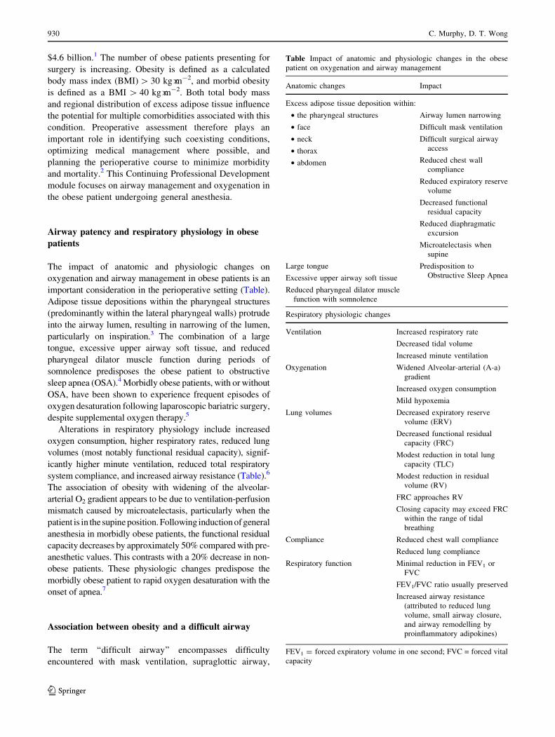
important consideration in the perioperative setting (Table).
Adipose tissue depositions within the pharyngeal structures
(predominantly within the lateral pharyngeal walls) protrude
into the airway lumen, resulting in narrowing of the lumen,
particularly on inspiration.3 The combination of a large
tongue, excessive upper airway soft tissue, and reduced
pharyngeal dilator muscle function during periods of
somnolence predisposes the obese patient to obstructive
sleep apnea (OSA).4 Morbidly obese patients, with or without
OSA, have been shown to experience frequent episodes of
oxygen desaturation following laparoscopic bariatric surgery,
despite supplemental oxygen therapy.5
Alterations in respiratory physiology include increased
oxygen consumption, higher respiratory rates, reduced lung
volumes (most notably functional residual capacity), signif-
icantly higher minute ventilation, reduced total respiratory
system compliance, and increased airway resistance (Table).6
The association of obesity with widening of the alveolar-
arterial O2 gradient appears to be due to ventilation-perfusion
mismatch caused by microatelectasis, particularly when the
patient is in the supine position. Following induction of general
anesthesia in morbidly obese patients, the functional residual
capacity decreases by approximately 50% compared with pre-
anesthetic values. This contrasts with a 20% decrease in non-
obese patients. These physiologic changes predispose the
morbidly obese patient to rapid oxygen desaturation with the
onset of apnea.7
Association between obesity and a difficult airway
The term ‘‘difficult airway’’ encompasses difficulty
encountered with mask ventilation, supraglottic airway,
Table Impact of anatomic and physiologic changes in the obese
patient on oxygenation and airway management
Anatomic changes Impact
Excess adipose tissue deposition within:
• the pharyngeal structures
• face
• neck
• thorax
• abdomen
Airway lumen narrowing
Difficult mask ventilation
Difficult surgical airway
access
Reduced chest wall
compliance
Reduced expiratory reserve
volume
Decreased functional
residual capacity
Reduced diaphragmatic
excursion
Microatelectasis when
supine
Large tongue
Excessive upper airway soft tissue
Reduced pharyngeal dilator muscle
function with somnolence
Predisposition to
Obstructive Sleep Apnea
Respiratory physiologic changes
Ventilation Increased respiratory rate
Decreased tidal volume
Increased minute ventilation
Oxygenation Widened Alveolar-arterial (A-a)
gradient
Increased oxygen consumption
Mild hypoxemia
Lung volumes Decreased expiratory reserve
volume (ERV)
Decreased functional residual
capacity (FRC)
Modest reduction in total lung
capacity (TLC)
Modest reduction in residual
volume (RV)
FRC approaches RV
Closing capacity may exceed FRC
within the range of tidal
breathing
Compliance Reduced chest wall compliance
Reduced lung compliance
Respiratory function Minimal reduction in FEV1 or
FVC
FEV1/FVC ratio usually preserved
Increased airway resistance
(attributed to reduced lung
volume, small airway closure,
and airway remodelling by
proinflammatory adipokines)
FEV1 = forced expiratory volume in one second; FVC = forced vital
capacity
930 C. Murphy, D. T. Wong
123

tracheal intubation, and/or surgical airway. Excess fatty
tissue on the face, neck, breasts, thorax, and abdomen may
pose substantial airway challenges related to patient
positioning, neck extension, bag and mask ventilation,
tracheal intubation, oxygenation, and tracheotomy. The
United Kingdom Fourth National Audit Project (NAP 4)
reviewed major complications of airway management
(death, brain damage, emergency surgical airway,
unanticipated admission to the intensive care unit, and
prolonged stay in the intensive care unit) and reported a
fourfold increase in the risk of serious complications in the
morbidly obese patient when compared with non-obese
patients.8,9 Nineteen (25%) of the 77 obese patients
reviewed in that study suffered brain damage or death.
During anesthesia, difficult tracheal intubation was
reported in 23 patients, aspiration in eight, tracheal
extubation problems in seven, and trauma to the airway
in four cases.
Mask ventilation
Obesity has been identified as an independent predictor of
difficult mask ventilation. In a prospective study of 1,502
consecutive patients presenting for surgery under general
anesthesia, anesthesiologists encountered difficulty with
mask ventilation in 75 instances, but only 17% of these
cases (13 patients) were anticipated.10 Using multivariate
analysis, BMI [ 26 kg�m-2 was identified as one of five
independent predicators of difficult mask ventilation.10 A
review of 22,660 mask ventilation attempts identified
BMI [ 30 kg�m-2 to be an independent predictor of
difficult mask ventilation and an independent predictor of
difficult or impossible mask ventilation and difficult
tracheal intubation.11 Nevertheless, a further study of
over 50,000 anesthetics failed to show obesity as an
independent predictor of impossible mask ventilation.12 In
this study,12 radiotherapy to the neck, male sex, presence of
a beard, and Mallampati scores 3 or 4 were identified as
independent predictors of impossible mask ventilation. In
summary, obesity is associated with potential difficult
mask ventilation; therefore, in anticipated or unanticipated
difficult mask ventilation, supraglottic airways should be
readily available at induction of anesthesia.
Supraglottic airway devices (SGAs)
Supraglottic airway devices are commonly used in both
elective and rescue airway management, but how
successful or safe are these supraglottic devices in the
obese patient? In a study population of over 15,000
patients, obesity was found to be an independent predictor
of failed use of a Laryngeal Mask Airway (LMA)
UniqueTM, requiring device removal and endotracheal
intubation. Inadequate ventilation due to leak and airway
obstruction was found in 42.4% and 30% of airway events,
respectively.13 Double-lumen SGAs with gastric channels
have been shown to provide effective ventilation in
morbidly obese patients when used as a bridge before
laryngoscope-guided tracheal intubation, and when
compared with face-mask ventilation in novice hands,
SGAs were associated with significantly easier and
superior ventilation scores.14,15 Double-lumen SGAs,
such as the LMA ProSealTM and the LMA SupremeTM,
provide higher leak pressures and may be safer in patients
with obesity;16 however, SGAs do not protect the airway
from aspiration. Eight of the 23 cases of aspiration of
gastric contents during anesthesia—reported as the primary
airway event in NAP 4—occurred in obese patients.8
Overall, eight patients died and two patients suffered brain
damage as a result of gastric content aspiration.9 Obesity is
therefore a relative contraindication of SGA use and
caution is advised.
Direct laryngoscopy and intubation
Is direct laryngoscopy or tracheal intubation more difficult
in the obese patient? Juvin et al. reported a 15.5% rate of
difficult tracheal intubation in obese patients compared
with a 2.2% rate in lean patients.17 While some studies
showed an association between obesity and difficult
intubation, controversy still exists, and there is
insufficient evidence to support increased BMI alone as
an independent predictor of difficult laryngoscopy or
difficult intubation.17,18 In a cohort of 91,332 consecutive
patients, a BMI [ 35 kg�m2 was found to be a statistically
significant predictor of difficult and failed intubation.19
Conversely, Erzi et al. found that BMI was not an
independent predictor of direct laryngoscopy view;20
however, in this study, the combination of BMI and
abnormal upper teeth was a significant predictor of difficult
laryngoscopy.20 By using ultrasound to quantify anterior
neck soft tissue at the level of the vocal cords, Erzi et al.
observed that morbidly obese patients in whom difficult
laryngoscopy was encountered had more pre-tracheal soft
tissue and a greater neck circumference than patients who
experienced an uncomplicated laryngoscopy; pre-tracheal
soft tissue thickness was [mean (SD)] 28 (2.7) mm vs
17.5 (1.8) mm, respectively (P \ 0.001) and neck
circumference was 50 (3.8) cm vs 43.5 (2.2) cm,
respectively (P \ 0.001).21 Komatsu et al. did not find
increased pre-tracheal tissue to be a good predictor of
difficult laryngoscopy in obese patients;22 however, the
patients in this study had a comparatively smaller neck
Airway management and oxygenation in obese patients 931
123

circumference [43.5 (4.7) cm vs 44.3 (5.3) cm; P \ 0.5] in
the difficult and easy laryngoscopy groups, respectively.
Further studies are required to clarify the role of ultrasound
and pre-tracheal adipose tissue quantification as a predictor
of difficult laryngoscopy.
Direct laryngoscopic view may be affected by patient
positioning and can be optimized by aligning the external
auditory meatus with the sternal notch, as discussed later
in the review.23 Overall lack of agreement on whether
tracheal intubation is more difficult in the morbidly obese
may be due to particular confounders, including
variations in the definitions of difficult intubation (more
than three attempts, intubation difficulty scale [ 5) and
difficult laryngoscopy (Cormack and Lehane grade C 3),
patient position, operator experience, and laryngoscopy
technique (direct vs video laryngoscopy). There is
agreement, however, that a combination of features,
such as Mallampati score C 3 and larger neck
circumference, in the obese patient does increase the
risk of difficult laryngoscopy and difficult tracheal
intubation.24,25
Surgical airway
In a ‘‘can’t intubate, can’t ventilate’’ scenario, emergency
cricothyroidotomy can be particularly challenging in the
obese patient because of obscured anatomical landmarks.
Aslani et al. investigated the ability of physicians to
identify the cricothyroid membrane correctly in obese
(n = 15) and non-obese (n = 41) female patients in whom
the cricothyroid membrane had been identified beforehand
with ultrasound. The cricothyroid membrane was correctly
identified in 1/15 obese patients and 12/41 non-obese
patients.26 The emerging role of ultrasound in identifying
airway anatomy presents the advantage of turning a blind
technique into one that is guided27 and offering the
possibility to identify the cricothyroid membrane
accurately in patients with large neck circumference and
impalpable landmarks, irrespective of physician
experience.28
As challenges may be encountered in mask ventilation,
supraglottic airway use, tracheal intubation, and
cricothyroidotomy in the morbidly obese patient, a
cautious approach is advised and awake tracheal
intubation should be considered in patients with known
or anticipated difficult airways.
Figure Positioning the patient to improve laryngeal inlet view during
direct laryngoscopy. a) Supine position in the ‘‘sniff’’ position, b)
horizontal alignment of the sternal notch with the external auditory
meatus in the ‘‘ramped’’ position using elevation pillows, and c)
‘‘ramped’’ table position achieved using the beach chair position
Figure Positionnement du patient pour ameliorer la visualisation du
larynx pendant une laryngoscopie directe. a) Position dorsale en
position de ‘reniflement’, b) alignement horizontal de l’echancrure
sternale et du meat auditif externe en position ‘inclinee’ a l’aide
d’oreillers, et c) position ‘inclinee’ de la table realisee a l’aide de la
position de chaise de plage
b
932 C. Murphy, D. T. Wong
123

Patient optimization for induction of anesthesia
Positioning and preoxygenation
Optimal positioning of the obese patient is vital prior to
induction of general anesthesia. The 25� head-up and
reverse Trendelenburg positions have been shown to
increase the duration of apnea without desaturation
compared with the supine position, thus increasing the
time for tracheal intubation.29,30 In a randomized
controlled trial, preoxygenation of morbidly obese
patients in the 25� head-up position led to a 23% higher
oxygen tension than in the supine position [442 (104)
mmHg vs 360 (99) mmHg, respectively; P = 0.012) and a
significant increase in the duration of apnea prior to
desaturation [201 (55) sec vs 155 (69) sec, respectively;
P = 0.023].29 The head-elevated laryngoscopy position
improves laryngeal exposure during direct laryngoscopy.31
In the obese patient population this is achieved by the
horizontal alignment of the sternal notch with the external
auditory meatus in the ‘‘ramped’’ position. To achieve this
visual alignment, the use of multiple folded blankets and
commercially available elevation pillows under the upper
body, shoulders, and head or the ‘‘ramped’’ table position
have been advocated.23 The beach chair position has also
been shown to facilitate intubation18 (Figure).
Induction of general anesthesia is a high-risk period for
hypoxemia. Even with preoxygenation, the duration of
apnea prior to desaturation can be anticipated to be
significantly shorter in the morbidly obese than in
patients of normal weight.32 Preoxygenation is considered
to be sufficient when the end-tidal oxygen fraction is
[ 90%.33 Tidal volume breathing through a well-sealed
face mask for three minutes or four vital capacity breaths
are common techniques of preoxygenation. Evidence
suggests that obese patients desaturate faster with the
latter technique.34 Additional techniques for peri-induction
oxygenation include supplemental nasopharyngeal oxygen
insufflation, semi-recumbent position, continuous positive
airway pressure (CPAP), positive end-expiratory pressure
(PEEP), and pressure support ventilation applied before
induction of general anesthesia in the spontaneous
ventilating patient.28-31 Baraka et al. compared
preoxygenation followed by nasopharyngeal oxygen
insufflation (study group) with preoxygenation alone
(control group) in morbidly obese patients placed in a
25� head-up position.35 In the study group, 16 of 17
patients maintained oxygen saturation at 100% during four
minutes of apnea. All 17 patients in the control group
experienced oxygen desaturation from 100 to 95% in a
mean time of 145 (27) sec. The use of 10 cm H2O
continuous positive airway pressure/positive end-
expiratory pressure (CPAP/PEEP) throughout the
induction period (CPAP when breathing and PEEP after
apnea) has been shown to increase the duration of apnea
prior to desaturation by 50% in morbidly obese
patients.36,37 Noninvasive positive pressure ventilation
using pressure support ventilation with PEEP via a face
mask has also been shown to enhance preoxygenation,
achieving a more rapid increase and higher end-tidal O2
when compared with preoxygenation with spontaneous
ventilation.38
Choice of intubation techniques
Direct vs indirect laryngoscopy
Optimal positioning of the patient will serve to optimize
success of tracheal intubation with direct laryngoscopy.
The use of a tracheal tube introducer (EschmannTM or
FrovaTM) or an intubating malleable stylet as an adjunct to
assist intubation should be considered when suboptimal
laryngeal inlet views are obtained.39,40
Alternatively, indirect laryngoscopy can be performed
using video and optical laryngoscopes, thus avoiding the need
to align oral and pharyngeal axes. In a study comparing video-
assisted with direct laryngoscopy in 318 morbidly obese
patients, the AirtraqTM and LMA CTrachTM provided an
earlier definitive airway and shortest apnea time, respectively,
when compared with the conventional Macintosh
laryngoscope.41 Video-assisted techniques resulted in
significantly better percentage of glottis opening (POGO)
scores compared with direct laryngoscopy (97% vs 75%,
respectively; P \ 0.01). Andersen et al. also found that the
Glidescope� video laryngoscope provided better glottic views
and lower intubation difficulty scores when compared with the
Macintosh laryngoscope in morbidly obese patients.42 While
an optimal laryngeal inlet view does not necessarily translate
into easier intubation, advancement of the endotracheal tube
may be assisted using a guiding channel on the device or an
introducer. Despite the increasing number of available airway
devices, no single video laryngoscope has shown superiority
for use in the obese patient; however, video laryngoscopes do
provide better laryngeal visualization and should be readily
available for airway management.
Intubating adjuncts: use of SGAs
In the anesthetized patient, fibreoptic intubation may be
hampered by loss of upper airway tone and collapsibility of
the airway passage. Supraglottic airway devices not only
facilitate oxygenation and ventilation of the lungs, but also
act as conduits or stents to maintain an open airway and
possibly improve visualization of and access to the
Airway management and oxygenation in obese patients 933
123

laryngeal inlet in the presence of a soiled and/or bloody
field.43 Several techniques have been described. An
endotracheal tube may be railroaded over a bronchoscope
through the SGA and positioned under direct vision prior to
removal of the bronchoscope. Alternatively an Aintree
Intubating Catheter (Cook Critical Care, Bloomington, IN,
USA) may be advanced over the bronchoscope into the
trachea through the SGA. The bronchoscope and SGA are
sequentially removed leaving the intubating catheter in situ
to serve as a ‘‘railroading’’ device for endotracheal tube
placement (maximum tube size, 7.0 mm internal diameter).
This technique has been described in both awake and
anesthetized patients with difficult airways.44,45 When
comparing use of an intubating laryngeal mask airway
(i.e., LMA-FastrachTM) in both lean and morbidly obese
patients, it was shown to be an efficient airway device
in both populations, with obese patients requiring
significantly fewer airway adjustment maneuvers.46
Awake bronchoscopic and video laryngoscopic
intubation
Awake tracheal intubation enables securing the airway in a
spontaneously breathing patient before induction of
anesthesia and therefore bypassing some of the difficulties
that may be encountered under general anesthesia. Awake
intubation should therefore be considered for patients
with either a documented history or clinical predictors of
difficult mask ventilation, laryngoscopy, and surgical
cricothyroidotomy. Flexible bronchoscopic intubation
requires training and experience and may be challenging in
the morbidly obese patient because of the anatomic changes
discussed above. Preparation of the patient for awake tracheal
intubation includes optimal positioning (‘‘ramped’’ or reverse
Trendelenburg) and preoxygenation, as mentioned above.
Tongue protrusion, jaw thrust maneuver, or oral airway
adjuncts (e.g., Ovassapian, Berman, and Williams airways)
may assist pharyngeal airway access and visualization. The
benefit of a more direct passage of the bronchoscope through
the nasal route into the pharynx should be weighed against the
risk of epistaxis. Local anesthetic topicalization of the airway
to blunt airway responses, with or without conscious sedation,
will improve patient tolerance. The use of awake video
laryngoscopy-assisted tracheal intubation has also been
described in the literature as an alternate to flexible
bronchoscopic intubation.47
Ventilator strategies and intraoperative considerations
Mechanical ventilation and muscle paralysis in the obese
patient under general anesthesia have been shown to impair
pulmonary function, lung compliance, and gas exchange
as a result of reduced lung volume and atelectasis.48
These consequences may be further exacerbated by
pneumoperitoneum and patient positioning for surgery
(supine, lithotomy, and Trendelenburg position).49 Special
attention should be paid to ventilatory strategies aimed at
minimizing these complications. In a computerized
tomography study of morbidly obese patients under
general anesthesia, the combination of a recruitment
maneuver at 55 cm H2O for ten seconds followed by
10 cm H2O PEEP reduced atelectasis and improved
oxygenation more than PEEP or a recruitment maneuver
alone.50 Obese patients receiving a vital capacity maneuver
(VCM) 40 cm H2O for seven to eight seconds followed by
PEEP 10 cm H2O were observed to have improved
intraoperative and postoperative oxygenation, and their
atelectasis scores were lower on chest computerized
tomography at two hours postoperatively when compared
with obese patients receiving VCM alone.51 Almarakbi
et al. also observed superior respiratory compliance and
PaO2 in obese patients undergoing laparoscopic gastric
banding with repeated inspiratory recruitment maneuvres
(40 cm H2O for 15 sec repeated every ten minutes)
followed by PEEP (10 cm H2O).52 Obese patients may
require high peak airway pressures with the application of
PEEP 10 cm H2O after recruitment maneuvers. While
barotrauma was not reported in these studies, caution
should be exercised. In a review of mechanical ventilation
in obese patients by Silva et al., the authors recommended
stepwise incremental recruitment maneuvers to reduce
hemodynamic instability and found no evidence supporting
one mode of ventilation over another (pressure vs volume
control ventilation).53 While they did report that pressure
support ventilation was found to be superior to pressure
control ventilation in terms of oxygenation and
postoperative lung function, this study was carried out in
obese patients undergoing minor surgery with LMAs as the
primary airway. The use of LMAs as the primary airway
device during minor surgery is not routinely practised in
many centres, and as mentioned previously, obesity is
considered a relative contraindication to the use of LMAs.
Extubation and postoperative oxygenation
Emergence from anesthesia, extubation, and recovery
Airway complications and issues with oxygenation may
present immediately on emergence from anesthesia at the
time of tracheal extubation or may become manifest only
in the postanesthesia care unit, resulting in significant
morbidity and mortality.8 Analysis of the American
Society of Anesthesiologists Closed Claims Project
934 C. Murphy, D. T. Wong
123

database of causes of death or brain death showed that 17%
of the cases (26/156) occurred at time of tracheal
extubation and recovery, and 58% of the extubation
claims (15/26) were encountered in obese patients.54
Consequently, careful consideration should be made for
the planning and execution of this procedure and for
management during the recovery period to minimize
patient risk.
The Difficult Airway Society published guidelines in
2012 for management of tracheal extubation using a
stepwise approach.55 Patients with obesity and
obstructive sleep apnea are stratified into a category of
extubation ‘‘at risk’’ of a major complication.
Recommendations for awake tracheal extubation in this
patient population include patient optimization (full
reversal of neuromuscular blockade and return of
protective airway reflexes), preoxygenation, placing the
patient in a reverse Trendelenburg or semi-recumbent
position, and suctioning of the oropharynx under direct
vision. Logistic factors to be considered include selecting
the operating room as the location for extubation and
having skilled assistance, equipment (difficult airway
trolley), and monitoring (in particular capnography)
available. The Difficult Airway Society guidelines also
advocate the placement of an airway exchange catheter in
patients for whom tracheal re-intubation is likely to be
difficult.
Postoperative atelectasis and management
Morbidly obese patients are at risk of significant atelectasis
under general anesthesia that persists into the immediate
postoperative period. Eichenberger et al. compared
pulmonary atelectasis in non-obese and morbidly obese
patients undergoing laparoscopic surgery using
computerized tomography before induction, immediately
post tracheal intubation, and 24 hr after general
anesthesia.56 Morbidly obese patients showed
significantly more atelectasis at all three stages compared
with non-obese patients. Importantly, the amount of
atelectasis (9.7% total lung area) at 24 hr remained
unchanged in the morbidly obese group, while there was
complete resolution in the non-obese patient group.
Frequent episodes of hypoxemia in the first 24 hr post
laparoscopic bariatric surgery have also been observed in
morbidly obese patients (with or without obstructive sleep
apnea) receiving supplemental oxygen therapy.5 Studies
comparing interventions to improve postoperative lung
function include the application of CPAP via the
Boussignac system and the use of respiratory physical
therapy in the postanesthesia care unit. Wong et al.
compared the Boussignac CPAP mask with the Venturi
face mask in morbidly obese patients undergoing
laparoscopic bariatric surgery, and study results showed
an improved postoperative PaO2/FIO2 ratio in the former
group, while the percent of forced expiratory volume and
the percent of forced vital capacity were comparable in
both groups.57 These findings suggest that improvement in
oxygenation using the Boussignac system resulted from a
combination of higher FIO2 and superior ventilation/
perfusion matching. In a study comparing the timing of
application of the Boussignac system, better maintenance
of spirometry lung function at 24 hr was observed when the
system was applied immediately post tracheal extubation
vs a delayed start in the postanesthesia care unit.58
Incentive spirometry performed immediately following
extubation and within the first two postoperative hours
has been shown to enhance pulmonary function in obese
patients up to 24 hr post surgery.59 This simple effective
deep breathing exercise warrants consideration when
CPAP systems are not readily available. To summarize,
morbidly obese patients are at high risk of hypoxemia up to
24 hr postoperatively. Vigilance should be exercised,
incentive spirometry provided, and CPAP administered as
indicated in this patient population.
Conclusion
In the obese patient, reduced functional residual capacity
and atelectasis predispose to rapid oxygen desaturation,
and challenges in oxygenation and ventilation may persist
throughout the perioperative period. Techniques to
optimize oxygenation include various preoxygenation
methods, head-up positioning at induction, use of
intraoperative alveolar recruitment maneuvers combined
with PEEP, CPAP mask application post tracheal
extubation, and use of incentive spirometry.
Difficulties in mask ventilation, SGA use, tracheal
intubation, and cricothyroidotomy should be assessed and
anticipated. Awake tracheal intubation should be considered
in cases where a higher suspicion of difficulty is anticipated
with these four aspects of airway management. Strategies to
optimize airway management include ramp position for
intubation, availability of multiple airway equipment, and
application of an algorithm for difficult airway management.
Tracheal extubation over an airway exchange catheter
should be considered for those patients in whom difficult
tracheal intubation was encountered.
Clinical case
A 54-yr-old morbidly obese woman (weight, 97 kg; height,
152 cm; body mass index, 42 kg�m-2) is scheduled for
Airway management and oxygenation in obese patients 935
123

elective laparoscopic hemicolectomy for colon carcinoma.
She has a history of controlled hypertension and
hypothyroidism. She denies any reflux, respiratory, or
cardiac symptoms. Airway assessment reveals a
Mallampati grade 3, but the examination is otherwise
unremarkable. Preoperative investigations reveal normal
electrocardiogram and chest radiograph findings, and
mildly obstructive pulmonary function tests (forced
expiratory flow in one second/forced vital capacity is 70%).
Upon arrival in the operating room, the patient is placed in
a 25� head-up ramped position, routine monitoring is
applied, and intravenous and arterial cannulae are placed.
Using a firmly fitting face mask, the patient is preoxygenated
for three minutes with an oxygen inspired fraction (FIO2) of
1.0. Vital signs show heart rate 74 beats�min-1, O2 saturation
99%, blood pressure 142/82 mmHg, and end-tidal carbon
dioxide (ETCO2) 34 mmHg. For induction of anesthesia, the
patient receives midazolam 2 mg, fentanyl 100 lg, propofol
200 mg, and rocuronium 60 mg. Bag and mask ventilation is
then commenced.
Instructions for completing the continuing professional
development (CPD) module
1. Read the current article and the references indicated in
bold.
2. Go to: http://www.cas.ca/Members/CPD-Online and
select the current module (Airway management and
oxygenation in obese patients).
3. Answer the multiple choice questions regarding the
case scenario.
4. Once you have entered all of your answers, you will
have access to experts’ explanations for all the possible
choices.
5. Participants may claim up to four hours of CPD, for a
total of 12 credits under Section 3 of the CPD program
of the Royal College of Physicians and Surgeons of
Canada.
La prise en charge des voiesaeriennes et l’oxygenationdu patient obese
Resume
Objectif L’objectif de ce module de developpement
professionnel continu est de decrire les difficultes
anatomiques et physiologiques rencontrees lors de la
prise en charge de patients obeses, de passer en revue
leurs effets sur l’oxygenation et la prise en charge des voies
aeriennes, et de proposer des strategies de prise en charge
pour la periode perioperatoire.
Constatations principales La combinaison d’une
accumulation excessive de tissu adipeux, de l’augmentation
de la consommation d’oxygene, de la reduction de la capacite
pulmonaire et d’une resistance accrue des voies aeriennes
chez les patients obeses augmente le risque de voies aeriennes
difficiles et de desaturation en oxygene rapide au cours de la
periode perioperatoire. La preoxygenation peut etre optimisee
en elevant la tete du patient ou en le placant en position de
Trendelenburg inversee, en exercant une ventilation a
pression positive continue et en fournissant une aide
inspiratoire. Des difficultes peuvent survenir lors de la
ventilation au ballon et au masque. La meilleure facon de
parvenir a une bonne exposition laryngee pendant la
laryngoscopie directe consiste a mettre le patient en position
‘inclinee’. Les bougies et mandrins d’intubation peuvent
faciliter l’intubation tracheale lorsque la vue laryngee est
sous-optimale, et la videolaryngoscopie pourrait ameliorer la
vue de la glotte et augmenter les chances de succes de
l’intubation tracheale. Les dispositifs supraglottiques a double
lumiere de nouvelle generation permettent des pressions de
fuite plus elevees et pourraient etre plus securitaires pour la
prise en charge des patients obeses; en outre, ils peuvent
egalement servir de guide pour l’intubation avec
bronchoscope. Dans le cas de patients chez lesquels on
prevoit des voies aeriennes difficiles, une intubation tracheale
vigile devrait etre planifiee. En periode peroperatoire, les
strategies de ventilation telles que les manœuvres de
recrutement en pression positive tele-expiratoire pourraient
reduire l’atelectasie et ameliorer l’oxygenation. Lors de
l’extubation tracheale, une position de la tete elevee et une
ventilation a pression positive continue reduisent l’hypoxemie
en postoperatoire. Apres une intubation tracheale difficile,
l’extubation sur un echangeur de tube devrait etre envisagee.
Conclusion Une desaturation en oxygene rapide est
possible chez le patient obese. Les difficultes potentielles
de prise en charge des voies aeriennes devraient etre
evaluees et anticipees, et des strategies d’oxygenation, de
ventilation et de prise en charge des voies aeriennes
devraient etre optimisees pendant la periode perioperatoire.
Objectifs de ce module de developpement professionnel
continu (DPC)
Apres avoir lu ce module, le lecteur devrait etre en mesure
de:
1. Decrire les changements de l’anatomie et de la
physiologie respiratoire associes a l’obesite.
2. Reconnaıtre les defis possibles en matiere de
ventilation au masque, d’utilisation de dispositifs
936 C. Murphy, D. T. Wong
123

supraglottiques, de l’intubation tracheale, et d’acces
chirurgical aux voies aeriennes chez le patient obese.
3. Appliquer certaines techniques visant a optimiser
l’induction de l’anesthesie generale chez le patient
obese.
4. Prevoir la methode, le materiel et les dispositifs de
secours necessaires a etablir la permeabilite des voies
aeriennes chez le patient obese.
5. Anticiper les difficultes a parvenir a une ventilation
optimale du patient obese sous anesthesie generale et
envisager des strategies de ventilation pour minimiser
les complications.
6. Optimiser les conditions pour realiser une extubation
tracheale securitaire chez le patient obese.
Selon le rapport Obesite au Canada 2011,
approximativement un adulte sur quatre au Canada
(24,3-25,4 %) est obese. En 2008, on estimait l’impact
economique de l’obesite a 4,6 milliards $.1 Le nombre de
patients obeses devant subir une chirurgie ne cesse
d’augmenter. On definit l’obesite comme un indice de
masse corporelle (IMC) calcule [ 30 kg�m-2, et l’obesite
morbide comme un IMC [ 40 kg�m-2. La masse
corporelle totale et la distribution regionale du tissu
adipeux en exces influencent le risque qu’une personne
court de souffrir des nombreuses comorbidites associees a
l’obesite. L’evaluation preoperatoire joue donc un role
crucial pour identifier de telles comorbidites et permet
d’optimiser la prise en charge medicale, le cas echeant,
ainsi que de planifier le deroulement de la periode
perioperatoire afin de minimiser la morbidite et la
mortalite.2 Ce module de developpement professionnel
continu traite principalement de la prise en charge des
voies aeriennes et de l’oxygenation du patient obese
subissant une anesthesie generale.
Permeabilite des voies aeriennes et physiologie
respiratoire du patient obese
Chez le patient obese, l’impact des changements
anatomiques et physiologiques sur l’oxygenation et la
prise en charge des voies aeriennes est une consideration
importante dans le contexte perioperatoire (tableau). Les
depots de tissu adipeux dans les structures pharyngees
(principalement le long des parois pharyngees laterales)
debordent dans la lumiere des voies aeriennes, ce qui
entraıne un retrecissement de la filiere, particulierement
lors de l’inspiration.3 La combinaison d’une grosse langue,
d’un exces de tissu mou dans les voies aeriennes
superieures et d’une reduction du fonctionnement du
muscle dilatateur du pharynx pendant les periodes de
somnolence predispose le patient obese a l’apnee
Tableau Impact des changements anatomiques et physiologiques
chez le patient obese sur l’oxygenation et la prise en charge des voies
aeriennes
Changements anatomiques Impact
Depot de tissu adipeux en exces sur:
• les structures pharyngees
• le visage
• le cou
• le thorax
• l’abdomen
Retrecissement de la lumiere
des voies aeriennes
Ventilation au masque
difficile
Acces chirurgical aux voies
aeriennes difficile
Reduction de la compliance de
la paroi abdominale
Reduction du volume de
reserve expiratoire
Reduction de la capacite
residuelle fonctionnelle
Reduction de l’excursion
diaphragmatique
Microatelectasie en decubitus
dorsal
Grosse langue
Exces de tissu mou dans les voies
aeriennes superieures
Reduction de la fonction du muscle
dilatateur pharynge lors de
somnolence
Predisposition a une apnee
obstructive du sommeil
Changements physiologiques
au niveau respiratoire
Ventilation Augmentation de la frequence
respiratoire
Reduction du volume courant
Augmentation de la ventilation
minute
Oxygenation Elargissement du gradient
alveolaire-arteriel (A-a)
Augmentation de la consommation
d’oxygene
Hypoxemie legere
Capacite pulmonaire Reduction du volume de reserve
expiratoire (VRE)
Reduction de la capacite residuelle
fonctionnelle (CRF)
Reduction modeste de la capacite
pulmonaire totale (CPT)
Reduction modeste du volume
residuel (VR)
La CRF s’approche du VR
La capacite de fermeture pourrait
depasser la CRF lors de
respiration normale
Compliance Reduction de la compliance de la
paroi abdominale
Reduction de la compliance
pulmonaire
Airway management and oxygenation in obese patients 937
123

obstructive du sommeil (AOS).4 Il a ete demontre que les
patients obeses morbides, qu’ils soient atteints d’AOS ou
non, souffraient d’episodes frequents de desaturation en
oxygene apres une chirurgie bariatrique par laparoscopie et
ce, malgre l’oxygenotherapie.5
Parmi les changements au niveau de la physiologie
respiratoire, citons une augmentation de la consommation
d’oxygene, une frequence respiratoire plus elevee, une
reduction de la capacite pulmonaire (et tout
particulierement de la capacite residuelle fonctionnelle),
une ventilation minute significativement plus elevee, une
reduction de la compliance totale du systeme respiratoire et
une resistance accrue des voies aeriennes (tableau).6
L’association entre obesite et elargissement du gradient
d’O2 alveolaire-arteriel semble etre due a une inadequation
ventilation-perfusion provoquee par une microatelectasie,
particulierement lorsque le patient est en position dorsale.
Apres l’induction de l’anesthesie generale chez les patients
obeses morbides, la capacite residuelle fonctionnelle
diminue d’environ 50 % par rapport aux valeurs
preanesthesie. Cette diminution est nettement plus
importante que celle de 20 % observee chez les patients
non obeses. Ces changements physiologiques predisposent
le patient obese morbide a une desaturation en oxygene
rapide avec l’apparition de l’apnee.7
Association entre obesite et voies aeriennes difficiles
Le terme de « voies aeriennes difficiles » englobe les
difficultes rencontrees lors d’une ventilation au masque,
avec les dispositifs supraglottiques, lors de l’intubation
tracheale, et/ou pour l’acces chirurgical aux voies
aeriennes. L’exces de tissu adipeux sur le visage, le cou,
la poitrine, le thorax et l’abdomen peut poser des defis
considerables en ce qui touche aux voies aeriennes: le
positionnement du patient, l’extension du cou, la
ventilation au ballon et au masque, l’intubation tracheale,
l’oxygenation et la tracheotomie peuvent tous etre
problematiques. Le quatrieme Projet d’audit national
(National Audit Project – NAP4) du Royaume-Uni a
passe en revue les complications majeures de prise en
charge des voies aeriennes (deces, lesion cerebrale, acces
chirurgical d’urgence aux voies aeriennes, admission
imprevue aux soins intensifs, et sejour prolonge a l’unite
des soins intensifs) et rapporte un risque quatre fois plus
eleve de complications graves chez les patients obeses
morbides que chez les patients non obeses.8,9 Dix-neuf
(25 %) des 77 patients obeses passes en revue dans cette
etude ont souffert d’une lesion cerebrale ou sont decedes.
L’etude rapporte qu’au cours de l’anesthesie, l’intubation
tracheale a ete difficile chez 23 patients, une inhalation est
survenue chez huit patients, sept patients ont connu des
problemes lors de l’extubation tracheale, et quatre patients
ont souffert d’un traumatisme des voies aeriennes.
La ventilation au masque
L’obesite a ete identifiee comme un predicteur independant
de ventilation au masque difficile. Dans une etude
prospective portant sur 1502 patients consecutifs devant
subir une chirurgie sous anesthesie generale, les
anesthesiologistes ont eprouve des difficultes lors de la
ventilation au masque dans 75 cas, mais les difficultes
n’avaient ete anticipees que pour 17 % de ces cas (13
patients).10 En se fondant sur une analyse multivariee, on a
determine qu’un IMC [ 26 kg�m-2 etait l’un de cinq
predicteurs independants de difficultes lors de la ventilation
au masque.10 Un compte rendu rapportant 22 660
tentatives de ventilation au masque a determine qu’un
IMC [ 30 kg�m-2 etait un predicteur independant de
ventilation au masque difficile et un predicteur independant
de ventilation au masque difficile ou impossible et
d’intubation tracheale difficile.11 Toutefois, une autre
etude portant sur plus de 50 000 anesthesies n’a pas
identifie l’obesite comme predicteur independant de
ventilation au masque impossible.12 Dans cette etude,12
une radiotherapie du cou, le sexe masculin, la presence
d’une barbe et des scores de Mallampati de 3 ou 4 ont ete
identifies comme predicteurs independants d’une
ventilation au masque impossible. Pour resumer, l’obesite
est associee a une ventilation au masque potentiellement
difficile; par consequent, qu’on anticipe ou non une
ventilation au masque difficile, des dispositifs
supraglottiques devraient etre a portee de votre main lors
de l’induction de l’anesthesie.
Tableau continued
Changements physiologiques
au niveau respiratoire
Fonction respiratoire Reduction minime de la VEMS ou
de la CVF
Rapport VEMS/CVF en general
preserve
Augmentation de la resistance des
voies aeriennes (attribuee a la
reduction de la capacite
pulmonaire, a la fermeture des
voies aeriennes de petit calibre,
et au remodelage des voies
aeriennes par les adipokines
pro-inflammatoires)
VEMS = volume expiratoire maximal en une seconde;
CVF = capacite vitale forcee
938 C. Murphy, D. T. Wong
123

Les dispositifs supraglottiques
Les dispositifs supraglottiques sont couramment utilises lors
de la prise en charge des voies aeriennes non urgente et
urgente, mais que sont le taux de reussite et la securite de tels
dispositifs chez le patient obese? Dans une population
d’etude de plus de 15 000 patients, on a decouvert que
l’obesite etait un predicteur independant d’echec
d’utilisation du masque larynge UniqueTM, necessitant le
retrait de l’instrument et une intubation endotracheale. La
ventilation etait inadaptee en raison de fuites ou
d’obstruction des voies aeriennes dans 42,4 % et 30 % des
cas de complications au niveau des voies aeriennes,
respectivement.13 Il a ete demontre que les dispositifs
supraglottiques a double lumiere avec conduit gastrique
offraient une ventilation efficace chez les patients obeses
morbides lorsqu’ils etaient utilises comme pont avant une
intubation tracheale a l’aide d’un laryngoscope. En outre,
lorsque les dispositifs supraglottiques etaient compares a une
ventilation par masque facial realisee par des novices, les
dispositifs supraglottiques entraınaient de meilleurs scores
de ventilation et permettaient une ventilation
considerablement plus facile.14,15 Les dispositifs
supraglottiques a double lumiere, comme les masques
larynges ProSealTM et SupremeTM, fournissent des
pressions de fuite plus elevees et pourraient donc etre plus
securitaires chez les patients obeses;16 toutefois, il faut
retenir que les dispositifs supraglottiques ne protegent pas les
voies aeriennes de l’inhalation. Huit des 23 cas d’inhalation
de contenu gastrique pendant l’anesthesie – la principale
complication au niveau des voies aeriennes rapportee dans
l’etude NAP4 – sont survenus chez des patients obeses.8 En
tout, huit patients sont decedes et deux patients ont souffert
de lesions cerebrales a la suite d’inhalation de contenu
gastrique.9 L’obesite est donc une contre-indication relative
a l’utilisation de dispositifs supraglottiques et il faut faire
preuve de prudence lors de leur utilisation.
La laryngoscopie directe et l’intubation
La laryngoscopie directe ou l’intubation tracheale sont-elles
plus difficiles chez un patient obese? Juvin et coll. ont
rapporte un taux de 15,5 % d’intubation tracheale difficile
chez les patients obeses, comparativement a 2,2 % chez des
patients sveltes.17 Alors que certaines etudes ont montre une
association entre obesite et intubation difficile, la
controverse demeure. En effet, les donnees probantes sont
insuffisantes pour soutenir que l’IMC seul est un predicteur
independant de laryngoscopie difficile ou d’intubation
difficile.17,18 Dans une etude de cohorte portant sur 91 332
patients consecutifs, on a observe qu’un IMC [ 35 kg�m2
etait un predicteur significatif d’un point de vue statistique
d’intubation difficile ou d’echec a l’intubation.19 A l’inverse,
Erzi et coll. ont decouvert que l’IMC n’etait pas un predicteur
independant d’une mauvaise visualisation de la glotte par
laryngoscopie directe;20 toutefois, dans cette etude, la
combinaison de l’IMC et d’une anomalie au niveau des
dents superieures etait un predicteur significatif de
laryngoscopie difficile.20 Erzi et coll. ont fait appel a
l’echographie pour determiner la quantite de tissu mou du
cou anterieur au niveau des cordes vocales. Ce faisant, les
auteurs ont observe que les patients obeses morbides chez qui
la laryngoscopie etait difficile avaient davantage de tissu
mou pretracheal et une circonference du cou plus importante
que les patients chez qui la laryngoscopie n’avait pas pose de
probleme. L’epaisseur du tissu mou pretracheal etait
[moyenne (ET)] 28 (2,7) mm vs. 17,5 (1,8) mm,
respectivement (P \ 0,001), et la circonference du cou
etait de 50 (3,8) cm vs. 43,5 (2,2) cm, respectivement (P\0,001).21 Selon Komatsu et coll., l’augmentation du tissu
pretracheal ne constitue pas un bon predicteur de
laryngoscopie difficile chez les patients obeses; toutefois,
la circonference du cou des patients de cette etude etait
comparativement plus basse [43,5 (4,7) cm vs. 44,3 (5,3) cm;
P \ 0,5] dans les groupes laryngoscopie difficile et
laryngoscopie facile, respectivement. Des etudes
supplementaires sont necessaires pour clarifier le role de
l’echographie et de la quantification du tissu adipeux
pretracheal en tant que predicteur de laryngoscopie difficile.
La vue obtenue par laryngoscopie directe pourrait etre
affectee par le positionnement du patient et peut etre
amelioree en alignant le meat auditif externe a
l’echancrure sternale, comme nous l’expliquerons plus
bas.23 La mesentente generale quant a savoir si l’intubation
tracheale est plus difficile chez les patients obeses morbides
pourrait etre due a des facteurs confondants particuliers,
notamment des variations au niveau de la definition d’une
intubation difficile (plus de trois tentatives, echelle de
difficulte d’intubation[5) et d’une laryngoscopie difficile
(grade C 3 selon la classification de Cormack et Lehane), de
la position du patient, de l’experience de l’operateur, et de la
technique de laryngoscopie (laryngoscopie directe ou
videolaryngoscopie). Toutefois, les specialistes s’entendent
pour dire qu’une combinaison de plusieurs elements, tels un
score de Mallampati C 3 et une circonference plus
importante du cou chez le patient obese, augmente le
risque de laryngoscopie difficile et d’intubation tracheale
difficile.24,25
L’acces chirurgical aux voies aeriennes
Dans les situations ou il est « impossible d’intuber,
impossible de ventiler », une cricothyroıdotomie peut etre
particulierement difficile a realiser chez un patient obese en
Airway management and oxygenation in obese patients 939
123

raison de la difficulte a identifier les reperes anatomiques
usuels. Aslani et coll. ont etudie la capacite des medecins a
reperer correctement la membrane cricothyroıdienne chez
des patientes obeses (n = 15) et non obeses (n = 41) chez
lesquelles la membrane avait ete identifiee au prealable
par echographie. La membrane cricothyroıdienne a ete
correctement identifiee chez 1/15 patiente obese et 12/41
patientes non obeses.26 Le nouveau role que l’echographie
joue pour l’identification de l’anatomie des voies aeriennes
presente l’avantage qu’elle transforme une technique
jusque-la realisee en aveugle en une technique guidee.27
En outre, l’echographie permet d’identifier precisement la
membrane cricothyroıdienne chez les patients possedant une
imposante circonference du cou et des reperes impalpables,
independamment de l’experience du medecin.28
Etant donne les defis auxquels nous pourrions etre
confrontes lors de la ventilation au masque, de l’utilisation
de dispositifs supraglottiques, de l’intubation tracheale et
de la cricothyroıdotomie chez le patient obese morbide, il
est recommande de faire preuve de prudence et d’envisager
une intubation tracheale vigile pour les patients chez
lesquels on anticipe des voies aeriennes difficiles, ou dont
les voies aeriennes sont connues comme etant difficiles.
La preparation optimale du patient pour l’induction de
l’anesthesie
Positionnement et preoxygenation
Un positionnement optimal du patient obese est essentiel
avant l’induction de l’anesthesie generale. Il a ete demontre
que les positions tete elevee a 25� et Trendelenburg inversee
augmentaient la duree de l’apnee sans desaturation
comparativement au decubitus dorsal, augmentant ainsi le
temps disponible pour l’intubation tracheale.29,30 Dans une
etude randomisee controlee, la preoxygenation de patients
obeses morbides en position tete elevee a 25� a entraıne une
tension en oxygene plus elevee de 23 % qu’en decubitus
dorsal [442 (104) mmHg vs. 360 (99) mmHg, respectivement;
P = 0,012) et une augmentation significative de la duree
d’apnee avant desaturation [201 (55) sec vs. 155 (69) sec,
respectivement; P = 0,023].29 La position de laryngoscopie
avec la tete surelevee ameliore l’exposition laryngee pendant
la laryngoscopie directe.31 Chez le patient obese, on y parvient
en alignant horizontalement l’echancrure sternale et le meat
auditif externe en position ‘inclinee’. Afin d’obtenir
cet alignement visuel, l’utilisation de plusieurs couvertures
pliees et d’oreillers speciaux disponibles sur le marche places
sous le haut du corps, les epaules et la tete ou le
positionnement ‘incline’ de la table ont ete preconises.23 Il a
aussi ete demontre que la position dite de la ‘chaise de plage’
facilitait l’intubation (figure).18
L’induction de l’anesthesie generale est une periode a
risque eleve d’hypoxemie. Meme avec une preoxygenation,
on peut prevoir que la duree de l’apnee avant desaturation
sera significativement plus courte chez le patient obese
morbide que chez un patient de poids normal.32 La
preoxygenation est consideree comme suffisante lorsque la
fraction d’oxygene tele-expiratoire est [ 90 %.33 La
respiration en volume courant via un masque facial bien
scelle pendant trois minutes ou quatre respirations en
capacite vitale sont deux techniques de preoxygenation
courantes. Les donnees probantes laissent penser que les
patients obeses desaturent plus rapidement avec la seconde
technique.34 Parmi les autres techniques d’oxygenation
peri-induction, citons l’insufflation nasopharyngee d’oxygene
supplementaire, la position semi-assise, la ventilation a
pression positive continue (CPAP), la pression positive
tele-expiratoire (PEEP) et l’aide inspiratoire – appliquees
avant l’induction de l’anesthesie generale chez un patient
respirant spontanement.28-31 Baraka et coll. ont compare une
technique de preoxygenation suivie par l’insufflation
nasopharyngee d’oxygene (groupe a l’etude) a une
technique de preoxygenation seule (groupe temoin) chez
des patients obeses morbides positionnes avec la tete elevee
a un angle de 25�.35 Dans le groupe a l’etude, 16 des 17
patients ont maintenu leur saturation en oxygene a 100 %
pendant quatre minutes d’apnee. Les 17 patients du groupe
temoin ont subi une desaturation en oxygene de 100 a 95 %
en un temps moyen de 145 (27) sec. Il a ete demontre que le
recours a une ventilation en pression positive continue /
pression positive tele-expiratoire de 10 cm H2O (CPAP /
PEEP) tout au long de la periode d’induction (CPAP lors de
la respiration et PEEP apres l’apnee) augmentait la duree de
l’apnee avant desaturation de 50 % chez les patients obeses
morbides.36,37 En outre, il a egalement ete demontre que la
ventilation en pression positive non effractive utilisant une
aide inspiratoire et une PEEP via un masque facial ameliorait
la preoxygenation, resultant en une augmentation plus rapide
et un O2 tele-expiratoire plus eleve comparativement a une
preoxygenation en ventilation spontanee.38
Choix de la technique d’intubation
Laryngoscopie directe ou indirecte
Le positionnement optimal du patient augmentera les
chances de reussite de l’intubation tracheale par
laryngoscopie directe. L’utilisation d’une bougie
(EschmannTM ou FrovaTM) ou d’un mandrin souple pour
favoriser l’intubation devrait etre envisagee lorsqu’on
n’obtient qu’une vue sous-optimale de l’ouverture du
larynx.39,40
940 C. Murphy, D. T. Wong
123

Sinon, une laryngoscopie indirecte peut etre realisee a
l’aide d’un videolaryngoscope ou d’un laryngoscope optique.
On contourne ainsi la necessite d’aligner les axes oral et
pharynge. Dans une etude comparant la videolaryngoscopie a
la laryngoscopie directe chez 318 patients obeses morbides, le
AirtraqTM et le masque larynge CTrachTM ont fourni un acces
definitif aux voies aeriennes plus rapidement et un temps
d’apnee plus court, respectivement, que le laryngoscope
conventionnel Macintosh.41 Lors du recours aux techniques
assistees par video, les scores d’ouverture glottique (POGO)
etaient significativement plus eleves que lors de l’utilisation
de la laryngoscopie directe (97 % vs. 75 %, respectivement;
P \ 0,01). Andersen et coll. ont egalement observe que le
videolaryngoscope Glidescope� fournissait de meilleures
vues de la glotte et des scores de difficulte d’intubation plus
bas que le laryngoscope Macintosh chez les patients obeses
morbides.42 Bien qu’une vue optimale de l’ouverture laryngee
ne se traduise pas forcement par une intubation plus facile, la
progression de la sonde endotracheale peut beneficier de
l’utilisation d’un canal guide place sur le dispositif larynge ou
sur une bougie. Malgre le nombre croissant d’outils
d’intubation disponibles, aucun videolaryngoscope ne s’est
avere superieur aux autres pour l’intubation du patient obese;
toutefois, les videolaryngoscopes fournissent une meilleure
vue laryngee et devraient etre disponibles pour la prise en
charge des voies aeriennes.
Les aides a l’intubation: le recours aux dispositifs
supraglottiques
Chez un patient anesthesie, l’intubation par fibroscope
pourrait etre entravee par la perte de tonus des voies
aeriennes superieures et la tendance a l’effondrement des
voies aeriennes. Non seulement les dispositifs supraglottiques
facilitent-ils l’oxygenation et la ventilation des poumons, mais
ils servent egalement de guides ou de tuteurs pour maintenir
les voies aeriennes permeables et, possiblement, permettent
d’ameliorer la visualisation des cordes vocales et l’acces au
larynx en presence de secretions et/ou de sang.43 Plusieurs
techniques ont ete decrites. Une sonde endotracheale peut etre
guidee sur un bronchoscope a travers le dispositif
supraglottique et positionnee en vision directe avant de
retirer le bronchoscope. Autrement, un echangeur de tube
Aintree (Cook Critical Care, Bloomington, IN, USA) peut etre
guide sur le bronchoscope dans la trachee a travers le dispositif
supraglottique. Le bronchoscope, puis le dispositif
supraglottique sont retires, laissant l’echangeur en place afin
de servir de guide pour le positionnement de la sonde
endotracheale (taille maximale de la sonde, 7,0 mm de
diametre interne). Cette technique a ete decrite chez le patient
eveille et le patient anesthesie avec voies aeriennes
difficiles.44,45 Lorsqu’on a compare l’utilisation d’un
masque larynge d’intubation (c.-a-d. le FastrachTM) chez
des patients sveltes et obeses morbides, le dispositif s’est avere
efficace pour l’intubation dans les deux populations de
patients, les patients obeses necessitant significativement
moins de manœuvres de repositionnement du dispositif.46
Utilisation d’un bronchoscope et d’un
videolaryngoscope pour l’intubation vigile
En pratiquant une intubation tracheale vigile, on permet
l’acces aux voies aeriennes d’un patient respirant
spontanement avant l’induction de l’anesthesie. Cette
approche permet de contourner certaines des difficultes qui
pourraient se presenter sous anesthesie generale. C’est
pourquoi une intubation vigile devrait etre envisagee chez
les patients presentant des antecedents ou des predicteurs
cliniques de ventilation au masque difficile, de laryngoscopie
difficile, ou de cricothyroıdotomie chirurgicale. L’intubation
avec un bronchoscope flexible necessite a la fois formation et
experience et pourrait s’averer difficile chez le patient obese
morbide en raison des changements anatomiques presentes
plus haut. La preparation du patient pour une intubation
tracheale vigile comprend son positionnement optimal (en
position ‘inclinee’ ou de Trendelenburg inversee) et la
preoxygenation, comme cela a deja ete mentionne. On peut
faciliter l’acces et la visualisation des voies aeriennes
pharyngees en degageant la langue, en deplacant la
machoire inferieure vers l’avant et en inserant une canule
(par ex. les canules Ovassapian, Berman et Williams). Les
avantages que presente un passage plus direct du
bronchoscope par voie nasale dans le pharynx doivent etre
soupeses avec le risque d’epistaxis. L’utilisation d’un
anesthesique local topique sur les voies aeriennes pour en
emousser les reflexes, avec ou sans sedation consciente,
ameliorera la tolerance du patient. Le recours a une intubation
tracheale vigile par videolaryngoscopie a egalement ete decrit
dans la litterature comme alternative a l’intubation avec un
bronchoscope flexible.47
Strategies de ventilation et considerations
peroperatoires
Il a ete demontre que la ventilation mecanique et la
curarisation entravaient la fonction pulmonaire, la
compliance pulmonaire et l’echange gazeux chez le patient
obese sous anesthesie generale, en raison de la reduction de
la capacite pulmonaire et de l’atelectasie.48 Ces
consequences pourraient etre encore exacerbees par un
pneumoperitoine et le positionnement du patient pour la
chirurgie (decubitus dorsal, lithotomie et position de
Trendelenburg). Il faut porter une attention particuliere aux
Airway management and oxygenation in obese patients 941
123

strategies de ventilation destinees a minimiser ces
complications. Dans une etude de tomodensitometrie
portant sur des patients obeses morbides sous anesthesie
generale, la combinaison d’une manœuvre de recrutement a
55 cm H2O pendant 10 sec suivie d’une PEEP a 10 cm H2O a
reduit l’atelectasie et ameliore l’oxygenation davantage que
l’une ou l’autre manœuvre realisee seule.50 Les patients
obeses chez lesquels on realise une manœuvre de capacite
vitale (MCV) de 40 cm H2O pendant sept a huit secondes
suivie de PEEP a 10 cm H2O ont affiche une meilleure
oxygenation peroperatoire et postoperatoire, et leurs scores
d’atelectasie etaient plus bas sur la tomodensitometrie
thoracique deux heures apres l’operation comparativement
a des patients obeses ne beneficiant que d’une MCV.51 Chez
les patients obeses subissant une chirurgie laraposcopique
d’anneau gastrique, Almarakbi et coll. ont egalement
observe une compliance respiratoire et une PaO2
superieures avec des manœuvres de recrutement
inspiratoire repetees (40 cm H2O pendant 15 sec toutes les
dix minutes) suivies de PEEP (10 cm H2O).52 Les patients
obeses peuvent necessiter des pressions de pointe lors de
l’application d’une PEEP de 10 cm H2O apres des
manœuvres de recrutement. Alors qu’aucun
barotraumatisme n’a ete rapporte dans ces etudes, il
convient d’etre prudent. Dans un compte rendu de Silva et
coll. examinant la ventilation mecanique chez des patients
obeses, les auteurs recommandent des manœuvres de
recrutement progressives afin de reduire l’instabilite
hemodynamique et n’ont decouvert aucune donnee
probante appuyant l’utilisation d’un mode de ventilation
plutot que d’un autre (ventilation en pression controlee vs. en
volume controle).53 Bien que les auteurs aient rapporte que
l’aide inspiratoire etait superieure a la ventilation en pression
controlee en termes d’oxygenation et de fonction pulmonaire
postoperatoire, cette etude portait sur des patients obeses
subissant une chirurgie mineure avec un masque larynge
comme principal dispositif pour les voies aeriennes.
L’utilisation d’un masque larynge pour la prise en charge
des voies aeriennes pour une chirurgie mineure n’est pas une
pratique courante dans de nombreux centres et, comme nous
l’avons mentionne precedemment, l’obesite est consideree
comme une contre-indication relative a son utilisation.
L’extubation et l’oxygenation postoperatoire
L’emergence de l’anesthesie, l’extubation et le reveil
Les complications au niveau des voies aeriennes et
les problemes d’oxygenation pourraient survenir
immediatement a l’emergence de l’anesthesie, au moment
de l’extubation tracheale, ou elles pourraient se manifester
seulement en salle de reveil, entraınant une morbidite et
une mortalite significatives.8 L’analyse de la base de
donnees du Closed Claims Project, litteralement le ‘Projet
des reclamations reglees’, de l’American Society of
Anesthesiologists portant sur les causes de deces ou de
mort cerebrale a montre que 17 % des cas (26/156) sont
survenus au moment de l’extubation tracheale et du reveil,
et 58 % des plaintes en matiere d’extubation (15/26) sont
survenues chez des patients obeses.54 Il est donc important
de bien reflechir a la planification et a l’execution de cette
intervention et a la prise en charge pendant la periode de
reveil, afin de minimiser les risques pour le patient.
En 2012, la Difficult Airway Society a publie des
directives pour la prise en charge de l’extubation tracheale
avec une approche progressive.55 Les patients souffrant
d’obesite et d’apnee obstructive du sommeil sont stratifies
dans une categorie d’extubation ‘a risque’ de complications
majeures. Parmi les recommandations pour l’extubation
tracheale vigile chez cette population de patients, citons la
preparation optimale du patient (neutralisation complete du
bloc neuromusculaire et retablissement des reflexes
protecteurs des voies aeriennes), la preoxygenation, le
positionnement du patient en position de Trendelenburg
inversee ou en position semi-assise, et l’aspiration de
l’oropharynx sous vision directe. Les facteurs logistiques
dont il faut en outre tenir compte comprennent le choix de
la salle d’operation comme lieu d’extubation, la presence
de personnes competentes, la disponibilite de materiel
adequat (chariot pour les voies aeriennes difficiles), et le
monitorage (en particulier la capnographie). Les directives
de la Difficult Airway Society recommandent egalement de
placer un echangeur de tube chez les patients pour lesquels
une reintubation tracheale serait probablement difficile.
L’atelectasie postoperatoire et sa prise en charge
Les patients obeses morbides courent le risque de souffrir
d’atelectasie grave sous anesthesie generale, laquelle
persiste en periode postoperatoire immediate.
Eichenberger et coll. ont compare l’atelectasie pulmonaire
chez des patients non obeses et des patients obeses morbides
subissant une chirurgie par laparoscopie a l’aide d’une
tomodensitometrie avant l’induction, immediatement
apres l’intubation tracheale, et 24 h apres l’anesthesie
generale.56 Il y a eu significativement plus d’atelectasie
chez les patients obeses morbides aux trois temps de mesure
que chez les patients non obeses. Fait important, la quantite
d’atelectasie (9,7 % de la surface pulmonaire totale) a 24 h
est demeuree inchangee dans le groupe obese morbide, alors
qu’une resolution complete a ete observee dans le groupe de
patients non obeses. Des episodes frequents d’hypoxemie au
cours des premieres 24 h apres une chirurgie bariatrique par
laparoscopie ont egalement ete observes chez les patients
942 C. Murphy, D. T. Wong
123

obeses morbides (avec ou sans apnee obstructive du sommeil)
beneficiant d’une oxygenotherapie.5 Les etudes comparant les
interventions pour ameliorer la fonction pulmonaire
postoperatoire incluent l’application d’une CPAP via le
systeme Boussignac et l’utilisation de physiotherapie
respiratoire en salle de reveil. Wong et coll. ont compare le
masque de CPAP de Boussignac au masque facial Venturi
chez des patients obeses morbides subissant une chirurgie
bariatrique par laparoscopie, et l’etude a montre une
amelioration du rapport PaO2/FIO2 en postoperatoire dans le
premier groupe, alors que le pourcentage de volume
expiratoire maximal en une seconde et le pourcentage de
capacite vitale forcee etaient comparables dans les deux
groupes.57 Ces resultats suggerent que l’amelioration de
l’oxygenation observee avec le systeme de Boussignac etait le
resultat combine d’une FIO2 plus elevee et d’une meilleure
adequation ventilation/perfusion. Dans une etude visant a
determiner le moment ou mettre en place le systeme de
Boussignac, un meilleur maintien de la fonction pulmonaire
telle que mesuree par spirometrie a 24 h a ete observe
lorsqu’on appliquait le systeme immediatement
apres l’extubation tracheale plutot que de retarder son debut
en salle de reveil.58 Il a ete demontre qu’une spirometrie
incitative realisee immediatement apres l’extubation et au
cours des deux premieres heures postoperatoires ameliorait la
fonction pulmonaire chez les patients obeses jusqu’a 24 h
apres la chirurgie.59 Cet exercice simple et efficace de
respiration profonde merite d’etre envisage lorsque les
systemes de CPAP ne sont pas a portee de main. Pour
resumer, les patients obeses morbides courent un risque eleve
d’hypoxemie jusqu’a 24 h apres leur operation. Il faut faire
preuve de vigilance, fournir une spirometrie incitative et
administrer une CPAP tel qu’indiquee chez cette population
de patients.
Conclusion
La reduction de la capacite residuelle fonctionnelle et
l’atelectasie observees chez un patient obese le
predisposent a une desaturation en oxygene rapide. En
outre, l’oxygenation et la ventilation peuvent demeurer
difficiles tout au long de la periode perioperatoire. Parmi
les techniques contribuant a ameliorer l’oxygenation,
citons diverses methodes de preoxygenation, un
positionnement avec la tete elevee lors de l’induction,
l’utilisation de manœuvres peroperatoires de recrutement
alveolaire combinees a une PEEP, l’application d’un
masque de CPAP apres l’extubation tracheale et le
recours a une spirometrie incitative.
Les difficultes potentielles lors de la ventilation au
masque, de l’utilisation de dispositifs supraglottiques, de
l’intubation tracheale et de la cricothyroıdotomie devraient
etre evaluees et anticipees. L’intubation tracheale vigile
devrait etre envisagee dans les cas ou les difficultes pour
ces quatre aspects de la prise en charge des voies aeriennes
semblent plus importantes. Parmi les strategies optimisant
la prise en charge des voies aeriennes, citons le
positionnement incline du patient pour l’intubation, la
mise a disposition de plusieurs dispositifs de prise en
charge des voies aeriennes et l’application d’un algorithme
pour la prise en charge des voies aeriennes difficiles.
L’extubation tracheale sur un echangeur de tube devrait
etre envisagee chez les patients dont l’intubation tracheale
a ete difficile.
Cas clinique
Une femme obese morbide de 54 ans (poids, 97 kg; taille,
152 cm; indice de masse corporelle, 42 kg�m-2) se
presente pour subir une hemicolectomie non urgente
par laparoscopie pour traiter un carcinome du colon.
Elle a des antecedents d’hypertension controlee et
d’hypothyroıdisme. Elle nie souffrir de reflux ou de
symptomes respiratoires ou cardiaques. L’evaluation des
voies aeriennes revele un score de Mallampati de grade 3,
mais l’examen ne releve rien d’autre. Les examens
preoperatoires revelent un electrocardiogramme et des
radiographies des poumons normaux, ainsi que des tests de
la fonction pulmonaire legerement obstructifs (le volume
expiratoire maximal en une seconde / capacite vitale forcee
est de 70 %).
A son arrivee en salle d’operation, la tete de la patiente est
relevee a 25�, le monitorage habituel est mis en place, et des
canules intraveineuses et arterielles sont placees. A l’aide d’un
masque facial ajuste, la patiente est preoxygenee pendant trois
minutes avec une fraction d’oxygene inspire (FIO2) de 1,0. Les
signes vitaux montrent une frequence cardiaque de 74
battements�min-1, une saturation en O2 a 99 %, une
pression arterielle de 142/82 mmHg, et un dioxyde de
carbone tele-expiratoire (ETCO2) de 34 mmHg. Pour
l’induction de l’anesthesie, la patiente recoit 2 mg de
midazolam, 100 lg de fentanyl, 200 mg de propofol et
60 mg de rocuronium. On commence alors la ventilation au
ballon et au masque.
Directives pour completer le module de developpement
professionnel continu (DPC)
1. Lisez cet article et les references en gras.
2. Allez a: http://www.cas.ca/Membres/modules-de-DPC
et choisissez le module actuel (La prise en charge des
voies aeriennes et l’oxygenation du patient obese).
Airway management and oxygenation in obese patients 943
123

3. Repondez aux questions a choix de reponses
concernant le cas clinique.
4. Une fois que vous avez saisi toutes vos reponses, vous
aurez acces aux explications d’experts pour tous les
choix possibles.
5. Les participants peuvent reclamer un maximum de
quatre heures de DPC pour un total de 12 credits sous
la Section 3 du programme de DPC du College royal
des medecins et chirurgiens du Canada.
Supported in part by the Department of Anesthesia, University ofToronto.Finance en partie par le departement d’anesthesie, Universite de
Toronto.
Conflicts of interest None declared.
Conflit d’interet Aucun.
References
1. Canadian Institute for Health Information, Public Health Agency
of Canada. Obesity in Canada: a joint report from the Public
Health Agency of Canada and the Canadian Institute for Health
Information. [Ottawa]: Public Health Agency of Canada; 2011.
Available from URL: http://www.phac-aspc.gc.ca/hp-ps/hl-mvs/
oic-oac/index-eng.php (accessed April 18, 2013) [Institut cana-
dien d’information sur la sante, Agence de la sante publique du
Canada. Obesite au Canada: rapport conjoint de l’Agence de la
sante publique du Canada et de l’Institut canadien d’information
sur la sante. [Ottawa]: Agence de la sante publique du Canada;
2011. Disponible a l’adresse: http://www.phac-aspc.gc.ca/hp-ps/
hl-mvs/oic-oac/index-fra.php (consulte en anglais le 18 avril
2013)].
2. Cullen A, Ferguson A. Perioperative management of theseverely obese patient: a selective pathophysiological review.Can J Anesth 2012; 59: 974-96.
3. Horner RL, Mohiaddin RH, Lowell DG, et al. Sites and sizes of
fat deposits around the pharynx in obese patients with obstructive
sleep apnoea and weight matched controls. Eur Respir J 1989; 2:
613-22.
4. Benumof JL. Obesity, sleep apnea, the airway and anesthesia.
Curr Opin Anaesthesiol 2004; 17: 21-30.
5. Ahmad S, Nagle A, McCarthy RJ, Fitzgerald PC, Sullivan JT,
Prystowsku J. Postoperative hypoxemia in morbidly obese
patients with and without obstructive sleep apnea undergoing
laparoscopic bariatric surgery. Anesth Analg 2008; 107: 138-43.
6. Littleton SW. Impact of obesity on respiratory function.
Respirology 2012; 17: 43-9.
7. Pelosi P, Croci M, Ravagnan I, et al. The effects of body mass on
lung volumes, respiratory mechanics, and gas exchange during
general anesthesia. Anesth Analg 1998; 87: 654-60.
8. Cook TM, Woodall N, Frerk C; Fourth National Audit Project.
Major complications of airway management in the UK: results of
the Fourth National Audit Project of the Royal College of
Anaesthetists and the Difficult Airway Society. Part 1:
anaesthesia. Br J Anaesth 2011; 106: 617-31.
9. Cook TM, Woodall N, Harper J, Benger J; Fourth National Audit
Project. Major complications of airway management in the UK:
results of the Fourth National Audit Project of the Royal College
of Anaesthetists and the Difficult Airway Society. Part 2:
intensive care and emergency departments. Br J Anaesth 2011;
106: 632-42.
10. Langeron O, Masso E, Huraux C, et al. Prediction of difficult
mask ventilation. Anesthesiology 2000; 92: 1229-36.
11. Kheterpal S, Han R, Tremper KK, et al. Incidence and predictors
of difficult and impossible mask ventilation. Anesthesiology
2006; 105: 885-91.
12. Kheterpal S, Martin L, Shanks AM, Tremper KK. Predictionand outcomes of impossible mask ventilation: a review of50,000 anesthetics. Anesthesiology 2009; 110: 891-7.
13. Ramachandran SK, Mathis MR, Tremper KK, Shanks AM,
Kheterpal S. Predictors and clinical outcomes from failed
Laryngeal Mask Airway UniqueTM: a study of 15,795 patients.
Anesthesiology 2012; 116: 1217-26.
14. Keller C, Brimacombe J, Kleinsasser A, Brimacombe L. The
Laryngeal Mask Airway ProSeal(TM) as a temporary ventilatory
device in grossly and morbidly obese patients before laryngoscope-
guided tracheal intubation. Anesth Analg 2002; 94: 737-40.
15. Abdi W, Dhonneur G, Amathieu R, et al. LMA supreme versus
facemask ventilation performed by novices: a comparative study
in morbidly obese patients showing difficult ventilation
predictors. Obes Surg 2009; 19: 1624-30.
16. Wong DT, Yang JJ, Jagannathan N. Brief review: The LMA
SupremeTM supraglottic airway. Can J Anesth 2012; 59: 483-93.
17. Juvin P, Lavaut E, Dupont H, et al. Difficult tracheal intubation is
more common in obese than in lean patients. Anesth Analg 2003;
97: 595-600.
18. Fox WT, Harris S, Kennedy NJ. Prevalence of difficult intubation
in a bariatric population, using the beach chair position.
Anaesthesia 2008; 63: 1339-42.
19. Lundstrom LH, Moller AM, Rosenstock C, Astrup G, Wetterslev J.
High body mass index is a weak predictor for difficult and failed
tracheal intubation: a cohort study of 91,332 consecutive patients
scheduled for direct laryngoscopy registered in the Danish
Anesthesia Database. Anesthesiology 2009; 110: 266-74.
20. Ezri T, Medalion B, Weisenberg M, Szmuk P, Warters RD,
Charuzi I. Increased body mass index per se is not a predictor of
difficult laryngoscopy. Can J Anesth 2003; 50: 179-83.
21. Ezri T, Gewurtz G, Sessler DI, et al. Prediction of difficult
laryngoscopy in obese patients by ultrasound quantification of
anterior neck soft tissue. Anaesthesia 2003; 58: 1111-4.
22. Komatsu R, Sengupta P, Wadhwa A, et al. Ultrasound
quantification of anterior soft tissue thickness fails to predict
difficult laryngoscopy in obese patients. Anaesth Intensive Care
2007; 35: 32-7.
23. Rao SL, Kunselman AR, Schuler HG, DesHarnais S. Laryngoscopy
and tracheal intubation in the head-elevated position in obese
patients: a randomized, controlled, equivalence trial. Anesth Analg
2008; 107: 1912-8.
24. Gonzalez H, Minville V, Delanoue K, Mazerolles M, Concina D,
Fourcade O. The importance of increased neck circumference to
intubation difficulties in obese patients. Anesth Analg 2008; 106:
1132-6.
25. Brodsky JB, Lemmens HJ, Brock-Utne JG, Vierra M, Saidman
LJ. Morbid obesity and tracheal intubation. Anesth Analg 2002;
94: 732-6.
26. Aslani A, Ng SC, Hurley M, McCarthy KF, McNicholas M,
McCaul CL. Accuracy of identification of the cricothyroid
membrane in female subjects using palpation: an observational
study. Anesth Analg 2012; 114: 987-92.
27. Dinsmore J, Heard AM, Green RJ. The use of ultrasound to guide
time-critical cannula tracheotomy when anterior neck airway
anatomy is unidentifiable. Eur J Anaesthesiol 2011; 28: 506-10.
28. Nicholls SE, Sweeney TW, Ferre RM, Strout TD. Bedside
sonography by emergency physicians for the rapid identification
944 C. Murphy, D. T. Wong
123

of landmarks relevant to cricothyrotomy. Am J Emerg Med 2008;
26: 852-6.
29. Dixon BJ, Dixon JB, Carden JR, et al. Preoxygenation is more
effective in the 25 degrees head-up position than in the supine
position in severely obese patients: a randomized controlled
study. Anesthesiology 2005; 102: 1110-5; discussion 5A.
30. Boyce JR, Ness T, Castroman P, Gleysteen JJ. A preliminary
study of the optimal anesthesia positioning for the morbidly obese
patient. Obes Surg 2003; 13: 4-9.
31. Levitan RM, Mechem CC, Ochroch EA, Shofer FS, Hollander JE.
Head-elevated laryngoscopy position: improving laryngeal
exposure during laryngoscopy by increasing head elevation.
Ann Emerg Med 2003; 41: 322-30.
32. Berthoud MC, Peacock JE, Reilly CS. Effectiveness of
preoxygenation in morbidly obese patients. Br J Anaesth 1991;
67: 464-6.
33. Tanoubi I, Drolet P, Donati F. Optimizing preoxygenation in
adults. Can J Anesth 2009; 56: 449-66.
34. Benumof JL. Preoxygenation: best method for both efficacy and
efficiency. Anesthesiology 1999; 91: 603-5.
35. Baraka AS, Taha SK, Siddik-Sayyid SM, et al. Supplementation of
pre-oxygenation in morbidly obese patients using nasopharyngeal
oxygen insufflation. Anaesthesia 2007; 62: 769-73.
36. Coussa M, Proietti S, Schnyder P, et al. Prevention of atelectasis
formation during the induction of general anesthesia in morbidly
obese patients. Anesth Analg 2004; 98: 1491-5.
37. Gander S, Frascarolo P, Suter M, Spahn DR, Magnusson L.
Positive end-expiratory pressure during induction of general
anesthesia increases duration of nonhypoxic apnea in morbidly
obese patients. Anesth Analg 2005; 100: 580-4.
38. Delay JM, Sebbane M, Jung B, et al. The effectiveness of
noninvasive positive pressure ventilation to enhance
preoxygenation in morbidly obese patients: a randomized
controlled study. Anesth Analg 2008; 107: 1707-13.
39. American Society of Anesthesiologists Task Force on Management
of the Difficult Airway. Practice guidelines for management of the
difficult airway: an updated report by the American Society of
Anesthesiologists Task Force on Management of the Difficult
Airway. Anesthesiology 2003; 98: 1269-77.
40. Henderson JJ, Popat MT, Latto IP, Pearce AC; Difficult AirwaySociety. Difficult Airway Society guidelines for managementof the unanticipated difficult intubation. Anaesthesia 2004;59: 675-94.
41. Dhonneur G, Abdi W, Ndoko SK, et al. Video-assisted versus
conventional tracheal intubation in morbidly obese patients. Obes
Surg 2009; 19: 1096-101.
42. Andersen LH, Rovsing L, Olsen KS. GlideScope videolaryngoscope
vs. Macintosh direct laryngoscope for intubation of morbidly obese
patients: a randomized trial. Acta Anaesthesiol Scand 2011; 55:
1090-7.
43. Wong DT, Yang JJ, Mak HY, Jagannathan N. Use of intubation
introducers through a supraglottic airway to facilitate tracheal
intubation: a brief review. Can J Anesth 2012; 59: 704-15.
44. Cook TM, Seller C, Gupta K, Thornton M, O’Sullivan E. Non-
conventional uses of the Aintree Intubating Catheter in
management of the difficult airway. Anaesthesia 2007; 62: 169-74.
45. Berkow LC, Schwartz JM, Kan K, Corridore M, Heitmiller ES.
Use of the Laryngeal Mask Airway-Aintree Intubating Catheter-
fiberoptic bronchoscope technique for difficult intubation. J Clin
Anesth 2011; 23: 534-9.
46. Combes X, Sauvat S, Leroux B, et al. Intubating laryngeal mask
airway in morbidly obese and lean patients: a comparative study.
Anesthesiology 2005; 102: 1106-9; discussion 5A.
47. Moore AR, Schricker T, Court O. Awake videolaryngoscopy-
assisted tracheal intubation of the morbidly obese. Anaesthesia
2012; 67: 232-5.
48. Andersson LE, Baath M, Thorne A, Aspelin P, Odeberg-
Wernerman S. Effect of carbon dioxide pneumoperitoneum on
development of atelectasis during anesthesia, examined by spiral
computed tomography. Anesthesiology 2005; 102: 293-9.
49. Brodsky JB. Positioning the morbidly obese patient for
anesthesia. Obes Surg 2002; 12: 751-8.
50. Reinius H, Jonsson L, Gustafsson S, et al. Prevention of
atelectasis in morbidly obese patients during general anesthesia
and paralysis: a computerized tomography study. Anesthesiology
2009; 111: 979-87.
51. Talab HF, Zabani IA, Abdelrahman HS, et al. Intraoperative
ventilatory strategies for prevention of pulmonary atelectasis in
obese patients undergoing laparoscopic bariatric surgery. Anesth
Analg 2009; 109: 1511-6.
52. Almarakbi WA, Fawzi HM, Alhashemi JA. Effects of four
intraoperative ventilatory strategies on respiratory compliance
and gas exchange during laparoscopic gastric banding in obese
patients. Br J Anaesth 2009; 102: 862-8.
53. Leme Silva P, Pelosi P, Rocco PR. Mechanical ventilation in
obese patients. Minerva Anestesiol 2012; 78: 1136-45.
54. Peterson GN, Domino KB, Caplan RA, Posner KL, Lee LA,
Cheney FW. Management of the difficult airway: a closed claims
analysis. Anesthesiology 2005; 103: 33-9.
55. Membership of the Difficult Airway Society ExtubationGuidelines Group: Popat M, Mitchell V, Dravid R, Patel A,Schampillai C, Higgs A. Difficult Airway Society Guidelinesfor the management of tracheal extubation. Anaesthesia 2012;67: 318-40.
56. Eichenberger A, Proietti S, Wicky S, et al. Morbid obesity and
postoperative pulmonary atelectasis: an underestimated problem.
Anesth Analg 2002; 95: 1788-92.
57. Wong DT, Adly E, Ip HY, Thapar S, Maxted GR, Chung FF. A
comparison between the BoussignacTM continuous positive
airway pressure mask and the venturi mask in terms of
improvement in the PaO2/F(I)O2 ratio in morbidly obese
patients undergoing bariatric surgery: a randomized controlled
trial. Can J Anesth 2011; 58: 532-9.
58. Neligan PJ, Malhotra G, Fraser M, et al. Continuous positive
airway pressure via the Boussignac system immediately after
extubation improves lung function in morbidly obese patients
with obstructive sleep apnea undergoing laparoscopic bariatric
surgery. Anesthesiology 2009; 110: 878-84.
59. Zoremba M, Dette F, Gerlach L, Wolf U, Wulf H. Short-term
respiratory physical therapy treatment in the PACU and influence
on postoperative lung function in obese adults. Obes Surg 2009;
19: 1346-54.
Airway management and oxygenation in obese patients 945
123