Embed Size (px)
Citation preview

7/31/2019 AHA Guidelines CABG
http://slidepdf.com/reader/full/aha-guidelines-cabg 1/24
AHA Guidelines
for CABG

7/31/2019 AHA Guidelines CABG
http://slidepdf.com/reader/full/aha-guidelines-cabg 2/24
Indications for CABG
7/31/2019 AHA Guidelines CABG
http://slidepdf.com/reader/full/aha-guidelines-cabg 3/24
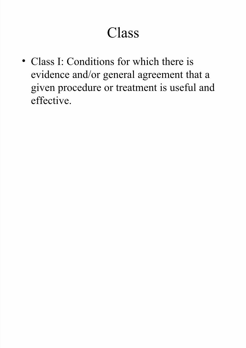
Class
• Class I: Conditions for which there is
evidence and/or general agreement that a
given procedure or treatment is useful andeffective.
7/31/2019 AHA Guidelines CABG
http://slidepdf.com/reader/full/aha-guidelines-cabg 4/24
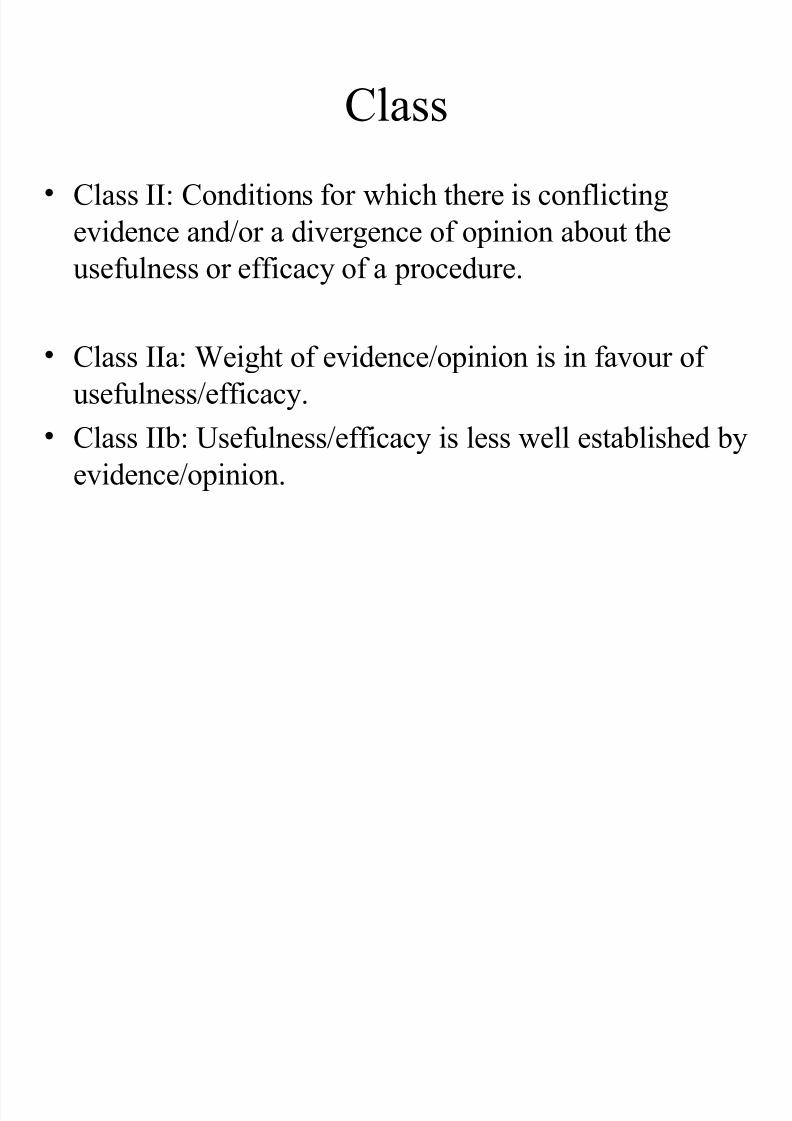
Class
• Class II: Conditions for which there is conflicting
evidence and/or a divergence of opinion about the
usefulness or efficacy of a procedure.
• Class IIa: Weight of evidence/opinion is in favour of
usefulness/efficacy.
• Class IIb: Usefulness/efficacy is less well established by
evidence/opinion.

7/31/2019 AHA Guidelines CABG
http://slidepdf.com/reader/full/aha-guidelines-cabg 5/24
Class
• Class III: Conditions for which there is
evidence and/or general agreement that the
procedure/treatment is not useful/effectiveand in some cases may be harmful.
7/31/2019 AHA Guidelines CABG
http://slidepdf.com/reader/full/aha-guidelines-cabg 6/24
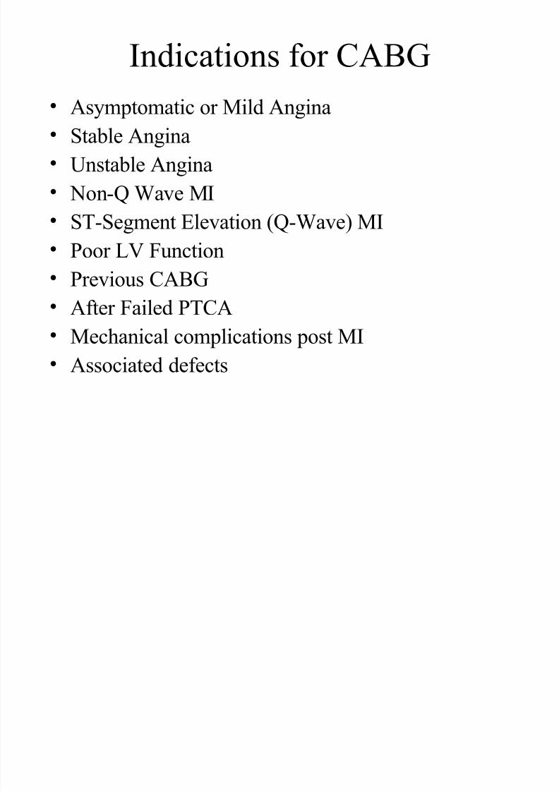
Indications for CABG
• Asymptomatic or Mild Angina
• Stable Angina
• Unstable Angina
• Non-Q Wave MI• ST-Segment Elevation (Q-Wave) MI
• Poor LV Function
• Previous CABG
• After Failed PTCA
• Mechanical complications post MI
• Associated defects
7/31/2019 AHA Guidelines CABG
http://slidepdf.com/reader/full/aha-guidelines-cabg 7/24
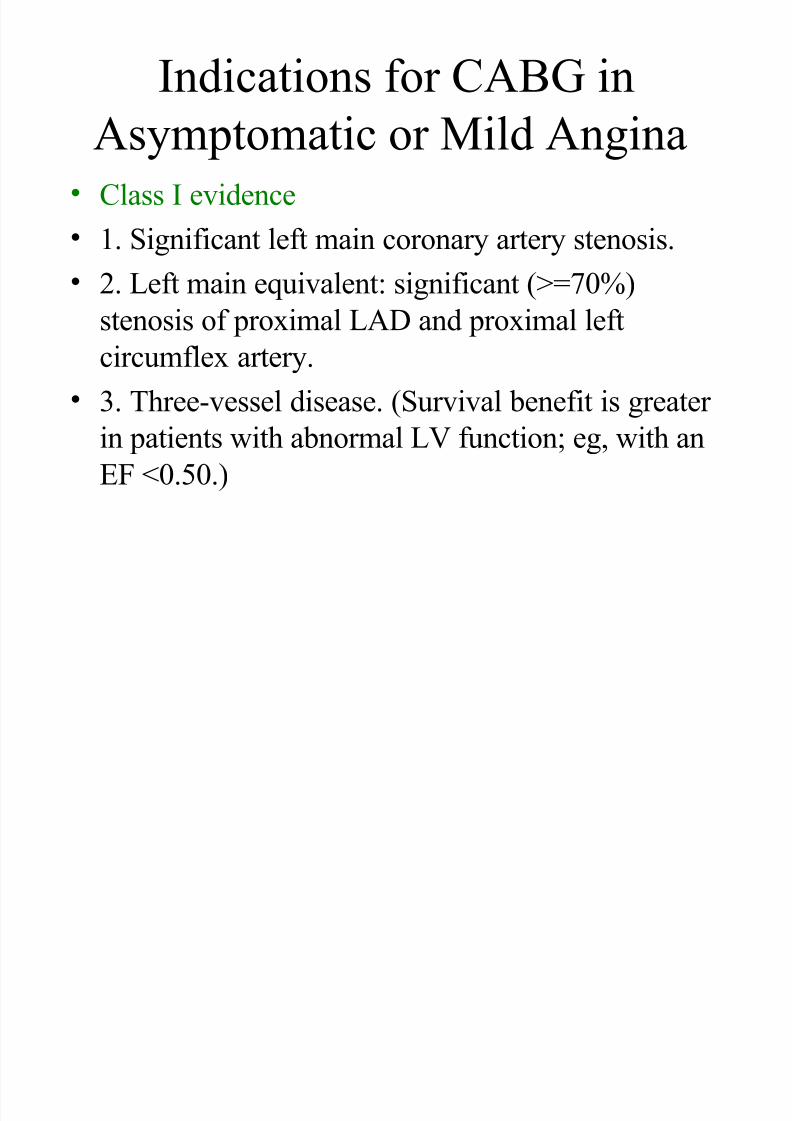
Indications for CABG in
Asymptomatic or Mild Angina• Class I evidence
• 1. Significant left main coronary artery stenosis.
• 2. Left main equivalent: significant (>=70%)stenosis of proximal LAD and proximal left
circumflex artery.
• 3. Three-vessel disease. (Survival benefit is greater in patients with abnormal LV function; eg, with an
EF <0.50.)
7/31/2019 AHA Guidelines CABG
http://slidepdf.com/reader/full/aha-guidelines-cabg 8/24
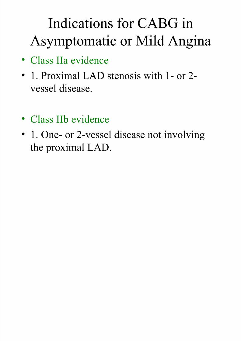
Indications for CABG in
Asymptomatic or Mild Angina• Class IIa evidence
• 1. Proximal LAD stenosis with 1- or 2-
vessel disease.
• Class IIb evidence
• 1. One- or 2-vessel disease not involving
the proximal LAD.
7/31/2019 AHA Guidelines CABG
http://slidepdf.com/reader/full/aha-guidelines-cabg 9/24
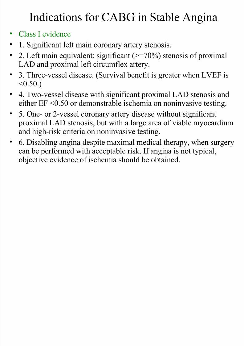
Indications for CABG in Stable Angina • Class I evidence
• 1. Significant left main coronary artery stenosis.• 2. Left main equivalent: significant (>=70%) stenosis of proximal
LAD and proximal left circumflex artery.
• 3. Three-vessel disease. (Survival benefit is greater when LVEF is
<0.50.)• 4. Two-vessel disease with significant proximal LAD stenosis and
either EF <0.50 or demonstrable ischemia on noninvasive testing.
• 5. One- or 2-vessel coronary artery disease without significant proximal LAD stenosis, but with a large area of viable myocardiumand high-risk criteria on noninvasive testing.
• 6. Disabling angina despite maximal medical therapy, when surgerycan be performed with acceptable risk. If angina is not typical,objective evidence of ischemia should be obtained.
7/31/2019 AHA Guidelines CABG
http://slidepdf.com/reader/full/aha-guidelines-cabg 10/24
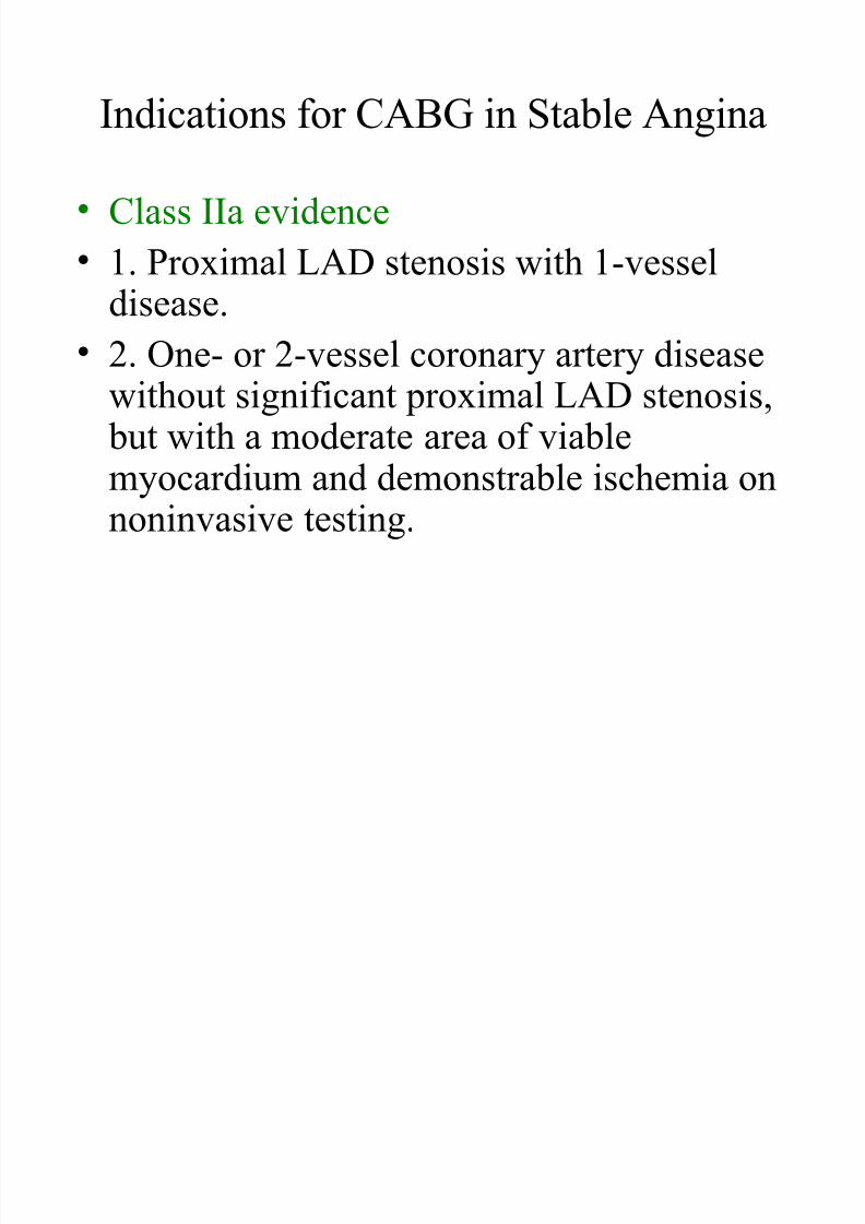
Indications for CABG in Stable Angina
• Class IIa evidence
• 1. Proximal LAD stenosis with 1-vessel
disease.• 2. One- or 2-vessel coronary artery disease
without significant proximal LAD stenosis,
but with a moderate area of viablemyocardium and demonstrable ischemia onnoninvasive testing.
7/31/2019 AHA Guidelines CABG
http://slidepdf.com/reader/full/aha-guidelines-cabg 11/24
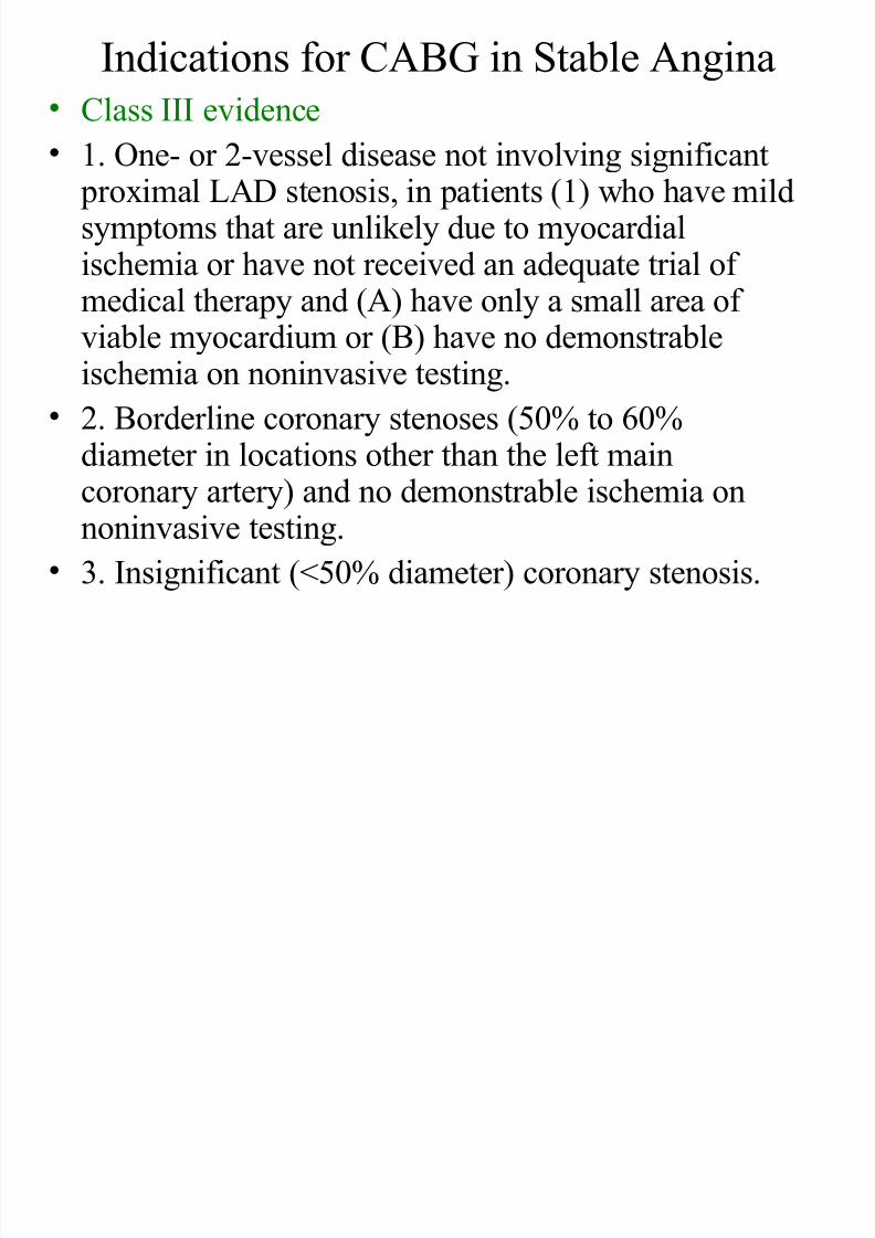
Indications for CABG in Stable Angina• Class III evidence
• 1. One- or 2-vessel disease not involving significant proximal LAD stenosis, in patients (1) who have mildsymptoms that are unlikely due to myocardialischemia or have not received an adequate trial of
medical therapy and (A) have only a small area of viable myocardium or (B) have no demonstrableischemia on noninvasive testing.
• 2. Borderline coronary stenoses (50% to 60%
diameter in locations other than the left maincoronary artery) and no demonstrable ischemia onnoninvasive testing.
• 3. Insignificant (<50% diameter) coronary stenosis.
di i f i
7/31/2019 AHA Guidelines CABG
http://slidepdf.com/reader/full/aha-guidelines-cabg 12/24
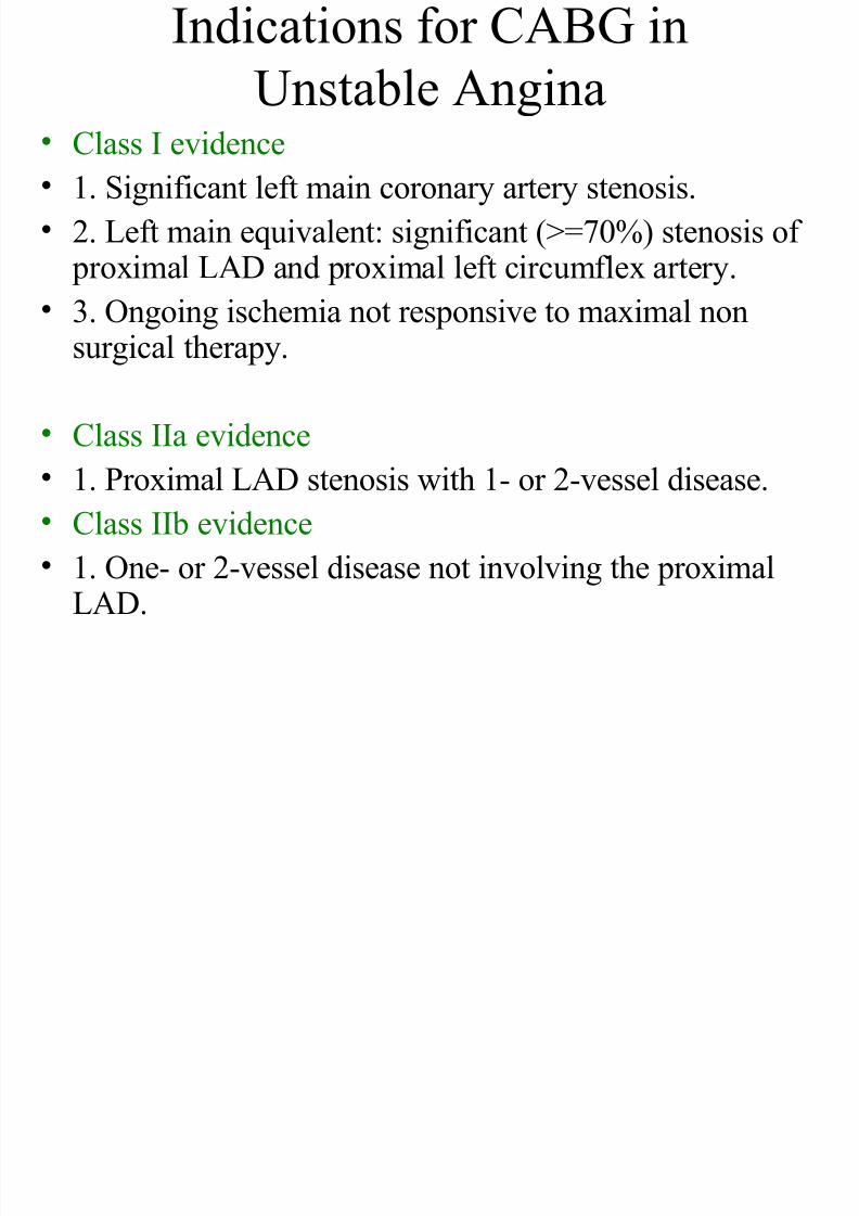
Indications for CABG in
Unstable Angina
• Class I evidence • 1. Significant left main coronary artery stenosis.
• 2. Left main equivalent: significant (>=70%) stenosis of proximal LAD and proximal left circumflex artery.
• 3. Ongoing ischemia not responsive to maximal nonsurgical therapy.
•Class IIa evidence • 1. Proximal LAD stenosis with 1- or 2-vessel disease.
• Class IIb evidence
• 1. One- or 2-vessel disease not involving the proximal
LAD.
7/31/2019 AHA Guidelines CABG
http://slidepdf.com/reader/full/aha-guidelines-cabg 13/24
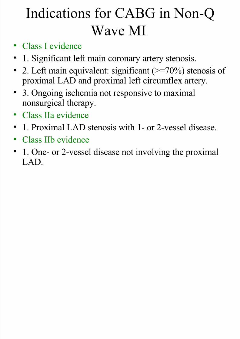
Indications for CABG in Non-Q
Wave MI• Class I evidence
• 1. Significant left main coronary artery stenosis.
• 2. Left main equivalent: significant (>=70%) stenosis of
proximal LAD and proximal left circumflex artery.• 3. Ongoing ischemia not responsive to maximal
nonsurgical therapy.
• Class IIa evidence
• 1. Proximal LAD stenosis with 1- or 2-vessel disease.• Class IIb evidence
• 1. One- or 2-vessel disease not involving the proximalLAD.
7/31/2019 AHA Guidelines CABG
http://slidepdf.com/reader/full/aha-guidelines-cabg 14/24
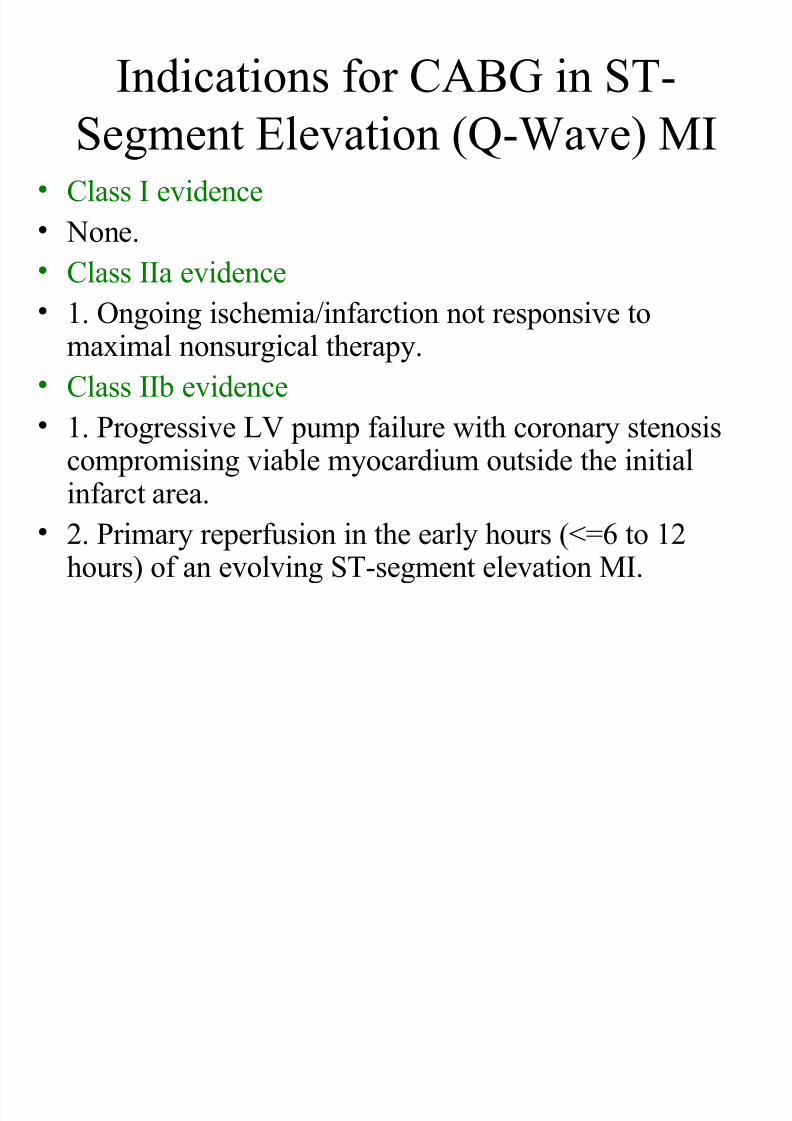
Indications for CABG in ST-
Segment Elevation (Q-Wave) MI• Class I evidence
• None.
• Class IIa evidence • 1. Ongoing ischemia/infarction not responsive to
maximal nonsurgical therapy.
• Class IIb evidence
• 1. Progressive LV pump failure with coronary stenosiscompromising viable myocardium outside the initialinfarct area.
• 2. Primary reperfusion in the early hours (<=6 to 12hours) of an evolving ST-segment elevation MI.
7/31/2019 AHA Guidelines CABG
http://slidepdf.com/reader/full/aha-guidelines-cabg 15/24
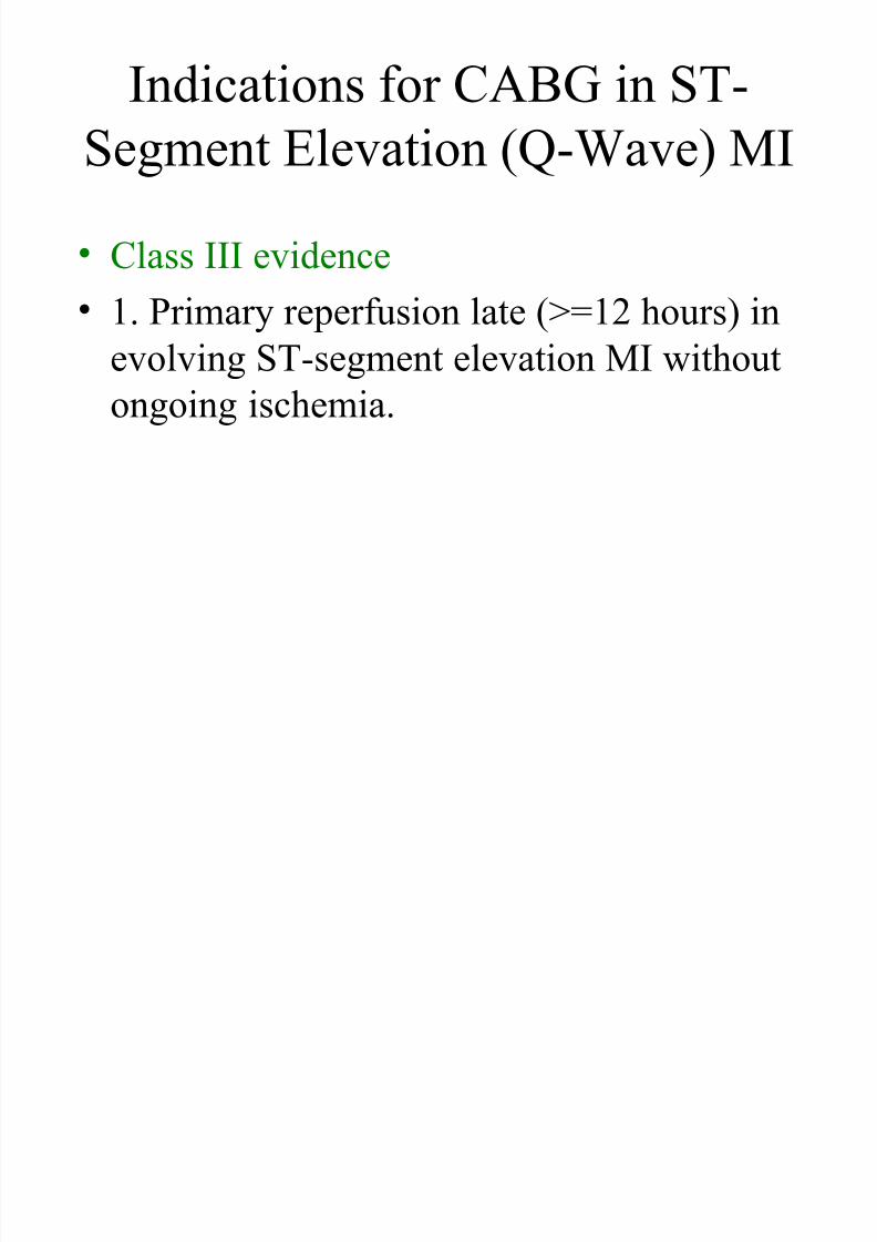
Indications for CABG in ST-
Segment Elevation (Q-Wave) MI
• Class III evidence
• 1. Primary reperfusion late (>=12 hours) inevolving ST-segment elevation MI without
ongoing ischemia.
7/31/2019 AHA Guidelines CABG
http://slidepdf.com/reader/full/aha-guidelines-cabg 16/24
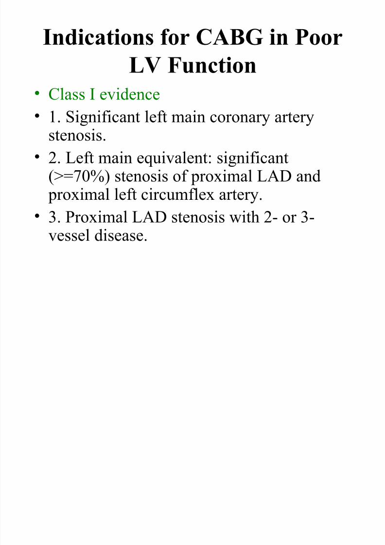
Indications for CABG in Poor
LV Function• Class I evidence
• 1. Significant left main coronary artery
stenosis.• 2. Left main equivalent: significant
(>=70%) stenosis of proximal LAD and
proximal left circumflex artery.• 3. Proximal LAD stenosis with 2- or 3-vessel disease.
7/31/2019 AHA Guidelines CABG
http://slidepdf.com/reader/full/aha-guidelines-cabg 17/24
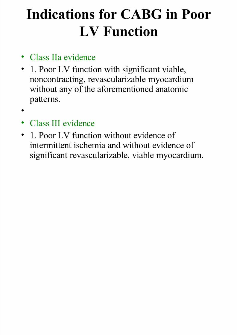
Indications for CABG in Poor
LV Function
• Class IIa evidence
• 1. Poor LV function with significant viable,
noncontracting, revascularizable myocardiumwithout any of the aforementioned anatomic patterns.
•
• Class III evidence • 1. Poor LV function without evidence of
intermittent ischemia and without evidence of significant revascularizable, viable myocardium.
7/31/2019 AHA Guidelines CABG
http://slidepdf.com/reader/full/aha-guidelines-cabg 18/24
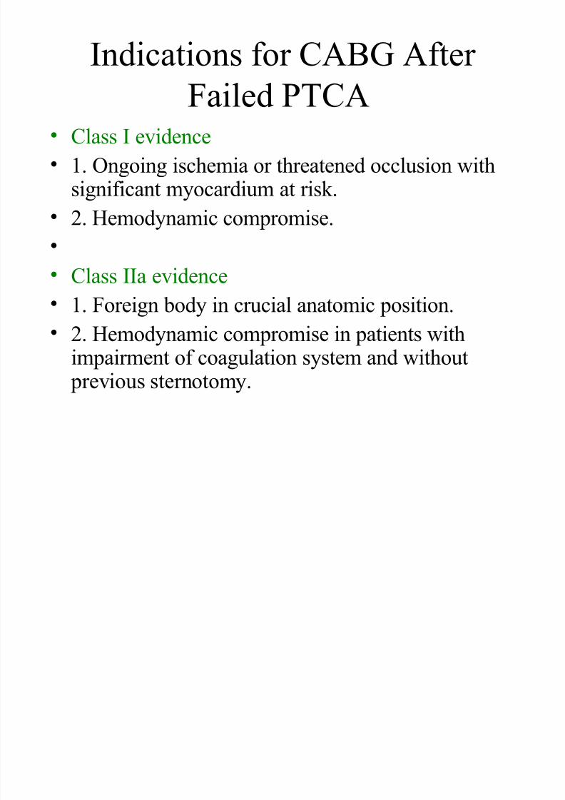
Indications for CABG After
Failed PTCA• Class I evidence
• 1. Ongoing ischemia or threatened occlusion with
significant myocardium at risk.• 2. Hemodynamic compromise.
•
• Class IIa evidence
• 1. Foreign body in crucial anatomic position.
• 2. Hemodynamic compromise in patients withimpairment of coagulation system and without
previous sternotomy.
7/31/2019 AHA Guidelines CABG
http://slidepdf.com/reader/full/aha-guidelines-cabg 19/24
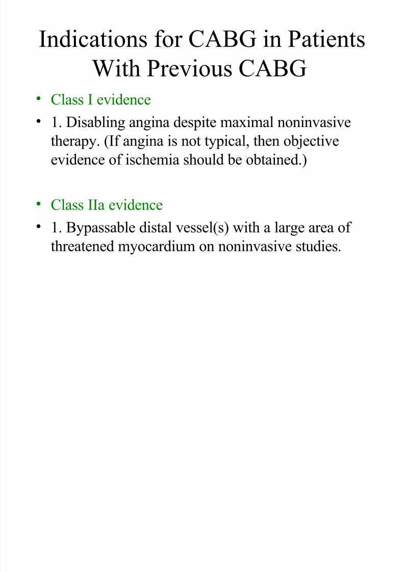
Indications for CABG in Patients
With Previous CABG• Class I evidence
• 1. Disabling angina despite maximal noninvasive
therapy. (If angina is not typical, then objectiveevidence of ischemia should be obtained.)
•Class IIa evidence
• 1. Bypassable distal vessel(s) with a large area of
threatened myocardium on noninvasive studies.
7/31/2019 AHA Guidelines CABG
http://slidepdf.com/reader/full/aha-guidelines-cabg 20/24
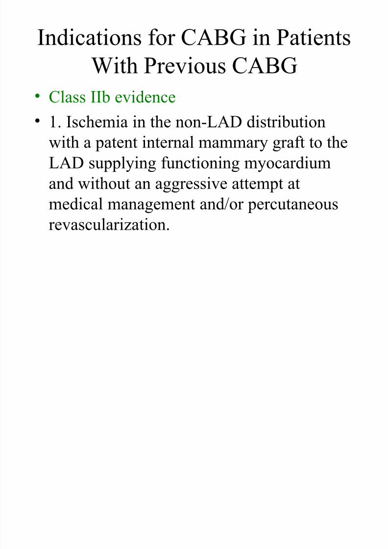
Indications for CABG in Patients
With Previous CABG• Class IIb evidence
• 1. Ischemia in the non-LAD distribution
with a patent internal mammary graft to the
LAD supplying functioning myocardium
and without an aggressive attempt at
medical management and/or percutaneousrevascularization.
7/31/2019 AHA Guidelines CABG
http://slidepdf.com/reader/full/aha-guidelines-cabg 21/24
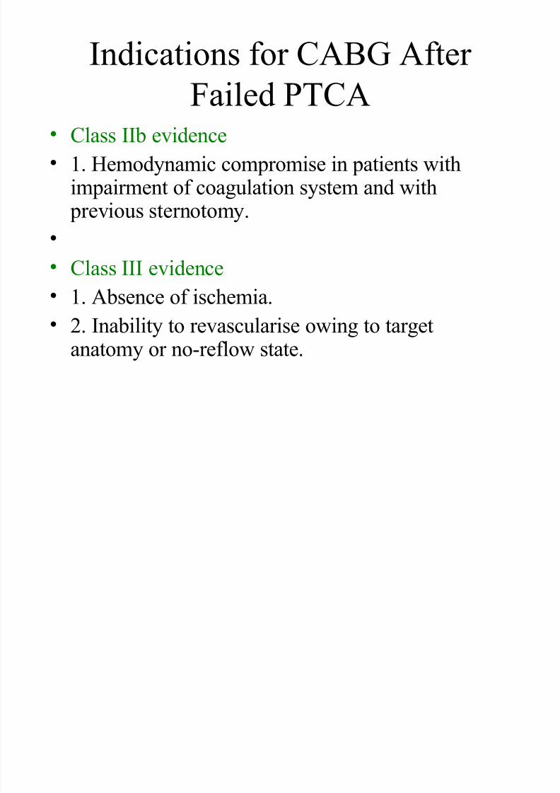
Indications for CABG After
Failed PTCA• Class IIb evidence
• 1. Hemodynamic compromise in patients with
impairment of coagulation system and with previous sternotomy.
•
• Class III evidence
• 1. Absence of ischemia.• 2. Inability to revascularise owing to target
anatomy or no-reflow state.
7/31/2019 AHA Guidelines CABG
http://slidepdf.com/reader/full/aha-guidelines-cabg 22/24
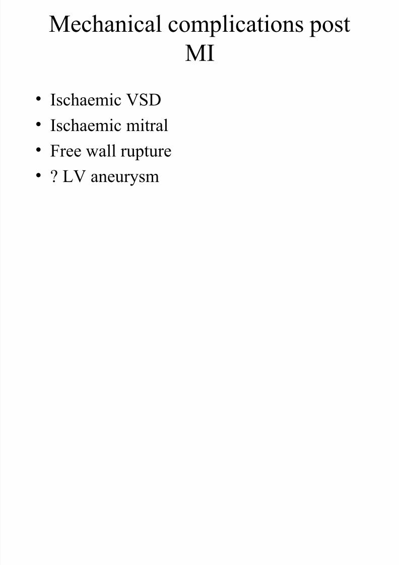
Mechanical complications post
MI
• Ischaemic VSD
• Ischaemic mitral
• Free wall rupture
• ? LV aneurysm

7/31/2019 AHA Guidelines CABG
http://slidepdf.com/reader/full/aha-guidelines-cabg 23/24
Associated defects
• Eg Valve, root etc etc

7/31/2019 AHA Guidelines CABG
http://slidepdf.com/reader/full/aha-guidelines-cabg 24/24
Viable myocardium
• What is best test for it ?