Embed Size (px)
Citation preview

Age-related BMAL1 change affects mouse bone marrowstromal cell proliferation and osteo-differentiationpotential
Yijia Chen1,2, Xiaomei Xu3, Zhen Tan2,4, Cui Ye1,2, Qing Zhao1,2, Yangxi Chen1,2
A b s t r a c t
Introduction: Aging people’s bone regeneration potential is always impaired.Bone marrow stromal cells (MSCs) contain progenitors of osteoblasts. Donorage may affect MSCs’ proliferation and differentiation potential, but the genom-ic base is still unknown. Due to recent research’s indication that a core circadi-an component, brain and muscle ARNT-like 1 protein (BMAL1), has a role in pre-mature aging, we investigated the normal aging mechanism in mice with theirMSCs and Bmal1 gene/protein level.Material and methods: 1, 6 and 16 month old C57BL/6 mice were used and thebone marrow stromal cells were gained and cultured at early passage. Bmal1gene and protein level were detected in these cells. Marrow stromal cells werealso induced to differentiate to osteoblasts or adipocytes. Three groups of miceMSCs were compared on proliferation by flow cytometry, on cell senescence bySA-β-gal expression and after osteo-induction on osteogenic potential by theexpression of osterix (Osx), alkaline phosphatase (ALP) and osteocalcin (OCN).Results: Bmal1 gene and protein level as well as S-phase fraction of the cell cycledecreased in MSCs along with the aging process. At the same time, SA-β-gal+levels increased, especially in the aged mice MSCs. When induced to be osteogenic,Osx gene expression and ALP activity declined in the mid-age and aged mice MSCs,while OCN protein secretion deteriorated in the aged mice MSCs.Conclusions: These findings demonstrate that mouse MSCs changed with theirproliferation and osteo-differentiation abilities at different aging stages, andthat Bmal1 is related to the normal aging process in MSCs.
Key words: brain and muscle ARNT-like 1 protein, bone marrow stromal cells, aging,differentiation, proliferation.
Introduction
In the elderly, consequences of teeth loss for the maxilla and mandibleare well known [1]. They are manifested by loss of jaw bone, especiallybone that forms the socket of the teeth on the jaw, which is termed “alve-olar bone”, the same as other bones in the human body. The poor boneregeneration potential will prolong or even ruin the result of orthodontic
Corresponding author:Qing ZhaoNo. 14, Section 3Renmin Nan RoadChengdu, Sichuan610041, ChinaPhone: +86 028 85502207E-mail: [email protected]
Basic research
1Orthodontic Centre, West China College of Stomatology, Sichuan University, Chengdu,China
2State Key Laboratory of Oral Diseases, Sichuan University, Chengdu, China3Department of Orthodontics, Stomatological Hospital of Luzhou Medical College,Luzhou, China
4Oral Implant Centre, West China College of Stomatology, Sichuan University, Chengdu,China
Submitted: 19 October 2010Accepted: 24 February 2011
Arch Med Sci 2012; 8, 1: 30-38DOI: 10.5114/aoms.2012.27277Copyright © 2012 Termedia & Banach
Arch Med Sci 1, February / 2012 31
Age-related BMAL1 change affects mouse bone marrow stromal cell proliferation and osteo-differentiation potential
treatment for adult and elderly people. Losing bonecan cause an aged morphological change on theface and impair possible implant or denture place-ment. The aged bone loss is due to the diminishedactivity of bone forming cells. Osteoblasts arederived from progenitors residing in the bone stro-ma. These cells are normally called bone marrowstromal cells (MSCs) [2]. When aging, while MSCsstill have self-renewal ability and multi-differentia-tion ability, these abilities, but the mechanism ofthe aging change is still being explored.
Recently, studies have focused on BMAL1, a corecomponent of circadian rhythm, concerning its rolein the aging process of mammals [3, 4]. Bmal–/–mice show early aging symptoms compared withtheir wild-type littermates. These symptoms includedecreased life span, arthropathy, osteoporosis, etc.,and the same defects do not occur in other circa-dian gene knock-out animals. Researchers placeBMAL1 as another osteo-differentiation-related genechanging with age, highlighting its non-circadianfunctions. While formerly found in the suprachias-matic nucleus (SCN) and controlling daily rhythm,circadian genes are also involved in control ofhomeostasis in peripheral tissues and organs. Theperipheral clocks, such as in the liver, skeletal mus-cle, etc, while synchronized with the central circa-dian pacemaker, can play a tissue-specific role inde-pendently from the SCN [5]. Circadian genes areexpressed in many kinds of cells in mammalian tis-sues, including the bone marrow stromal cells [6].
While early-aging symptoms of Bmal1–/– micearoused great interest, the normal aging changesof Bmal1 expression in MSCs or other cells relatedto their vitality are still unknown. Bmal1’s role inaging osteogenesis change of MSCs is also lackingany evidence.
The purpose of the present study was to inves-tigate the age-related effects in mouse MSCs,namely proliferation and osteo-differentiationpotential as well as the relationship between thesechanges and Bmal1 expression.
Material and methods
Animals and cell culture
Young adult (1 month old), middle-aged (6 monthsold) and aged (16 months old) male C57BL/6 micewere obtained from the Animal Centre of SichuanUniversity and the operations in the study havebeen approved by the University’s Bioethics Com-mittee. Animals were housed with free access towater and were maintained at a constant temper-ature on a 12-h light-dark cycle.
The cell isolation method was reported previ-ously [7]. Briefly, both femora and tibias wereremoved and soft tissues were detached. The meta-physes from both ends of the bone were resected
and the marrow cavities were flushed with sterilemedium using a #25 gauge needle. After washingwith phosphate buffered saline (PBS) by centrifuge,the culture was established in T25 flasks in freshalpha-MEM (Gibco) with 10% fetal bovine serum(Hyclone), 100 U/ml penicillin, and 100 mg/ml strep-tomycin (as basic medium). Cells were maintainedat 37°C in a humidified atmosphere containing 5%CO2. After 48 h, the non-adherent cells wereremoved by gentle rinsing with sterile PBS whichwas pre-warmed to 37°C, and the cells maintainedin basic medium with 2~3 times per week mediumchange until confluent. When confluent, the MSCswere passaged by 0.25% trypsin and 0.01% ethyl-enediaminetetraacetic acid (EDTA). Cells of passage3 or 4 were used in the following experiments.
Marrow stromal cells identification, osteo- and adipo-differentiation procedure
Marrow stromal cells were identified as CD29(+),Stro-1(+), CD45(–) and CD34(–) by cell immunohis-tochemistry as other studies suggested [8]. Con-fluent cultures of MSCs were induced to undergoosteogenesis by basic medium supplemented with10 nM dexamethasone, 10 mM β-glycerophosphate,and 50 μg/ml L-ascorbate (all from Sigma, as OSmedium). After 21 days in OS medium, the osteocultures were rinsed in 0.9% NaCl, fixed in 70%ethanol, and stained with 1% Alizarin Red for detec-tion of calcium phosphate mineralization. The cul-tures were also processed with routine von Kossastaining.
Marrow stromal cells were induced to undergoadipogenesis by replacing the basic medium withadipocyte induction medium (as AD medium) com-posed of α-MEM with 10% fetal bovine serum (FBS),supplemented with 100 nM dexamethasone,0.5 mM isobutylmethylxanthine (IBMX), 10 μg/mlinsulin, 0.2 mM indomethacin, 100 U/ml penicillin,and 100 mg/ml streptomycin. After 3 days of cul-ture with AD medium, the medium was replacedwith AD medium without IBMX and indomethacinup to 10 days for maintenance. After that, fixed cellswere stained with 0.5% oil red O for 1 h for obser-vation of adipogenesis.
Flow cytometry assay
When passage 2 cells reached 80% confluence,they were passaged 1 : 2 to a T25 flask. After 3 days, the cells were detached with trypsin andEDTA, twice centrifuged at 500 g for 5 min con-taining PBS wash and then fixed with 70% alco-hol overnight. Then the S-phase fraction of totalcells (SPF) in each sample was analysed and cal-culated through flow cytometry (Beckman, US)and according to the formula: SPF (%) = S/(G0/G1+ S + G2/M) × 100%.

32 Arch Med Sci 1, February / 2012
Assessment of senescence-associated β-galactosidase staining
Marrow stromal cells s were seeded in a 6-wellplate with 2 × 10³ cell/cm2. After 2 days of culture,the medium was discarded, cells were rinsed withPBS once, 1 ml of fixative per well was added for 15 min, and subsequently rinsed with PBS 3 times.Then 1 ml per well of working solution of β-galac-tosidase with X-Gal was placed and the plate wasmaintained at 37°C overnight (senescence-associ-ated β-galactosidase staining kit from BeyotimeChina). The senescent cells were observed in anoptical microscope and counted from 5 randomfields of vision.
Real-time RT-PCR analysis
For real-time RT-PCR analysis, cells were har-vested and maintained in RNA preservation solu-tion (RNAsafeguard, Keygen, China) before the fol-lowing experiment. The total RNA was extractedusing a simple P total RNA extraction kit (Bioer, Chi-na). Total RNA was quantified, in a spectropho-tometer, at an absorbance (A) of 260 nm. The RNAsamples had an A260 : A280 ratio of 2.0 to guar-antee high purity. Two miligrams of total RNA fromeach sample was subjected to reverse transcrip-tion using the SYBR_ PrimeScriptTM RT-PCR Kit(TaKaRa, China) according to the manufacturer’sprotocol. Each real time PCR was carried out in trip-licate in a total of 20 μl reaction mixture in an ABIPRISM 7300 Real-time PCR System (Applied Biosys-tems, US). Primers used for real-time PCR analysisare presented in Table I. The PCR program was con-ducted according to the suggestion of the Takaramanual. The starting copy numbers of unknownsamples were calculated by the 7300 System SDSSoftware (Applied Biosystems) from the standardcurve. The housekeeping gene β-actin was con-currently amplified in each sample as a control andwas used for normalization. The cDNA of youngadult MSCs with or without osteo-induction nor-malized to the level of β-actin was ascribed a foldinduction of 1. Melting curves for each PCR reac-tion were generated to ensure the purity of theamplification product.
Quantitative alkaline phosphatase and proteinassays
For quantitative analysis of alkaline phosphataseactivities, cells after 14 days of osteo-induction werewashed with PBS, prepared following the instruc-tion of the alkaline phosphatase (ALP) assay kit(Merit Choice, China). Briefly, cells were lysed with10 mM Tris-HCl and 0.1% Triton X-100, pH 7.4 andrepeating freeze-thaw cycle for 3 times. After son-ication, the lysates were taken for assay ALP ina microtitre plate using p-nitrophenyl phosphateas the substrate. The absorbance of p-nitrophenolformed by the hydrolysis of p-nitrophenyl phos-phate, catalysed by ALP, was measured at 405 nmby a microplate reader (HTS 7000 Plus, PerkinElmer,US). Total protein content was measured by theBradford method, read at 595 nm and calculatedaccording to the bovine γ globulin standards. Alka-line phosphatase activity was expressed as unit/gprotein.
Enzyme-linked immunosorbent assay for osteocalcin
To quantify and compare the concentration ofosteocalcin (OCN) in cell cultured supernatant ofthe three groups of MSCs, commercially availableenzyme linked immunosorbent assay (ELISA) kitswere used according to the manufacturer’s recom-mendations (R&D, US). Constitutive OCN secretionwas analysed after 2 weeks of osteogenic induc-tion. Prior to collection, the supernatant of themedium was centrifuged to remove cell debris.
Western blotting assay for BMAL1 protein level
To obtain whole-cell protein extracts, cells werewashed twice with ice-cold PBS and then lysed ina lysis buffer (Keygen total protein extraction kit,Keygen Biotech, China). The supernatant was col-lected after centrifugation at 14,000g at 4°C for 15 min and assayed quantitatively with the BCAmethod. The total protein extracts were subjectedto the standard procedure of SDS-PAGE and sub-sequent transfer to a PVDF membrane. After block-ing, the membranes were probed with dilution ofthe anti-BMAL and anti-β-actin primary antibody
Yijia Chen, Xiaomei Xu, Zhen Tan, Cui Ye, Qing Zhao,Yangxi Chen
Target gene Primers Sequence Fragment size [bp]
Bmal1 Forward primer 5’-AACCTTCCCGCAGCTAACAG-3’ 79
Reverse primer 5’-AGTCCTCTTTGGGCCACCTT-3
Osx Forward primer 5’-TATGCTCCGACCTCCTCAAC-3’ 120
Reverse primer 5’-AATAGGATTGGGAAGCAGAAA-3’
β-actin Forward primer 5’-GGGCTGTATTCCCCTCCATCG-3’ 201
Reverse primer 5’-GCAGCTCATTGTAGAAGGTGTGGTG-3’
Table I. qRT-PCR primers

Arch Med Sci 1, February / 2012 33
Age-related BMAL1 change affects mouse bone marrow stromal cell proliferation and osteo-differentiation potential
(Abcam), followed by the addition of horseradishperoxidase (HRP)-conjugated secondary antibody.Immunoreactive proteins were visualized usinga chemiluminescence kit (Millipore). Band intensi-ties were determined using the ChemiDoc XRS Geldocumentation system and Quantity One software(Bio-Rad).
Statistical analysis
All experiments were conducted a minimum of3 times. Measurements are expressed as mean ± SD. Statistical comparisons were made using fac-torial analysis of variance (ANOVA), followed by mul-tiple comparisons using the SNK test. A value of p < 0.05 was considered statistically significant.
Results
Cell culture and osteoblast and adipogenic differentiation
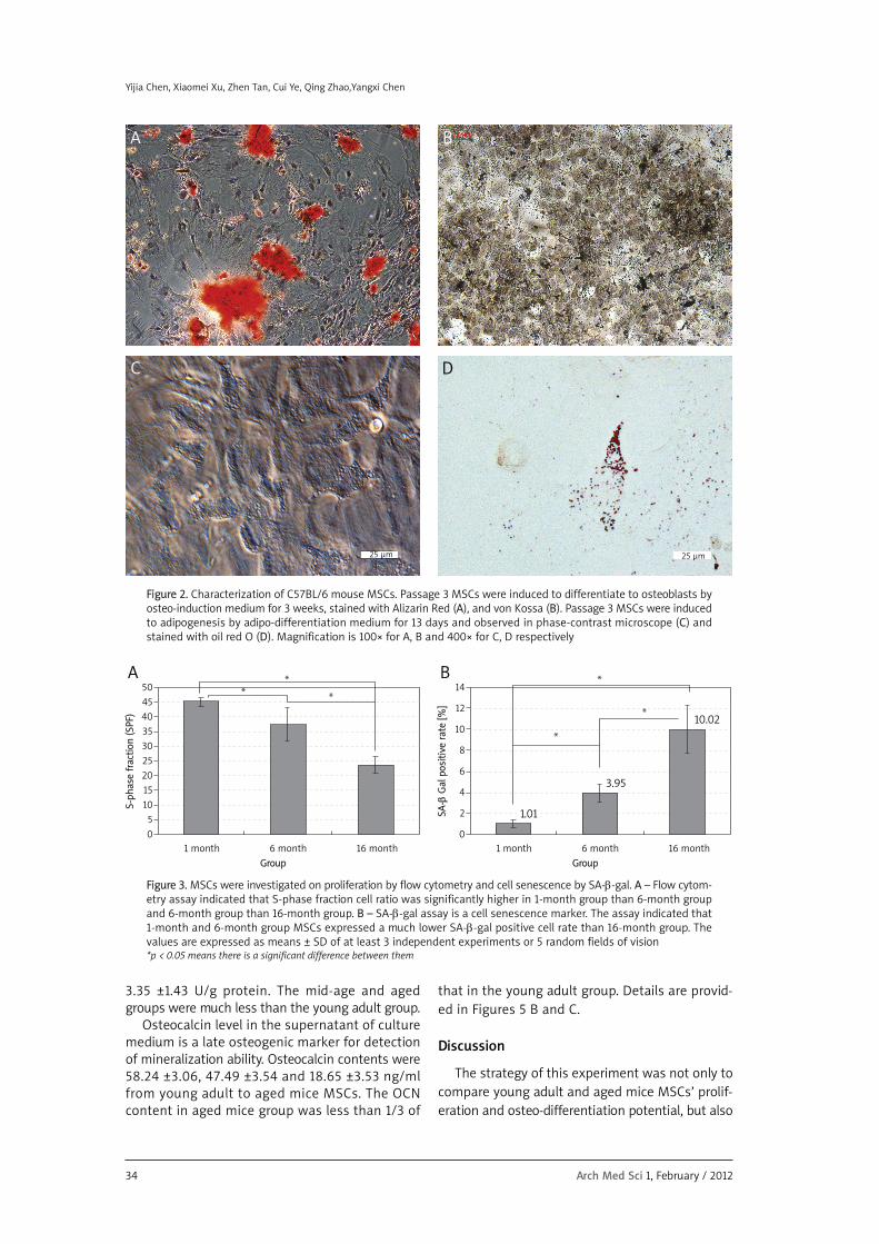
Marrow stromal cells primary culture and pas-sage 3 culture are shown in Figures 1 A-B. As canbe seen, the shape of the cells was more spindle-like after passaging, but we cannot observe anymorphological difference among the three groupsof MSCs. Besides identification of CD (data notshown), the MSCs isolated from three different agesof animal can all be induced to differentiate toosteoblasts or adipocytes at the stated time by thedifferentiation medium. The common view underphase contrast microscope after adipo-inductionand the result of Alizarin Red, von Kossa and oil redO staining of a sample of MSCs are presented inFigures 2 A-D.
Effects of age on cell proliferation marker and cellular senescence
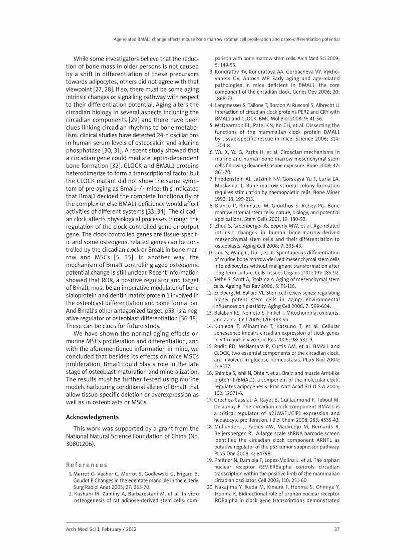
Through flow cytometry for detection of cell cyclephase proportion, the results showed that the S-phase fraction rates in 1, 6 and 16 month old miceMSCs were 45.38 ±1.52, 37.64 ±5.72, 23.8 ±2.91 per-
cent respectively. The aged mice MSCs containedan S-phase fraction rate which was only about halfof that in young adult MSCs. The result is present-ed in Figure 3 A.
Marrow stromal cells isolated from young adult,mid-age and aged mice have a growing rate of SAβ-gal+ cells. But both young adult and mid-age miceMSCs presented an SA-β-gal+ rate lower than 5%,which can be treated as not significant as otherresearchers suggested [9]. In aged mice, the SA-β-gal+ cells were more than 10% of the totalcount, and showed a slower proliferation rate whenpassaging (data not shown), which is a sign of sus-ceptibility to toxin in serum or capacity for senes-cence (see Figure 3 B).
Bmal1 and Osx expression affected by age
Bmal1 gene and protein level dropped along withage, but was much lower in aged mice MSCs. Theaged mice MSCs Bmal1 gene and protein level wasless than half of the young adult mice MSCs. Andthe aged mice MSCs Bmal1/β-actin protein rate waseven less than 1 (Figures 4 A-C).
Osx is a transcription factor that is more strong-ly expressed in the progenitors of osteoblasts thanchondrocytes and also can be detected in MSCs,the progenitors of osteoblasts, chondrocytes andadipocytes. The result of RT-qPCR showed thatwhen MSCs were differentiated into osteoblasts bydexamethasone, β-glycerophosphate, and L-ascor-bate lasting for 2 weeks, their Osx gene level washigher in the young adult and mid-age mice groupthan the aged mice group (Figure 5 A).
Alkaline phosphatase activity and osteocalcinconcentration after 2 weeks of osteogenicinduction
Alkaline phosphatase activity level is an earlyosteoblast marker. When comparing the threegroups of cells and detecting the ALP activity level,the results were 15.05 ±1.81 U/g, 8 ±1.44 U/g,
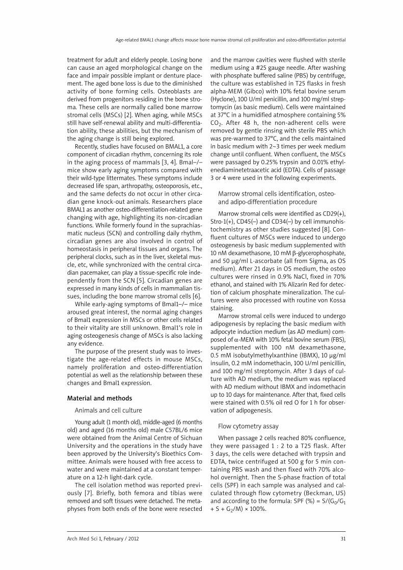
Figure 1. C57BL/6 mouse bone marrow stromal cells primary culture (A) and passage 3 culture (B) maintained inbasic culture medium were observed by phase-contrast microscope. Magnification is 100×
A B
100 μm
100 μm
34 Arch Med Sci 1, February / 2012
3.35 ±1.43 U/g protein. The mid-age and agedgroups were much less than the young adult group.
Osteocalcin level in the supernatant of culturemedium is a late osteogenic marker for detectionof mineralization ability. Osteocalcin contents were58.24 ±3.06, 47.49 ±3.54 and 18.65 ±3.53 ng/mlfrom young adult to aged mice MSCs. The OCNcontent in aged mice group was less than 1/3 of
that in the young adult group. Details are provid-ed in Figures 5 B and C.
Discussion
The strategy of this experiment was not only tocompare young adult and aged mice MSCs’ prolif-eration and osteo-differentiation potential, but also
Yijia Chen, Xiaomei Xu, Zhen Tan, Cui Ye, Qing Zhao,Yangxi Chen
Figure 2. Characterization of C57BL/6 mouse MSCs. Passage 3 MSCs were induced to differentiate to osteoblasts byosteo-induction medium for 3 weeks, stained with Alizarin Red (A), and von Kossa (B). Passage 3 MSCs were inducedto adipogenesis by adipo-differentiation medium for 13 days and observed in phase-contrast microscope (C) andstained with oil red O (D). Magnification is 100× for A, B and 400× for C, D respectively
A B
DC
50
45
40
35
30
25
20
15
10
5
0
14
12
10
8
6
4
2
01 month 6 month 16 month
Group
1 month 6 month 16 month
Group
Figure 3. MSCs were investigated on proliferation by flow cytometry and cell senescence by SA-β-gal. A – Flow cytom-etry assay indicated that S-phase fraction cell ratio was significantly higher in 1-month group than 6-month groupand 6-month group than 16-month group. B – SA-β-gal assay is a cell senescence marker. The assay indicated that1-month and 6-month group MSCs expressed a much lower SA-β-gal positive cell rate than 16-month group. Thevalues are expressed as means ± SD of at least 3 independent experiments or 5 random fields of vision*p < 0.05 means there is a significant difference between them
S-ph
ase
frac
tion
(SPF
)
SA-β
Gal
pos
itiv
e ra
te [%
]
A B
1.01
3.95
10.02
25 μm25 μm

Arch Med Sci 1, February / 2012 35
Age-related BMAL1 change affects mouse bone marrow stromal cell proliferation and osteo-differentiation potential
to add a mid-age group, because we believe theaging procedure advances gradually. As expected,the results of this study revealed that along withaging, MSCs of C57BL/6 mice expressed more SA-β-gal, especially in aged mice; with the reversetrend, the S-phase fraction ratio of all cell cycles andthe Bmal1 gene and protein level dropped. Whencells were induced to differentiate to osteoblasts,the three osteo-differentiation markers Osx, ALP
1.2
1.0
0.8
0.6
0.4
0.2
0
1.8
1.6
1.4
1.2
1.0
0.8
0.6
0.4
0.2
0
1 month 6 month 16 month
Group
1 month 6 month 16 month
Group
Bm
al1
gene
exp
ress
ion
Prot
ein
(Bm
al/a
ctin
)A
B
C
Figure 4. Bmal1 gene and protein level in the threegroups of C57BL/6 mouse MSCs. A – qRT-PCR resultsindicate there was much lower Bmal1 mRNA expres-sion in 6-month and 16-month groups of MSCs than1-month group. The 16-month group’s Bmal1 mRNAlevel was less than 1/3 of that in 1 month group. ThecDNA of 1-month group MSCs normalized to the lev-el of β-actin was ascribed as a fold induction of 1. B, C – Western blotting results showed the sametrend of Bmal1 expression in the three groups. Theprotein levels were calculated by Bmal1/β-actin pro-tein expression ratio. The values are expressed asmeans ± SD of at least 3 independent experiments*p < 0.05 means there is a significant difference between them
1.2
1.0
0.8
0.6
0.4
0.2
0
70
60
50
40
30
20
10
0
1 month 6 month 16 month
Group
1 month 6 month 16 month
Group
Osx
gen
e ex
pres
sion
18
16
14
12
10
8
6
4
2
01 month 6 month 16 month
Group
ALP
act
ivit
y (U
/E p
rote
in)
OCN
[ng/
ml]
A
B
C
Figure 5. Osteoblast marker Osx, ALP and OCNexpression on cultured cells or supernatant of cul-ture medium after 2 weeks of maintenance in osteo-induction medium. A – Osx is an osteogenesis relat-ed transcription factor. The MSCs Osx mRNA levelshowed a decreasing trend, 6-month to 1-monthgroup and 16-month to 6-month group. The cDNAof 1-month group MSCs normalized to the level ofβ-actin was ascribed as a fold induction of 1. B – ALPis an early marker of osteogenesis. The ALP activityin MSCs cell lysates indicated that the level in 6-month group and 16-month group were about halfand below 1/4 of that in 1-month group respective-ly. C – The OCN is a late osteoblast maturation mark-er. The OCN protein level in the supernatant of culture medium was calculated by ELISA. The OCNconcentration was about 18.65 ng/ml in 16-monthgroup MSCs compared to 47.49 ng/ml in 6-monthgroup and 58.24 ng/ml in 1-month group. The values are expressed as means ± SD of at least 3 independent experiments*p < 0.05 means there is a significant difference between them
15.05
58.24
47.49
18.65
8.00
3.35

36 Arch Med Sci 1, February / 2012
and OCN presented a consistent trend, but the pro-portion of each age group was not exactly the same.The Osx level decreased along with the agingprocess, ALP activity was much higher in MSCs ofyoung adult mice, and OCN protein synthesis wasdramatically lower in MSCs of aged mice.
To find out whether there are intrinsic, senes-cence-associated changes of MSCs in vivo, we eval-uated the freshly isolated stromal cells at very ear-ly passage. This strategy was intended to avoidchanges in cell behaviours that are related to pro-longed culture [10]. Like many other types of mam-malian cells, MSCs with repeated passaging display“in vitro senescence”, such as decreased prolifera-tion, replicative quiescence, enlargement andincreased SA-β-gal activity [11]. This in vitro methodwas also intended to eliminate the aged-relatedextrinsic somatic environment change, i.e. declinesin circulating hormones [12] when applying in vivostudy, and to focus on the intrinsic aging changesthemselves.
In the present study, there was a much lower fre-quency of SA-β-gal positive cells and much higherS-phase fraction of the cell cycle in young adult andmid-age mice MSCs, compared with the aged one.In fact, the frequency of SA-β-gal positive cells inthe former two age groups can be ignored becausethe frequency was less than 5%. In older mice, theearly passage MSCs showed a frequency of 10% ofSA-β-gal positive cells, suggesting an age thresh-old of abundantly showing this marker, suggestingthat when aging, some MSCs fail to protect them-selves from metabolic waste or an increased levelof reactive oxygen species, etc [13]. The S-phasefraction is a measure of the percentage of cells thatare in the phase of DNA synthesis and representsa proliferative index of cell culture. The result of SPFdetection was consistent with our observation thataged mice MSCs needed more time to reach con-fluence than the other two groups under the sameculture conditions.
We detected the Bmal1 mRNA and protein levelin MSCs of the three groups. Its trend was as theSPF ratio and as the reverse transformation of SA-β-gal+ ratio. The core circadian protein BMAL1,in addition to its known functions in the circadianoscillator, plays essential non-redundant roles inthe control of tissue homeostasis and aging. Oneof the most striking phenomena was prematureaging of Bmal1–/– animals [3]. While there are nomorphological differences between Bmal1–/– ani-mals and their wild-type littermates at birth andearly age, 18-week-old Bmal1–/– mice showed signsof growth retardation and 36-week-old knockoutmice morphologically resembled aged mice. Thestudy suggested that the control of aging is a nov-el function of Bmal1, relatively independent fromits role in the circadian oscillator. Along with our
study results, the Bmal1 gene and protein level weremuch lower in older mice’s MSCs, and the fre-quency of SA-β-gal positive cells was much higheras well. We can speculate that Bmal1 reduction ormalfunction can also be a candidate reason ofMSCs in vivo ageing or cell senescence [14]. BecauseBMAL1 was reported to participate in controllingglucose and fat metabolism and homeostasis [15,16], and MSCs are the progenitors of adipocytes,chondrocytes and osteoblasts, the hypothesis maybe convincing. Recently, studies showed that cyclin-dependent kinase inhibitor p21, which negativelyregulates the G1/S phase transition, is antagonizedby Bmal1 through the orphan nuclear receptorROR/REV-ERB pathway with or without a relation-ship with p21’s regulator, p53 [17, 18]. These inves-tigations established a novel molecular linkbetween clock and cell cycle genes. Because RORand REV-ERB are BMAL1’s activator and repressorrespectively, the three transcriptional factors canform an interlock loop that plays a stabilizing rolein circadian rhythm [19, 20]. This may be the rea-son why the aged mice MSCs showed a much moredecreased Bmal1 level and SPF rate as well.
In aging, bone formation activity is significantlyreduced. It has been postulated that a deficiencyin osteoprogenitor cells is the main reason for thedecline in bone induction activity [21]. This isbecause osteoprogenitor cells play a major role notonly in bone formation but also in bone resorption[22, 23]. In contrast, a few researchers found noeffects of age-related decline in osteoblast poten-tial when applying colony assays for the numbersof colonies that stain for alkaline phosphatase [24].Osteoblast differentiation is regulated by a com-plex network of transcription factors and differentsignalling pathways. Osteoblastic differentiation invitro is also characterized by three distinct stagesof cellular activity: proliferation, extracellular matrix(ECM) maturation and mineralization [25, 26]. Sowe recruited three indices of osteoblast differenti-ation: transcriptional factor Osx, early osteogenicmarker ALP and late osteogenic marker OCN,hypothesizing that their aging evolvement wouldbe different. As we expected, the Osx level in MSCsdecreased gradually with aging of the mice, reveal-ing a different response to osteogenic activation.The early osteoblast marker ALP activity was alsohigher in young adult mice MSCs, but much lowerin mid-age and aged mice MSCs. However, whileALP level was much lower in mid-age MSCs thanyoung adult MSCs, the late osteoblast marker, OCNlevel, was not statistically different between thetwo groups, and it needs to be stressed that theaged mice showed the dramatically lowest level ofOCN. Our research results showed a gradual changeof osteogenic ability along with aging with osteo-differentiation indices.
Yijia Chen, Xiaomei Xu, Zhen Tan, Cui Ye, Qing Zhao,Yangxi Chen
Arch Med Sci 1, February / 2012 37
Age-related BMAL1 change affects mouse bone marrow stromal cell proliferation and osteo-differentiation potential
While some investigators believe that the reduc-tion of bone mass in older persons is not causedby a shift in differentiation of these precursorstowards adipocytes, others did not agree with thatviewpoint [27, 28]. If so, there must be some agingintrinsic changes or signalling pathway with respectto their differentiation potential. Aging alters thecircadian biology in several aspects including thecircadian components [29] and there have beenclues linking circadian rhythms to bone metabo-lism: clinical studies have detected 24-h oscillationsin human serum levels of osteocalcin and alkalinephosphatase [30, 31]. A recent study showed thata circadian gene could mediate leptin-dependentbone formation [32]. CLOCK and BMAL1 proteinsheterodimerize to form a transcriptional factor butthe CLOCK mutant did not show the same symp-tom of pre-aging as Bmal1–/– mice; this indicatedthat Bmal1 decided the complete functionality ofthe complex or else BMAL1 deficiency would affectactivities of different systems [33, 34]. The circadi-an clock affects physiological processes through theregulation of the clock-controlled gene or outputgene. The clock-controlled genes are tissue-specif-ic and some osteogenic related genes can be con-trolled by the circadian clock or Bmal1 in bone mar-row and MSCs [5, 35]. In another way, themechanism of Bmal1 controlling aged osteogenicpotential change is still unclear. Recent informationshowed that ROR, a positive regulator and targetof Bmal1, must be an imperative modulator of bonesialoprotein and dentin matrix protein 1 involved inthe osteoblast differentiation and bone formation.And Bmal1’s other antagonized target, p53, is a neg-ative regulator of osteoblast differentiation [36-38].These can be clues for future study.
We have shown the normal aging effects onmurine MSCs proliferation and differentiation, andwith the aforementioned information in mind, weconcluded that besides its effects on mice MSCsproliferation, Bmal1 could play a role in the latestage of osteoblast maturation and mineralization.The results must be further tested using murinemodels harbouring conditional alleles of Bmal1 thatallow tissue-specific deletion or overexpression aswell as in osteoblasts or MSCs.
Acknowledgments
This work was supported by a grant from theNational Natural Science Foundation of China (No.30801206).
Re f e r e n c e s 1. Merrot O, Vacher C, Merrot S, Godlewski G, Frigard B,
Goudot P. Changes in the edentate mandible in the elderly.Surg Radiol Anat 2005; 27: 265-70.
2. Kashani IR, Zaminy A, Barbarestani M, et al. In vitroosteogenesis of rat adipose-derived stem cells: com -
parison with bone marrow stem cells. Arch Med Sci 2009;5: 149-55.
3. Kondratov RV, Kondratova AA, Gorbacheva VY, Vykho -vanets OV, Antoch MP. Early aging and age-relatedpathologies in mice deficient in BMAL1, the corecomponent of the circadian clock. Genes Dev 2006; 20:1868-73.
4. Langmesser S, Tallone T, Bordon A, Rusconi S, Albrecht U.Interaction of circadian clock proteins PER2 and CRY withBMAL1 and CLOCK. BMC Mol Biol 2008; 9: 41-56.
5. McDearmon EL, Patel KN, Ko CH, et al. Dissecting thefunctions of the mammalian clock protein BMAL1 by tissue-specific rescue in mice. Science 2006; 314: 1304-8.
6. Wu X, Yu G, Parks H, et al. Circadian mechanisms inmurine and human bone marrow mesenchymal stemcells following dexamethasone exposure. Bone 2008; 42:861-70.
7. Friedenstein AJ, Latzinik NV, Gorskaya Yu F, Luria EA,Moskvina IL. Bone marrow stromal colony formationrequires stimulation by haemopoietic cells. Bone Miner1992; 18: 199-213.
8. Bianco P, Riminucci M, Gronthos S, Robey PG. Bonemarrow stromal stem cells: nature, biology, and potentialapplications. Stem Cells 2001; 19: 180-92.
9. Zhou S, Greenberger JS, Epperly MW, et al. Age-relatedintrinsic changes in human bone-marrow-derivedmesenchymal stem cells and their differentiation toosteoblasts. Aging Cell 2008; 7: 335-43.
10. Gou S, Wang C, Liu T, et al. Spontaneous differentiationof murine bone marrow-derived mesenchymal stem cellsinto adipocytes without malignant transformation afterlong-term culture. Cells Tissues Organs 2010; 191: 185-92.
11. Sethe S, Scutt A, Stolzing A. Aging of mesenchymal stemcells. Ageing Res Rev 2006; 5: 91-116.
12. Edelberg JM, Ballard VL. Stem cell review series: regulatinghighly potent stem cells in aging: environmentalinfluences on plasticity. Aging Cell 2008; 7: 599-604.
13. Balaban RS, Nemoto S, Finkel T. Mitochondria, oxidants,and aging. Cell 2005; 120: 483-95.
14. Kunieda T, Minamino T, Katsuno T, et al. Cellularsenescence impairs circadian expression of clock genesin vitro and in vivo. Circ Res 2006; 98: 532-9.
15. Rudic RD, McNamara P, Curtis AM, et al. BMAL1 andCLOCK, two essential components of the circadian clock,are involved in glucose homeostasis. PLoS Biol 2004; 2: e377.
16. Shimba S, Ishii N, Ohta Y, et al. Brain and muscle Arnt-likeprotein-1 (BMAL1), a component of the molecular clock,regulates adipogenesis. Proc Natl Acad Sci U S A 2005;102: 12071-6.
17. Grechez-Cassiau A, Rayet B, Guillaumond F, Teboul M,Delaunay F. The circadian clock component BMAL1 isa critical regulator of p21WAF1/CIP1 expression andhepatocyte proliferation. J Biol Chem 2008; 283: 4535-42.
18. Mullenders J, Fabius AW, Madiredjo M, Bernards R,Beijersbergen RL. A large scale shRNA barcode screenidentifies the circadian clock component ARNTL asputative regulator of the p53 tumor suppressor pathway.PLoS One 2009; 4: e4798.
19. Preitner N, Damiola F, Lopez-Molina L, et al. The orphannuclear receptor REV-ERBalpha controls circadiantranscription within the positive limb of the mammaliancircadian oscillator. Cell 2002; 110: 251-60.
20. Nakajima Y, Ikeda M, Kimura T, Honma S, Ohmiya Y,Honma K. Bidirectional role of orphan nuclear receptorRORalpha in clock gene transcriptions demonstrated

38 Arch Med Sci 1, February / 2012
by a novel reporter assay system. FEBS Lett 2004; 565:122-6.
21. Wlodarski KH, Wlodarski PK, Galus R. Senescence of osteo -genic cells. Review [Polish]. Ortop Traumatol Rehabil 2007;9: 63-7.
22. Fili S, Karalaki M, Schaller B. Therapeutic implications of osteoprotegerin. Cancer Cell Int 2009; 9; 26.
23. Fili S, Karalaki M, Schaller B. Mechanism of bonemetastasis: the role of osteoprotegerin and of the host-tissue microenvironment-related survival factors. CancerLett 2009; 283: 10-9.
24. Dobson K, Reading L, Scutt A. A cost-effective method forthe automatic quantitative analysis of fibroblastic colony-forming units. Calcif Tissue Int 1999; 65: 166-72.
25. Lian JB, Stein GS, Javed A, et al. Networks and hubs forthe transcriptional control of osteoblastogenesis. RevEndocr Metab Disord 2006; 7: 1-16.
26. Sila-Asna M, Bunyaratvej A, Maeda S, Kitaguchi H, Bunya -ratavej N. Osteoblast differentiation and bone formationgene expression in strontium-inducing bone marrowmesenchymal stem cell. Kobe J Med Sci 2007; 53: 25-35.
27. Moerman EJ, Teng K, Lipschitz DA, Lecka-Czernik B. Agingactivates adipogenic and suppresses osteogenic programsin mesenchymal marrow stroma/stem cells: the role ofPPAR-gamma2 transcription factor and TGF-beta/BMPsignaling pathways. Aging Cell 2004; 3: 379-89.
28. Kretlow JD, Jin YQ, Liu W, et al. Donor age and cell passageaffects differentiation potential of murine bone marrow-derived stem cells. BMC Cell Biol 2008; 9: 60-72.
29. Kolker DE, Fukuyama H, Huang DS, Takahashi JS, HortonTH, Turek FW. Aging alters circadian and light-inducedexpression of clock genes in golden hamsters. J BiolRhythms 2003; 18: 159-69.
30. Gundberg CM, Markowitz ME, Mizruchi M, Rosen JF.Osteocalcin in human serum: a circadian rhythm. J ClinEndocrinol Metab 1985; 60: 736-9.
31. Shao P, Ohtsuka-Isoya M, Shinoda H. Circadian rhythmsin serum bone markers and their relation to the effect ofetidronate in rats. Chronobiol Int 2003; 20: 325-36.
32. Fu L, Patel MS, Bradley A, Wagner EF, Karsenty G. Themolecular clock mediates leptin-regulated bone formation.Cell 2005; 122: 803-15.
33. Kwon I, Lee J, Chang SH, et al. BMAL1 shuttling controlstransactivation and degradation of the CLOCK/BMAL1heterodimer. Mol Cell Biol 2006; 26: 7318-30.
34. Kondratov RV. A role of the circadian system and circadianproteins in aging. Ageing Res Rev 2007; 6: 12-27.
35. Liu AC, Tran HG, Zhang EE, Priest AA, Welsh DK, Kay SA.Redundant function of REV-ERBalpha and beta and non-essential role for Bmal1 cycling in transcriptional regu -lation of intracellular circadian rhythms. PLoS Genet 2008;4: e1000023.
36. Miyamoto S, Cooper L, Watanabe K, et al. Role of retinoicacid-related orphan receptor-alpha in differentiation ofhuman mesenchymal stem cells along with osteoblasticlineage. Pathobiology 2010; 77: 28-37.
37. Wang X, Kua HY, Hu Y, et al. p53 functions as a negativeregulator of osteoblastogenesis, osteoblast-dependentosteoclastogenesis, and bone remodeling. J Cell Biol 2006;172: 115-25.
38. Lengner CJ, Steinman HA, Gagnon J, et al. Osteoblastdifferentiation and skeletal development are regulatedby Mdm2-p53 signaling. J Cell Biol 2006; 172: 909-21.
Yijia Chen, Xiaomei Xu, Zhen Tan, Cui Ye, Qing Zhao,Yangxi Chen


![Research Paper Small Heterodimer Partner Regulates ... · chemotherapeutic drug)-induced toxicity is determined by the functional status of CLOCK/ BMAL1 complex [ 12 ]. CLOCK controls](https://img.dokumen.tips/doc/110x75/6050d078b0257f6a2d216f90/research-paper-small-heterodimer-partner-regulates-chemotherapeutic-drug-induced.jpg)