Embed Size (px)
Citation preview

Virus Research 173 (2013) 58– 75
Contents lists available at SciVerse ScienceDirect
Virus Research
journa l h o me pag e: www.elsev ier .com/ locate /v i rusres
Review
African swine fever virus controls the host transcription and cellular machinery
of protein synthesis
Elena G. Sáncheza,1, Ana Quintasa,1, Marisa Nogala, Alfredo Castellób, Yolanda Revillaa,∗
a Centro de Biología Molecular Severo Ochoa, CSIC-UAM, Madrid, Spainb European Molecular Biology Laboratory (EMBL), Heidelberg, Germany
a r t i c l e i n f o
Article history:
Available online 12 November 2012
Keywords:
ASFV
Cell transcription
Cell translation
A238L
eIF4F
c-Myc
a b s t r a c t
Throughout a viral infection, the infected cell reprograms the gene expression pattern in order to establish
a satisfactory antiviral response. African swine fever virus (ASFV), like other complex DNA viruses, sets up
a number of strategies to evade the host’s defense systems, such as apoptosis, inflammation and immune
responses. The capability of the virus to persist in its natural hosts and in domestic pigs, which recover
from infection with less virulent isolates, suggests that the virus displays effective mechanisms to escape
host defense systems. ASFV has been described to regulate the activation of several transcription factors,
thus regulating the activation of specific target genes during ASFV infection.
Whereas some reports have concerned about anti-apoptotic ASFV genes and the molecular mechanisms
by which ASFV interferes with inducible gene transcription and immune evasion, less is yet known
regarding how ASFV regulates the translational machinery in infected cells, although a recent report has
shown a mechanism for favored expression of viral genes based on compartmentalization of viral mRNA
and ribosomes with cellular translation factors within the virus factory.
The viral mechanisms involved both in the regulation of host genes transcription and in the control of
cellular protein synthesis are summarized in this review.
© 2012 Elsevier B.V. All rights reserved.
Contents
1. Regulation of cellular gene transcription by ASFV . . . . . . . . . . . . . . . . . . . . . . . . . . . . . . . . . . . . . . . . . . . . . . . . . . . . . . . . . . . . . . . . . . . . . . . . . . . . . . . . . . . . . . . . . . . . . . . . . . 591.1. Introduction . . . . . . . . . . . . . . . . . . . . . . . . . . . . . . . . . . . . . . . . . . . . . . . . . . . . . . . . . . . . . . . . . . . . . . . . . . . . . . . . . . . . . . . . . . . . . . . . . . . . . . . . . . . . . . . . . . . . . . . . . . . . . . . . . . 591.2. African swine fever virus: a unique viral model of host cell transcription interference . . . . . . . . . . . . . . . . . . . . . . . . . . . . . . . . . . . . . . . . . . . . . . . . . . . . 591.3. ASFV gene A238L regulates the activation of transcription factors NFkB and NFAT . . . . . . . . . . . . . . . . . . . . . . . . . . . . . . . . . . . . . . . . . . . . . . . . . . . . . . . . 601.4. Control of pro-inflammatory and immunomodulatory molecules by A238L . . . . . . . . . . . . . . . . . . . . . . . . . . . . . . . . . . . . . . . . . . . . . . . . . . . . . . . . . . . . . . . 60
1.4.1. Transcriptional regulation of cyclooxygenase-2 (COX-2) . . . . . . . . . . . . . . . . . . . . . . . . . . . . . . . . . . . . . . . . . . . . . . . . . . . . . . . . . . . . . . . . . . . . . . . . . . 601.4.2. Transcriptional regulation of TNF-� promoter by A238L: involvement of CBP/p300 . . . . . . . . . . . . . . . . . . . . . . . . . . . . . . . . . . . . . . . . . . . . . 611.4.3. Downregulation of iNOS promoter by A238L: a viral mechanism that concurrently blocks CBP/p300 and NF�B . . . . . . . . . . . . . . . 621.4.4. Molecular mechanism of inhibition of CBP/p300 transcriptional pathway by A238L: role of p300–Ser384 as a new regulator
of the transactivation controlled by ASFV . . . . . . . . . . . . . . . . . . . . . . . . . . . . . . . . . . . . . . . . . . . . . . . . . . . . . . . . . . . . . . . . . . . . . . . . . . . . . . . . . . . . . . . . . . . . . . . . . . . . . . . . . . . . . . . . 621.5. Other ASFV proteins involved in host gene transcription regulation. . . . . . . . . . . . . . . . . . . . . . . . . . . . . . . . . . . . . . . . . . . . . . . . . . . . . . . . . . . . . . . . . . . . . . . . 641.6. New data about the transcriptional control of eIF4F components by ASFV . . . . . . . . . . . . . . . . . . . . . . . . . . . . . . . . . . . . . . . . . . . . . . . . . . . . . . . . . . . . . . . . . 65
2. Translational regulation by ASFV . . . . . . . . . . . . . . . . . . . . . . . . . . . . . . . . . . . . . . . . . . . . . . . . . . . . . . . . . . . . . . . . . . . . . . . . . . . . . . . . . . . . . . . . . . . . . . . . . . . . . . . . . . . . . . . . . . . . 662.1. Introduction . . . . . . . . . . . . . . . . . . . . . . . . . . . . . . . . . . . . . . . . . . . . . . . . . . . . . . . . . . . . . . . . . . . . . . . . . . . . . . . . . . . . . . . . . . . . . . . . . . . . . . . . . . . . . . . . . . . . . . . . . . . . . . . . . . 662.2. The cellular protein synthesis machinery: hijacking cellular sources by viruses . . . . . . . . . . . . . . . . . . . . . . . . . . . . . . . . . . . . . . . . . . . . . . . . . . . . . . . . . . . 662.3. Regulation of eukaryotic initiation factors by African swine fever virus . . . . . . . . . . . . . . . . . . . . . . . . . . . . . . . . . . . . . . . . . . . . . . . . . . . . . . . . . . . . . . . . . . . . 672.4. Monopolizing the sources: ASFV recruits eIFs, ribosomes and mitochondria to viral factories . . . . . . . . . . . . . . . . . . . . . . . . . . . . . . . . . . . . . . . . . . . . 692.5. Regulation of RNA metabolism by ASFV . . . . . . . . . . . . . . . . . . . . . . . . . . . . . . . . . . . . . . . . . . . . . . . . . . . . . . . . . . . . . . . . . . . . . . . . . . . . . . . . . . . . . . . . . . . . . . . . . . . . . 702.6. Future perspectives. . . . . . . . . . . . . . . . . . . . . . . . . . . . . . . . . . . . . . . . . . . . . . . . . . . . . . . . . . . . . . . . . . . . . . . . . . . . . . . . . . . . . . . . . . . . . . . . . . . . . . . . . . . . . . . . . . . . . . . . . . . 71Acknowledgments . . . . . . . . . . . . . . . . . . . . . . . . . . . . . . . . . . . . . . . . . . . . . . . . . . . . . . . . . . . . . . . . . . . . . . . . . . . . . . . . . . . . . . . . . . . . . . . . . . . . . . . . . . . . . . . . . . . . . . . . . . . . . . . . . . . 71References . . . . . . . . . . . . . . . . . . . . . . . . . . . . . . . . . . . . . . . . . . . . . . . . . . . . . . . . . . . . . . . . . . . . . . . . . . . . . . . . . . . . . . . . . . . . . . . . . . . . . . . . . . . . . . . . . . . . . . . . . . . . . . . . . . . . . . . . . . . 71
∗ Corresponding author at: c/ Nicolás Cabrera 1, Campus de la Universidad Autónoma de Madrid, 28049 Madrid, Spain. Tel.: +34 911964570; fax: +34 911964420.
E-mail address: [email protected] (Y. Revilla).1 Both these authors equally contributed to the work.
0168-1702/$ – see front matter © 2012 Elsevier B.V. All rights reserved.http://dx.doi.org/10.1016/j.virusres.2012.10.025

E.G. Sánchez et al. / Virus Research 173 (2013) 58– 75 59
1. Regulation of cellular gene transcription by ASFV
1.1. Introduction
Immune response against pathogens depends on the ability of
the infectious agent to modulate cytokines and other factors that
help raise the inflammatory response and recruit immune cells to
the site of infection. Viruses have been shown for a long time to
deploy a variety of strategies not only to alter the host metabolism
via their signaling proteins but also to hijack cellular signaling
pathways and transcription factors to control them to their own
advantage.
Gene expression is regulated by the functions of cis-DNA ele-
ments, enhancers and promoters, which respond to cellular signals.
The modulator functions mediated by enhancers and promoters
need the combination of DNA sequence units, which bind one or
more transcription factors. Structural and functional studies have
indicated that transcription factors are complex proteins with dif-
ferent regions to execute specific functions: a DNA-binding domain
focusing the protein to a specific DNA, a multimerization domain,
and an effector domain to modulate the activation or repression
of the transcription (Tjian and Maniatis, 1994). Both the structural
complexity of transcription factors, together with the architecture
of enhancers and promoters, provide the combinatorial form of
gene expression.
The nuclear factor-B (NFkB) is a shared term referring to a class
of dimeric transcription factors belonging to the Rel family. In res-
ting cells, NFkB subsists in the cytoplasm as an inactive complex
bound to inhibitory proteins of the IkB family (Beg and Baldwin,
1993; Ghosh et al., 1998). IkB proteins undergo phosphorylation
in response to a variety of stimuli, followed by ubiquitination
and degradation in the proteasome, thus making perceptible the
nuclear localization sequence of the transactivating heterodimers
and allowing translocation of active NFkB to the nucleus, to bind
there to specific regions of the promoters (Brockman et al., 1995;
Brown et al., 1995).
Proteins belonging to the nuclear factor of activated T cells
(NFAT) are a family of transcription factors that regulate the
expression of several inducible genes during the immune response,
being expressed in a variety of immune competent cells, including
macrophages, as well as in endothelial cells (de la Pompa et al.,
1998; Shaw et al., 1995).
The distinguishing characteristic of NFAT is its regulation by
Ca2+ and the Ca2+/calmodulin dependent serine phosphatase cal-
cineurin. In resting cells, phosphorylated NFAT proteins localize in
the cytoplasm and further stimulation they are dephosphorylated
by calcineurin, translocated to the nucleus, and turn into transcrip-
tionally active (Timmerman et al., 1996).
Either NFkB or NFAT appear to be attractive targets for common
viral pathogens, due to their ability to promote the expression of
numerous proteins involved in both innate and adaptative immu-
nity (Li and Verma, 2002). A number of viruses, including hepatitis
C virus, immunodeficiency virus, Herpes viruses, and African swine
fever virus, have been shown to modulate the activation of NFAT
or NFkB (Bergqvist and Rice, 2001; Kinoshita et al., 1998; Miskin
et al., 1998; Revilla et al., 1998; Rodriguez et al., 2002; Scott et al.,
2001).
Additionally, the transcription process is regulated by the
transcriptional coactivators CBP/p300, which do not specifically
interact with promoter elements of target genes, but they are
recruited to promoters by interaction with DNA bound tran-
scription factors. Then, they directly interact with the RNA
polymerase II complex. In this regard, it has been demonstrated
that CBP/p300 interact with multiple transcription factors, includ-
ing p53, E2F, CREB, NFAT, NFkB, c-Jun and c-Fos (Bannister et al.,
1995; Garcia-Rodriguez and Rao, 1998; Gerritsen et al., 1997).
The p300 complex achieves this function through diverse func-
tional domains integrated in its amino-terminal (C/H1 and KIX
domains) and carboxyl-terminal regions (C/H2 and C/H3 domains)
(see Fig. 5).
1.2. African swine fever virus: a unique viral model of host cell
transcription interference
African swine fever virus (ASFV) is a large and complex cytoplas-
mic DNA virus of icosahedral morphology that has been classified
as the sole member of the new Asfarviridae family (Dixon et al.,
2004; Vinuela, 1985; Yanez et al., 1995). African swine fever (ASF)
causes severe economic losses and expansion threats. No specific
protection or vaccine against ASF is available so far, regardless of
the high hazard that the recent outbreak in the Caucasus in 2007,
the subsequent dissemination through Russia and the potential
dissemination to neighboring countries represents. ASFV naturally
replicates mainly in swine macrophages and monocytes, and, after
adaptation, can be growth in a number of established cell lines.
It has been recently shown that ASFV uses macropinocytosis to
enter cells, both in macrophages and in Vero cells (Sanchez et al.,
2012), although other mechanisms involving clathrin has been also
reported (Hernaez and Alonso, 2010). The entry of virus into cells
has been shown to directly stimulate EGFR, PI3K–Akt, Pak1 and
Rac1 activation (Sanchez et al., 2012), which could potentially rep-
resent one of the first steps for viral regulation of cellular genes
transcription.
Furthermore, it has been analyzed by quantitative PCR and
microarrays experiments, the modulation of about 150 cellular
mRNA during ASFV infection in pig macrophages. The results
showed several differences among the level of expression of rele-
vant cytokines, such as IL-1 and TNF-�, depending on the virulence
of ASFV strains used (Zhang et al., 2006).
In connection to this, it has also been reported by quantitative
RT-PCR and ELISA tests, differences in the level of several cytokines
found in pig macrophages after infection with the attenuated ASFV
strain NHV or the virulent strain Lisbon60 (L60). In this study
the authors demonstrated that the low-virulent ASFV/NH/P68
induced enhanced expression and production of relevant regu-
latory cytokines, such as IFN-�, TNF-� and IL-12p40 on porcine
macrophages in comparison to the highly virulent ASFV/L60 (Gil
et al., 2008).
Moreover, changes in gene expression were observed in Vero
cells as a consequence of ASFV infection, when the authors
searched for infection-associated proteins to determine target pro-
teins for pathogenesis studies. The alterations in cellular protein
profile after ASFV infection was analyzed by two-dimensional
electrophoresis and proteomics analysis, allowing the identi-
fication of twelve over-expressed cellular proteins. The most
significant changes were found in redox-related proteins, nucle-
oside diphosphate kinases, heat shock proteins, members of the
Ran–Gppnhp–Ranbd1 complex and apolipoproteins. These cel-
lular protein modifications were hypothetically engaged by the
authors with viral-induced transcriptional modulation mecha-
nisms (Alfonso et al., 2004).
The examination of the complete 170-kbp DNA sequence of
ASFV has revealed genes coding for structural proteins, enzymes
with functions related to DNA replication, gene transcription and
protein modification (Yanez et al., 1995) as well as proteins func-
tionally involved in virus–host interactions (Borca et al., 1998;
Miskin et al., 1998; Nogal et al., 2001; Revilla et al., 1998; Rodriguez
et al., 2002). Among these genes, A238L ORF contains ankyrin
repeats homologous to those described in the IkB family and has
been shown to act as a bona fide IkB-� viral homolog, because it
binds p65–NFkB (Revilla et al., 1998).

60 E.G. Sánchez et al. / Virus Research 173 (2013) 58– 75
1.3. ASFV gene A238L regulates the activation of transcription
factors NFkB and NFAT
One of the mechanisms of viral evasion consists of the abil-
ity of the infectious agent to regulate proinflammatory molecules
and cytokines that provoke the inflammatory response and the
recruitment of immune cells to the site of infection. Work devel-
oped by different labs in the past several years has identified
NF�B and NFAT as two of the most important factors coordinat-
ing such responses (Kopp and Ghosh, 1995). Concerning to this, it
has been suggested that differences among the sequences encoding
for p65–NFkB found in the wild host (warthogs) and in domestic
pigs may influence the virulence of ASFV infection, causing either
persistence or acute forms of the disease (Palgrave et al., 2011).
ASFV has developed mechanisms to evade the inflammatory
and immunological responses during the infection (Dixon et al.,
2004). One of these mechanisms evolves from the function of
the viral protein A238L, which contains ankyrin repeats similar
to those of cellular I�B. The structural homology between A238L
and IkB suggests that this viral product might act inhibiting the
NFkB activation, thus modulating the transcriptional activation of
genes dependent on NFkB in the infected cells. This hypothesis was
first demonstrated by ectopic expression of A238L into Jurkat cells
that had been previously stimulated to activate the NFkB pathway.
Through the analysis of an NFkB-dependent luciferase reporter
gene in cells expressing the viral protein, it was assessed that A238L
inhibits the expression of genes under the control of NFkB. No effect
of A238L expression was found on an AP-1-dependent reporter
gene or using a construct with a mutation in the kB site, thus
demonstrating that the inhibition observed was specific for NFkB
(Powell et al., 1996; Revilla et al., 1998).
It has been reported that A238L interacts with the p65 sub-
unit of NFkB during the infection, likely explaining the mechanism
by which the viral protein inhibits NFkB. This result indicates
that A238L is present in a complex together with NFkB (Revilla
et al., 1998). In support of this, purified recombinant A238L protein
added to nuclear extracts from PMA/ionomycin-stimulated cells
inhibited binding of NFkB to target DNA sequences and displaced
preformed NFkB complexes from DNA (Revilla et al., 1998). Super-
shift assays, using antibodies specific for different NFkB family
members, demonstrated that formation of p50/p65 heterodimers
was inhibited by recombinant A238L, rather than formation of
p50/p50 complex (Revilla et al., 1998).
Remarkably, A238L lacks of the residues phosphorylated by IkB
kinase, which are needed for the regulation of cellular I�B degrada-
tion, suggesting that the viral protein is a natural, constitutive and
potent suppressor of NF�B activity. Consistent with this observa-
tion, the protein could be neither phosphorylated nor ubiquitinated
and therefore resistant to stimulus-induced degradation (Tait et al.,
2000).
A second unexpected function for the A238L protein was
revealed by using the yeast two-hybrid system to recognize host
proteins that join A238L (Miskin et al., 1998). The results showed by
this report demonstrated that A238L binds to the catalytic subunit
of the serine threonine protein phosphatase calcineurin so inhibi-
ting calcineurin phosphatase activity. Among other functions in the
cell, calcineurin modulates the activation of NFAT transcription fac-
tor family (Crabtree and Olson, 2002; Lopez-Rodriguez et al., 1999;
Rao et al., 1997). The domain necessary for inhibiting calcineurin
phosphatase was found at the C-terminus of A238L downstream
from the ankyrin repeats, and the PxIxITxC/S motif located in this
sequence is essential for binding to calcineurin (Miskin et al., 1998).
They have also shown that transfection of a plasmid express-
ing A238L into cells inhibits expression of an NFAT-dependent
luciferase reporter gene (Miskin et al., 1998). It is noteworthy that
the critical motif necessary for A238L binding to calcineurin is very
Fig. 1. Inhibition of calcineurin phosphatase activity by A238L viral protein. A238L
binds catalytic domain of the serine/threonine phosphatase calcineurin and inhibits
its activity impairing NFAT activation and translocation to the nucleus. This inhibi-
tion is carried out by the PxIxITxC/S motif located in the carboxyl terminal of A238L
(Miskin et al., 1998).
similar to that required for NFAT binding to calcineurin (Aramburu
et al., 1998) and it is not competed either by cyclosporine A or
cyclophilin, signifying that the binding sites are different (Miskin
et al., 1998).
The consequences of A238L inhibition of calcineurin during
ASFV infection can be so far only suggested from knowledge of
the effects of cyclosporine A on cell function. A key role for cal-
cineurin is in activating NFAT-dependent gene transcription mainly
in T lymphocytes, where the target genes activated by NFAT have
been studied and these include several cytokines including IL-2, IL-
4, GM-CSF (Rao et al., 1997). Although it has been shown that NFAT
is expressed in porcine macrophages (Miskin et al., 1998), the tar-
get genes that NFAT is involved in activating in these cells are still
unknown. Calcineurin has also been shown to negatively regulate
activity of the Elk-1 transcription factor which is phosphorylated
by MAP kinase (Tian and Karin, 1999). It has been suggested that
A238L could consequently increase Elk1-dependent transcription
in ASFV-infected cells. Therefore, many potential benefits for ASFV
could result from the inhibition of calcineurin by A238L during the
infection (Dixon et al., 2004) (Fig. 1).
1.4. Control of pro-inflammatory and immunomodulatory
molecules by A238L
1.4.1. Transcriptional regulation of cyclooxygenase-2 (COX-2)
The inhibitory effect of A238L on NF�B and NFAT transcrip-
tion factors likely enable the virus to control the expression
of immunomodulatory factors dependent on both pathways of
signaling in infected macrophages, and thus probably modulating
the expression of genes involved in the development of a protec-
tive immune response against the virus (Gil et al., 2003; Salguero
et al., 2008). Monocytes/macrophages play a key role in the devel-
opment of the immune response presenting antigens and secreting
bioactive molecules. One such secreted product is prostaglandin
E.G. Sánchez et al. / Virus Research 173 (2013) 58– 75 61
E2 (PGE2), a strong lipid mediator of inflammation and modula-
tor of the immune response, whose synthesis is tightly controlled
by cyclooxigenase-2 (COX-2) (Janelle et al., 2002). Abundant evi-
dence shows that some viruses regulate COX-2 expression and the
production of prostaglandins (PGs) (Fang et al., 2012; Janelle et al.,
2002; Murono et al., 2001; Pollara et al., 2012; Steer et al., 2003;
Tung et al., 2011).
The COX-2 promoter contains binding sites for the transcription
factors NF�B and NFAT/AP-1 (Iniguez et al., 2000), nuclear factor
IL-6/CCAAT-enhancer protein, and cyclic AMP responsive element
CRE-binding proteins (Appleby et al., 1994). We have shown that
COX-2 transcription is inhibited during ASFV infection, and pro-
moter studies indicate that NFAT sites are involved in this process.
We have also found that the viral protein A238L down-regulates
COX-2 transcription both during infection in Vero cells and when
is ectopically expressed in transfected T cells. Besides, we demon-
strated that the inhibition of COX-2 promoter induced by A238L in
T cells occurs in a NF�B-independent manner, as the NF�B site is
not required for A238L inhibition and p65–NF�B did not recover
this inhibition. Results obtained with COX-2 promoter deletion
constructs, or with promoter containing distal or proximal NFAT
mutated sites, as well as the results of overexpression of NFAT or of
a constitutively active version of calcineurin, all together demon-
strated that NFAT is the target of A238L-mediated down-regulation
of COX-2 promoter (Granja et al., 2004a).
As it has been mentioned above, it is commonly recognized
that NFAT activation is controlled at several levels, such as nuclear
import and export, DNA binding, and regulation of the transactivat-
ing activity (Hogan et al., 2003). Hence, it is well established that
nuclear import of NFAT factor requires dephosphorylation by the
calcineurin phosphatase. The mechanism by which dephosphory-
lation mediates NFAT regulation has been clearly established (Beals
et al., 1997; Okamura et al., 2000). Removal of five phosphates from
a conserved serine-rich sequence located immediately adjacent to
the PXIXIT calcineurin-binding motif in NFAT exposes a nuclear
localization signal in the regulatory domain and renders an addi-
tional phosphoserine residue in the regulatory domain significantly
more accessible to calcineurin (Okamura et al., 2000). Thus, the
available data demonstrated that dephosphorylation by calcineurin
plays a conserved role in activating all four NFAT proteins at multi-
ple levels, including translocation to the nucleus, DNA binding, and
transcriptional activity. Inhibition of calcineurin phosphatase activ-
ity by A238L both in ASFV-infected macrophages and in Vero cells
has been described (Miskin et al., 1998, 2000; Silk et al., 2007). The
authors cotransfected IBRS-2 cells with a vector expressing A238L
along with an NFAT-dependent reporter gene. They reported a con-
sistent reduction of the NFAT-dependent activity, similar to that
described by our lab (Granja et al., 2004a).
However, the precise mechanism of action of A238L on the
process of NFAT translocation was not fully established in those
studies. Matsuda et al. (Matsuda et al., 2000) have reported that
ectopically expression of A238L induced the cytoplasmic accumu-
lation of GFP–NFAT4 in BHK cells upon stimulation with ionomycin
and that the viral protein binds immunophilin in vitro. Yet, no
data were available about the subcellular localization of NFAT dur-
ing ASFV infection. In regard to this, we have reported that both
wild type and A238L deletion Ba71V virus induced translocation of
NFAT in ASFV-infected Vero cells, clearly indicating that the pres-
ence of A238L does not impair NFAT translocation to the nucleus
of the infected cell. These apparent differences can be ascribed to
the different cellular systems used, mainly to the fact that Mat-
suda et al. transfected both proteins NFAT and A238L in BHK cells,
whereas we have employed both A238L stably expressing T cells
and ASFV-infected Vero cells to investigate endogenous NFAT acti-
vation, thus adding information about the function of A238L during
the infection (Granja et al., 2004a). In addition, a motif similar to
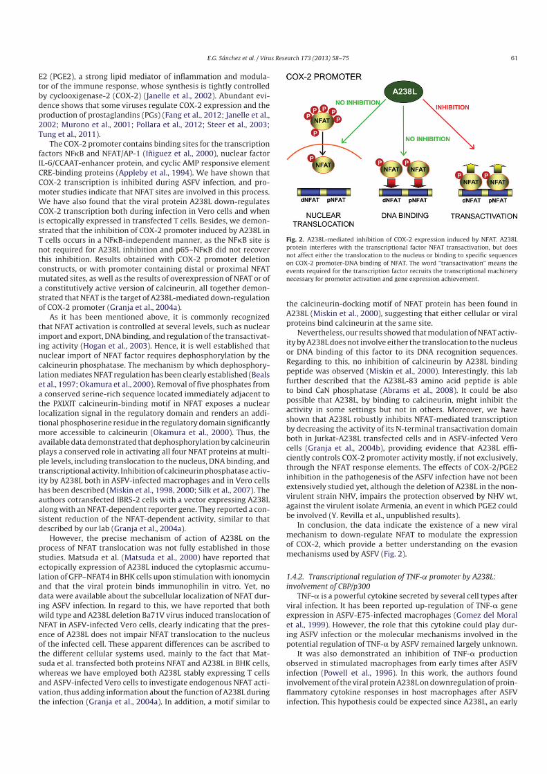
Fig. 2. A238L-mediated inhibition of COX-2 expression induced by NFAT. A238L
protein interferes with the transcriptional factor NFAT transactivation, but does
not affect either the translocation to the nucleus or binding to specific sequences
on COX-2 promoter-DNA binding of NFAT. The word “transactivation” means the
events required for the transcription factor recruits the transcriptional machinery
necessary for promoter activation and gene expression achievement.
the calcineurin-docking motif of NFAT protein has been found in
A238L (Miskin et al., 2000), suggesting that either cellular or viral
proteins bind calcineurin at the same site.
Nevertheless, our results showed that modulation of NFAT activ-
ity by A238L does not involve either the translocation to the nucleus
or DNA binding of this factor to its DNA recognition sequences.
Regarding to this, no inhibition of calcineurin by A238L binding
peptide was observed (Miskin et al., 2000). Interestingly, this lab
further described that the A238L-83 amino acid peptide is able
to bind CaN phosphatase (Abrams et al., 2008). It could be also
possible that A238L, by binding to calcineurin, might inhibit the
activity in some settings but not in others. Moreover, we have
shown that A238L robustly inhibits NFAT-mediated transcription
by decreasing the activity of its N-terminal transactivation domain
both in Jurkat-A238L transfected cells and in ASFV-infected Vero
cells (Granja et al., 2004b), providing evidence that A238L effi-
ciently controls COX-2 promoter activity mostly, if not exclusively,
through the NFAT response elements. The effects of COX-2/PGE2
inhibition in the pathogenesis of the ASFV infection have not been
extensively studied yet, although the deletion of A238L in the non-
virulent strain NHV, impairs the protection observed by NHV wt,
against the virulent isolate Armenia, an event in which PGE2 could
be involved (Y. Revilla et al., unpublished results).
In conclusion, the data indicate the existence of a new viral
mechanism to down-regulate NFAT to modulate the expression
of COX-2, which provide a better understanding on the evasion
mechanisms used by ASFV (Fig. 2).
1.4.2. Transcriptional regulation of TNF- promoter by A238L:
involvement of CBP/p300
TNF-� is a powerful cytokine secreted by several cell types after
viral infection. It has been reported up-regulation of TNF-� gene
expression in ASFV-E75-infected macrophages (Gomez del Moral
et al., 1999). However, the role that this cytokine could play dur-
ing ASFV infection or the molecular mechanisms involved in the
potential regulation of TNF-� by ASFV remained largely unknown.
It was also demonstrated an inhibition of TNF-� production
observed in stimulated macrophages from early times after ASFV
infection (Powell et al., 1996). In this work, the authors found
involvement of the viral protein A238L on downregulation of proin-
flammatory cytokine responses in host macrophages after ASFV
infection. This hypothesis could be expected since A238L, an early

62 E.G. Sánchez et al. / Virus Research 173 (2013) 58– 75
Fig. 3. A238L hampers the recruitment of p300 to the trascription complex. Viral
protein A238L interacts with p300 at the nucleus thus impairing its interaction with
the transcription factors bound to the specific regions on DNA promoter. The figure
represents the transcriptional complex or enhanceosome CRE/�3 on TNF-� pro-
moter. As a consequence of the blockage induced by the viral protein, the expression
of TNF-� is strongly inhibited.
protein synthesized from 6 hpi during the viral cycle, had been
shown to inhibit NF�B/NFAT-dependent gene activation. These
results were supported by experiments that showed the connec-
tion between the expression at mRNA level of cytokines and A238L
gene in porcine macrophages infected with ASFV isolates of dif-
ferent virulence (Gil et al., 2003). Furthermore, we identified the
cAMP-responsive element (CRE) and �3 site as responsible of gene
regulation during ASFV infection (Granja et al., 2006a). The CRE
site has been shown to bind the ATF-2/Jun heterodimer forming a
composite element with the �3 site, which can bind NF�B as well
as NFAT factors (Falvo et al., 2000b; Goldfeld and Maniatis, 1989;
Leitman et al., 1992; Rhoades et al., 1992). Through these studies,
we demonstrated that A238L inhibits TNF-� expression acting at
specific DNA binding sites and the transcription factors involved
(Granja et al., 2006a).
In order to explain the considerable activity displayed by A238L
on different boxes on both COX-2 and TNF-� promoters, we deeply
investigated the molecular mechanism subjacent to A238L func-
tion. Cellular transcriptional coactivators CBP/p300 are members
of a family of transcriptional co-adaptor molecules with distinct
functional domains that have been shown to interact with several
DNA sequences bound by different transcription factors (Garcia-
Rodriguez and Rao, 1998; Gerritsen et al., 1997; Goodman and
Smolik, 2000). Furthermore, CBP/p300 has been reported to be
bound by several viral proteins such as the adenovirus protein E1A,
SV40 large T antigen, and herpes virus E6 and E7 (Arany et al., 1995;
Kwok et al., 1996). The consequence of this interaction on the bio-
logical effects on p300 functions changes depending on the specific
viral proteins and so, both adenovirus E1A and SV40 large T Ag
interact with p300 in overlapping locations, large T antigen inhibits,
whereas E1A enhances the phosphorylation of p300 (Chakravarti
et al., 1999; Goodman and Smolik, 2000). On the other hand, CBP
and p300 have been shown to be essential for the optimal transcrip-
tional activity of TNF-� and COX-2 (Deng et al., 2004; Falvo et al.,
2000a), two genes controlled by A238L. It is interesting to speculate
that a viral gene such as A238L, which inhibits the trans-activation
of NFAT, NF�B, and c-Jun in response to PMA/ion, may have evolved
this level of flexibility to accomplish novel patterns of gene regu-
lation to evade the host response. Our results demonstrated that
A238L, which localizes in the nucleus of infected cells after PMA/ion
stimulation of transfected cells, binds to the CRE/�3 complex on
the TNF-� promoter and displaces the coactivators CBP/p300, thus
inhibiting the activation of associated factors such as NFAT, NF�B,
and c-Jun (Fig. 3) (Granja et al., 2006a). In support of these data, we
found that a deletion mutant of ASFV E70, lacking of A238L gene,
increases the synthesis of TNF-� and other cytokines during the
in vivo infection (Salguero et al., 2008).
1.4.3. Downregulation of iNOS promoter by A238L: a viral
mechanism that concurrently blocks CBP/p300 and NF�B
Further to the results described above, we investigate other
components of the inflammatory cascade that could be involved
in ASFV pathogenesis, possibly controlled by A238L and involv-
ing p300. The expression of iNOS in macrophages is induced by
lipopolysaccharides (LPS) and inflammatory cytokines, such as
interferons (IFNs), and is modulated by a number of transcription
factors, including NF�B. By using a recombinant ASFV lacking of
the A238L gene, we found that A238L strongly down regulates iNOS
promoter activation, as well as the levels of iNOS transcripts (Granja
et al., 2006b). In this work we showed that A238L down-regulates
iNOS transcription by interfering NF�B-mediated activation of the
promoter. We also found that overexpression of the p65 subunit of
NF�B neutralized the inhibitory effect of the viral protein. The com-
plex regulation of iNOS gene transcription includes the interaction
of these transcription factors with the coactivators CBP/p300. This
idea was reinforced since we demonstrated that A238L-mediated
inhibition of iNOS promoter activity and iNOS level were reverted
by overexpression of p300. Both p300 and CBP contain a his-
tone acetyltransferase (HAT) enzymatic activity that regulates gene
expression through acetylation of the N-terminal tails of histones.
In addition to modifying histones, p300/CBP directly acetylates sev-
eral transcription factors, including p65 and p50 (Bannister and
Miska, 2000; Berger, 1999). It is remarkable that, in our hands,
the p300 HAT deletion mutant construct was unable to restore the
iNOS protein level inhibited by A238L, suggesting that iNOS inhi-
bition by A238L might be related to the acetylase activity of p300.
Our results also indicated that p300 and p65 were displaced from
the iNOS enhanceosome in cells expressing the A238L protein. In
agreement with this, increased doses of p65 as well as of p300, not
only induced iNOS promoter transcription but, more importantly,
reverted the inhibition of iNOS promoter induced by A238L, sup-
porting the involvement of these proteins in the control of iNOS by
the viral protein. It is known that p65 subunit of NF�B interacts
with p300 to recruit this coactivator to the transcriptional acti-
vation complex on iNOS promoter. As described in our work, the
presence of A238L impaired this interaction, suggesting that the
viral protein might suppress the transcriptional activation of the
iNOS/p65 signal transduction pathway by competing with p65 for
binding to p300.
Taken together, the data presented by Granja et al. (2006a,b)
established a new viral mechanism of p300 transcription coacti-
vator activity downregulation to modulate iNOS activation. It is
important to note that the sustained high output of nitric oxide
accounts for its antimicrobial effects on a variety of pathogens,
including viruses (Karupiah et al., 1993). Thus, the regulation of the
iNOS promoter activity by A238L might be an important checkpoint
in the virus cycle, which could affect the virulence of the virus. In
contrast, it has been reported that deletion of the ASFV A238L gene
from the highly virulent Malawi Lil-20/1 strain does not affect the
virulence phenotype in domestic pigs (Neilan et al., 1997). Taken
into account the results obtained in vivo, it can be speculated that
immunomodulation by A238L could play a significant role in suba-
cute and chronic infections both in wild and in domestic pigs, rather
than in acute ASF, which cause a fulminating death of the animal
after 5 days of infection.
1.4.4. Molecular mechanism of inhibition of CBP/p300
transcriptional pathway by A238L: role of p300–Ser384 as a new
regulator of the transactivation controlled by ASFV
Many signal-activated pathways assemble in the transcriptio-
nal coactivator proteins CBP and p300, which join together these
signals to coordinate and promote the expression of specific sets of
genes in response to diverse physiological stimuli (Goodman and
Smolik, 2000; Vo and Goodman, 2001).

E.G. Sánchez et al. / Virus Research 173 (2013) 58– 75 63
Fig. 4. Subcellular localization of A238L and colocalization with p300. (A) Vero cells were transiently transfected with the pcDNA-A238L-SV5 expression plasmid (kindly
gifted by L. Dixon). Twenty-four hours after transfection, cells were unstimulated or stimulated with 15 ng/ml PMA plus 1 �M ion for 15 or 30 min. The cells were labeled with
an anti-SV5 Ab and examined by confocal microscopy, showing that after stimulation, A238L is mainly located at the nucleus. (B) Vero cells were transiently transfected with
pcDNA-A238L-SV5 and pCMV-p300-HA expression plasmids, and incubated in the absence or presence of 15 ng/ml PMA plus 1 �M ion during 15, 30, or 60 min. Then the cells
were labeled with anti-SV5 (green) and with anti-p300 (red) Abs and examined by confocal microscopy. The figure shows images corresponding to the co-localization of p300
and A238L at the nucleus of the cells. (C) Nuclear extracts from 107 Jurkat cells transiently transfected with pcDNA3.1 or pcDNA-A238L-SV5, treated or not with PMA/ion
for 4 h, were incubated and immunoprecipitated with rabbit polyclonal Ab against p300, or rabbit preimmune normal IgG as a negative control of immunoprecipitation
(IgG). Immunoprecipitates were analyzed by Western blot using the same Ab anti p300 to determine levels of this protein in the precipitate, and anti-SV5 to detect levels of
A238L-SV5 associated with p300.
We have shown that A238L specifically inhibits the transac-
tivation of transcription factors that require the activity of the
amino-terminal TAD (transactivation domain) of p300. In contrast,
the transactivation of carboxyl-terminal TAD-dependent transcrip-
tion factors, such as Sp-1, a p300-independent factor, was not
affected by the viral protein. We further demonstrated that A238L
modulation involves the autoacetylation activity of p300 that has
been shown to be essential to its intrinsic transcriptional activ-
ity (Santos-Rosa et al., 2003). Several viruses encode proteins that
interact with CBP and/or p300 modulating their activity, such as
SV40 T large antigen (Eckner et al., 1996), Adenovirus E1A protein
(Felzien et al., 1999) or E6 and E7 proteins from human papillo-
mavirus (Bernat et al., 2003; Patel et al., 1999), and in fact, p300
was described as an E1A-interacting protein (Eckner et al., 1994).
The results further obtained in our lab demonstrated that A238L
colocalizes in the nucleus with endogenous p300 in structures com-
patible with transcription initiation complexes, and associates with
p300 (Fig. 4). We found that the viral protein interacts with the
amino terminus of p300, but it does not bind the carboxyl-terminal
region of the coactivator (Granja et al., 2008). In this regard, it
is noteworthy that most of the viral proteins that regulate p300
inhibit the HAT activity and the activation of p300–CH3-interacting
transcription factors, such as p53 or E2F. In contrast, in this work
we have shown that A238L is inhibiting the amino-terminal TAD
without altering carboxyl-terminal activity (Fig. 5). We additionally
analyzed whether the interaction of A238L with CH1/KIX domain
of p300 interfered with phosphorylation in this domain. We iden-
tified a potential PKC site of phosphorylation of p300 at Ser384.
Our data demonstrated that this residue is necessary in the activa-
tion of the amino-terminal TAD of p300, because mutation of this
serine completely abrogated the autoacetylation and the transcrip-
tional activity of the p300 amino terminus. As it had been previously
described that PKC-� activates the signal transduction pathways of
NFATc2, p65–NF�B, and AP-1 (Manicassamy et al., 2006) and in
order to identify the PKC isotype involved in Ser384 phosphory-
lation, we achieved experiments to determine that neither PKC-�nor PKC-� phosphorylate the amino terminus of p300. In contrast,
we found that PKC-� efficiently phosphorylated Gal4–p300 fusion
protein and in addition, a constitutively active mutant of PKC-�(pEF-PKC-� A/E) fully recovered the inhibition induced by A238L,
thus enhancing the relevance of PKC-� in the functional mechanism
of the viral protein and enlightening for the first time the impor-
tance of PKC-� in the phosphorylation of this regulatory domain of
p300. Therefore, A238L might represent a viral model to find new
targets for the control of T cell activation in several pathological
processes and immunological diseases.

64 E.G. Sánchez et al. / Virus Research 173 (2013) 58– 75
Fig. 5. Map of the p300 coactivator protein showing the functional domains. The
amino-terminal region contains the CH1 and KIX functional domains and a bromo
domain, and the carboxyl-terminal region contains the CH2 and CH3 domains, which
are part of the HAT catalytic domain. Both regulatory regions may act independently
and interact simultaneously with the transcriptional machinery and/or with dif-
ferent transcription factors and viral products to build the transcriptional activity
mediated by the coactivator. According to our model, ASFV A238L protein interacts
with the CH1 domain of p300.
The results described above using ectopically or recombinant
expressed A238L in different cells, prompted us to study the possi-
bility of A238L might also block the transcriptional activity of p300
during ASFV infection. In this regard, we showed that the viral pro-
tein regulates the transcriptional transactivation mediated by p300
during the viral infection through the C/H1 and KIX regulatory
regions of the coactivator, by using recombinant ASFV lacking of
A238L (Granja et al., 2009). We have used the site-directed mutant
p300 constructs in which Ser384 was substituted to alanine or
aspartic acid, to explore whether the signaling pathway involving
this residue was interfered by ASFV. Aspartic acid and alanine are
generally accepted as standard substitutions of serine to mimic the
phosphorylated and non-phosphorylated state, respectively (Kock
et al., 2003). This experimental approach demonstrated that the
transcriptional activity of p300 was completely abrogated when
Ser384 was substituted to alanine, whereas substitution by aspar-
tic acid resulted in a dramatically increased of p300 activity during
the infection. In fact, we demonstrated that the presence of the viral
protein impairs the association of PKC-� and the amino-terminal
192–703 region of p300, thus blocking the amino terminal trans-
activation activity of p300 in porcine macrophages infected with
ASFV E70wt, but not during the infection with E70�A238L. We
established the relevance of PKC-� in the activation of the amino-
terminal domain of p300 via phosphorylation of the residue Ser384,
suggesting that this mechanism is part of a complex signaling net-
work regulating p300 under pathological conditions, such as viral
infection. Our model concludes that during ASFV infection the p300
transactivation is efficiently blocked by the viral product A238L to
inhibit the synthesis of proinflammatory molecules as a mechanism
of virus evasion (Fig. 6).
1.5. Other ASFV proteins involved in host gene transcription
regulation
Other ASFV proteins modulating host gene transcription, thus
interfering with the function of infected macrophages, have been
identified. These proteins include the ASFV inhibitor of apoptosis
A224L (Nogal et al., 2001), that has been also reported to be involved
in the activation of NFkB (Rodriguez et al., 2002). The mechanism by
which A224L activates this transcription factor is likely dependent
Fig. 6. Model for A238L-mediated inhibition of immune and inflammatory genes
transcription. Viral infection and cell activation trigger different signaling path-
ways which lead to the activation of several kinases. We have found that PKC-�translocates to the nucleus during ASFV infection where it is able to bind the p300
CH1/KIX domain to activate p300. This event usually up-regulates the transcriptio-
nal activity of p300, enhancing the transactivation mediated by NFAT, NF�B and
AP-1 transcription factors. According with our results, viral protein A238L impairs
the phosphorylation of p300–Ser384 by PKC-� subsequently inhibiting the p300
activity and the expression of several pro-inflammatory genes.
on the activation of IKK kinases that induce the phosphorylation of
NFkB-inhibitor IkB, allowing the translocation of p65–NFkB to the
nucleus, where it activates its specific target genes. It is important
to note that A224L, also known as IAPv (Nogal et al., 2001), is a late
protein in ASFV viral cycle, in contrast with A238L, which is an early
protein. Thus, by the expression of these two regulators of the tran-
scription at different times of the infection, ASFV might control the
expression of cellular genes to interfere with pathways that could
counteract different steps of the viral cycle. Studies from C. Mar-
tins lab have also characterized the expression of A224L in porcine
macrophages infected with different virulence isolates (Portugal
et al., 2009).
Secondly, ASFVj4R protein, which has been described to bind to
the host �-NAC protein (Goatley et al., 2002), is also a candidate
to regulate host transcription. �-NAC was first reported to play a
role in translation by preventing non-specific targeting of proteins
lacking signal peptides to the secretory pathway (Wiedmann et al.,
1994). Later, the finding that the � subunit of the NAC complex
is a yeast transcription factor, BTF3, led to investigate the possi-
ble function of �-NAC in transcription, which was finally found to
be the regulator of the transcription of c-Jun target-genes (Moreau
et al., 1998; Yotov et al., 1998). Thus, it has been speculated that,
by binding to �-NAC, ASFVj4R might interfere with the ability of
this cellular factor to act as a transcriptional co-activator. Besides,
�-NAC has also been shown to interacts with FADD (Fas associ-
ated death domain), possibly impairing FADD oligomerization and
assembly of the DISC complex in the absence of TNF-� (Stilo et al.,
2003). Hypothetical interaction of j4R with �-NAC could regulate
apoptosis induced by TNF-� (Dixon et al., 2004).

E.G. Sánchez et al. / Virus Research 173 (2013) 58– 75 65
Fig. 7. Effect of ASFV infection in the activation of c-Myc and transcriptional activation of eIF4G1, eIF4E and eIF4A promoters. Vero cells were transfected with p-Ebox-luc
(A), pGL3-eIF4G1-luc (B), pGL3-eIF4E-luc (C), and pGL3-eIF4A-luc (D) reporter plasmids (300 ng/106 cells). Sixteen hours after transfection, cells were mock-infected or
infected with the Vero-adapted isolate Ba71V at a MOI of 5 pfu/cell in 2% FCS medium. Whole extracts were prepared at indicated times post infection and assayed for
luciferase activity. Extracts were normalized to Renilla luciferase. Results from triplicate assays are shown in RLU/�g protein or Fold induction relative to mock-infected cells
(mean ± SD).
Also, the ASFV ubiquitin conjugating (UBCv) enzyme has been
reported as playing a possible role in regulating host gene tran-
scription as it was shown to bind to a host nuclear protein SMCy,
which contains an ARID DNA binding domain and is involved in
transcription regulation (Bulimo et al., 2000).
1.6. New data about the transcriptional control of eIF4F
components by ASFV
The Myc/Max/Mad network of transcriptional regulators con-
trols multiple aspects of cell behavior, including proliferation,
apoptosis and differentiation (Grandori et al., 2000). This family
of proteins binds DNA to E-box sequence motifs (5′-CACGTG-3′)modulating the transcriptional activity of several genes through
chromatin compaction. Both Myc and Mad1 proteins are able
to bind Max, forming heterodimers that display different func-
tions. Myc:Max dimers activate the transcriptional activity whereas
Mad:Max dimers act as repressors. As a consequence of their antag-
onic function, Myc activates cell growing and protein synthesis,
and Mad1 regulates negatively the cell cycle (Ayer et al., 1993;
Rottmann and Luscher, 2006). Therefore, since Myc and Mad1 com-
pete for Max protein, the availability of Max to Myc is profoundly
depending on the expression levels of Mad1 and vice versa. Mad
proteins are expressed preferentially in non-proliferating cells and
Myc proteins are present almost exclusively in proliferating cells.
There are numerous genes regulated by Myc/Max/Mad network
such as cdc25, CdK4 and Cyclin D2 (Luscher, 2001). Recently, it
has been described that the translation initiation factors eIF4A,
eIF4GI and eIF4E are also targets of c-Myc (Coller et al., 2000;
Mao et al., 2003; Roeding et al., 2009). It has been also proposed
that c-Myc regulates the rate-limiting step of translation initiation
and thereby induces eIF4F activity (Lin et al., 2008). Since ASFV
stimulates cap-dependent translation to increase the initiation of
viral mRNA translation by activating the eIF4F (Castello et al.,
2009b), we hypothesized if the virus regulates the transcriptio-
nal expression of translational initiation factors of eIF4F complex
through c-Myc activation. To confirm this hypothesis, we first stud-
ied whether ASFV induces c-Myc trans-activation in infected Vero
cells by transfection of a reporter plasmid containing canonical
E-box sequence motif. (Plasmids were kindly provided by Dr. J.
Pelletier, McGill University, Montreal, Canada). The results show
that ASFV strongly induces c-Myc activation from early times after
infection (Fig. 7A). Moreover, by using the reporter plasmids con-
taining E-box sites in eIF4G1, eIF4E and eIF4A promoter, we found
that the transcriptional activation of these factors was up regulated
in infected Vero cells likely as a consequence of ASFV-induced c-
Myc activation (Fig. 7B–D). These new results suggest that, further
to the recruitment of translational factors within viral factories dur-
ing ASFV infection (Castello et al., 2009b), the virus is able to control
the expression of the components of eIF4F complex at transcrip-
tional level to guarantee the viral protein synthesis. Noteworthy,
the ASFV-induced expression of eIF4G1, eIF4E and eIF4A factors is
mostly induced at late times after infection, suggesting that the
virus might use the cellular factors pool available early in the viral
cycle, whereas induce the novo synthesis of these factors at late
times of infection.
As mentioned above, Mad1 should be inhibited to allow the
expression of c-Myc-regulated genes. It has been described that
Mad1 inhibition can be achieved through two different mech-
anisms. First, phosphorylation-dependent degradation through

66 E.G. Sánchez et al. / Virus Research 173 (2013) 58– 75
Fig. 8. Role of A224L viral protein in the transcriptional activation of eIF4G1 and eIF4E promoters. Vero cells were transfected with pGL3-eIF4G1-luc (A) and pGL3-eIF4E-luc
(B) reporter plasmids (300 ng/106 cells). Sixteen hours after transfection, the cells were mock-infected or infected with the Vero-adapted isolate Ba71VWT or Ba71V�A224L at
a MOI of 5 pfu/cell in 2% fetal calf serum medium. Whole extracts were prepared at indicated times post infection and assayed for luciferase activity. Extracts were normalized
to Renilla luciferase. Results from triplicate assays are shown in RLU/�g protein or fold induction relative to mock-infected cells (mean ± SD). (C) BIRC2 mRNA expression
during ASFV infection. Vero cells were mock-infected or infected with the Vero adapted isolated Ba71V at a MOI of 5 pfu/cell. At different times post infection total RNA was
isolated by the TRIzol reagent and was analyzed by quantitative RT-PCR assays (1 �g) with specific primers for BIRC2 mRNA. The levels of mRNA were represented relative
to mock expression (n = 2; mean ± SD). a.u.: arbitrary units.
PI3K/Akt/mTOR and MAPK activation pathways (Zhu et al., 2008),
and second, via ubiquitination by c-IAP1 that triggers degradation
by 26S proteasome pathway (Xu et al., 2007). Since the viral pro-
tein A224L has been described to be an IAP homolog that inhibits
caspase activation and promotes cell survival (Nogal et al., 2001),
we speculated whether this viral protein could be involved in the
transcriptional control of eIF4G1 and eIF4E. To analyze this, Vero
cells were transfected with the specific reporter plasmids contain-
ing the promoters regions of eIF4G1 and eIF4E and infected either
with Ba71V-WT or with the deletion mutant Ba71V-�A224L. As
Fig. 8 shows, the transcriptional activation of both eIF4G1 (A) and
eIF4E (B) induced by ASFV was strongly reduced when cells were
infected with Ba71V-�A224L, indicating that this viral protein has
an important role on the expression of these genes. Further exper-
iments are needed to clarify the molecular mechanisms by which
A224L cooperates with c-Myc to stimulate the expression of eIF4G1
and eIF4E, thus promoting the viral protein synthesis. It can be
speculated that A224L could promote the degradation of Mad1 to
achieve the transcriptional activation of these translational factors,
as it has been described for cellular IAPs (Xu et al., 2007). On the
other hand, as shown in Fig. 8A and B, when the infection is car-
ried out with the A224L deletion mutant virus, the eIF4G1 and eIF4E
transcriptional activation is still higher than in mock-infected cells.
This result suggests that other factors, apart from A224L, could be
involved in the control of these factors. In this regard we hypoth-
esized whether ASFV might induce the expression of cellular IAPs
which could complement the A224L function during viral infection.
Thus, we have investigated the mRNA expression level of BIRC2, a
member of cellular IAPs family. As shown in Fig. 8C, the transcrip-
tional level of this gene diminished in response to the virus, making
difficult to establish any conclusion about our hypothesis. Other
possible candidates will be study in the next future to explore this
interesting possibility.
2. Translational regulation by ASFV
2.1. Introduction
Viruses have developed mechanisms to monopolize the cellular
translation activity in order to synthesize their own proteins. Most
of these strategies are based on switching on/off the activity of key
initiation factors essentials for host protein synthesis. African swine
fever virus, in analogy to other DNA virus, hijacks the translation
machinery by affecting not only the activity of the translation initia-
tion factors, but also their localization. Furthermore, ASFV impacts
on RNA metabolism promoting the degradation of cellular RNAs,
process in which a putative viral decapping enzyme could play an
important role. The ASFV-specific mechanisms to overcome cellular
function will be dissected below.
2.2. The cellular protein synthesis machinery: hijacking cellular
sources by viruses
The initiation of translation consists in the recruitment of the
ribosome to the messenger RNA (mRNA) and is one of the most
regulated steps in gene expression. The eukaryotic initiation factors
(eIFs) play a central role in this process and are usually targets for
fine tune regulation. Due to their key role in protein synthesis, many
viruses target these factors during the infection. Frequently, viral
infection modulates two important events in initiation of transla-
tion: (i) the phosphorylation of eIF2 and (ii) the recruitment of the
ribosome to the mRNA by targeting eIF4F and 4E-BPs. The phos-
phorylation of eIF2 represents one of the most important steps
in the regulation of the cellular translation since it allows rapid
and reversible modulation of translation initiation. eIF2 is a het-
erotrimeric initiation factor composed by 3 subunits: � is involved
in regulation, while � and are implicated in tRNA and GTP bind-
ing. The phosphorylation of the � subunit inhibits eIF2 activity
and is performed by several cellular protein kinases (Proud, 2005;
Wek et al., 2006), and among them, the double stranded RNA-
activated kinase (PKR), the PKR-like endoplasmic reticulum (ER)
kinase (PERK) and general control non-derepressible-2 (GCN2) play
a major role in response to viral infections (Barber et al., 1993;
Berlanga et al., 2006; Cheng et al., 2005; Garcia et al., 2007). eIF2
binds Met-tRNA and GTP to form a ternary complex necessary for
the formation of the pre-initiation complex. Following the recog-
nition of the AUG start codon and the joining of the ribosomal
subunit 60S, eIF2–GDP releases the Initiation Complex and GDP
is exchanged to GTP by eIF2B. The phosphorylated eIF2� (P-eIF2�)
has greater affinity than its non-phosphorylated counterpart for
eIF2B and inhibits eIF2B GDP–GTP exchange activity, causing a
decreased in the eIF2–GTP pools and inhibiting translation (Van
Der Kelen et al., 2009).
eIF2� phosphorylation is one of the most important host
defense mechanisms against viral infections. For this reason, sev-
eral viruses have developed mechanisms to evade the activation
of PKR and the phosphorylation of this factor. Some viruses, as
adenovirus (AdV) and Epstein–Barr virus (EBV), encodes double
stranded (ds) RNAs that bind PKR but do not trigger the activa-
tion of the kinase (Schneider and Mohr, 2003; Walsh and Mohr,
2011). Herpes simplex virus-1 (HSV-1), vaccinia virus (VV), reovirus
and influenza virus encodes dsRNA binding proteins that mask or
sequester dsRNA and prevent activation of PKR (Beattie et al., 1995;
Khoo et al., 2002; Lloyd and Shatkin, 1992; Mulvey et al., 1999;

E.G. Sánchez et al. / Virus Research 173 (2013) 58– 75 67
Salvatore et al., 2002). In the case of poliovirus, PKR is degraded
during the infection (Gale et al., 2000). Often, viruses employ more
than one mechanism to assure the inhibition of this pathway. One
example is HSV-1, which in addition to the previous mechanism
encodes for proteins that avoid the accumulation of P-eIF2� by
targeting the phosphatases involved in the regulation of this factor
(Mulvey et al., 2003). Moreover, HSV-1 and VV also possess viral
proteins (glycoprotein B and K3L, respectively) that are able to pre-
vent the phosphorylation of eIF2� by PERK (Mulvey et al., 2003;
Sood et al., 2000).
On the other hand, not all the virus avoids phosphorylation of
eIF2�. Some viruses as hepatitis C virus (HCV), Sindbis virus, pes-
tivirus, poliovirus, cricket paralysis virus and Semilinki Forest virus
induce eIF2� phosphorylation as a mechanism to impair cellular
protein synthesis, being able to translate its own mRNA in an eIF2-
independent manner (Beckham and Parker, 2008; Garaigorta and
Chisari, 2009; Garrey et al., 2010; Jordan et al., 2002; O’Neill and
Racaniello, 1989; Ventoso et al., 2006).
The other key point involves the recruitment of ribosomes and
eIF4F formation. eIF4F, as central component of the cap-dependent
translation machinery, is fine regulated in response to extracellular
stimuli, stress and viral infections. eIF4F is a complex composed by
three proteins: eIF4A, eIF4E and eIF4G (Prevot et al., 2003). eIF4A
is a RNA helicase implicated in unwinding the secondary structure
of the 5′-end of the mRNA together with eIF4B; eIF4E binds the
cap structure at the mRNA 5′-terminus, and eIF4G is a scaffold-
ing protein that forms a molecular bridge between the mRNA and
the small ribosomal subunit 40S. eIF4G coordinate the initiation
of translation via protein–protein interactions: (i) the N-terminus
domain is involved in the recruitment of the mRNA by its inter-
action with the cap-binding factor eIF4E and the poly (A) binding
protein (PABP); (ii) simultaneously, the C-terminal domain recruits
the small ribosomal subunit by means of its interaction with eIF3
(Jackson et al., 2010). Furthermore, eIF4G is also interacting with
other viral and cellular proteins implicated in the regulation of the
translation machinery, as the mitogen activated kinase 1 (Mnk-
1, which phosphorylates eIF4E), the non-structural protein 1 of
influenza or the 100 kDa protein of adenovirus (Gingras et al., 1999;
Prevot et al., 2003).
Some RNA viruses, such as retroviruses, calciviruses and picor-
naviruses, encodes for viral proteases that cleavage eIF4G (Alvarez
et al., 2003; Castello et al., 2011; Lloyd, 2006; Ventoso et al., 2001).
eIF4G cleavage by picornavirus proteases hydrolyze this factor in
two moieties, which decouples the capacity of this host factor to
recruit the mRNA (by interaction with eIF4E and PABP in the N-
terminus) and the ribosome (via eIF3 interaction in the C-terminus)
(Castello et al., 2011). However, viral RNAs can drive translation
by non-canonical mechanisms. During the last two decades many
laboratories worldwide joined efforts to better understand the
mechanisms that viruses display to initiate translation, and one
of the most important discoveries was the existence of the inter-
nal ribosome entry sites (IRES) in picornavirus RNAs (Jang et al.,
1990). These RNA elements, which were later discovered in many
virus families, drive translation initiation in the absence of key ini-
tiation factors such as eIF4E or when eIF4G is cleaved, allowing
viral translation under conditions where host protein synthesis is
inhibited (Hellen, 2009). Other virus such as AdV, influenza virus
or vesicular stomatitis virus (VSV) (Burgui et al., 2007; Connor and
Lyles, 2002; Cuesta et al., 2000; Welnowska et al., 2009) promote
the dephosphorylaton of 4E-BP and eIF4E to repress the host mRNA
translation. Some of these viruses transcribe capped mRNAs, which
cannot be distinguished among cellular mRNAs. Nevertheless, pro-
teins of such viruses can be synthesized in spite of the inactivation
of eIF4E and the subsequent cellular shutoff. How these “cellular-
like” viral RNAs drive translation under these conditions is still
unclear.
Complex DNA viruses constitute one of the most intriguing
cases. Conversely to most of the viruses studied so far, they enhance
the assembly of eIF4F but still are able to inhibit cellular protein
synthesis (Walsh et al., 2008; Walsh and Mohr, 2004; Walsh et al.,
2005). This review will try to shed light in this “abnormality” in the
virus kingdom, with special focus on the molecular mechanisms
displayed by ASFV to regulate the cellular machinery of protein
synthesis.
2.3. Regulation of eukaryotic initiation factors by African swine
fever virus
ASFV mRNAs are structurally similar to the cellular mRNAs.
In vitro transcribed ASFV early mRNAs possess a cap structure in its
5′-UTR and a poly (A) tail of 33 nucleotides in average (Salas et al.,
1981). The cap structure is predominantly the type m7G (5′) pppAm,
which suggests that an enzymatic activity is required for RNA cap-
ping to occur. In this regard, ASFV encodes a guanylyltransferase
(NP868R) able to exert triphosphatase and guanylyltransferase
activities (Pena et al., 1993; Yanez et al., 1995). The fact that ASFV
mRNAs are capped indicates that they drive translation by a canon-
ical cap-dependent mechanism, as happens with most of cellular
mRNAs.
We have recently described that, similarly to VV infection, P-
eIF2� levels decrease at early times post infection, and remain
undetectable throughout the infection (Castello et al., 2009b), sug-
gesting a viral mechanism to ensure eIF2 availability for viral
protein synthesis. In this regard, ASFV–Ba71V genome encodes a
protein, DP71L, which possesses a characteristic binding Protein
Phosphatase 1 motif (VxF) (Cohen, 2002). Furthermore, it shares
an N-terminal sequence of basic residues and is similar in its
C-terminal domain to the Herpes simplex virus-encoded neurovir-
ulence factor ICP34.5 (Goatley et al., 1999). Indeed, DP71L is able
to interact with PP1 in vitro (Rivera et al., 2007) and in vivo (Zhang
et al., 2010) (Fig. 9A). Individual expression of DP71L induces a
decrease of phosphorylated eIF2� and enhances the expression of
co-transfected reporters, suggesting that DP71L plays a role keep-
ing the translation machinery active to allow viral protein synthesis
(Zhang et al., 2010). Furthermore, it has been predicted that DP71L,
like ICP34.5, could act as a regulatory subunit of PP1 and targets
it to dephosphorylate-specific substrates in the nucleus of infected
cells altering their function. ASFV–Ba71V DP71L gene is named l14L
in ASFV E70 and 23NL in Malawi Lil 20/1 isolates, respectively.
Deletion of the gene 114L from the genome of E70 reduced vir-
ulence in domestic pigs (Zsak et al., 1996) whereas deletion of the
gene 23NL from the virulent Malawi Lil 20/1 isolate did not reduce
virulence, suggesting that Malawi isolate may encode another addi-
tional gene, with a similar function (Afonso et al., 1998). Moreover,
the depletion of DP71L in the viral strains Malawi Lil 20/1 and E70
not leads to an increase in the levels of P-eIF2�, suggesting that as
VV, ASFV possess multiple mechanisms to avoid eIF2� phosphory-
lation (Zhang et al., 2010).
Interestingly, new preliminary data from our lab reveal that PKR
is found phosphorylated in murine Raw cells at 6 h after virus addi-
tion inducing eIF2�s phosphorylation (Fig. 9B), in contrast to that
was observed in productive infection in Vero cells. It is important to
realize that Raw cell is a murine macrophage line, which only allows
the expression of early ASFV proteins, such as p32. Neither late viral
protein (p72) (Fig. 9B), nor viral production could be detected, even
though more than 80% of the murine macrophages were shown to
express ASFV p32 protein, as found by confocal experiments (data
not shown). Thus, the sustained eIF2� phosphorylation observed in
this murine system could be involved in the impairment of eIF2�binding to the translational initiation complex, causing the block-
age of the infection in these cells and partially explaining the ASFV
tropism for swine macrophages. Moreover, the mRNA levels of PKR

68 E.G. Sánchez et al. / Virus Research 173 (2013) 58– 75
Fig. 9. Regulation of eIF2� and PKR phosphorylation by ASFV. (A) ASFV DP71L protein has been shown to bind the catalytic subunit of protein phosphatase 1 (PP1), leading
to PP1 activation and, consequently, eIF2a dephosphorylation (Rivera et al., 2007; Zhang et al., 2010), therefore allowing the participation of this factor in the initiation of
translation. (B and C) Raw cells were treated with the virulent isolate E70 at a MOI of 5 pfu/cell for 6 and 16 h. (B) Cellular extracts (50 �g) were lysed with RIPA modified
buffer, subjected to SDS-PAGE and phospho-PKR, phospho-eIF2�, p32, p72 and �-actin proteins were detected by immunoblotting with specific antibodies. (C) At indicated
times post virus addition total RNA was isolated by the TRIzol reagent and was analyzed by quantitative RT-PCR assays (1 �g) with specific primers for PKR mRNA. The levels
of mRNA were represented relative to mock expression. Representative experiment is shown. a.u.: arbitrary units.
were found to increase from early times after virus addition in
Raw cells (6 h) until 16 h (Fig. 9C), whereas the phosphorylation
of the kinase strongly decreased at late times of the infection. Fur-
ther experiments will be developed to better understand and to
complete these data.
ASFV induces apoptotic response at late times post infec-
tion, and consequently, caspase-3 activation (Granja et al., 2004a;
Ramiro-Ibanez et al., 1996). eIF4G has been reported to be a sub-
strate for caspase-3 (Bushell et al., 1999; Prevot et al., 2003),
being its proteolytic cleavage a potential cause of the shut off dur-
ing apoptosis (Marissen and Lloyd, 1998). Surprisingly, eIF4G was
refractory to caspase-3 cleavage in ASFV-infected cells (Castello
et al., 2009b), indicating that (i) the cleavage sites are not accessible
to the protease (perhaps protected by protein–protein interac-
tions); (ii) eIF4G is not at the same location than caspase-3; or (iii)
caspase-3 activity is abrogated. Regarding the last possibility, ASFV
encodes for an inhibitor of the apoptosis (IAP)-like protein (A224L),
which has been reported to be an inhibitor of caspase-3 (Nogal et al.,
2001). The possibility that this factor protects translation machin-
ery from caspase-3-mediated degradation should be explored in
the future. Conversely, ASFV induces a rapid mTOR-mediated phos-
phorylation of eIF4G at Ser1108 (Castello et al., 2009b), which is has
been associated to “translational activation” (Kimball et al., 2000;
Raught et al., 2000) (Fig. 10).
In parallel, we showed that ASFV triggers the phosphorylation of
eIF4E at Ser209 by Mnk-1. Although the biological relevance of the
eIF4E phosphorylation is still controversial (Morley and Naegele,
2002; Richter and Sonenberg, 2005), its importance for some viral
infections has been demonstrated. For instance, during Influenza
virus, VSV and AdV infection, the dephosphorylation of eIF4E
correlates with the inhibition of the cellular protein synthesis
(Connor and Lyles, 2002; Feigenblum and Schneider, 1993; Xi et al.,
2004). During ASFV infection, as occurs with HSV-1, HCMV and VV,
eIF4E phosphorylation is associated to an enhancement of the viral
replication and protein synthesis (Buchkovich et al., 2008; Castello
et al., 2009b). eIF4E phosphorylation takes place after 8 h post ASFV
infection (hpi) and reaches its maximum levels at 14–18 hpi. This
phosphorylation in infected cells is avoided in the presence of the
Mnk-1 inhibitor CGP57380, and correlates with the phosphoryla-
tion of this kinase, suggesting that it relies on Mnk1 activation as
reported before (Pyronnet, 2000; Pyronnet et al., 1999).
The eIF4E-binding proteins (4E-BPs) are well-known nega-
tive regulators of the cap-dependent translation (Sonenberg and
Hinnebusch, 2009). In its hypo-phosphorylated state, 4E-BPs are
able to associate with eIF4E and compete the interaction of the cap-
binding factor with eIF4G, impairing eIF4F assembly. Conversely,
4E-BPs are inactivated by mTOR-mediated hyperphosphoryla-
tion, allowing cap-dependent translation (Bhandari et al., 2001;
Richter and Sonenberg, 2005). Similarly to VV and other DNA
viruses (Buchkovich et al., 2008), ASFV infection promotes 4E-
BP1-phosphorylation at early times post infection, but in this
case it is progressively hypo-phosphorylated at later times (from
14 hpi). eIF4G, eIF4E and 4E-BP1 phosphorylations are concomitant
with a boost of eIF4F assembly (Castello et al., 2009b). The hypo-
phosphorylation observed at late times post infection may be due
to a viral mechanism to stop the viral protein synthesis when late
morphogenesis stage is taking place (Fig. 10).
Nevertheless, we have shown that phosphorylation of eIF4G,
eIF4E and 4E-BP1 by specific kinases, is important but not essen-
tial to the ASFV infection in cultured Vero cells, since the presence

E.G. Sánchez et al. / Virus Research 173 (2013) 58– 75 69
Fig. 10. ASFV infection promotes cap-dependent translation. Activation of eIF4E and eIF4G during ASFV infection. ASFV promotes phosphorylation and of eIF4E, eIF4G and
the repressor of eIF4E, 4E-BP, which is inactivated by hyperphosphorylation. However, at late times post infection, 4E-BP is hypophosphorylated. eIF, eukaryotic translation
initiation factor; 4E-BP, eIF4E binding protein, Mnk-1, mitogen activated kinase 1; ASFV, African Swine Fever Virus.
of inhibitors of Mnk-1 (CGP57380) and mTOR (rapamycin) affects
only moderately to the viral protein synthesis and virus production
(Castello et al., 2009b). In contrast, the viral protein synthesis was
found to be abrogated by inhibitors of mTOR in other DNA virus
infections (Moorman and Shenk, 2010). Interestingly, the activi-
ties of eIF4G and eIF4E themselves are essential for ASFV infection
as depletion of those factors by specific siRNAs, strongly abrogates
viral protein synthesis, viral factory formation and virus production
(Castello et al., 2009b).
2.4. Monopolizing the sources: ASFV recruits eIFs, ribosomes and
mitochondria to viral factories
Re-localization of eIFs during viral infection has been described
for some viruses as tobacco mosaic virus, poliovirus, Sindbis virus,
and poxvirus (Katsafanas and Moss, 2007; Sanz et al., 2009;
Thivierge et al., 2008; Walsh et al., 2008). Interestingly, it seems
like only factors that are required are recruited to viral factories
(Sanz et al., 2009; Katsafanas and Moss, 2007).
Data from our lab showed that during the ASFV infection, all
the components of the translation machinery examined (eIF4G,
eIF4E, eIF2, eIF3b and the eukaryotic elongation factor 2 [eEF2])
are relocated from a diffused distribution throughout the cyto-
plasm of infected cells to the viral factories, were viral replication
and virus morphogenesis take place (Fig. 11A). At 8 hpi, eIF4E and
eIF4G are clustered together to the viral DNA and in proximity but
not overlapping with the ASFV protein p72; suggesting that “active
translation” and morphogenesis areas are in near but independent
environments (Castello et al., 2009b). However, both translation
factors are found at the periphery of the factories at 16 and 24 hpi,
correlating with a clear accumulation of DNA in the central area of
the replication foci. These facts point to the idea that ASFV acti-
vates and recruits eIF4F to areas where active viral translation
takes place. Mobilization of eIFs to ASFV replication foci relies on
late viral proteins since treatment with AraC prevents all these
effects; although the viral proteins involved in this process are still
unknown. During ASFV infection, not only eIFs are recruited to the
viral factories but also ribosomes, which strongly support the cou-
pling of viral replication and translation by attracting the protein
synthesis machinery to ASFV replication sites. Also viral RNAs local-
ize at the periphery of the viral factories, supporting the idea that
active translation is limited to these foci in infected cells. Finally,
the mitochondria network are mobilized together with ribosomes
to the viral factories, showing that ATP production, translation
and viral replication are in proximity (Castello et al., 2009b)
(Fig. 11B).
The more obvious role of the virus-mediated redistribution of
all these key host machineries may be to increase the availability
of the host sources needed for the viral biological cycle in the loca-
tions where they are required, i.e. viral factories. Simultaneously,
accumulation of the protein synthesis machinery at the viral repli-
cation foci may induce its depletion in the cytoplasm, which may
result in the shut off of the host mRNA translation. In the future,
this attractive hypothesis should be explored.

70 E.G. Sánchez et al. / Virus Research 173 (2013) 58– 75
Fig. 11. (A) eIF4E and eIF4GI redistribution during ASFV time-infection steps. Vero cells were cultured on glass coverslips and mock infected or infected with ASFVBa71V. Cells
were permeabilized and fixed at 4 and 24 hpi. Cellular factors eIF4E and eIF4GI were detected by indirect immunofluorescence. Cell nuclei and ASFV factories were stained with
To-Pro-3. Cells were visualized by confocal microscopy and the cell outline was defined by phase contrast microscopy. Images were obtained under restricted conditions and
processed with Huygens 3.0 software. (B) Mobilization of the cellular translation machinery and mitochondria to viral factories. During ASFV infection, eukaryotic initiation
factors (eIFs), ribosomes and mitochondria are recruited to the periphery of the viral factories and cytoplasmic poly (A) RNAs are degraded. Simultaneously, poly (A) RNAs
accumulates in the nucleus of infected cells.
2.5. Regulation of RNA metabolism by ASFV
Several viruses strongly alter RNA metabolism (Castello et al.,
2009b; Covarrubias et al., 2011; Cheng et al., 2005; Park et al.,
2008; Parrish and Moss, 2007; Parrish et al., 2007; Richner et al.,
2011; Sandri-Goldin, 2011; Satterly et al., 2007; von Kobbe et al.,
2000). Due the genetic complexity of ASFV, it is not surprising that
it displays multiple mechanisms to alter transcription and, most
probably, nuclear RNA export and RNA stability.
During infection viruses can alter the distribution and quantity
of cellular mRNAs, decreasing the competence for the transla-
tional machinery. This can be achieved by altering cellular RNA
metabolism functions. In the case of HSV-1, the splicing machin-
ery is inhibited leading to an accumulation of cellular mRNAs at
the nucleus of infected cells (Sandri-Goldin, 2011), while in the
case of AdV (Yatherajam et al., 2011), VSV (von Kobbe et al., 2000),
poliovirus (Castello et al., 2009a; Park et al., 2008) and influenza
virus (Satterly et al., 2007) host mRNAs export from the nucleus to
the cytoplasm is impaired by targeting the nuclear pore complex. In
other cases, viruses stimulate mRNA degradation, as occur in HSV-
1 (Cheng et al., 2005) and gammaherpesvirus (Covarrubias et al.,
2011; Richner et al., 2011) that encode for ribonucleases. Vaccinia
virus also induced RNA degradation and, even though the mech-
anism is still unclear, it is proposed that viral-encoded decapping
enzymes could trigger RNA degradation (Parrish and Moss, 2007;
Parrish et al., 2007).
A progressive decrease in the amount of cytoplasmic polyadeny-
lated mRNAs, together with an increment of the pool of nuclear
RNAs was observed during ASFV infection (Castello et al., 2009a),
suggesting that poly (A) mRNA stability and nuclear RNA export
could be altered. Nuclear organization is damaged as a consequence
of ASFV infection, which includes nucleoli and nuclear envelope

E.G. Sánchez et al. / Virus Research 173 (2013) 58– 75 71
disorganization (Ballester et al., 2011). At early times post infection,
laminin A/C is phosphorylated and the lamina network is disassem-
bled at the proximity of the viral factories, showing lamin A/C in
the cytoplasm of infected cells at late times post infection. Simi-
larly, both nucleoporin p62, a marker of the nuclear envelope; and
B-23, a marker of the nucleolus, are found in the cytoplasm of ASFV
infected cells at late times post infection, suggesting an important
damage in nuclear functions. Although lamin A/C localizes in the
cytoplasm, a pool of this protein have been found to co-localize
with the viral protein p54 (a marker of the viral factories). Never-
theless, no direct interaction between lamin A/C and viral proteins
was found (Ballester et al., 2011). In addition to the cytoplasmic
distribution, the lamin A/C that remains in the nucleus is reorga-
nized into nucleoplasmic sites together with other transcriptionally
related markers as SC35 at early times post-infection. Simulta-
neously, RNApol II is early dephosphorylated and then degraded at
late times post infection. This suggests the existence of a viral mech-
anism to inactivate global cellular transcription (Ballester et al.,
2011). Collectively, all these data support that ASFV is altering
important RNA metabolism steps within the nucleus that should
impact in host gene expression.
ASFV genome encodes for a protein called g5R that harbors
a Nudix motif, which is comparable to other Nudix hydrolases
such as the host decapping enzyme 2 (Dcp2) or the viral pro-
teins D9 and D10 of VV (McLennan, 2007). g5R exerts in vitro
decapping activity (Parrish et al., 2009), although it preferentially
degrades diphosphoinositol polyphosphates under this conditions
(Cartwright et al., 2002). Nevertheless, it is still unknown the role
of this protein in the viral biological cycle, but its high homology
(at least for the Nudix motif) with Dcp2 suggests its involvement in
mRNA decapping. Therefore, this viral enzyme represents a candi-
date to be a viral factor involved in the degradation of the cellular
mRNAs, which may contribute in the cellular shut off.
Unpublished data from our lab shows the predicted structure of
g5R and its structural homology with the cellular decaping enzyme
Dcp2 from Schizosaccharomyces pombe, but further studies have
been planned to demonstrate the function of this ASFV protein in
vivo.
2.6. Future perspectives
During the last decades, numerous studies focused on how
viruses manipulate and control the host translational machinery.
However, the molecular mechanism that complex DNA viruses
such as ASFV develop to monopolize host cell translation is still
unclear and requires extensive research.
Even though it has been described the state and distribution
of the eIFs in ASFV-infected cells (Castello et al., 2009b), the exact
mechanism by which these events are accomplished remain elu-
sive. Future studies should face this biological problem at the
molecular level, i.e. (i) identification of the signaling pathways trig-
gered by ASFV and their activation mechanism; (ii) determination
of the viral mechanisms that promote the redistribution of cellular
sources (eIFs, ribosomes, mitochondria and other host factors) to
the viral factories; and (iii) decipher the role of these events in host
and viral gene expression.
Understanding how ASFV mRNA translation takes place, will
give further insights into new strategies for antiviral treatments. A
recent study has reported the atlas of RNA-binding proteins of pro-
liferating HeLa cells, adding hundreds of novel RBPs (Castello et al.,
2012). This represents a useful tool for in silico identification of
new RNA-binding proteins and domains in the ASFV proteome. g5R
represents a viral-encoded putative RBP and this discovery raises
several questions: (i) Is g5R a bona fide RBP? (ii) Does it harbor
decapping activity in vivo? (iii) What is the role of this protein in
viral and host gene expression? and (iv) Could it be an alternative
target for antiviral therapy?
Acknowledgments
We thank colleagues for reagents and Susana Barroso for excel-
lent technical assistance. The helpful advice of Drs. Ricardo Madrid
and Daniel López Núnez is also very much appreciated.
This work was supported by grants from the Spanish Mini-
sterio de Ciencia e Innovación (BFU2007-63110/BFU2010-17794),
from the European Community’s Seventh Framework Programme
(FP7/2007–2013), under grant agreement KBBE-211691-ASFRISK
and by an institutional grant from the Fundación Ramón Areces.
References
Abrams, C.C., Chapman, D.A., Silk, R., Liverani, E., Dixon, L.K., 2008. Domains involvedin calcineurin phosphatase inhibition and nuclear localisation in the Africanswine fever virus A238L protein. Virology 374 (2), 477–486.
Afonso, C.L., Zsak, L., Carrillo, C., Borca, M.V., Rock, D.L., 1998. African swine fevervirus NL gene is not required for virus virulence. Journal of General Virology 79(Pt 10), 2543–2547.
Alfonso, P., Rivera, J., Hernaez, B., Alonso, C., Escribano, J.M., 2004. Identification ofcellular proteins modified in response to African swine fever virus infection byproteomics. Proteomics 4 (7), 2037–2046.
Alvarez, E., Menendez-Arias, L., Carrasco, L., 2003. The eukaryotic translation initi-ation factor 4GI is cleaved by different retroviral proteases. Journal of Virology77 (23), 12392–12400.
Appleby, S.B., Ristimaki, A., Neilson, K., Narko, K., Hla, T., 1994. Structure of the humancyclo-oxygenase-2 gene. Biochemical Journal 302 (Pt 3), 723–727.
Aramburu, J., Garcia-Cozar, F., Raghavan, A., Okamura, H., Rao, A., Hogan, P.G., 1998.Selective inhibition of NFAT activation by a peptide spanning the calcineurintargeting site of NFAT. Molecular Cell 1 (5), 627–637.
Arany, Z., Newsome, D., Oldread, E., Livingston, D.M., Eckner, R., 1995. A family oftranscriptional adaptor proteins targeted by the E1A oncoprotein. Nature 374(6517), 81–84.
Ayer, D.E., Kretzner, L., Eisenman, R.N., 1993. Mad: a heterodimeric partner for Maxthat antagonizes Myc transcriptional activity. Cell 72 (2), 211–222.
Ballester, M., Rodriguez-Carino, C., Perez, M., Gallardo, C., Rodriguez, J.M., Salas, M.L.,Rodriguez, F., 2011. Disruption of nuclear organization during the initial phaseof African swine fever virus infection. Journal of Virology 85 (16), 8263–8269.
Bannister, A.J., Miska, E.A., 2000. Regulation of gene expression by transcriptionfactor acetylation. Cellular and Molecular Life Sciences 57 (8–9), 1184–1192.
Bannister, A.J., Oehler, T., Wilhelm, D., Angel, P., Kouzarides, T., 1995. Stimulationof c-Jun activity by CBP: c-Jun residues Ser63/73 are required for CBP inducedstimulation in vivo and CBP binding in vitro. Oncogene 11 (12), 2509–2514.
Barber, G.N., Wambach, M., Wong, M.L., Dever, T.E., Hinnebusch, A.G., Katze, M.G.,1993. Translational regulation by the interferon-induced double-stranded-RNA-activated 68-kDa protein kinase. Proceedings of the National Academy ofSciences of the United States of America 90 (10), 4621–4625.
Beals, C.R., Clipstone, N.A., Ho, S.N., Crabtree, G.R., 1997. Nuclear localization ofNF-ATc by a calcineurin-dependent, cyclosporin-sensitive intramolecular inter-action. Genes and Development 11 (7), 824–834.
Beattie, E., Denzler, K.L., Tartaglia, J., Perkus, M.E., Paoletti, E., Jacobs, B.L., 1995.Reversal of the interferon-sensitive phenotype of a vaccinia virus lacking E3Lby expression of the reovirus S4 gene. Journal of Virology 69 (1), 499–505.
Beckham, C.J., Parker, R., 2008. P bodies, stress granules, and viral life cycles. CellHost and Microbe 3 (4), 206–212.
Beg, A.A., Baldwin Jr., A.S., 1993. The I kappa B proteins: multifunctional regula-tors of Rel/NF-kappa B transcription factors. Genes and Development 7 (11),2064–2070.
Berger, S.L., 1999. Gene activation by histone and factor acetyltransferases. CurrentOpinion in Cell Biology 11 (3), 336–341.
Bergqvist, A., Rice, C.M., 2001. Transcriptional activation of the interleukin-2 pro-moter by hepatitis C virus core protein. Journal of Virology 75 (2), 772–781.
Berlanga, J.J., Ventoso, I., Harding, H.P., Deng, J., Ron, D., Sonenberg, N., Carrasco, L.,de Haro, C., 2006. Antiviral effect of the mammalian translation initiation factor2alpha kinase GCN2 against RNA viruses. EMBO Journal 25 (8), 1730–1740.
Bernat, A., Avvakumov, N., Mymryk, J.S., Banks, L., 2003. Interaction between theHPV E7 oncoprotein and the transcriptional coactivator p300. Oncogene 22 (39),7871–7881.
Bhandari, B.K., Feliers, D., Duraisamy, S., Stewart, J.L., Gingras, A.C., Abboud, H.E.,Choudhury, G.G., Sonenberg, N., Kasinath, B.S., 2001. Insulin regulation of proteintranslation repressor 4E-BP1, an eIF4E-binding protein, in renal epithelial cells.Kidney International 59 (3), 866–875.
Borca, M.V., Carrillo, C., Zsak, L., Laegreid, W.W., Kutish, G.F., Neilan, J.G., Burrage,T.G., Rock, D.L., 1998. Deletion of a CD2-like gene, 8-DR, from African swinefever virus affects viral infection in domestic swine. Journal of Virology 72 (4),2881–2889.
Brockman, J.A., Scherer, D.C., McKinsey, T.A., Hall, S.M., Qi, X., Lee, W.Y., Ballard,D.W., 1995. Coupling of a signal response domain in I kappa B alpha to

72 E.G. Sánchez et al. / Virus Research 173 (2013) 58– 75
multiple pathways for NF-kappa B activation. Molecular and Cellular Biology15 (5), 2809–2818.
Brown, K., Gerstberger, S., Carlson, L., Franzoso, G., Siebenlist, U., 1995. Control ofI kappa B-alpha proteolysis by site-specific, signal-induced phosphorylation.Science 267 (5203), 1485–1488.
Buchkovich, N.J., Yu, Y., Zampieri, C.A., Alwine, J.C., 2008. The TORrid affairs ofviruses: effects of mammalian DNA viruses on the PI3K–Akt–mTOR signallingpathway. Nature Reviews Microbiology 6 (4), 266–275.
Bulimo, W.D., Miskin, J.E., Dixon, L.K., 2000. An ARID family protein binds to theAfrican swine fever virus encoded ubiquitin conjugating enzyme, UBCv1. FEBSLetters 471 (1), 17–22.
Burgui, I., Yanguez, E., Sonenberg, N., Nieto, A., 2007. Influenza virus mRNAtranslation revisited: is the eIF4E cap-binding factor required for viral mRNAtranslation? Journal of Virology 81 (22), 12427–12438.
Bushell, M., McKendrick, L., Janicke, R.U., Clemens, M.J., Morley, S.J., 1999. Caspase-3is necessary and sufficient for cleavage of protein synthesis eukaryotic initiationfactor 4G during apoptosis. FEBS Letters 451 (3), 332–336.
Cartwright, J.L., Safrany, S.T., Dixon, L.K., Darzynkiewicz, E., Stepinski, J., Burke, R.,McLennan, A.G., 2002. The g5R (D250) gene of African swine fever virus encodesa Nudix hydrolase that preferentially degrades diphosphoinositol polyphos-phates. Journal of Virology 76 (3), 1415–1421.
Castello, A., Alvarez, E., Carrasco, L., 2011. The multifaceted poliovirus 2A protease:regulation of gene expression by picornavirus proteases. Journal of Biomedicineand Biotechnolog 2011, 369648.
Castello, A., Fischer, B., Eichelbaum, K., Horos, R., Beckmann, B.M., Strein, C., Davey,N.E., Humphreys, D.T., Preiss, T., Steinmetz, L.M., Krijgsveld, J., Hentze, M.W.,2012. Insights into RNA biology from an atlas of mammalian mRNA-bindingproteins. Cell 149 (6), 1393–1406.
Castello, A., Izquierdo, J.M., Welnowska, E., Carrasco, L., 2009a. RNA nuclear exportis blocked by poliovirus 2A protease and is concomitant with nucleoporin cleav-age. Journal of Cell Science 122 (Pt 20), 3799–3809.
Castello, A., Quintas, A., Sanchez, E.G., Sabina, P., Nogal, M., Carrasco, L., Revilla, Y.,2009b. Regulation of host translational machinery by African swine fever virus.PLoS Pathogens 5 (8), e1000562.
Cohen, P.T., 2002. Protein phosphatase targeted in many directions. Journal of CellScience 115 (Pt 2), 241–256.
Coller, H.A., Grandori, C., Tamayo, P., Colbert, T., Lander, E.S., Eisenman, R.N., Golub,T.R., 2000. Expression analysis with oligonucleotide microarrays reveals thatMYC regulates genes involved in growth, cell cycle, signaling, and adhesion.Proceedings of the National Academy of Sciences of the United States of America97 (7), 3260–3265.
Connor, J.H., Lyles, D.S., 2002. Vesicular stomatitis virus infection alters the eIF4Ftranslation initiation complex and causes dephosphorylation of the eIF4E bind-ing protein 4E-BP1. Journal of Virology 76 (20), 10177–10187.
Covarrubias, S., Gaglia, M.M., Kumar, G.R., Wong, W., Jackson, A.O., Glaunsinger, B.A.,2011. Coordinated destruction of cellular messages in translation complexesby the gammaherpesvirus host shutoff factor and the mammalian exonucleaseXrn1. PLoS Pathogens 7 (10), e1002339.
Crabtree, G.R., Olson, E.N., 2002. NFAT signaling: choreographing the social lives ofcells. Cell 109 (Suppl), S67–S79.
Cuesta, R., Xi, Q., Schneider, R.J., 2000. Adenovirus-specific translation by displace-ment of kinase Mnk1 from cap-initiation complex eIF4F. EMBO Journal 19 (13),3465–3474.
Chakravarti, D., Ogryzko, V., Kao, H.Y., Nash, A., Chen, H., Nakatani, Y., Evans, R.M.,1999. A viral mechanism for inhibition of p300 and PCAF acetyltransferase activ-ity. Cell 96 (3), 393–403.
Cheng, G., Feng, Z., He, B., 2005. Herpes simplex virus 1 infection activates theendoplasmic reticulum resident kinase PERK and mediates eIF-2alpha dephos-phorylation by the gamma(1)34.5 protein. Journal of Virology 79 (3), 1379–1388.
de la Pompa, J.L., Timmerman, L.A., Takimoto, H., Yoshida, H., Elia, A.J., Samper, E.,Potter, J., Wakeham, A., Marengere, L., Langille, B.L., Crabtree, G.R., Mak, T.W.,1998. Role of the NF-ATc transcription factor in morphogenesis of cardiac valvesand septum. Nature 392 (6672), 182–186.
Deng, W.G., Zhu, Y., Wu, K.K., 2004. Role of p300 and PCAF in regulatingcyclooxygenase-2 promoter activation by inflammatory mediators. Blood 103(6), 2135–2142.
Dixon, L.K., Abrams, C.C., Bowick, G., Goatley, L.C., Kay-Jackson, P.C., Chapman, D.,Liverani, E., Nix, R., Silk, R., Zhang, F., 2004. African swine fever virus pro-teins involved in evading host defence systems. Veterinary Immunology andImmunopathology 100 (3–4), 117–134.
Eckner, R., Ewen, M.E., Newsome, D., Gerdes, M., DeCaprio, J.A., Lawrence, J.B., Liv-ingston, D.M., 1994. Molecular cloning and functional analysis of the adenovirusE1A-associated 300-kD protein (p300) reveals a protein with properties of atranscriptional adaptor. Genes and Development 8 (8), 869–884.
Eckner, R., Ludlow, J.W., Lill, N.L., Oldread, E., Arany, Z., Modjtahedi, N., DeCaprio, J.A.,Livingston, D.M., Morgan, J.A., 1996. Association of p300 and CBP with simianvirus 40 large T antigen. Molecular and Cellular Biology 16 (7), 3454–3464.
Falvo, J.V., Brinkman, B.M., Tsytsykova, A.V., Tsai, E.Y., Yao, T.P., Kung, A.L., Goldfeld,A.E., 2000a. A stimulus-specific role for CREB-binding protein (CBP) in T cellreceptor-activated tumor necrosis factor alpha gene expression. Proceedingsof the National Academy of Sciences of the United States of America 97 (8),3925–3929.
Falvo, J.V., Uglialoro, A.M., Brinkman, B.M., Merika, M., Parekh, B.S., Tsai, E.Y., King,H.C., Morielli, A.D., Peralta, E.G., Maniatis, T., Thanos, D., Goldfeld, A.E., 2000b.Stimulus-specific assembly of enhancer complexes on the tumor necrosis factoralpha gene promoter. Molecular and Cellular Biology 20 (6), 2239–2247.
Fang, J., Hao, Q., Liu, L., Li, Y., Wu, J., Huo, X., Zhu, Y., 2012. Epigenetic changes medi-ated by microRNA miR29 activate cyclooxygenase 2 and lambda-1 interferonproduction during viral infection. Journal of Virology 86 (2), 1010–1020.
Feigenblum, D., Schneider, R.J., 1993. Modification of eukaryotic initiation factor 4Fduring infection by influenza virus. Journal of Virology 67 (6), 3027–3035.
Felzien, L.K., Farrell, S., Betts, J.C., Mosavin, R., Nabel, G.J., 1999. Specificity of cyclinE-Cdk2, TFIIB, and E1A interactions with a common domain of the p300 coacti-vator. Molecular and Cellular Biology 19 (6), 4241–4246.
Gale Jr., M., Tan, S.L., Katze, M.G., 2000. Translational control of viral gene expressionin eukaryotes. Microbiology and Molecular Biology Reviews 64 (2), 239–280.
Garaigorta, U., Chisari, F.V., 2009. Hepatitis C virus blocks interferon effector func-tion by inducing protein kinase R phosphorylation. Cell Host and Microbe 6 (6),513–522.
Garcia-Rodriguez, C., Rao, A., 1998. Nuclear factor of activated T cells (NFAT)-dependent transactivation regulated by the coactivators p300/CREB-bindingprotein (CBP). Journal of Experimental Medicine 187 (12), 2031–2036.
Garcia, M.A., Meurs, E.F., Esteban, M., 2007. The dsRNA protein kinase PKR: virus andcell control. Biochimie 89 (6–7), 799–811.
Garrey, J.L., Lee, Y.Y., Au, H.H., Bushell, M., Jan, E., 2010. Host and viral translationalmechanisms during cricket paralysis virus infection. Journal of Virology 84 (2),1124–1138.
Gerritsen, M.E., Williams, A.J., Neish, A.S., Moore, S., Shi, Y., Collins, T., 1997. CREB-binding protein/p300 are transcriptional coactivators of p65. Proceedings of theNational Academy of Sciences of the United States of America 94 (7), 2927–2932.
Ghosh, S., May, M.J., Kopp, E.B., 1998. NF-kappa B and Rel proteins: evolutionarilyconserved mediators of immune responses. Annual Review of Immunology 16,225–260.
Gil, S., Sepulveda, N., Albina, E., Leitao, A., Martins, C., 2008. The low-virulent Africanswine fever virus (ASFV/NH/P68) induces enhanced expression and productionof relevant regulatory cytokines (IFNalpha, TNFalpha and IL12p40) on porcinemacrophages in comparison to the highly virulent ASFV/L60. Archives of Virol-ogy 153 (10), 1845–1854.
Gil, S., Spagnuolo-Weaver, M., Canals, A., Sepulveda, N., Oliveira, J., Aleixo, A., Allan,G., Leitao, A., Martins, C.L., 2003. Expression at mRNA level of cytokines andA238L gene in porcine blood-derived macrophages infected in vitro with Africanswine fever virus (ASFV) isolates of different virulence. Archives of Virology 148(11), 2077–2097.
Gingras, A.C., Raught, B., Sonenberg, N., 1999. eIF4 initiation factors: effectors ofmRNA recruitment to ribosomes and regulators of translation. Annual Reviewof Biochemistry 68, 913–963.
Goatley, L.C., Marron, M.B., Jacobs, S.C., Hammond, J.M., Miskin, J.E., Abrams, C.C.,Smith, G.L., Dixon, L.K., 1999. Nuclear and nucleolar localization of an Africanswine fever virus protein, I14L, that is similar to the herpes simplex virus-encoded virulence factor ICP34.5. Journal of General Virology 80 (Pt 3), 525–535.
Goatley, L.C., Twigg, S.R., Miskin, J.E., Monaghan, P., St-Arnaud, R., Smith, G.L.,Dixon, L.K., 2002. The African swine fever virus protein j4R binds to the alphachain of nascent polypeptide-associated complex. Journal of Virology 76 (19),9991–9999.
Goldfeld, A.E., Maniatis, T., 1989. Coordinate viral induction of tumor necrosis factoralpha and interferon beta in human B cells and monocytes. Proceedings of theNational Academy of Sciences of the United States of America 86 (5), 1490–1494.
Gomez del Moral, M., Ortuno, E., Fernandez-Zapatero, P., Alonso, F., Alonso, C.,Ezquerra, A., Dominguez, J., 1999. African swine fever virus infection inducestumor necrosis factor alpha production: implications in pathogenesis. Journalof Virology 73 (3), 2173–2180.
Goodman, R.H., Smolik, S., 2000. CBP/p300 in cell growth, transformation, and devel-opment. Genes and Development 14 (13), 1553–1577.
Grandori, C., Cowley, S.M., James, L.P., Eisenman, R.N., 2000. The Myc/Max/Mad net-work and the transcriptional control of cell behavior. Annual Review of Cell andDevelopmental Biology 16, 653–699.
Granja, A.G., Nogal, M.L., Hurtado, C., Del Aguila, C., Carrascosa, A.L., Salas, M.L.,Fresno, M., Revilla, Y., 2006a. The viral protein A238L inhibits TNF-alphaexpression through a CBP/p300 transcriptional coactivators pathway. Journalof Immunology 176 (1), 451–462.
Granja, A.G., Nogal, M.L., Hurtado, C., Salas, J., Salas, M.L., Carrascosa, A.L., Revilla, Y.,2004a. Modulation of p53 cellular function and cell death by African swine fevervirus. Journal of Virology 78 (13), 7165–7174.
Granja, A.G., Nogal, M.L., Hurtado, C., Vila, V., Carrascosa, A.L., Salas, M.L., Fresno, M.,Revilla, Y., 2004b. The viral protein A238L inhibits cyclooxygenase-2 expressionthrough a nuclear factor of activated T cell-dependent transactivation pathway.Journal of Biological Chemistry 279 (51), 53736–53746.
Granja, A.G., Perkins, N.D., Revilla, Y., 2008. A238L inhibits NF-ATc2, NF-kappa B, andc-Jun activation through a novel mechanism involving protein kinase C-theta-mediated up-regulation of the amino-terminal transactivation domain of p300.Journal of Immunology 180 (4), 2429–2442.
Granja, A.G., Sabina, P., Salas, M.L., Fresno, M., Revilla, Y., 2006b. Regulation ofinducible nitric oxide synthase expression by viral A238L-mediated inhibitionof p65/RelA acetylation and p300 transactivation. Journal of Virology 80 (21),10487–10496.
Granja, A.G., Sanchez, E.G., Sabina, P., Fresno, M., Revilla, Y., 2009. African swinefever virus blocks the host cell antiviral inflammatory response through a directinhibition of PKC-theta-mediated p300 transactivation. Journal of Virology 83(2), 969–980.
Hellen, C.U., 2009. IRES-induced conformational changes in the ribosome and themechanism of translation initiation by internal ribosomal entry. Biochimica etBiophysica Acta 1789 (9–10), 558–570.

E.G. Sánchez et al. / Virus Research 173 (2013) 58– 75 73
Hernaez, B., Alonso, C., 2010. Dynamin- and clathrin-dependent endocytosis inAfrican swine fever virus entry. Journal of Virology 84 (4), 2100–2109.
Hogan, P.G., Chen, L., Nardone, J., Rao, A., 2003. Transcriptional regula-tion by calcium, calcineurin, and NFAT. Genes and Development 17 (18),2205–2232.
Iniguez, M.A., Martinez-Martinez, S., Punzon, C., Redondo, J.M., Fresno, M., 2000. Anessential role of the nuclear factor of activated T cells in the regulation of theexpression of the cyclooxygenase-2 gene in human T lymphocytes. Journal ofBiological Chemistry 275 (31), 23627–23635.
Jackson, R.J., Hellen, C.U., Pestova, T.V., 2010. The mechanism of eukaryotic trans-lation initiation and principles of its regulation. Nature Reviews Molecular CellBiology 11 (2), 113–127.
Janelle, M.E., Gravel, A., Gosselin, J., Tremblay, M.J., Flamand, L., 2002. Activation ofmonocyte cyclooxygenase-2 gene expression by human herpesvirus 6, Role forcyclic AMP-responsive element-binding protein and activator protein-1. Journalof Biological Chemistry 277 (34), 30665–30674.
Jang, S.K., Pestova, T.V., Hellen, C.U., Witherell, G.W., Wimmer, E., 1990. Cap-independent translation of picornavirus RNAs: structure and function of theinternal ribosomal entry site. Enzyme 44 (1–4), 292–309.
Jordan, R., Wang, L., Graczyk, T.M., Block, T.M., Romano, P.R., 2002. Replication of acytopathic strain of bovine viral diarrhea virus activates PERK and induces endo-plasmic reticulum stress-mediated apoptosis of MDBK cells. Journal of Virology76 (19), 9588–9599.
Karupiah, G., Xie, Q.W., Buller, R.M., Nathan, C., Duarte, C., MacMicking, J.D., 1993.Inhibition of viral replication by interferon-gamma-induced nitric oxide syn-thase. Science 261 (5127), 1445–1448.
Katsafanas, G.C., Moss, B., 2007. Colocalization of transcription and translationwithin cytoplasmic poxvirus factories coordinates viral expression and subju-gates host functions. Cell Host and Microbe 2 (4), 221–228.
Khoo, D., Perez, C., Mohr, I., 2002. Characterization of RNA determinants recognizedby the arginine- and proline-rich region of Us11, a herpes simplex virus type1-encoded double-stranded RNA binding protein that prevents PKR activation.Journal of Virology 76 (23), 11971–11981.
Kimball, S.R., Jefferson, L.S., Nguyen, H.V., Suryawan, A., Bush, J.A., Davis, T.A.,2000. Feeding stimulates protein synthesis in muscle and liver of neonatalpigs through an mTOR-dependent process. American Journal of Physiology:Endocrinology and Metabolism 279 (5), E1080–E1087.
Kinoshita, S., Chen, B.K., Kaneshima, H., Nolan, G.P., 1998. Host control of HIV-1 parasitism in T cells by the nuclear factor of activated T cells. Cell 95 (5),595–604.
Kock, J., Kann, M., Putz, G., Blum, H.E., Von Weizsacker, F., 2003. Central role of aserine phosphorylation site within duck hepatitis B virus core protein for cap-sid trafficking and genome release. Journal of Biological Chemistry 278 (30),28123–28129.
Kopp, E.B., Ghosh, S., 1995. NF-kappa B and rel proteins in innate immunity.Advances in Immunology 58, 1–27.
Kwok, R.P., Laurance, M.E., Lundblad, J.R., Goldman, P.S., Shih, H., Connor, L.M., Marr-iott, S.J., Goodman, R.H., 1996. Control of cAMP-regulated enhancers by the viraltransactivator Tax through CREB and the co-activator CBP. Nature 380 (6575),642–646.
Leitman, D.C., Mackow, E.R., Williams, T., Baxter, J.D., West, B.L., 1992. The core pro-moter region of the tumor necrosis factor alpha gene confers phorbol esterresponsiveness to upstream transcriptional activators. Molecular and CellularBiology 12 (3), 1352–1356.
Li, Q., Verma, I.M., 2002. NF-kappaB regulation in the immune system. NatureReviews Immunology 2 (10), 725–734.
Lin, C.J., Cencic, R., Mills, J.R., Robert, F., Pelletier, J., 2008. c-Myc and eIF4F are com-ponents of a feedforward loop that links transcription and translation. CancerResearch 68 (13), 5326–5334.
Lopez-Rodriguez, C., Aramburu, J., Rakeman, A.S., Rao, A., 1999. NFAT5, a con-stitutively nuclear NFAT protein that does not cooperate with Fos and Jun.Proceedings of the National Academy of Sciences of the United States of America96 (13), 7214–7219.
Luscher, B., 2001. Function and regulation of the transcription factors of theMyc/Max/Mad network. Gene 277 (1–2), 1–14.
Lloyd, R.E., 2006. Translational control by viral proteinases. Virus Research 119 (1),76–88.
Lloyd, R.M., Shatkin, A.J., 1992. Translational stimulation by reovirus polypeptidesigma 3: substitution for VAI RNA and inhibition of phosphorylation of the alphasubunit of eukaryotic initiation factor 2. Journal of Virology 66 (12), 6878–6884.
Manicassamy, S., Gupta, S., Sun, Z., 2006. Selective function of PKC-theta in T cells.Cellular and Molecular Immunology 3 (4), 263–270.
Mao, D.Y., Watson, J.D., Yan, P.S., Barsyte-Lovejoy, D., Khosravi, F., Wong, W.W., Farn-ham, P.J., Huang, T.H., Penn, L.Z., 2003. Analysis of Myc bound loci identified byCpG island arrays shows that Max is essential for Myc-dependent repression.Current Biology 13 (10), 882–886.
Marissen, W.E., Lloyd, R.E., 1998. Eukaryotic translation initiation factor 4Gis targeted for proteolytic cleavage by caspase 3 during inhibition oftranslation in apoptotic cells. Molecular and Cellular Biology 18 (12),7565–7574.
Matsuda, S., Shibasaki, F., Takehana, K., Mori, H., Nishida, E., Koyasu, S., 2000. Twodistinct action mechanisms of immunophilin-ligand complexes for the blockadeof T-cell activation. EMBO Reports 1 (5), 428–434.
McLennan, A.G., 2007. Decapitation: poxvirus makes RNA lose its head. Trends inBiochemical Sciences 32 (7), 297–299.
Miskin, J.E., Abrams, C.C., Dixon, L.K., 2000. African swine fever virus protein A238Linteracts with the cellular phosphatase calcineurin via a binding domain similarto that of NFAT. Journal of Virology 74 (20), 9412–9420.
Miskin, J.E., Abrams, C.C., Goatley, L.C., Dixon, L.K., 1998. A viral mechanismfor inhibition of the cellular phosphatase calcineurin. Science 281 (5376),562–565.
Moorman, N.J., Shenk, T., 2010. Rapamycin-resistant mTORC1 kinase activity isrequired for herpesvirus replication. Journal of Virology 84 (10), 5260–5269.
Moreau, A., Yotov, W.V., Glorieux, F.H., St-Arnaud, R., 1998. Bone-specific expressionof the alpha chain of the nascent polypeptide-associated complex, a coactivatorpotentiating c-Jun-mediated transcription. Molecular and Cellular Biology 18(3), 1312–1321.
Morley, S.J., Naegele, S., 2002. Phosphorylation of eukaryotic initiation factor (eIF)4E is not required for de novo protein synthesis following recovery from hyper-tonic stress in human kidney cells. Journal of Biological Chemistry 277 (36),32855–32859.
Mulvey, M., Poppers, J., Ladd, A., Mohr, I., 1999. A herpesvirus ribosome-associated,RNA-binding protein confers a growth advantage upon mutants deficient in aGADD34-related function. Journal of Virology 73 (4), 3375–3385.
Mulvey, M., Poppers, J., Sternberg, D., Mohr, I., 2003. Regulation of eIF2alphaphosphorylation by different functions that act during discrete phases inthe herpes simplex virus type 1 life cycle. Journal of Virology 77 (20),10917–10928.
Murono, S., Inoue, H., Tanabe, T., Joab, I., Yoshizaki, T., Furukawa, M.,Pagano, J.S., 2001. Induction of cyclooxygenase-2 by Epstein–Barr viruslatent membrane protein 1 is involved in vascular endothelial growthfactor production in nasopharyngeal carcinoma cells. Proceedings of theNational Academy of Sciences of the United States of America 98 (12),6905–6910.
Neilan, J.G., Lu, Z., Kutish, G.F., Zsak, L., Lewis, T.L., Rock, D.L., 1997. A conservedAfrican swine fever virus IkappaB homolog, 5EL, is nonessential for growthin vitro and virulence in domestic swine. Virology 235 (2), 377–385.
Nogal, M.L., Gonzalez de Buitrago, G., Rodriguez, C., Cubelos, B., Carrascosa, A.L., Salas,M.L., Revilla, Y., 2001. African swine fever virus IAP homologue inhibits caspaseactivation and promotes cell survival in mammalian cells. Journal of Virology 75(6), 2535–2543.
O’Neill, R.E., Racaniello, V.R., 1989. Inhibition of translation in cells infectedwith a poliovirus 2Apro mutant correlates with phosphorylation of thealpha subunit of eucaryotic initiation factor 2. Journal of Virology 63 (12),5069–5075.
Okamura, H., Aramburu, J., Garcia-Rodriguez, C., Viola, J.P., Raghavan, A., Tahiliani,M., Zhang, X., Qin, J., Hogan, P.G., Rao, A., 2000. Concerted dephosphorylation ofthe transcription factor NFAT1 induces a conformational switch that regulatestranscriptional activity. Molecular Cell 6 (3), 539–550.
Palgrave, C.J., Gilmour, L., Lowden, C.S., Lillico, S.G., Mellencamp, M.A., Whitelaw,C.B., 2011. Species-specific variation in RELA underlies differences in NF-kappaBactivity: a potential role in African swine fever pathogenesis. Journal of Virology85 (12), 6008–6014.
Park, N., Katikaneni, P., Skern, T., Gustin, K.E., 2008. Differential targeting of nuclearpore complex proteins in poliovirus-infected cells. Journal of Virology 82 (4),1647–1655.
Parrish, S., Hurchalla, M., Liu, S.W., Moss, B., 2009. The African swine fever virus g5Rprotein possesses mRNA decapping activity. Virology 393 (1), 177–182.
Parrish, S., Moss, B., 2007. Characterization of a second vaccinia virus mRNA-decapping enzyme conserved in poxviruses. Journal of Virology 81 (23),12973–12978.
Parrish, S., Resch, W., Moss, B., 2007. Vaccinia virus D10 protein has mRNA decappingactivity, providing a mechanism for control of host and viral gene expression.Proceedings of the National Academy of Sciences of the United States of America104 (7), 2139–2144.
Patel, D., Huang, S.M., Baglia, L.A., McCance, D.J., 1999. The E6 protein of humanpapillomavirus type 16 binds to and inhibits co-activation by CBP and p300.EMBO Journal 18 (18), 5061–5072.
Pena, L., Yanez, R.J., Revilla, Y., Vinuela, E., Salas, M.L., 1993. African swine fever virusguanylyltransferase. Virology 193 (1), 319–328.
Pollara, J.J., Spesock, A.H., Pickup, D.J., Laster, S.M., Petty, I.T., 2012. Production ofprostaglandin E(2) in response to infection with modified vaccinia Ankara virus.Virology 428 (2), 146–155.
Portugal, R., Leitao, A., Martins, C., 2009. Characterization of African swine fevervirus IAP homologue expression in porcine macrophages infected with differentvirulence isolates. Veterinary Microbiology 139 (1–2), 140–146.
Powell, P.P., Dixon, L.K., Parkhouse, R.M., 1996. An IkappaB homolog encoded byAfrican swine fever virus provides a novel mechanism for downregulation ofproinflammatory cytokine responses in host macrophages. Journal of Virology70 (12), 8527–8533.
Prevot, D., Darlix, J.L., Ohlmann, T., 2003. Conducting the initiation of protein syn-thesis: the role of eIF4G. Biologie Cellulaire 95 (3–4), 141–156.
Proud, C.G., 2005. eIF2 and the control of cell physiology. Seminars in Cell andDevelopmental Biology 16 (1), 3–12.
Pyronnet, S., 2000. Phosphorylation of the cap-binding protein eIF4E by theMAPK-activated protein kinase Mnk1. Biochemical Pharmacology 60 (8),1237–1243.
Pyronnet, S., Imataka, H., Gingras, A.C., Fukunaga, R., Hunter, T., Sonenberg, N., 1999.Human eukaryotic translation initiation factor 4G (eIF4G) recruits mnk1 to phos-phorylate eIF4E. EMBO Journal 18 (1), 270–279.

74 E.G. Sánchez et al. / Virus Research 173 (2013) 58– 75
Ramiro-Ibanez, F., Ortega, A., Brun, A., Escribano, J.M., Alonso, C., 1996. Apopto-sis: a mechanism of cell killing and lymphoid organ impairment during acuteAfrican swine fever virus infection. Journal of General Virology 77 (Pt 9),2209–2219.
Rao, A., Luo, C., Hogan, P.G., 1997. Transcription factors of the NFAT family: regulationand function. Annual Review of Immunology 15, 707–747.
Raught, B., Gingras, A.C., Gygi, S.P., Imataka, H., Morino, S., Gradi, A., Aebersold, R.,Sonenberg, N., 2000. Serum-stimulated, rapamycin-sensitive phosphorylationsites in the eukaryotic translation initiation factor 4GI. EMBO Journal 19 (3),434–444.
Revilla, Y., Callejo, M., Rodriguez, J.M., Culebras, E., Nogal, M.L., Salas, M.L., Vin-uela, E., Fresno, M., 1998. Inhibition of nuclear factor kappaB activation by avirus-encoded IkappaB-like protein. Journal of Biological Chemistry 273 (9),5405–5411.
Rhoades, K.L., Golub, S.H., Economou, J.S., 1992. The regulation of the human tumornecrosis factor alpha promoter region in macrophage, T cell, and B cell lines.Journal of Biological Chemistry 267 (31), 22102–22107.
Richner, J.M., Clyde, K., Pezda, A.C., Cheng, B.Y., Wang, T., Kumar, G.R., Covarru-bias, S., Coscoy, L., Glaunsinger, B., 2011. Global mRNA degradation during lyticgammaherpesvirus infection contributes to establishment of viral latency. PLoSPathogens 7 (7), e1002150.
Richter, J.D., Sonenberg, N., 2005. Regulation of cap-dependent translation by eIF4Einhibitory proteins. Nature 433 (7025), 477–480.
Rivera, J., Abrams, C., Hernaez, B., Alcazar, A., Escribano, J.M., Dixon, L., Alonso, C.,2007. The MyD116 African swine fever virus homologue interacts with the cat-alytic subunit of protein phosphatase 1 and activates its phosphatase activity.Journal of Virology 81 (6), 2923–2929.
Rodriguez, C.I., Nogal, M.L., Carrascosa, A.L., Salas, M.L., Fresno, M., Revilla, Y., 2002.African swine fever virus IAP-like protein induces the activation of nuclear factorkappa B. Journal of Virology 76 (8), 3936–3942.
Roeding, F., Borner, J., Kube, M., Klages, S., Reinhardt, R., Burmester, T., 2009. A454 sequencing approach for large scale phylogenomic analysis of the commonemperor scorpion (Pandinus imperator). Molecular Phylogenetics and Evolution53 (3), 826–834.
Rottmann, S., Luscher, B., 2006. The Mad side of the Max network: antagonizing thefunction of Myc and more. Current Topics in Microbiology and Immunology 302,63–122.
Salas, M.L., Kuznar, J., Vinuela, E., 1981. Polyadenylation, methylation, and cappingof the RNA synthesized in vitro by African swine fever virus. Virology 113 (2),484–491.
Salguero, F.J., Gil, S., Revilla, Y., Gallardo, C., Arias, M., Martins, C., 2008. CytokinemRNA expression and pathological findings in pigs inoculated with Africanswine fever virus (E-70) deleted on A238L. Veterinary Immunology andImmunopathology 124 (1–2), 107–119.
Salvatore, M., Basler, C.F., Parisien, J.P., Horvath, C.M., Bourmakina, S., Zheng, H.,Muster, T., Palese, P., Garcia-Sastre, A., 2002. Effects of influenza A virus NS1protein on protein expression: the NS1 protein enhances translation and isnot required for shutoff of host protein synthesis. Journal of Virology 76 (3),1206–1212.
Sanchez, E.G., Quintas, A., Perez-Nunez, D., Nogal, M., Barroso, S., Carrascosa, A.L.,Revilla, Y., 2012. African Swine Fever virus uses macropinocytosis to enter hostcells. PLoS Pathogens 8 (6), e1002754.
Sandri-Goldin, R.M., 2011. The many roles of the highly interactive HSVprotein ICP27, a key regulator of infection. Future Microbiology 6 (11),1261–1277.
Santos-Rosa, H., Valls, E., Kouzarides, T., Martinez-Balbas, M., 2003. Mechanisms ofP/CAF auto-acetylation. Nucleic Acids Research 31 (15), 4285–4292.
Sanz, M.A., Castello, A., Ventoso, I., Berlanga, J.J., Carrasco, L., 2009. Dual mecha-nism for the translation of subgenomic mRNA from Sindbis virus in infectedand uninfected cells. PLoS One 4 (3), e4772.
Satterly, N., Tsai, P.L., van Deursen, J., Nussenzveig, D.R., Wang, Y., Faria,P.A., Levay, A., Levy, D.E., Fontoura, B.M., 2007. Influenza virus targets themRNA export machinery and the nuclear pore complex. Proceedings of theNational Academy of Sciences of the United States of America 104 (6),1853–1858.
Scott, E.S., Malcomber, S., O’Hare, P., 2001. Nuclear translocation and activation of thetranscription factor NFAT is blocked by herpes simplex virus infection. Journalof Virology 75 (20), 9955–9965.
Schneider, R.J., Mohr, I., 2003. Translation initiation and viral tricks. Trends in Bio-chemical Sciences 28 (3), 130–136.
Shaw, K.T., Ho, A.M., Raghavan, A., Kim, J., Jain, J., Park, J., Sharma, S., Rao, A.,Hogan, P.G., 1995. Immunosuppressive drugs prevent a rapid dephosphory-lation of transcription factor NFAT1 in stimulated immune cells. Proceedingsof the National Academy of Sciences of the United States of America 92 (24),11205–11209.
Silk, R.N., Bowick, G.C., Abrams, C.C., Dixon, L.K., 2007. African swine fever virusA238L inhibitor of NF-kappaB and of calcineurin phosphatase is importedactively into the nucleus and exported by a CRM1-mediated pathway. Journalof General Virology 88 (Pt 2), 411–419.
Sonenberg, N., Hinnebusch, A.G., 2009. Regulation of translation initiation in eukary-otes: mechanisms and biological targets. Cell 136 (4), 731–745.
Sood, R., Porter, A.C., Ma, K., Quilliam, L.A., Wek, R.C., 2000. Pancreatic eukary-otic initiation factor-2alpha kinase (PEK) homologues in humans, Drosophilamelanogaster and Caenorhabditis elegans that mediate translational control inresponse to endoplasmic reticulum stress. Biochemical Journal 346 (Pt 2),281–293.
Steer, S.A., Moran, J.M., Maggi Jr., L.B., Buller, R.M., Perlman, H., Corbett, J.A.,2003. Regulation of cyclooxygenase-2 expression by macrophages in responseto double-stranded RNA and viral infection. Journal of Immunology 170 (2),1070–1076.
Stilo, R., Liguoro, D., di Jeso, B., Leonardi, A., Vito, P., 2003. The alpha-chain of thenascent polypeptide-associated complex binds to and regulates FADD function.Biochemical and Biophysical Research Communications 303 (4), 1034–1041.
Tait, S.W., Reid, E.B., Greaves, D.R., Wileman, T.E., Powell, P.P., 2000. Mechanismof inactivation of NF-kappa B by a viral homologue of I kappa b alpha. Signal-induced release of i kappa b alpha results in binding of the viral homologue toNF-kappa B. Journal of Biological Chemistry 275 (44), 34656–34664.
Thivierge, K., Cotton, S., Dufresne, P.J., Mathieu, I., Beauchemin, C., Ide, C., Fortin,M.G., Laliberte, J.F., 2008. Eukaryotic elongation factor 1A interacts with Turnipmosaic virus RNA-dependent RNA polymerase and VPg-Pro in virus-inducedvesicles. Virology 377 (1), 216–225.
Tian, J., Karin, M., 1999. Stimulation of Elk1 transcriptional activity by mitogen-activated protein kinases is negatively regulated by protein phosphatase 2B(calcineurin). Journal of Biological Chemistry 274 (21), 15173–15180.
Timmerman, L.A., Clipstone, N.A., Ho, S.N., Northrop, J.P., Crabtree, G.R., 1996. Rapidshuttling of NF-AT in discrimination of Ca2+ signals and immunosuppression.Nature 383 (6603), 837–840.
Tjian, R., Maniatis, T., 1994. Transcriptional activation: a complex puzzle with feweasy pieces. Cell 77 (1), 5–8.
Tung, W.H., Hsieh, H.L., Lee, I.T., Yang, C.M., 2011. Enterovirus 71 modulates a COX-2/PGE2/cAMP-dependent viral replication in human neuroblastoma cells: roleof the c-Src/EGFR/p42/p44 MAPK/CREB signaling pathway. Journal of CellularBiochemistry 112 (2), 559–570.
Van Der Kelen, K., Beyaert, R., Inze, D., De Veylder, L., 2009. Translational controlof eukaryotic gene expression. Critical Reviews in Biochemistry and MolecularBiology 44 (4), 143–168.
Ventoso, I., Blanco, R., Perales, C., Carrasco, L., 2001. HIV-1 protease cleaves eukary-otic initiation factor 4G and inhibits cap-dependent translation. Proceedingsof the National Academy of Sciences of the United States of America 98 (23),12966–12971.
Ventoso, I., Sanz, M.A., Molina, S., Berlanga, J.J., Carrasco, L., Esteban, M., 2006.Translational resistance of late alphavirus mRNA to eIF2alpha phosphorylation:a strategy to overcome the antiviral effect of protein kinase PKR. Genes andDevelopment 20 (1), 87–100.
Vinuela, E., 1985. African swine fever virus. Current Topics in Microbiology andImmunology 116, 151–170.
Vo, N., Goodman, R.H., 2001. CREB-binding protein and p300 in transcriptional reg-ulation. Journal of Biological Chemistry 276 (17), 13505–13508.
von Kobbe, C., van Deursen, J.M., Rodrigues, J.P., Sitterlin, D., Bachi, A., Wu, X., Wilm,M., Carmo-Fonseca, M., Izaurralde, E., 2000. Vesicular stomatitis virus matrixprotein inhibits host cell gene expression by targeting the nucleoporin Nup98.Molecular Cell 6 (5), 1243–1252.
Walsh, D., Arias, C., Perez, C., Halladin, D., Escandon, M., Ueda, T., Watanabe-Fukunaga, R., Fukunaga, R., Mohr, I., 2008. Eukaryotic translation initiation factor4F architectural alterations accompany translation initiation factor redistribu-tion in poxvirus-infected cells. Molecular and Cellular Biology 28 (8), 2648–2658.
Walsh, D., Mohr, I., 2004. Phosphorylation of eIF4E by Mnk-1 enhances HSV-1translation and replication in quiescent cells. Genes and Development 18 (6),660–672.
Walsh, D., Mohr, I., 2011. Viral subversion of the host protein synthesis machinery.Nature Reviews Microbiology 9 (12), 860–875.
Walsh, D., Perez, C., Notary, J., Mohr, I., 2005. Regulation of the translation initiationfactor eIF4F by multiple mechanisms in human cytomegalovirus-infected cells.Journal of Virology 79 (13), 8057–8064.
Wek, R.C., Jiang, H.Y., Anthony, T.G., 2006. Coping with stress: eIF2 kinases andtranslational control. Biochemical Society Transactions 34 (Pt 1), 7–11.
Welnowska, E., Castello, A., Moral, P., Carrasco, L., 2009. Translation of mRNAs fromvesicular stomatitis virus and vaccinia virus is differentially blocked in cellswith depletion of eIF4GI and/or eIF4GII. Journal of Molecular Biology 394 (3),506–521.
Wiedmann, B., Sakai, H., Davis, T.A., Wiedmann, M., 1994. A protein complexrequired for signal-sequence-specific sorting and translocation. Nature 370(6489), 434–440.
Xi, Q., Cuesta, R., Schneider, R.J., 2004. Tethering of eIF4G to adenoviral mRNAs byviral 100k protein drives ribosome shunting. Genes and Development 18 (16),1997–2009.
Xu, L., Zhu, J., Hu, X., Zhu, H., Kim, H.T., LaBaer, J., Goldberg, A., Yuan, J., 2007. c-IAP1cooperates with Myc by acting as a ubiquitin ligase for Mad1. Molecular Cell 28(5), 914–922.
Yanez, R.J., Rodriguez, J.M., Nogal, M.L., Yuste, L., Enriquez, C., Rodriguez, J.F., Vinuela,E., 1995. Analysis of the complete nucleotide sequence of African swine fevervirus. Virology 208 (1), 249–278.
Yatherajam, G., Huang, W., Flint, S.J., 2011. Export of adenoviral late mRNA fromthe nucleus requires the Nxf1/Tap export receptor. Journal of Virology 85 (4),1429–1438.
Yotov, W.V., Moreau, A., St-Arnaud, R., 1998. The alpha chain of the nascentpolypeptide-associated complex functions as a transcriptional coactivator.Molecular and Cellular Biology 18 (3), 1303–1311.
Zhang, F., Hopwood, P., Abrams, C.C., Downing, A., Murray, F., Talbot, R., Archibald, A.,Lowden, S., Dixon, L.K., 2006. Macrophage transcriptional responses followingin vitro infection with a highly virulent African swine fever virus isolate. Journalof Virology 80 (21), 10514–10521.

E.G. Sánchez et al. / Virus Research 173 (2013) 58– 75 75
Zhang, F., Moon, A., Childs, K., Goodbourn, S., Dixon, L.K., 2010. The African swinefever virus DP71L protein recruits the protein phosphatase 1 catalytic subunit todephosphorylate eIF2alpha and inhibits CHOP induction but is dispensable forthese activities during virus infection. Journal of Virology 84 (20), 10681–10689.
Zhu, J., Blenis, J., Yuan, J., 2008. Activation of PI3K/Akt and MAPK pathways regulatesMyc-mediated transcription by phosphorylating and promoting the degradation
of Mad1. Proceedings of the National Academy of Sciences of the United Statesof America 105 (18), 6584–6589.
Zsak, L., Lu, Z., Kutish, G.F., Neilan, J.G., Rock, D.L., 1996. An African swine fevervirus virulence-associated gene NL-S with similarity to the herpes simplex virusICP34.5 gene. Journal of Virology 70 (12), 8865–8871.