Embed Size (px)
Citation preview

© 2
014
Bru
ker
Cor
pora
tion.
All
right
s re
serv
ed. B
ruke
r N
ano
Sur
face
s D
ivis
ion
is c
ontin
ually
impr
ovin
g its
pro
duct
s an
d re
serv
es t
he r
ight
to
chan
ge s
peci
ficat
ions
with
out
notic
e.
Bio
Sco
pe C
atal
yst,
Icon
, Inn
ova,
Pea
kFor
ce Q
NM
, and
Sca
nAsy
st a
re t
rade
mar
ks o
f B
ruke
r C
orpo
ratio
n. A
ll ot
her
trad
emar
ks a
re t
he p
rope
rty
of t
heir
resp
ectiv
e co
mpa
nies
. B07
8, R
ev. B
3
AFM-Raman SolutionsThe Highest Performance AFM-Raman Systems
Atomic Force MicroscopyInnovation with Integrity

Leading AFM-RamanTechnologyAided by Bruker technological advances, atomic force microscopy has progressed beyond providing only nanoscale topographical data. Today, true quantitative characterization of electrical, thermal, and mechanical information of sample surfaces is readily accessible. Similarly, Raman spectroscopy has emerged as the new method for direct, label-free nondestructive analysis of chemical matter—a method that augments the infrared approach to vibrational spectroscopy, providing not only higher resolution but also the ability to interrogate nonpolar bonds and in-situ systems in aqueous solutions. And these advances in AFM and spectroscopy techniques have proven to be complementary to each other, not only because different information can be obtained from each technique, but also because the data acquired are interrelated in terms of spatial resolution. Bruker now has combined the most advanced AFM and Raman techniques into powerful, seamlessly integrated research solutions.
Bruker’s wide range of optimized AFM-Raman solutions includes IRIS (Integrated AFM-Raman Imaging System) models that enable the emerging technique of tip-enhanced Raman spectroscopy (TERS), as well as AFM-Raman-dedicated, co-localized systems that integrate the highest performance, large-sample atomic force microscopy with uncompromised confocal micro-Raman spectroscopy.
The Bruker IRIS models are proven TERS systems, whereby the AFM tip becomes the light source and delivers the Raman chemical information at a significantly smaller spatial scale. The Innova-IRIS is best for opaque samples, and the Catalyst-IRIS is designed specifically for transparent samples. The Dimension Icon® AFM-Raman system, on the other hand, is a new milestone in co-localized surface characterization. This system combines advanced confocal μ-Raman imaging with Bruker’s proprietary ScanAsyst® ease of use and PeakForce QNM® quantitative nanomechanical mapping capability.
Bruker’s AFM-Raman solutions provide:
�Most complete TERS solution with Bruker-exclusive high-contrast IRIS TERS probes
� Highest performance, most complete AFM capabilities
� Bruker’s exclusive high contrast TERS probes enable measurements on a wide range of samples
� True nanoscale spectroscopy targeted to your application
Catalyst-IRIS AFM-Raman system for biology and transparent samples. (Image courtesy of Aleksander Balter, Physics Department, UMK, Torun, Poland.)
Dimension Icon co-localized AFM-Raman system for correlated property imaging.
Innova-IRIS TERS AFM-Raman system for materials science.
Renishaw inVia RAMAN MicroscopeFlexible Arm
Innova-IRIS AFM

Colocalized AFM-Raman SolutionDimension Icon-Raman with PeakForce QNM
The Dimension Icon AFM utilizes an open-access platform, large- or multiple-sample holders, and numerous ease-of-use features to deliver uncompromised performance, robustness, and flexibility at scales previously only possible with extensively customized systems. With the introduction of integrated Raman spectroscopy capability, the Icon again sets a new standard in high-performance surface characterization, enabling co-localized measurements with unsurpassed efficiency and ease.
The Icon-Raman’s full complement of techniques, advanced features, and μ-Raman capabilities perfectly address the needs of researchers investigating the mechanisms that composition or crystalline structures have on relevant material properties. The configuration enables the full complement of Icon upgrades, AFM modes, and ease-of-use features, including Bruker-exclusive ScanAsyst. Bruker’s proprietary PeakForce QNM instantly reveals the distinct mechanical signature of a given polymer phase, thus complementing the Raman map and effectively providing higher resolution chemical information.
Dimension Icon-Raman Key Features
� Fully integrated system delivers convenient correlation of advanced AFM data with information on chemical composition or crystallographic structure
� High-resolution X-Y stage permits fast and accurate positioning between the AFM and Raman instrument
� Full range of AFM capabilities provides more features than any other system
� Highest resolution PeakForce KPFM enables quantitative nanoelectrical characterization
� PeakForce QNM enables quantitative nanoscale mechanical property mapping
�Wide-open access to tip and sample accommodates a large variety of standard and customized experiments
� ScanAsyst enables dramatically more productive imaging with fully automatic parameter optimization, guaranteeing best results on the most delicate samples
Dimension Icon-Raman in AFM measurement position. Here acquiring quantitative nanomechanical data on a polymer blend.
Graphene flakes characterized by co-localized AFM (top row) and Raman spectroscopy (bottom raw). Only Bruker’s exclusive PeakForce QNM gives unambiguous information beyond Raman while highest resolution PeakForce KPFM pinpoints origin of property variation as the most quantitative approach.
Dimension Icon-Raman in Raman measurement position. Here acquiring a correlated Raman chemical map of the same polymer blend.
PeakForce QNM modulus image of a polystyrene/polypropylene structure (left), and Raman map of the same area (right, polystyrene in green, polypropylen in red). Variations in material properties are seen to correspond with different chemical compositions, allowing the quantitative nanomechanical information in the modulus image to serve effectively as a higher resolution chemical map.
Typography
G
PeakForce QNM
2D
PeakForce KPFM
D
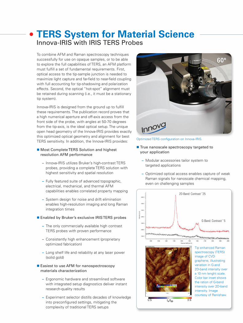
TERS System for Material ScienceInnova-IRIS with IRIS TERS Probes
To combine AFM and Raman spectroscopy techniques successfully for use on opaque samples, or to be able to explore the full capabilities of TERS, an AFM platform must fulfill a set of fundamental requirements. First, optical access to the tip-sample junction is needed to maximize light capture and far-field to near-field coupling with full accounting for tip-shadowing and polarization effects. Second, the optical “hot-spot” alignment must be retained during scanning (i.e., it must be a stationary tip system).
Innova-IRIS is designed from the ground up to fulfill these requirements. The publication record proves that a high numerical aperture and off-axis access from the front side of the probe, with angles at 50-70 degrees from the tip-axis, is the ideal optical setup. The unique open head geometry of the Innova-IRIS provides exactly this optimized optical geometry and alignment for best TERS sensitivity. In addition, the Innova-IRIS provides:
�Most Complete TERS Solution and highest resolution AFM performance
– Innova-IRIS utilizes Bruker’s high-contrast TERS probes, providing a complete TERS solution with highest sensitivity and spatial resolution
– Fully featured suite of advanced topographic, electrical, mechanical, and thermal AFM capabilities enables correlated property mapping
– System design for noise and drift elimination enables high-resolution imaging and long Raman integration times
� Enabled by Bruker’s exclusive IRIS TERS probes
– The only commercially available high contrast TERS probes with proven performance
– Consistently high enhancement (proprietary optimized fabrication)
– Long shelf life and reliability at any laser power (solid gold)
� Easiest to use AFM for nanospectroscopy materials characterization
– Ergonomic hardware and streamlined software with integrated setup diagnostics deliver instant research-quality results
– Experiment selector distills decades of knowledge into preconfigured settings, mitigating the complexity of traditional TERS setups
� True nanoscale spectroscopy targeted to your application
– Modular accessories tailor system to targeted applications
– Optimized optical access enables capture of weak Raman signals for nanoscale chemical mapping, even on challenging samples
Optimized TERS configuration on Innova-IRIS.
Tip enhanced Raman spectroscopy (TERS) image of CVD graphene, illustrating variation in G-and 2D-band intensity over a 10 nm lenght scale. Color bar inset shows the ration of G-band intensity over 2D-band intensity. Image courtesy of Renishaw.
2D-Band: Contrast ˜25
G-Band: Contrast ˜ 5

An Optimized Solution
The Innova-IRIS provides exceptionally easy setup and use, even with the full AFM-Raman integration in place. The system also exhibits the stringent performance required for both advanced atomic force microscopy and Raman spectroscopy, while preserving the tip to enable tip-scattering optical techniques. Modular integration software ideally coordinates the actions of the Innova AFM and spectrometer to allow combined experiments without technique interference.
At the same time, the hardware and software of the Innova-IRIS system preserve the full power and flexibility of both AFM and spectrometer, providing the most complete characterization possible. Combining Raman spectroscopy with AFM-based nanoscale electrical, mechanical, thermal characterization on the same sample is fast, simple, and effective.
Reliable high performance probes are the central and non-trivial part of any TERS solution. Commercially available, Bruker’s exclusive high-contrast TERS probes uniquely provide full access to measurement on a wide range of samples. Bruker’s reliable TERS-STM and TERS-AFM probes exhibit zero spectral interference (no feedback laser) delivering superior sensitivity with high confidence.
On the other hand, your application will benefit from the best tip preservation and lowest drift, guaranteeing that alignment is preserved even over the optical integration times necessary to interrogate weak Raman scatterers. Bruker’s integrated TERS solution enables researchers to focus on what is most important in their work: scientific discovery.
TERS of Nile Blue thin film using Innova-IRIS with IRIS TERS probes. TERS signal more than 100x stronger than far-field Raman signal.
Nanoscale strain in silicon and buried oxide is detected by TERS using Innova-IRIS system. Data courtesy of Dr. Daisuke Kosemura, Meiji University.
Innova-IRIS with IRIS TERS probes and a Raman spectrometer offers a complete AFM-Raman solution.
RAMAN
RAMANTOP
Simultaneous optical views enabling TERS alignment.

The creation of integrated Raman and AFM capabilities for transparent samples requires a design that allows the AFM to function seamlessly as an integral part of an inverted optical microscope. This opens the door to the full power of optical spectroscopy. Enabling TERS adds the fundamental requirement of retaining tip-sample alignment, and thus scanning the sample. Finally, a well-designed system addresses compatibility with samples and their carriers.
The innovative open head of the BioScope Catalyst-IRIS accomplishes all of these requirements, allowing unmatched and virtually unrestricted optical and physical access from below and above the sample to maximize objective and condenser access. Designed entirely for operation on top of an inverted microscope, the Catalyst AFM exhibits the force control and stability required to preserve tip and alignment in biological, combined-setup environments. The system is built from the ground up around a sample scanning geometry to retain alignment between tip and optics during imaging, which provides uncompromised performance for advanced applications such as TERS.
Catalyst-IRIS provides:
� Easiest to use AFM for spectroscopy in life sciences
– ScanAsyst makes in situ liquid imaging dramatically easier while retaining best force control for the most delicate samples
– Experiment selector distills decades of knowledge into preconfigured settings, mitigating the complexity of traditional TERS setups
� Highest performance, most complete AFM capabilities in the world
– Exclusive PeakForce QNM imaging mode uniquely provides highest resolution mechanical property mapping
– System design for noise and drift elimination enables high- resolution imaging and long Raman integration times
� Best TERS AFM-Raman system integration available
– Modular system integration interface provides tools for most effective setup optimization, real-time control, and data acquisition
– Flexible system architecture offers the widest compatibility with leading Raman microscopy suppliers
TERS System for BiologyCatalyst-IRIS Life Science AFM
TERS of Cresyl Blue using Catalyst-IRIS showing contrast factor of 99.532nm excitation at 64uW. (Data courtesy of L. Opilik, C. Blum, T. Schmid, and R. Zenobi.)
Co-localized high resolution topography (left), Bruker’s exclusive PeakForce QNM nanomechanical mapping (middle), and Raman spectroscopy (right) can be used to identify cancerous cells unambiguously using Catalyst-IRIS.
Topography PeakForce QNM Modulus Raman
Organelles
Nucleus

Optimized for Life Sciences
In addition to all these features, the Catalyst-IRIS supports a number of accessories to provide the complete solution for your biological samples:
� Perfusing Stage Incubator enables researchers to maintain ideal cell culture conditions for long- duration live cell studies
� Proprietary MIRO (Microscope Image Registration and Overlay) software utilizes optical images to guide AFM imaging and force measurements to targeted regions of interest, accurately registering optical and AFM images in real time, even when there are no obvious common features
� Exclusive ScanAsyst automatically adjusts scan parameters, such as setpoint, feedback gains, and scan rate, to make Bio-AFM imaging dramatically easier, even in fluids
Tailored Systems
Combine the Catalyst-IRIS with your choice of inverted optical microscope and leading Raman system to create the most integrated AFM-spectroscopy system optimized for your application. However you tailor your system, you will benefit from the best tip and sample preservation pushing the frontier to more fragile samples with smaller Raman scattering cross sections.
Catalyst-IRIS TERS AFM-Raman system for biology and transparent samples.
In situ nanomechanical map of fixed MDCK cell in PBS buffer acquired using Catalyst with ScanAsyst and PeakForce QNM.
AFM topography image of the 2D polymer film on glass (gray) overplayed by a confocal Raman map (3s/pixel acquisition time at 470 μW) using MIRO. Image courtesy of Prof. Renato Zenobi from ETH University.
Overlaid AFM ScanAsyst image (brown), Raman chemical map (green) and optical view (gray) of a polymer blend containing polystyrene and low-density polyethylene. (Raman data courtesy of J. Schreiber, Horiba.)

Bruker Nano Surfaces Division
Santa Barbara, CA • USA Phone +1.805.967.1400/800.873.9750 [email protected]/nano
Cover images: Spectra overlaid on the illustration show TERS of Malachite Green on gold acquired with Innova-IRIS.
© 2
014
Bru
ker
Cor
pora
tion.
All
right
s re
serv
ed. B
ruke
r N
ano
Sur
face
s D
ivis
ion
is c
ontin
ually
impr
ovin
g its
pro
duct
s an
d re
serv
es t
he r
ight
to
chan
ge s
peci
ficat
ions
with
out
notic
e.
Bio
Sco
pe C
atal
yst,
Icon
, Inn
ova,
Pea
kFor
ce Q
NM
, and
Sca
nAsy
st a
re t
rade
mar
ks o
f B
ruke
r C
orpo
ratio
n. A
ll ot
her
trad
emar
ks a
re t
he p
rope
rty
of t
heir
resp
ectiv
e co
mpa
nies
. B07
8, R
ev. B
3
An AFM-Raman System Designed Specifically for Your Research
Bruker’s IRIS solutions integrate the highest atomic force microscopy performance seamlessly with micro-Raman systems to address your specific nanoscale spectroscopy application needs. Bruker’s Icon-Raman systems provide an enormous wealth of information, from mechanical to electrical properties to chemical ID or structural information, taking the quality of material characterization to a new level.
Images above show multi-dimensional characterization of a polymer blend that contains polystyrene and polyisoprene and shows uniform and chemically pure domains with nanoscale sharp boundaries. Bruker’s Raman solution uniquely combines several characterization methods, including a complete solution for high contrast TERS chemical identification, co-localized highest resolution AFM imaging and Raman mapping, and quantitative nanomechanical mapping with access to the full force curve at each pixel—all of which creates a productive system that can be tailored to your specific research needs.
TERS ChemID Typography Adhesion PeakForce QNM
Raman
AFM-Raman ConfigurationsSystem Key Features Applications
Innova-IRIS with Renishaw inVia or Horiba LabRAM
AFM-Raman system for TERS and material science research applications
Bruker exclusive, high-contrast IRIS TERS probes
Materials research, nanoparticles, graphene
BioScope Catalyst-IRIS with Renishaw inVia or Horiba XploRA INV
Inverted optical microscope configuration for transparent samples and life science applications
Life science applications from single molecule DNA and protein investigations-to cell biology
Dimension Icon with Renishaw inVia or Horiba LabRAM
High-performance, research-grade instrument
Most advanced capabilities using PeakForce QNM, and ScanAsyst
Materials research and development, polymers, composites, semiconductors