Embed Size (px)
Citation preview

British Journal of Ophthalmology, 1987, 71, 449-453
A five-year survey of ocular shotgun injuries in IrelandDERMOT RODEN,' PHILIP CLEARY,2 AND PETER EUSTACE
From the 'Department of Ophthalmology, Mater Misericordiae Hospital, Dublin, and the 2Eye, Ear, andThroat Hospital, Cork, Ireland
SUMMARY Between November 1980 and September 1985 20 patients in Ireland sustainedaccidental ocular shotgun injuries severe enough to necessitate hospital admission. Eight patientshad contusion injuries and 12 perforating injuries. Contusion damage was disproportionate to thesize of the pellet. Through and through perforation of the globe occurred in eight patients; theremaining four patients in this group had retained intraocular pellets. Twelve patients sustainedperforating eye injuries. Those that were treated by primary closure alone lost the sight in that eye.Those treated by vitreoretinal surgery recovered vision directly related to where the pellet had itsexit from the eye. It was possible to contact 15 of the patients. All 15 were shooting pheasant. It wasnot possible to relate the distance of the patient from the gun to the severity of the ocular injury.
Accidental shotgun injuries regularly present toophthalmic departments in Ireland. Over 176000shotgun licences were issued in Ireland in 1985, andgame shooting is a popular sport. The injuriessustained have many distinctive features, contusion issevere, and the pellets travel towards the apex of theorbit. Perforation is common and often through andthrough. Vitreoretinal surgery is a prerequisite tosatisfactory management of the perforating injuries.Two-thirds of the victims were shot by experiencedgun handlers. Bullet and shrapnel injuries involvingthe eye have been extensively described by Duke-Elder,' and BB-gun injuries to the eye have beenreported (a BB-gun is an air rifle that shootspellets).2 I Bowen and Magauran4 reported 105 casesof ocular injuries caused by airgun pellets. Conway etal.' mention the surgical management of two patientswho suffered perforating eye injuries from shotgunpellets.The regular presentation of ocular shotgun injuries
to ophthalmologists in Ireland and the rarity ofreports in the literature prompted this survey.
Material and methods
All ophthalmologists in the Republic of Ireland werecircularised by letter. One of the authors (DR)telephoned each consultant, asking him if he couldrecall attending any patient involved in a shotgun
accident during the period November 1980 toJanuary 1985. Twenty patients were thus recalled.The charts were retrieved and scrutinised by the sameauthor. Particular attention was paid to the ocularinjury, its treatment, and ultimate best correctedvisual acuity. Fifteen of the 20 patients were con-tacted about the circumstances of the accident. Theage and experience of the person who shot them, thetype of the gun and cartridge used, and the distancefrom the patient were ascertained. It was not possibleto interview five patients as they could not becontacted.
Results
Twenty patients were involved in ocular shotgunaccidents from November 1980 to January 1985-anaverage rate of four cases a year from a population of3-8 million people. The findings of our survey aretabulated in Tables 1 and 2. Ten of the 20 patients(50%) presented in November, three (15%) inDecember, and four (20%) in January. All thepatients were male, their ages ranging from 10 to 65years, mean 30 years. Of the 20 patients threesustained bilateral eye injuries, accounting for 23eyes. Thirteen of the 17 uniocular injuries (76%)were to the right eye.
Clinically the injuries were divided into twogroups:
Correspondence to Professor P Eustace, Department of Ophthal- 1 CONTUSION INJURIESmology, Matcr Misericordiae Hospital, Dublin 7, Ireland. Eight patients had contusion eye injuries, one patient
449
on 15 May 2019 by guest. P
rotected by copyright.http://bjo.bm
j.com/
Br J O
phthalmol: first published as 10.1136/bjo.71.6.449 on 1 June 1987. D
ownloaded from

450
Table 1 Patients with contusion injuries
Dermot Roden, Philip Cleary, and Peter Eustace
Case Date of Sex Age Eye Injuriesno. presentation
1 2 Nov 1980 M 42 R Comotio retinae
2Nov1982 M 33 R
20Nov 1983 M 30 L
2OJan 1983 M 20 L
3Nov1984 M 19 R
L8Jan 1984 M 29 R
21Jan 1984 M 16 R
18Jan 1985 M 25 R
Comotio retina
Comotio retinae
III palsy
Hyphaema+comotioretinae
Eyelid entry woundIII, IV, V, VI palsy
Comotio retinae
Comotio retinae
Management Ultimate Interviewed Target,visual circumstances,acuity distance
Laser 6/9 Yes Pheasant, sallies,photocoagula- 12 yardstion
Conservative 6/6 Yes Pheasant, ditch,60 yards
Conservative 6/6 Yes Pheasant, bush,60 yards
Conservative 6/9 Yes Pheasant, ditch,15 yards
Conservative 6/6 Yes Pheasant, field,60 yards
Conservative 6/6Conservative 6/12 Yes Pheasant, ditch,
15 yardsConservative 6/6 Yes Pheasant, field,
20 yardsConservative 6/5 Yes Pheasant, ditch
Gun handler, age,sex, relationship topatient, experience
15, M, acquaintance,no experience
25, M, brother,experienced
32, M, relative, noexperience
35, M, friend,experienced
19, M, friend,experienced
35, M, acquaintance,experienced
23, M, brother,experienced
19, M, friend, noexperience
Metric conversion: 10 yards=9 m.
had bilateral injuries, Table 1. In no case did a orbital entry wounds, subconjunctival lacerations,perforation occur in this group. In contusion injuries and haemorrhages, hyphaema, comotio retinae, andthe pellet lodged in the retro-orbital tissue or in the transient vitreous haemorrhage (Fig. 1). In two casescavernous sinuses. Contusion injuries included peri- (Nos. 4 and 6) the patients suffered transient cranial
Table 2 Patients with perforating injuries
Case Date of Sex Age Eye Injuries Management Ultimate Interviewed Target, Gun handler, age,no. presentation visual circumstances, sex, relationship to
acuity distance patient, experience
9 2 Nov 1980 M 59 L Double perforation with 1'Closure HM Yes Pheasant, field, 55, M, friend,RD 100 yards experienced
10 4Dec 1980 M 45 R Single perforation, 1'Closure NPL Nophthisical response
11 4 Jan 1981 M 29 R Double perforation, P Closure NPL Yes Rabbit, ditch, 33, M, brother,phthisical 10 yards experienced
12 12 Nov 1982 M 65 L Double perforation, vit. P Closure HM Yes Pheasant, 20 yards 33, M, friend, nohaem. RD experience
13 13 Nov 1982 M 15 R Double perforation, P Closure NPL Yes Pheasant, field, 45, M, no experiencephthisical 30 yards
14 6May 1983 M ? R Double perforation, 1°Closure NPL Nophthisical response
15 22 Aug 1983 M 29 R Double, aphakia 1°Closure 6/9 No Family feud+ vitrectomy response
L Single, aphakia 1°Closure CF+ vitrectomy
16 4Nov 1983 M 14 R Single,vit.haem. 1°Closure 6/12 No+ vitrectomy response
17 27 Dec 1983 M 40 R Double perforation, 1°Closure NPL Yes Woodcock, ditch, 45, M, uncle,phthisical + vitrectomy 60 yards experienced
18 5 Nov 1983 M 25 R Double, aphakia, vit. 1°Closure 6/60haem. + vitrectomy
19 17 Nov 1984 M 10 R Single, phthisical 1°Closure NPL Yes Pheasant, ditch, 21, M, brother30 yards
20 4 Dec 1984 M 24 R Double, vit. haem., Vitrectomy 2/60 Yes Pheasant, ditch, 34, M, friend,aphakia 15 yards experienced
L Double, vit. haem., Vitrectomy 6/9aphakia
CF=counting fingers. HM= hand movements. NPL= no perception of light.
2
3
4
S
6
7
8
on 15 May 2019 by guest. P
rotected by copyright.http://bjo.bm
j.com/
Br J O
phthalmol: first published as 10.1136/bjo.71.6.449 on 1 June 1987. D
ownloaded from
Afive-year survey ofocular shotgun injuries in Ireland
.
iinjuries.picalpresentation ofapatient with shotgun
nerve palsies, which recovered within three months.All of the contusion injuries were managed conserva-tively. There was no attempt to remove the pelletsunless they were palpably superficial.
In all nine eyes the visual acuity recovered to 6/12or better without surgical intervention. Two yearsafter the initial injury a retinal tear developed in case1. This was satisfactorily treated by laser photo-coagulation.
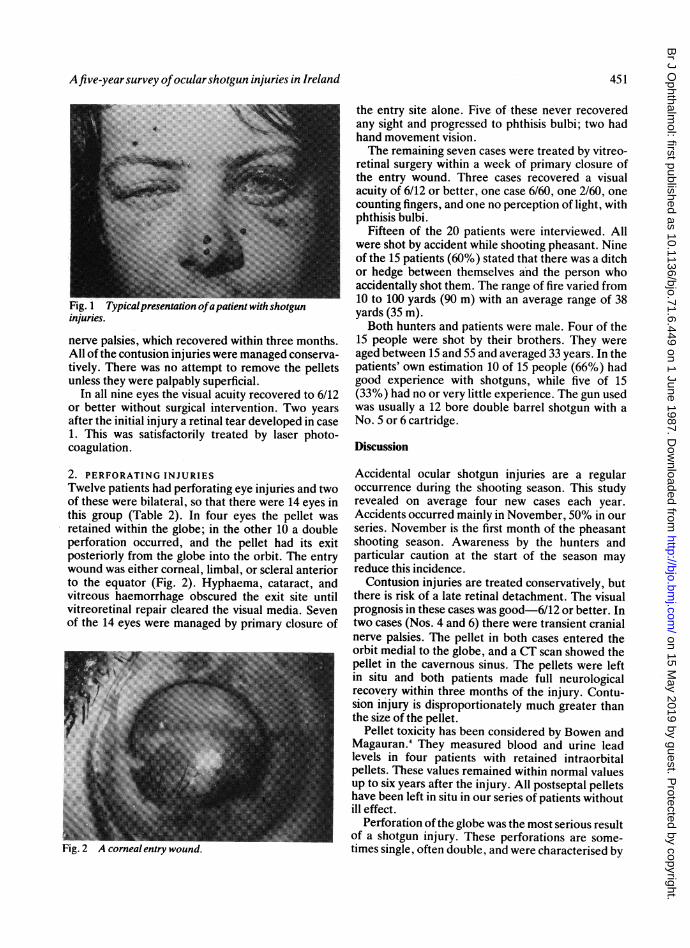
2. PERFORATING INJURIESTwelve patients had perforating eye injuries and twoof these were bilateral, so that there were 14 eyes inthis group (Table 2). In four eyes the pellet wasretained within the globe; in the other 10 a doubleperforation occurred, and the pellet had its exitposteriorly from the globe into the orbit. The entrywound was either corneal, limbal, or scleral anteriorto the equator (Fig. 2). Hyphaema, cataract, andvitreous haemorrhage obscured the exit site untilvitreoretinal repair cleared the visual media. Sevenof the 14 eyes were managed by primary closure of
Fig. 2 A corneal entry wound.
the entry site alone. Five of these never recoveredany sight and progressed to phthisis bulbi; two hadhand movement vision.The remaining seven cases were treated by vitreo-
retinal surgery within a week of primary closure ofthe entry wound. Three cases recovered a visualacuity of 6/12 or better, one case 6/60, one 2/60, onecounting fingers, and one no perception of light, withphthisis bulbi.
Fifteen of the 20 patients were interviewed. Allwere shot by accident while shooting pheasant. Nineof the 15 patients (60%) stated that there was a ditchor hedge between themselves and the person whoaccidentally shot them. The range of fire varied from10 to 100 yards (90 m) with an average range of 38yards (35 in).Both hunters and patients were male. Four of the
15 people were shot by their brothers. They wereaged between 15 and 55 and averaged 33 years. In thepatients' own estimation 10 of 15 people (66%) hadgood experience with shotguns, while five of 15(33%) had no or very little experience. The gun usedwas usually a 12 bore double barrel shotgun with aNo. 5 or 6 cartridge.
Discussion
Accidental ocular shotgun injuries are a regularoccurrence during the shooting season. This studyrevealed on average four new cases each year.Accidents occurred mainly in November, 50% in ourseries. November is the first month of the pheasantshooting season. Awareness by the hunters andparticular caution at the start of the season mayreduce this incidence.
Contusion injuries are treated conservatively, butthere is risk of a late retinal detachment. The visualprognosis in these cases was good-6/12 or better. Intwo cases (Nos. 4 and 6) there were transient cranialnerve palsies. The pellet in both cases entered theorbit medial to the globe, and a CT scan showed thepellet in the cavernous sinus. The pellets were leftin situ and both patients made full neurologicalrecovery within three months of the injury. Contu-sion injury is disproportionately much greater thanthe size of the pellet.
Pellet toxicity has been considered by Bowen andMagauran.4 They measured blood and urine leadlevels in four patients with retained intraorbitalpellets. These values remained within normal valuesup to six years after the injury. All postseptal pelletshave been left in situ in our series of patients withoutill effect.
Perforation of the globe was the most serious resultof a shotgun injury. These perforations are some-times single, often double, and were characterised by
451
on 15 May 2019 by guest. P
rotected by copyright.http://bjo.bm
j.com/
Br J O
phthalmol: first published as 10.1136/bjo.71.6.449 on 1 June 1987. D
ownloaded from

DermotRoden, Philip Cleary, and Peter Eustace
Fig.3 Ultrasonagraphicdemonstration ofan intravitrealhaemorrhage (arrow) in apatientwith a doubleperforating injury.
intraocular haemorrhage (Fig. 3), vitreous incarcera-tion, and a possible retinal detachment. It has beenshown experimentally67 and it is recognised clinically'that perforating injuries (with these complications)have poor prognosis when treated by conventionaltechniques. In this study seven eyes were treated byprimary closure alone and none recovered usefulvision, with the development of phthisis in five. Incontrast, the seven eyes treated by vitrectomy withinone week of primary closure had a much more
Fig. 4 Case20. The leftfundus showing an equatorial exit.The visual acuity is 619.
favourable visual outcome. Three of the sevenregained a visual acuity of 6/12 or better, and in theother four the eye was salvaged but the visualpotential had been destroyed by the pellet leaving theeye at or near the macula.
Perforating injuries involving the posterior seg-ment result in vitreous gel incarceration and vitreoushaemorrhage. This progresses to retinal traction andtractional retinal detachments. In those cases whichunderwent vitrectomy we consider the scaffolding onwhich the contractile fibroblasts proliferate wasremoved, aborting the retinal traction and detach-ment and salvaging the visual potential of the eye.
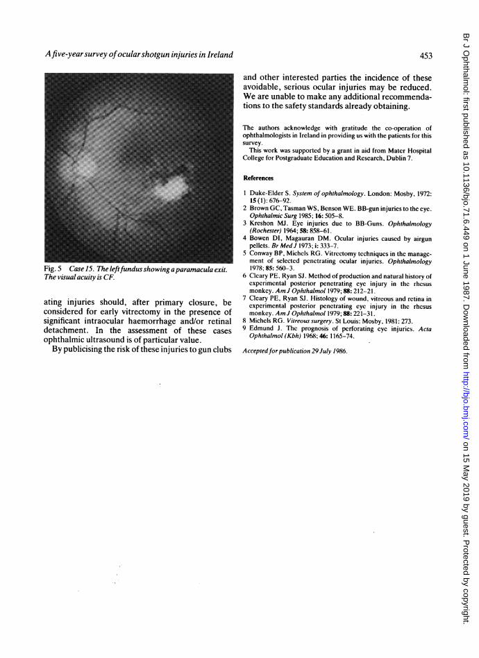
It has been suggested previously that visual out-come may be related to the entry site of the pellet.' Inour series the visual outcome was related to wherethe pellet had its exit from the globe. In the twobilateral perforation injuries (cases 15 and 16) thevisual acuity was 6/9 where the pellet had its exit atthe equator (Fig. 4). In contrast, the visual acuity wascounting fingers and 2/60 when the pellet left the eyeat or near the macula (Fig. 5).
Fifteen of the 20 patients were interviewed, nine ofthem described the accident occurring through aditch or hedge. We found no correlation between theseverity of the injury and the distance from whichthey were shot. Indeed one case (No. 9) was shotfrom 100 yards (90 m). He had a double perforationand the eye was lost. He received no other injuries. Itis noteworthy that 10 of the 15 were shot by peoplewho, in the patients' estimation, were experienced.
In conclusion, the results in this study suggeststhat, after a shotgun injury, contusion eye injuriesare best treated conservatively. But eyes with perfor-
452
on 15 May 2019 by guest. P
rotected by copyright.http://bjo.bm
j.com/
Br J O
phthalmol: first published as 10.1136/bjo.71.6.449 on 1 June 1987. D
ownloaded from
Afive-yearsurvey ofocularshotgun injuries in Ireland
.5 Case 15. The leftfundus showing aparamacula exit.e visual acuity is CF.
ating injuries should, after primary closure, beconsidered for early vitrectomy in the presence ofsignificant intraocular haemorrhage and/or retinaldetachment. In the assessment of these casesophthalmic ultrasound is of particular value.By publicising the risk of these injuries to gun clubs
and other interested parties the incidence of theseavoidable, serious ocular injuries may be reduced.We are unable to make any additional recommenda-tions to the safety standards already obtaining.
The authors acknowledge with gratitude the co-operation ofophthalmologists in Ireland in providing us with the patients for thissurvey.
This work was supported by a grant in aid from Mater HospitalCollege for Postgraduate Education and Research, Dublin 7.
References
1 Duke-Elder S. System of ophthalmology. London: Mosby, 1972:15 (1): 676-92.
2 Brown GC, Tasman WS, Benson WE. BB-gun injuries to the eye.Ophthalmic Surg 1985; 16: 505-8.
3 Kreshon MJ. Eye injuries due to BB-Guns. Ophthalmology(Rochester) 1964; 58: 858-61.
4 Bowen DI, Magauran DM. Ocular injuries caused by airgunpellets. Br Med J 1973; i: 333-7.
5 Conway BP, Michels RG. Vitrectomy techniques in the manage-ment of selected penetrating ocular injuries. Ophthalmology1978; 85: 560-3.
6 Cleary PE, Ryan SJ. Method of production and natural history ofexperimental posterior penetrating eye injury in the rhesusmonkey. Am J Ophthalmol 1979; 88: 212-21.
7 Cleary PE, Ryan SJ. Histology of wound, vitreous and retina inexperimental posterior penetrating eye injury in the rhesusmonkey. Am J Ophthalmol 1979; 88: 221-31.
8 Michels RG. Vitreous surgery. St Louis: Mosby, 1981: 273.9 Edmund J. The prognosis of perforating eye injuries. Acta
Ophthalmol (Kbh) 1968; 46: 1165-74.
Accepted for publication 29 July 1986.
453
on 15 May 2019 by guest. P
rotected by copyright.http://bjo.bm
j.com/
Br J O
phthalmol: first published as 10.1136/bjo.71.6.449 on 1 June 1987. D
ownloaded from