Embed Size (px)
Citation preview

J. med. Genet. (I964). I, 27.
A Family Apparently Showing Transmission of aTranslocation Between Chromosome 3 and One
of the 'X-6-I2' or 'C' Group*G. CLARKE, A. C. STEVENSON, PAMELA DAVIES, and C. E. WILLIAMS
From Medical Research Council Population Genetics Research Unit, and Radcliffe Infirmary, Oxford, and BorocourtHospital, Reading
The propositus (III.7 in the diagrammatic pedigreeFig. i) was born in hospital on September io, I956 afteran uneventful pregnancy lasting 39 weeks and a normallabour lasting 5 hours. Although nothing untoward wasnoticed at birth, within a few days it was noted that shewas rather flaccid and seldom opened her eyes. Sub-sequently her mother was worried at her 'lifelessness'and suspected that she had mongolism. Between theages of 3 months and 31 years the child was investigatedat 3 different hospitals, and at each the mother was toldsubstantially the same, namely, that she was definitelynot a mongol, but that she was not progressing as quicklyas she should and was likely to be mentally retarded.The child was able to sit unaided at about 2I months
and walked without support at about 2 years and 3months. Bladder control was established at about 3years. At 4 years and 3 months she had an epileptic fit.Mild seizures recurred at decreasing intervals, and shehas had only one fit in the last year.
Early in I96I the family moved to the Oxford area andthe child was seen by Dr Victoria Smallpeice. She hasbeen under observation since then. In I962 she beganto attend a day school for severely retarded children atBorocourt Hospital.At 7 years, she is a chubby child, 3 ft 91 in. (ii5 cm.)
in height which is more than one standard deviationbelow the mean for her age. Her head circumference is21 in. (22 cm.) which is just over average for her age.Fig. 2 and 3 show her appearance. She has a dull,rather expressionless, face and an alternating strabismus.Her fundi are normal. Her neck is short with somewebbing, and she has a high arched palate. There isgenuvalgum, pes planus, and some increase in the carry-ing angle of both arms. She has short 'stubby' handsshowing clinodactyly of both fifth fingers. However,this trait is shown also by her mother and her eldersister. Although the hands are short the fingers tapermarkedly, and there is limitation of full extension of theterminal interphalangeal joints, as can be seen in Fig. 3.Both hands show a single transverse palmar crease.There is marked clinodactyly of both feet, the second,
fourth, and fifth toes incurving towards the third. Thereis some dorsal scoliosis. Some of these features areevident in the photographs. The lower end of the
*Received February I7, I964.
sternum is depressed and the umbilicus is located ratherhigh on the abdomen with a brown line running down tothe pubis. On the right buttock is a flat cafd au lait spot.The external genitalia appear normal. The general
muscle tone is poor and she has some hyperextensibilityof joints. Cardiac examination reveals no abnormalityand both femoral pulses are palpable. Toxoplasmosisskin tests and Wassermann reactions are negative. Skulland lumbar region radiography reveal no abnormality.
Mentally the child is severely retarded, scoring 43 onthe Vineland Social Maturity Scale. She has, however,improved considerably since attending the day schooland can speak short sentences and laboriously print hername. She enjoys going to school and is a contentedhappy child, which to some extent reflects an excellenthome environment.Dr C. Ounsted of the Park Hospital for Children,
Oxford, kindly undertook an electroencephalographicinvestigation. He reports that 'neither the waking northe sleeping record shows any specific anomaly'.
Family HistoryA diagrammatic pedigree is set out in Fig. i. The
father and mother were aged 40 and 34 years respectivelywhen the propositus was born. Both parents are ofsuperior intelligence and have no physical stigmata.There is no history that appears relevant on the
father's side of the family. The last of i9 children in thefamily of the maternal grandmother of the child is saidto have been a mongol. All that is known of II.4 is that
l~~~~~~~~~ 6
i 6
((? f~~~~67
FIG. I. Pedigree chart of family.
27
on April 2, 2020 by guest. P
rotected by copyright.http://jm
g.bmj.com
/J M
ed Genet: first published as 10.1136/jm
g.1.1.27 on 1 Septem
ber 1964. Dow
nloaded from

Clarke, Stevenson, Davies, and Williams
FIG. 2 and 3. Appearance of propositus.
he is said to have been weakly at birth and to have diedwithin a few days. II.5 is a tall man (6 ft 2 in.) (i88 cm.)of more than average intelligence. He has extremelylong hands measuring 8j in. (22-2 cm.) from the distalwrist crease to the tip of the middle finger. He wasmarried when aged 34 to a woman of the same age, andthere have been no pregnancies in the ensuing eight yearsof the marriage. I.3, the mother's father, is a healthyelderly man; his wife, I.4, is dead.
N0 4
III.s, the eldest sister of the propositus, was born inMay 1952 and has passed her 'eleven-plus' examination.She appears normal in every way as does her 3-year-oldbrother, III.8. III.6 was born in September I954 anddied in February 1955. She was recognized at birth tohave a cardiac abnormality; when 2 months old she wasadmitted to hospital where an electrocardiogramshowed right axis deviation and x-ray examinationshowed enlargement of both ventricles with widening of
III&
A * *̂^ ^> x̂r~AW
XX As.
FIG. 4. Karyotype of 111.7.
A& ha2l -----22
28
on April 2, 2020 by guest. P
rotected by copyright.http://jm
g.bmj.com
/J M
ed Genet: first published as 10.1136/jm
g.1.1.27 on 1 Septem
ber 1964. Dow
nloaded from

Translocation between Chromosome 3 and 'X-6-i2' or 'C' Group
the upper mediastinum, mainly on the right side. Shewas regarded as having an anomaly of the great vessels,probably a coarctation of the aorta. There is no recordon the hospital notes of any bodily anomaly. She died inanother hospital at i8 months but there was no necropsy.III.8 was born in April I960, but he has always appearedto be a normal child, and there are not obvious mentalor physical anomalies. The mother had no abortions.A note on the dermatoglyphs in the family by Dr
Sarah Holt is given in the Appendix.
KaryotypesThe Denver Conference (I960) numbers are used in
identification of chromosomes. For brevity, however,and following the lead of the recent London Conferenceon the Normal Human Karyotype (I963), the group ofchromosomes numbers 6-I2 inclusive and the Xchromosome are referred to as group C.On examination of chromosomes from skin fibro-
blasts and white blood cell preparations from the propo-situs, III.7, she was found to have a modal number of 46chromosomes. Nos. i and 2 and I3 to 22 could beidentified and paired or grouped without difficulty.There were, however, (a) five large sub-metacentricchromosomes instead of the expected four from Nos. 4and 5 (the 'B' group); (b) only IS of the intermediatesize sub-metacentric group C chromosomes, instead ofthe expected i6 for a female child (Fig. 4).
0 X 1
____*_ ____ frt____
3 4 5
FIG. 4a. Part karyotypes-5 cells of III.7.
Fig. 4a shows relevant chromosomes from additionalfibroblast cells of the propositus III.7. These chromo-somes are the five large sub-metacentric chromosomesmentioned above and a pair of normal No. 3 chromo-somes.
Buccal smears appeared normal female with singleBarr bodies in about 40% of cells.Chromosomes from white blood cell preparations
from the child's mother, II.6, are shown in Fig. 5. Themodal chromosome number was 46. Chromosomescorresponding to Nos. i and 2 and I3 to 22 were readilyidentified and paired. As in her daughter, there were 5instead of the expected 4 large sub-metacentric chromo-somes like pairs 4 and 5. However, there were twodifferences from the karyotype of her daughter: (a) therewas only one large metacentric chromosome like chro-mosome No. 3 which is usually so easily identified, and(b) there was a full complement for a female of i6 chro-mosomes which, by morphology, belonged to group C.
Fig. Sa shows relevant chromosomes from fiveadditional leucocyte cells of the mother (II.6). Theyare (a) as in the propositus five large sub-metacentricchromosomes, (b) one unmistakable No. 3 chromosome,and (c) a chromosome with one pair of arms the samelength as in a No. 3 and one pair of shorter arms.There was no suggestion of mosaicism in the mother;
the changes described could be identified in all well-spread metaphases. Buccal smears were those of anormal female with single Barr bodies in a majority ofcells.
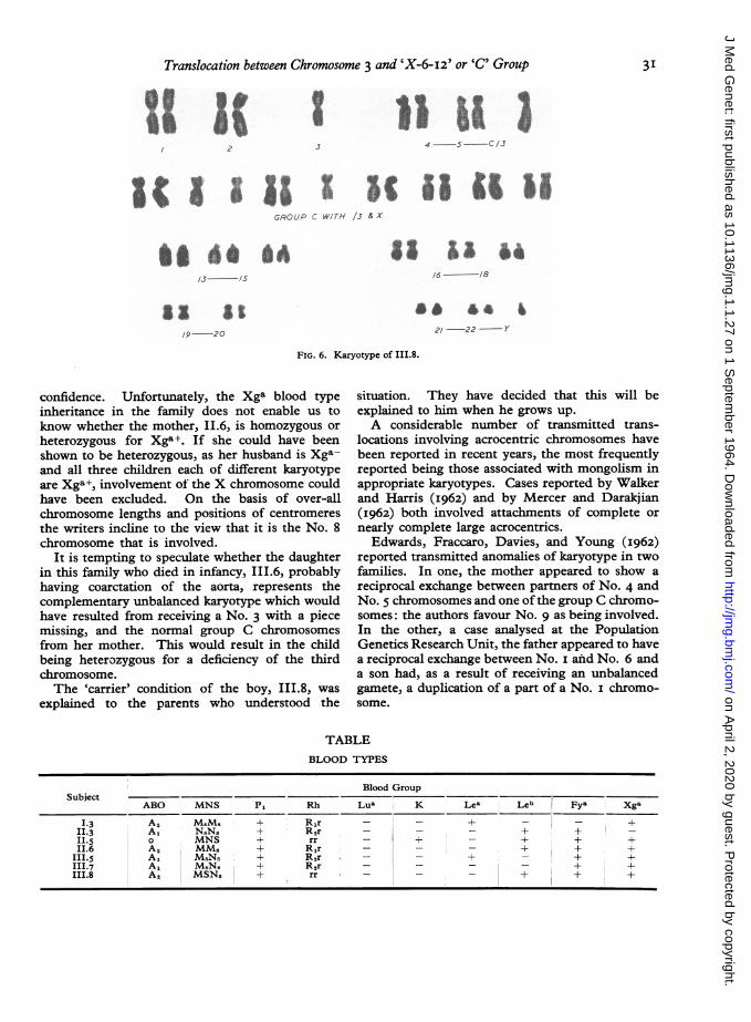
Karyotypes of the maternal uncle of the propositus,II.s, and of her elder sister, III.5, were normal in num-ber and morphology for the appropriate sexes. How-ever, that of her younger brother, III.8 (Fig. 6), wasexactly similar to that of his mother except that beingmale he had one fewer group C chromosome and asmall chromosome identified as a Y.The most probable interpretation of the chromosome
findings in the mother, taken in association with the factthat she appears normal phenotypically, is that there hasbeen reciprocal interchange of unequal terminal portionsof arms of a chromosome No. 3 and one of the group Cchromosomes. As chromosome No. 3 has a mediancentromere, it is impossible to say which arm is involved,but it is the long arm of the group C chromosome thatappears to have the attached portion of the No. 3 (Fig.5 and 5a). It is impossible to judge the size of the smallterminal portion of the group C chromosome which hasbeen transferred to the No. 3 chromosome. If only asmall acentric portion of the terminal section distal to abreak was involved, it might be 'lost' without thedeletion determining any ill phenotypic effects, par-ticularly if, as appears to be the situation in some otherorganisms, the telomere region is inert. In such a situ-ation the translocation would not be 'reciprocal'.The above explanation, whether postulating that the
telomere portion is lost or attached to No. 3, requirespostulation of only two breaks and seems inherentlymore probable than an alternative one requiring threebreaks, namely, that there have been two breaks in thearm of No. 3 and that the interstitial portion released was
29
on April 2, 2020 by guest. P
rotected by copyright.http://jm
g.bmj.com
/J M
ed Genet: first published as 10.1136/jm
g.1.1.27 on 1 Septem
ber 1964. Dow
nloaded from

(3 larke, Stevenson l)avies, and Williams
2 3 4 5 C/3
lGEXOGROUP C WITH /3 & x
&A# AA Afto X AS 4A
3 15
N9 20 21 - 22
FIG. 5. Karyotype of 11.6.
FIG. 5a. Pant karyotypes-S cells of 11.6.
inserted between the broken ends of a single break in thelong arm of the group C chromosome.
Accepting the first explanation, namely, two-breaksingle reciprocal interchange of terminal portions of twochromosomes, III.8, the son with a similar karyotypeto that of his mother, presumably received both of hismother's abnormal chromosomes. The unaffecteddaughter, III.5, must have received normal No. 3 andnormal group C chromosomes from her mother.The propositus, III.7, would have received from her
mother a group C chromosome with the relatively largeportion of an arm of a No. 3 attached and a completenormal No. 3 (Fig. 4 and 4a). On this basis thereforeshe is (a) effectively trisomic for part of one arm of aNo. 3 chromosome, and (b) deficient of a much smallerterminal portion of the group C chromosome which isinvolved.
Blood TypingThe Table below shows the blood types of members
ofthe family whose karyotypes have been analysed. Theredoes not appear to be any anomaly of an expected patternof inheritance.
DiscussionThe question as to which of the group C chromo-
somes is involved cannot be answered with complete
30
on April 2, 2020 by guest. P
rotected by copyright.http://jm
g.bmj.com
/J M
ed Genet: first published as 10.1136/jm
g.1.1.27 on 1 Septem
ber 1964. Dow
nloaded from
Translocation between Chromosome 3 and 'X-6-I2' or 'C' Group
O 22
I
4 5 cIJ
GROUP C WITH /3& X
01 6o *4/3 -5
'9-20
a 4'
21/-22 ~Yl
FIG. 6. Karyotype of III.8.
confidence. Unfortunately, the Xga blood typeinheritance in the family does not enable us toknow whether the mother, I1.6, is homozygous or
heterozygous for Xga+. If she could have beenshown to be heterozygous, as her husband is Xga-and all three children each of different karyotypeare Xga+, involvement of the X chromosome couldhave been excluded. On the basis of over-allchromosome lengths and positions of centromeresthe writers incline to the view that it is the No. 8chromosome that is involved.
It is tempting to speculate whether the daughterin this family who died in infancy, III.6, probablyhaving coarctation of the aorta, represents thecomplementary unbalanced karyotype which wouldhave resulted from receiving a No. 3 with a piecemissing, and the normal group C chromosomesfrom her mother. This would result in the childbeing heterozygous for a deficiency of the thirdchromosome.The 'carrier' condition of the boy, III.8, was
explained to the parents who understood the
situation. They have decided that this will beexplained to him when he grows up.
A considerable number of transmitted trans-locations involving acrocentric chromosomes havebeen reported in recent years, the most frequentlyreported being those associated with mongolism inappropriate karyotypes. Cases reported by Walkerand Harris (I962) and by Mercer and Darakjian(I962) both involved attachments of complete or
nearly complete large acrocentrics.Edwards, Fraccaro, Davies, and Young (I962)
reported transmitted anomalies of karyotype in twofamilies. In one, the mother appeared to show a
reciprocal exchange between partners of No. 4 andNo. 5 chromosomes and one of the group C chromo-somes: the authors favour No. 9 as being involved.In the other, a case analysed at the PopulationGenetics Research Unit, the father appeared to havea reciprocal exchange between No. i and No. 6 anda son had, as a result of receiving an unbalancedgamete, a duplication of a part of a No. i chromo-some.
TABLEBLOOD TYPES
Blood GroupSubject
ABO MNS Pi Rh Lua K Lea Leb Fya Xga
I.3 A2 MSMS + R1r - - + _ _ +II3 A1 NSNS + R2r - - - + +II.5 MNS + rr - + - + + +II.6 A2 MMS + R1r - - _ + + +
III.5 A1 MSNS + R2r - - + - + +111.7 A1 MSNS + R2r - - + +III.8 A, MSNS + rr - _ _ + + +
3I
on April 2, 2020 by guest. P
rotected by copyright.http://jm
g.bmj.com
/J M
ed Genet: first published as 10.1136/jm
g.1.1.27 on 1 Septem
ber 1964. Dow
nloaded from

32Clarke, Stevenson, Davies, and Williams
The family here described appears to be only thethird reported where translocations have not in-volved acrocentric chromosomes. Two have beeninvestigated at the Population Genetics ResearchUnit.
It will be interesting to see how many familieswith translocations, not involving acrocentrics, arereported in the future. They may not be as uncom-mon as is at present suggested, partly because severeaffections of the child may result from geneticimbalances determined by structural changes thatare close to the limits of optical recognition. It isperhaps noteworthy that, as in other complete or
partial trisomies so far described, this child isseverely retarded.
SummaryAn account is given of a family identified by a
mentally retarded girl. The chromosome anomaliesdescribed are interpreted as indicating that themother is a balanced translocate having a reciprocalexchange ofthe long arm ofa No. 3 chromosome anda terminal segment of one of X-6-_2 or C group,probably No. 8. The affected propositus is hetero-zygous for the C group plus large portion of No. 3compound chromosome and has two 'normal' No. 3chromosomes, being thus effectively trisomic forpart of the third chromosome. One female sib hasa normal karyotype and a male sib appears to be a
'balanced' translocate like his mother.
We are indebted to Dr Victoria Smallpeice who firstreferred this child; to Dr J. F. P. Asbury, the familydoctor, who made easy our approach to the family; toDr Ruth Sanger, of the M.R.C. Blood Group ResearchUnit for the serological findings; to Dr C. E. Ford, of theM.R.C. Radiobiological Research Unit, who read themanuscript and made many helpful suggestions whichwe have followed in writing this paper; and to Dr C.Ounsted who advised us on interpretation of the electro-encephalographic tracing.
REFRNCES
Denver Conference. A proposed standard system of nomenclatureof human mitotic chromosomes (Denver) (I960). Ann. hum.Genet., 24, 3I9.
Edwards, J. H., Fraccaro, M., Davies, P., and Young, R. B. (I962).Structural heterozygosis in man: analysis of two families. (Witha note on dermal ridge configuration by L. S. Penrose and Sarah B.Holt). ibid., 26, 157.
London Conference on the Normal Human Karyotype (I963).Cytogenetics, 2, 264.
Mercer, R. D., and Darakjian, G. (I962). Apparent translocationbetween chromosome 2 and an acrocentric in group I3-15.Lancet, 2, 784.
Walker, S., and Harris, R. (x962). Familial transmission of a trans-location between two chromosomes of the 13-15 group (Denverclassification). Ann. hum. Genet., 26, x51.
Appendix
A Note on the Dermal Ridge ConfigurationsSARAH B. HOLT
From The Galton Laboratory, University College, London
FINGER RIDGE COUNTS
DigitsSubject Side V IV III II Total
Mother (II.6) Left ?io/o 7/0 0/2 8/o 20/27 131Right 20/12 8/12 II/0 o0/0 24/?20
Daughter (III.7) Left 0/0 o/o 0/0 o/o I/O 9Right 0/0 0/0 0/0 0/0 8/o
Son (III.8) Left I/O o/o 0/0 0/0 6/12 26Right o/o 0/0 o/o o/o 5/13Left 18/13 20/16 I8/19 15/20 25/29
Mother's brother (II.5) Right 20/10 ?20/13 17/0 17/?21 27/26 2II
Key to figures; iolo indicates an ulnar loop, with a radial count of io ridges; 0/2 indicates a radial loop with an ulnar count of 2ridges; 20/27 indicates a whorl with a count of 20 ridges on the radial side and 27 ridges on the ulnar side. When both radial andulnar counts are zero, o/o, the pattern is an arch. The larger number for each finger is used to obtain the total ridge count.
32-
on April 2, 2020 by guest. P
rotected by copyright.http://jm
g.bmj.com
/J M
ed Genet: first published as 10.1136/jm
g.1.1.27 on 1 Septem
ber 1964. Dow
nloaded from

Translocation between Chromosome 3 and 'X-6-I2' or 'C' Group 33
FIG. A. Dermatoglyphic pattenas on palms and fingers of members of the family. Top left, II.6, mother; top right, II.s, mother'sbrother; bottom left, III.7, daughter; bottom right, III.8, son.
CommentaryThe total finger ridge count of the mother (II.6) is
near the population mean for females, I27 ridges. Incontrast, both her children have low total counts, owingto the patterns on most of their fingers being arches(Fig. A). In the general population only i-6 % of femaleshave total ridge counts under IO ridges, while i -6% ofmales have values under 30 ridges. The daughter(III.7) has ulnar loops on both thumbs and simplearches on her other fingers. This combination occurs ino-5 % of normal females. The son has a somewhatunusual type of whorl on both thumbs. The combina-tion of whorls on the thumbs and arches on all otherfingers does not occur in a series of 2,000 subjects fromthe general population.The mother's brother (II.5) has large whorls on all
save i of his fingers (Fig. A), resulting in a high totalridge count, well above the population mean for males,I45 ridges.
Maximal atd Angle a-b Ridge Counton Palm
Total TotalLeft Right (both Left Right (both
hands) hands)
Mother (II.6) 4I° 300 7I1 36 38 74Daughter (III.7) 390 380 770 34 30 64Son (III.8) 5°° 52° I02° 44 41 85Mother's brother (II.5) 43' 41 840 38 35 73
The palms of the mother show no particularly unusualfeatures. The arrangement of the main lines on the righthand is of a very common type. On the left hand onedigital triradius, c, in the distal palm is absent (c isabsent on one or both palms in nearly I3 % of females inthe population), and the axial triradius is duplicated, thedistal, t', triradius forming a hypothenar loop.There are transverse flexion creases on both the
daughter's palms. On the left hand main lines C and Dappear to join and such a configuration is uncommon.
on April 2, 2020 by guest. P
rotected by copyright.http://jm
g.bmj.com
/J M
ed Genet: first published as 10.1136/jm
g.1.1.27 on 1 Septem
ber 1964. Dow
nloaded from

Clarke, Stevenson, Davies, and Williams
FIG. B. Dermatoglyphic patterns on soles of the daughter, 111.7. Oneach foot there may be triradii in the distal sole under the toes, anarea that is not easy to print. The possible positions of these havebeen indicated by dotted lines.
There is duplication of triradius d in the distal palmof the right hand of the son. The t triradius is dupli-cated on both palms. The value of the maximal atdangle (sum both hands) is, consequently, higher than themean for boys of his age-group in the general population,920. On both hands, the proximal triradius is associatedwith a loop pattern. Neither mother nor children havethenar patterns, but these occur on both hands of themother's brother. On his right hand the axial triradiusis duplicated and the distal, t', triradius forms a hypo-thenar pattern similar to that on the left palm of themother.The soles of the daughter (Fig. B) show some unusual
features. In the hallucal area of the left sole there is ane triradius, but no pattern, and on the right anf triradiusalso without pattern. Both e and f triradii withoutassociated patterns in the hallucal area are uncommonin the general population. The distal radiants fromtriradius d (on the fibular side of the sole) usuallyembrace the little toe. In this case on both feet the distaltibial radiant is extended and courses across the sole toterminate in the fourth interdigital interval. The proxi-mal radiant continues to form the main line D, in theusual manner. Such an extension of the distal tibialradiant occurs in less than i % of normal females. Asomewhat similar configuration was present on the rightsole of a grossly abnormal female child with a reciprocaltranslocation, probably between chromosomes Nos. 9and 4 (Edwards et al., I962).
34
on April 2, 2020 by guest. P
rotected by copyright.http://jm
g.bmj.com
/J M
ed Genet: first published as 10.1136/jm
g.1.1.27 on 1 Septem
ber 1964. Dow
nloaded from