Embed Size (px)
Citation preview
Received: 2014.12.16Accepted: 2015.02.05
Published: 2015.02.15
3247 2 5 30
Advantage of Minimally Invasive Lateral Approach Relative to Conventional Deltopectoral Approach for Treatment of Proximal Humerus Fractures
ABCDEF Kuan Liu* ABCDEF Peng-cheng Liu* ABCDEF Run Liu ABCDEF Xing Wu
* These authors contributed equally to this work Corresponding Author: Xing Wu, e-mail: [email protected] Source of support: Departmental sources
Background: Despite the wide application of open reduction and internal fixation with locking plates for the treatment of proximal humeral fractures, the surgical invasive approach remains controversial. This study aimed to evalu-ate the pros and cons of the minimally invasive lateral approach for the treatment of proximal humeral frac-ture (PHF) in comparison with the deltopectoral approach.
Material/Methods: All patients who sustained a PHF and received open reduction and internal fixation (ORIF) surgery with lock-ing plate through either minimally invasive subacromial approach or conventional deltopectoral approach be-tween January 2008 and February 2012 were retrospectively analyzed. Patients were divided into the con-ventional group and min-group according to the surgical incision. Surgery-related information, postoperative radiography, complications, and shoulder functional measurement scores in a 2-year follow-up were collected and evaluated.
Results: Ninety-one patients meeting the inclusion criteria were included in this study. We observed a significant differ-ence in both surgery time (81.8±18.3 vs. 91.0±18.4) (p=0.021) and blood loss (172±54.2 vs. 205±73.6) (p=0.016) between the min-group and conventional group. Compared to the conventional group, the min-group had sig-nificantly better Constant-Murley score and DASH score at early follow-up (p<0.05) and higher patients sat-isfaction rate (8.1±1.1 vs. 7.6±1.2) (p= 0.019). The multiple linear regression analysis indicated that age, PHF types, surgical groups, surgery time, and blood loss have significant effect on the activity of affected shoulder in both abduction and forward flexion (p<0.05) except for gender factor. While larger range of movement of the affected shoulder, mainly in the 2-part and 3-part fractures, was observed in the min-group, the conven-tional group obtained better movement in the 4-part fractures.
Conclusions: The minimally invasive lateral approach is the optimal alternative for the treatment of Neer’s type 2 and 3 prox-imal humerus fractures.
MeSH Keywords: Internal Fixators • Retrospective Studies • Shoulder Fractures • Surgical Procedures, Minimally Invasive
Full-text PDF: http://www.medscimonit.com/abstract/index/idArt/893323
Authors’ Contribution: Study Design A
Data Collection B Statistical Analysis CData Interpretation D
Manuscript Preparation E Literature Search FFunds Collection G
Department of Orthopaedics, Shanghai Tenth People’s Hospital, Tongji University School of Medicine, Shanghai, P.R. China
e-ISSN 1643-3750© Med Sci Monit, 2015; 21: 496-504
DOI: 10.12659/MSM.893323
496Indexed in: [Current Contents/Clinical Medicine] [SCI Expanded] [ISI Alerting System] [ISI Journals Master List] [Index Medicus/MEDLINE] [EMBASE/Excerpta Medica] [Chemical Abstracts/CAS] [Index Copernicus]
CLINICAL RESEARCH
This work is licensed under a Creative CommonsAttribution-NonCommercial-NoDerivs 3.0 Unported License

Background
Except for distal radius fractures and those adjacent to the hip joint, proximal humerus fractures (PHF) are the most common fractures of the extremity, accounting for 4–5% of all fractures, with a rising incidence rate in recent years [1,2]. However, the therapeutic option of PHF is still controversial. A large number of interventions are applied routinely, ranging from conservative treatment to shoulder joint arthroplasty [3]. In the last decade, locking plate technology has been developed and heralded as a breakthrough for the treatment of PHF, especially in osteoporot-ic patients [4]. We therefore hypothesized that open reduction and internal fixation (ORIF) with a locking plate, which leads to a good clinical and functional outcome for even the most complex fractures [5], is a helpful option in treatment of PHF.
In addition to a reliable fixation system to stabilize fractures and an adequate incision facilitating the exposure of all frac-ture fragments, limited soft tissue damage is another impor-tant factor for the recovery of postoperative shoulder function and for decreasing the complication rate. Given its satisfacto-ry clinical outcome, the deltopectoral approach remains the most widely used treatment for these injuries [6,7]. Using this approach, surgeons benefit from the excellent exposure of the anterior structures, including the humeral head and lesser tuberosity, with limited concern about injuring axillary nerve branches. In addition, it facilitates an easy intraoper-ative conversion to arthroplasty [8]. However, this approach provides limited exposure of the posterior aspect of the prox-imal humerus due to the pull of the cuff muscles and the lat-eral aspect where the plate is placed [9]. Furthermore, exces-sive soft tissue stripping destroys the local blood supply and integrity of the deltoid, which may increase the risk of avas-cular necrosis and restrict postoperative functional recovery [7,8,10]. These limiting factors have triggered the exploration of a minimally invasive approach. A recent study showed that minimally invasive surgery (MIS) or minimally invasive plate osteosynthesis (MIPPO) for anterior ring fracture combined with pubic symphysis separation has the advantages of short operation time and less blood loss [11]. Percutaneous verte-broplasty (PV) and kyphoplasty (PK) are 2 vertebral augmen-tation procedures that have emerged as minimally invasive surgical options to treat painful vertebral compression frac-tures (VCF) during the last 2 decades. This review consists of a discussion of current research on clinical outcome of these 2 procedures, and it also sheds light on ongoing and future research to maximize the efficacy and safety of vertebral aug-mentation procedures [12]. This clinical operation is safe and feasible, with good therapeutic efficacy. According to the an-atomic characteristics of the proximal humerus, a sub-acro-mial lateral minimal incision that distinctly exposes the frac-ture sites and facilitates reduction during surgery with limited soft issue trauma has been utilized.
This retrospective study aimed to investigate the advantages and disadvantages of the sub-acromial lateral minimally inva-sive approach in comparison with the conventional deltopec-toral approach. We hope our results lead to the identification of a more suitable surgery incision for the treatment of PHF.
Material and Methods
Patients and inclusion criteria
We included patients aged 18 or older who had PHF and re-ceived ORIF surgery with locking plate through either the min-imally invasive subacromial approach or the conventional del-topectoral approach between January 2008 and February 2012. The diagnosis and severity of PHF were determined by preop-erative radiographs, including standard anteroposterior, later-al X-ray film of the shoulder, and CT scan with 3-dimension-al reconstruction. Fracture type was defined according to the Neer classification system [13]. Biochemical and other medi-cal technological reports were employed to evaluate the phys-ical condition of the patients. The indications for surgery were defined according to the modified Neer criteria (displacement of the tuberosity of >5 mm and angulation of the head frag-ment of >45°) [13]. All patients had data available on preop-erative and postoperative evaluations (including radiographic and biochemical assessment), statistical data of series mark-ing system, and basic information. All the patients involved in this study had 4 follow-up evaluations during the first 2 years after surgery. This retrospective study was approved by the Institutional Review Board and was performed based on the understanding and agreement from the patients. Patients with the following situations were excluded: open, patholog-ical fractures or refractures; pseudarthrosis; 1-part-fracture (displacement less than 30° and 5 mm); concomitant ipsilat-eral fractures of the distal part of the humerus or the elbow joint; time from the injury more than 4 weeks; having been treated with other therapy instead of ORIF; no prior treatment through either the minimal-invasive lateral approach or con-ventional deltopectoral incision; and follow-up frequency less than 4 times or a follow-up time shorter than 2 years.
Surgical techniques and physiotherapy
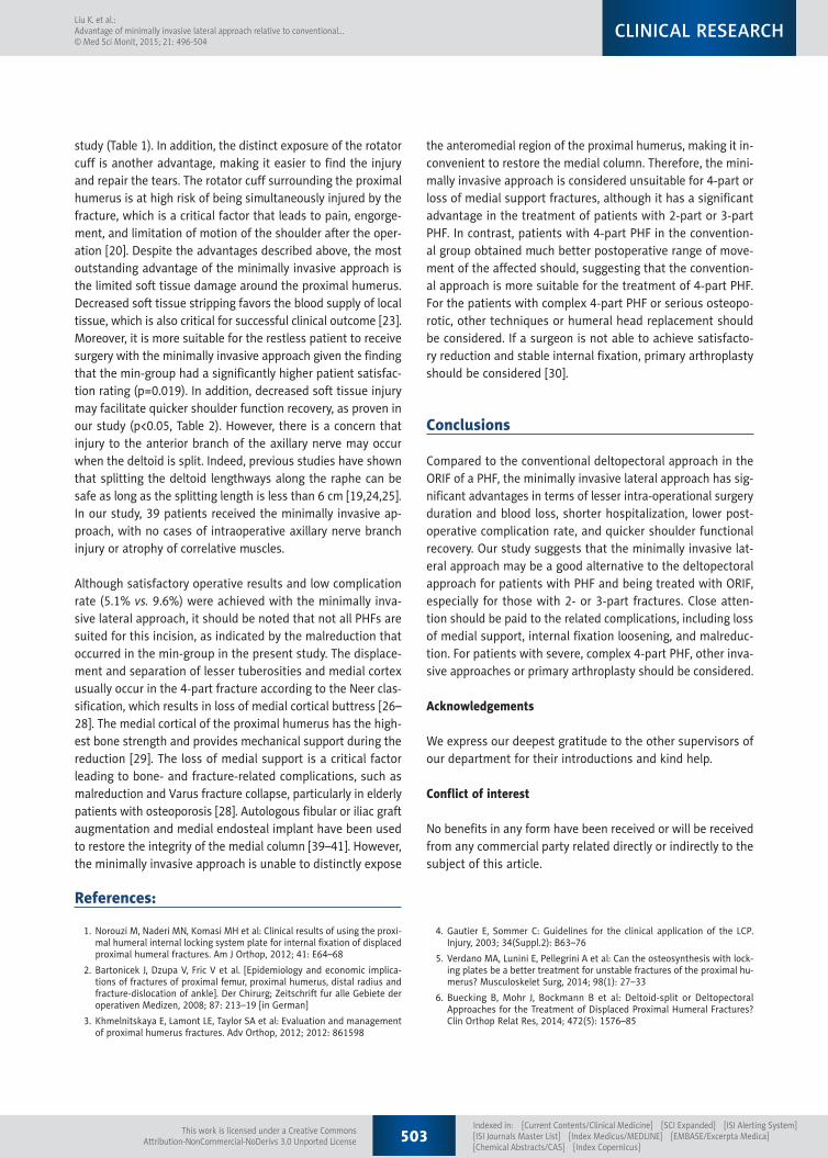
All the operations were performed by professor Wu X either with deltopectoral approach or with subacromial minimally invasive lateral approach. Patients were divided into 2 differ-ent groups based on their incisions, the min-group (minimal-ly invasive surgery group) and the conventional group. Both groups of patients were placed in a beach-chair position with the affected extremity draped free and the image intensifier included in the sterile surgical field. In the conventional group, a standard deltopectoral incision (Figure 1A) starting from the
497Indexed in: [Current Contents/Clinical Medicine] [SCI Expanded] [ISI Alerting System] [ISI Journals Master List] [Index Medicus/MEDLINE] [EMBASE/Excerpta Medica] [Chemical Abstracts/CAS] [Index Copernicus]
Liu K. et al.: Advantage of minimally invasive lateral approach relative to conventional…© Med Sci Monit, 2015; 21: 496-504
CLINICAL RESEARCH
This work is licensed under a Creative CommonsAttribution-NonCommercial-NoDerivs 3.0 Unported License

lateral edge of the coracoid and 8–10 cm length along the del-topectoral groove was performed. The definitive operative pro-cedure has been well described previously [10,14,6].
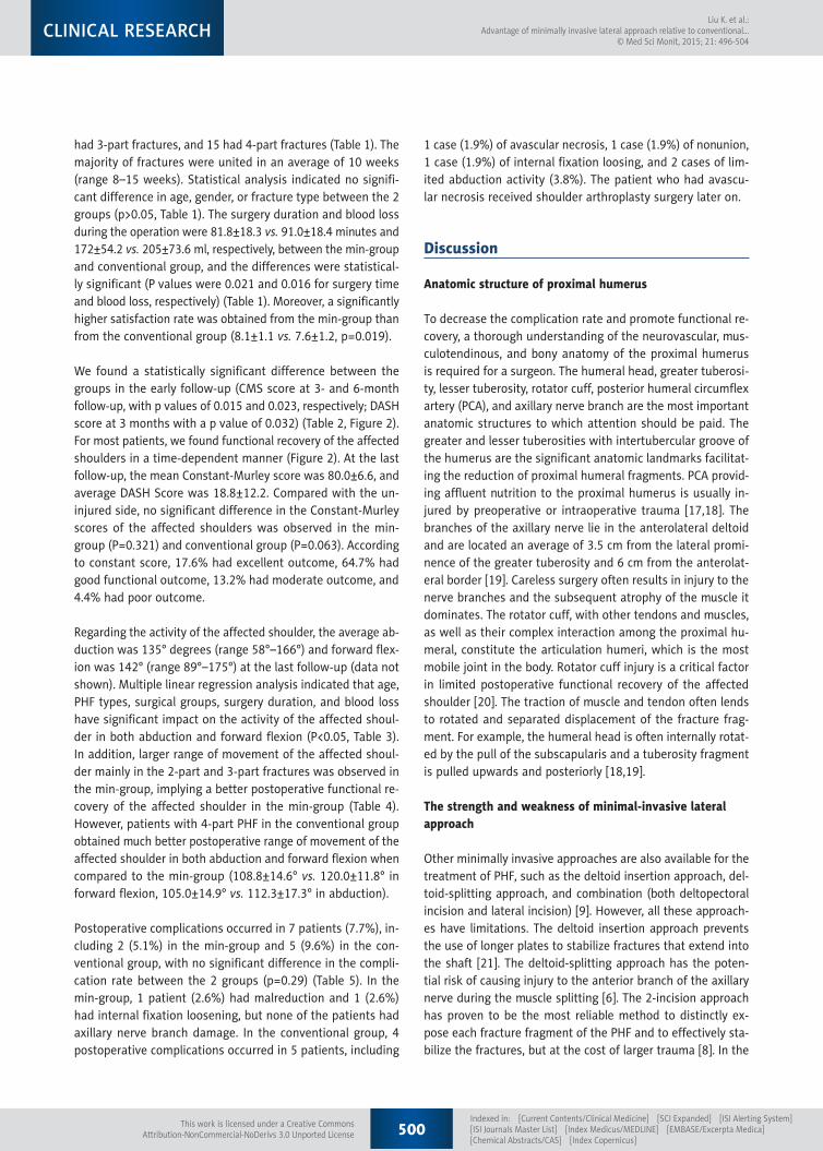
In the min-group, subacromial lateral laterigrade incision was used as an invasive approach, and the locking plan and inci-sion are shown in Figure 1B. The crosscut incision was 4–5 cm in length and 1 finger below the acromion. Muscle fiber was split bluntly and carefully along the deltoid, and excessive split-ting of the deltoid muscle was avoided to protect the anterior motor branch of the axillary nerve from injury. A longitudinal
split was made in the deltoid and was retracted to identify the subdeltoid bursa, and the greater tuberosity and humeral fracture sites were identified. Anatomic reduction was max-imally achieved via manipulation of pulling traction and le-verage using a periosteal elevator. Kirschner wires were also used to facilitate reduction and to temporarily fix the fracture. After reduction and fixation were verified with C-arm image-intensified fluoroscopy, an appropriate-length locking plate (Synthes, Johnson & Johnson, USA) was safely inserted along the submuscular tunnel to prevent the axillary nerve from being
A
B
C
D
E
Figure 1. The different surgical incision, locking plate placement, and the internal fixation during the operation between the 2 groups. (A) The conventional deltopectoral approach. (B) The lateral minimally invasive approach. (C) The placement of a locking plate through the minimally invasive approach. (D) The anatomic reduction, plate position, fixation, and screw length were verified as satisfactory under the C-arm image-intensified fluorocopy before the operation was finished. (E) Suture of the incision after the operation.
498Indexed in: [Current Contents/Clinical Medicine] [SCI Expanded] [ISI Alerting System] [ISI Journals Master List] [Index Medicus/MEDLINE] [EMBASE/Excerpta Medica] [Chemical Abstracts/CAS] [Index Copernicus]
Liu K. et al.: Advantage of minimally invasive lateral approach relative to conventional…
© Med Sci Monit, 2015; 21: 496-504CLINICAL RESEARCH
This work is licensed under a Creative CommonsAttribution-NonCommercial-NoDerivs 3.0 Unported License
trapped under the plate (Figure 1C). The plate was placed lat-erally between the greater and lesser tuberosity, and proxi-mally below the apex of the greater tuberosity. Another lon-gitudinal incision below the branch of the axillary nerve was made to visualize the distal part of the plate. Multiple-angle locking screws were used to fix the distal part of the fracture. C-arm image-intensified fluoroscopy was used again to veri-fy the reduction, plate position, and screw length (Figure 1D). The incision was closed after irrigating the wound and hemo-stasis was achieved (Figure 1E).
Physiotherapy was initiated within the first week post-surgery, which is important for the recovery of shoulder function. We requested all the patients to flex their elbow joint to 90°and to use a sling for 3–4 weeks. Passive pendulum exercise of the affected shoulder was initiated 2–3 days after the opera-tion, and the range of activity was gradually increased. After 2 weeks, positive shoulder exercise was executed and much higher strength of exercise, including superduction, abduc-tion, post-stretch, and ante-flexion, was initiated. The time and method of all the postoperative exercises were strictly moni-tored by a physiotherapist.
Intraoperative and postoperative assessment method
The surgery duration and amount of bleeding during the opera-tion were recorded. Patients were followed up radiographically and clinically with a detailed clinical evaluation and shoulder function assessed during the visit at 3, 6, 12, and 24 months post-surgery, respectively. All the patients were examined by the same surgeon in a blinded manner. At each follow-up, an-teroposterior and lateral view radiographies were performed to monitor fracture healing, and the recovery of shoulder function
was evaluated by measuring the shoulder functional score and range of motion of the affected shoulder. The Constant-Murley score (CMS) [15], graded as poor (0–64), moderate (65–74), good (75–85), or excellent (86–100), was used to evaluate the functional outcome of the affected shoulder in compari-son with the uninjured contralateral joint. A questionnaire on disabilities of arm, shoulder, and hand (DASH) [16] score was used to evaluate any limitations in the activities of daily living. The satisfaction of the patients associated with pain, cosmet-ic appearance, and ability to return to work, as well as over-all outcome of the affected shoulder after the operation, were investigated and graded as unsatisfied (0–6), satisfied (6–8), or very satisfied (8–10). Complications related to the opera-tion were recorded and analyzed at the time they occurred.
Statistical analysis
SPSS 11.0 for Windows (SPSS Inc., Chicago, IL, USA) was used for statistical analysis. The 2 independent-sample T test, chi-square test, and Mann-Whitney U test were used for examin-ing the statistical difference in preoperative and postoperative evaluation between the 2 groups. Multiple linear regression analysis was used to evaluate the strength of associations be-tween surgical approaches and clinical end result. The level of significance was set at 0.05.
Results
We analyzed 91 patients with PHF who met the inclusion cri-teria. These patients were divided into 2 groups: 39 in the min-group and 52 in the conventional group. According to the Neer classification [13], 33 patients had 2-part fractures, 43
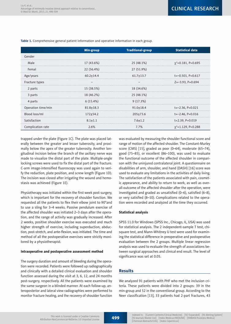
Min-group Traditional-group Statistical data
Gender
Male 17 (43.6%) 25 (48.1%) c2=0.181, P=0.695
Femal 22 (56.4%) 27 (51.9%)
Age/years 60.2±14.4 61.7±13.7 t=–0.501, P=0.617
Fracture types – – Z=–3.93, P=0.694
2 parts 15 (38.5%) 18 (34.6%)
3 parts 18 (46.2%) 25 (48.1%)
4 parts 6 (15.4%) 9 (17.3%)
Operation time/min 81.8±18.3 91.0±18.4 t=–2.36, P=0.021
Blood loss/ml 172±54.2 205±73.6 t=–2.46, P=0.016
Satisfaction 8.1±1.1 7.6±1.2 t=2.39, P=0.019
Complication rate 2.6% 7.7% c2=1.129, P=0.288
Table 1. Comprehensive general patient information and operative information in each group.
499Indexed in: [Current Contents/Clinical Medicine] [SCI Expanded] [ISI Alerting System] [ISI Journals Master List] [Index Medicus/MEDLINE] [EMBASE/Excerpta Medica] [Chemical Abstracts/CAS] [Index Copernicus]
Liu K. et al.: Advantage of minimally invasive lateral approach relative to conventional…© Med Sci Monit, 2015; 21: 496-504
CLINICAL RESEARCH
This work is licensed under a Creative CommonsAttribution-NonCommercial-NoDerivs 3.0 Unported License
had 3-part fractures, and 15 had 4-part fractures (Table 1). The majority of fractures were united in an average of 10 weeks (range 8–15 weeks). Statistical analysis indicated no signifi-cant difference in age, gender, or fracture type between the 2 groups (p>0.05, Table 1). The surgery duration and blood loss during the operation were 81.8±18.3 vs. 91.0±18.4 minutes and 172±54.2 vs. 205±73.6 ml, respectively, between the min-group and conventional group, and the differences were statistical-ly significant (P values were 0.021 and 0.016 for surgery time and blood loss, respectively) (Table 1). Moreover, a significantly higher satisfaction rate was obtained from the min-group than from the conventional group (8.1±1.1 vs. 7.6±1.2, p=0.019).
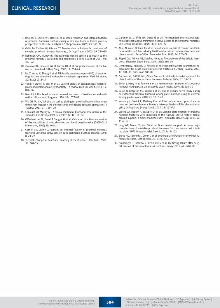
We found a statistically significant difference between the groups in the early follow-up (CMS score at 3- and 6-month follow-up, with p values of 0.015 and 0.023, respectively; DASH score at 3 months with a p value of 0.032) (Table 2, Figure 2). For most patients, we found functional recovery of the affected shoulders in a time-dependent manner (Figure 2). At the last follow-up, the mean Constant-Murley score was 80.0±6.6, and average DASH Score was 18.8±12.2. Compared with the un-injured side, no significant difference in the Constant-Murley scores of the affected shoulders was observed in the min-group (P=0.321) and conventional group (P=0.063). According to constant score, 17.6% had excellent outcome, 64.7% had good functional outcome, 13.2% had moderate outcome, and 4.4% had poor outcome.
Regarding the activity of the affected shoulder, the average ab-duction was 135° degrees (range 58°–166°) and forward flex-ion was 142° (range 89°–175°) at the last follow-up (data not shown). Multiple linear regression analysis indicated that age, PHF types, surgical groups, surgery duration, and blood loss have significant impact on the activity of the affected shoul-der in both abduction and forward flexion (P<0.05, Table 3). In addition, larger range of movement of the affected shoul-der mainly in the 2-part and 3-part fractures was observed in the min-group, implying a better postoperative functional re-covery of the affected shoulder in the min-group (Table 4). However, patients with 4-part PHF in the conventional group obtained much better postoperative range of movement of the affected shoulder in both abduction and forward flexion when compared to the min-group (108.8±14.6° vs. 120.0±11.8° in forward flexion, 105.0±14.9° vs. 112.3±17.3° in abduction).
Postoperative complications occurred in 7 patients (7.7%), in-cluding 2 (5.1%) in the min-group and 5 (9.6%) in the con-ventional group, with no significant difference in the compli-cation rate between the 2 groups (p=0.29) (Table 5). In the min-group, 1 patient (2.6%) had malreduction and 1 (2.6%) had internal fixation loosening, but none of the patients had axillary nerve branch damage. In the conventional group, 4 postoperative complications occurred in 5 patients, including
1 case (1.9%) of avascular necrosis, 1 case (1.9%) of nonunion, 1 case (1.9%) of internal fixation loosing, and 2 cases of lim-ited abduction activity (3.8%). The patient who had avascu-lar necrosis received shoulder arthroplasty surgery later on.
Discussion
Anatomic structure of proximal humerus
To decrease the complication rate and promote functional re-covery, a thorough understanding of the neurovascular, mus-culotendinous, and bony anatomy of the proximal humerus is required for a surgeon. The humeral head, greater tuberosi-ty, lesser tuberosity, rotator cuff, posterior humeral circumflex artery (PCA), and axillary nerve branch are the most important anatomic structures to which attention should be paid. The greater and lesser tuberosities with intertubercular groove of the humerus are the significant anatomic landmarks facilitat-ing the reduction of proximal humeral fragments. PCA provid-ing affluent nutrition to the proximal humerus is usually in-jured by preoperative or intraoperative trauma [17,18]. The branches of the axillary nerve lie in the anterolateral deltoid and are located an average of 3.5 cm from the lateral promi-nence of the greater tuberosity and 6 cm from the anterolat-eral border [19]. Careless surgery often results in injury to the nerve branches and the subsequent atrophy of the muscle it dominates. The rotator cuff, with other tendons and muscles, as well as their complex interaction among the proximal hu-meral, constitute the articulation humeri, which is the most mobile joint in the body. Rotator cuff injury is a critical factor in limited postoperative functional recovery of the affected shoulder [20]. The traction of muscle and tendon often lends to rotated and separated displacement of the fracture frag-ment. For example, the humeral head is often internally rotat-ed by the pull of the subscapularis and a tuberosity fragment is pulled upwards and posteriorly [18,19].
The strength and weakness of minimal-invasive lateral approach
Other minimally invasive approaches are also available for the treatment of PHF, such as the deltoid insertion approach, del-toid-splitting approach, and combination (both deltopectoral incision and lateral incision) [9]. However, all these approach-es have limitations. The deltoid insertion approach prevents the use of longer plates to stabilize fractures that extend into the shaft [21]. The deltoid-splitting approach has the poten-tial risk of causing injury to the anterior branch of the axillary nerve during the muscle splitting [6]. The 2-incision approach has proven to be the most reliable method to distinctly ex-pose each fracture fragment of the PHF and to effectively sta-bilize the fractures, but at the cost of larger trauma [8]. In the
500Indexed in: [Current Contents/Clinical Medicine] [SCI Expanded] [ISI Alerting System] [ISI Journals Master List] [Index Medicus/MEDLINE] [EMBASE/Excerpta Medica] [Chemical Abstracts/CAS] [Index Copernicus]
Liu K. et al.: Advantage of minimally invasive lateral approach relative to conventional…
© Med Sci Monit, 2015; 21: 496-504CLINICAL RESEARCH
This work is licensed under a Creative CommonsAttribution-NonCommercial-NoDerivs 3.0 Unported License
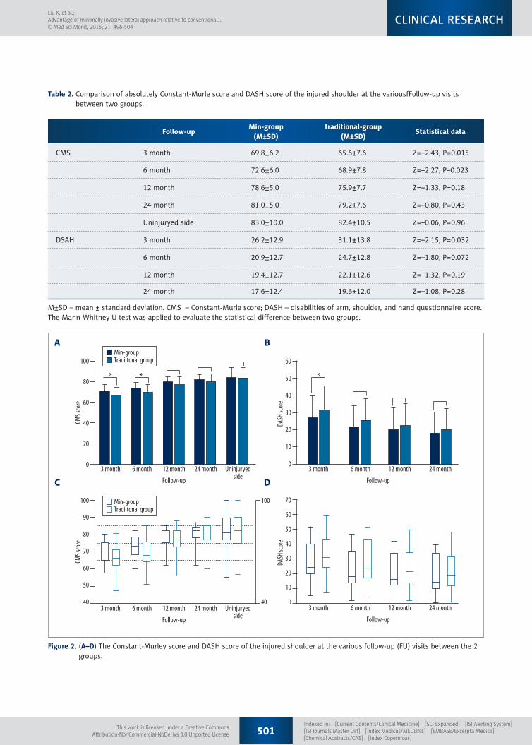
Follow-upMin-group
(M±SD)traditional-group
(M±SD)Statistical data
CMS 3 month 69.8±6.2 65.6±7.6 Z=–2.43, P=0.015
6 month 72.6±6.0 68.9±7.8 Z=–2.27, P–0.023
12 month 78.6±5.0 75.9±7.7 Z=–1.33, P=0.18
24 month 81.0±5.0 79.2±7.6 Z=–0.80, P=0.43
Uninjuryed side 83.0±10.0 82.4±10.5 Z=–0.06, P=0.96
DSAH 3 month 26.2±12.9 31.1±13.8 Z=–2.15, P=0.032
6 month 20.9±12.7 24.7±12.8 Z=–1.80, P=0.072
12 month 19.4±12.7 22.1±12.6 Z=–1.32, P=0.19
24 month 17.6±12.4 19.6±12.0 Z=–1.08, P=0.28
Table 2. Comparison of absolutely Constant-Murle score and DASH score of the injured shoulder at the variousfFollow-up visits between two groups.
M±SD – mean ± standard deviation. CMS – Constant-Murle score; DASH – disabilities of arm, shoulder, and hand questionnaire score. The Mann-Whitney U test was applied to evaluate the statistical difference between two groups.
100
80
60
40
20
0
Min-groupTradiitonal group
Min-groupTradiitonal group
3 month 6 month 12 month
Follow-up
24 month Uninjuryedside
* *
CMS s
core
100
90
80
70
60
50
40
100
403 month 6 month 12 month
Follow-up
24 month Uninjuryedside
CMS s
core
60
50
40
30
20
10
03 month 6 month 12 month
Follow-up
24 month
*
DASH
scor
e
70
60
50
40
30
20
10
03 month 6 month 12 month
Follow-up
24 month
DASH
scor
e
A
C
B
D
Figure 2. (A–D) The Constant-Murley score and DASH score of the injured shoulder at the various follow-up (FU) visits between the 2 groups.
501Indexed in: [Current Contents/Clinical Medicine] [SCI Expanded] [ISI Alerting System] [ISI Journals Master List] [Index Medicus/MEDLINE] [EMBASE/Excerpta Medica] [Chemical Abstracts/CAS] [Index Copernicus]
Liu K. et al.: Advantage of minimally invasive lateral approach relative to conventional…© Med Sci Monit, 2015; 21: 496-504
CLINICAL RESEARCH
This work is licensed under a Creative CommonsAttribution-NonCommercial-NoDerivs 3.0 Unported License
present study, a special minimally invasive lateral approach was applied. An average of 1-finger lower than the subacromi-al and 4–5-cm length laterigrade incision was made to cut-off the skin and the subcutaneous tissues, so as to expose del-toid muscles, but the muscle fiber was split lengthways along the deltoid. This technique was designed to facilitate the ex-tension of the incision in case the fracture is too complex to re-duce and fix. Moreover, the lateral incision is able to distinctly expose laterally fractured structures, especially the displaced
greater tuberosity, thereby facilitating reduction and fixation with a locking plate. Anatomic reduction of the greater tuber-osity is important for the recovery of shoulder function [22]. As the locking plate is positioned on the greater tuberosity and lat-eral proximal humeral, this technique offers direct access to the laterally fractured planes for the placement of a locking plate, which is considered a difficult procedure with the deltopectoral approach [8]. Explicit exposure and convenient surgery result in decreased surgery duration and blood loss, as shown in our
Gender Age Surgery time Blood loss Groups Fracture types
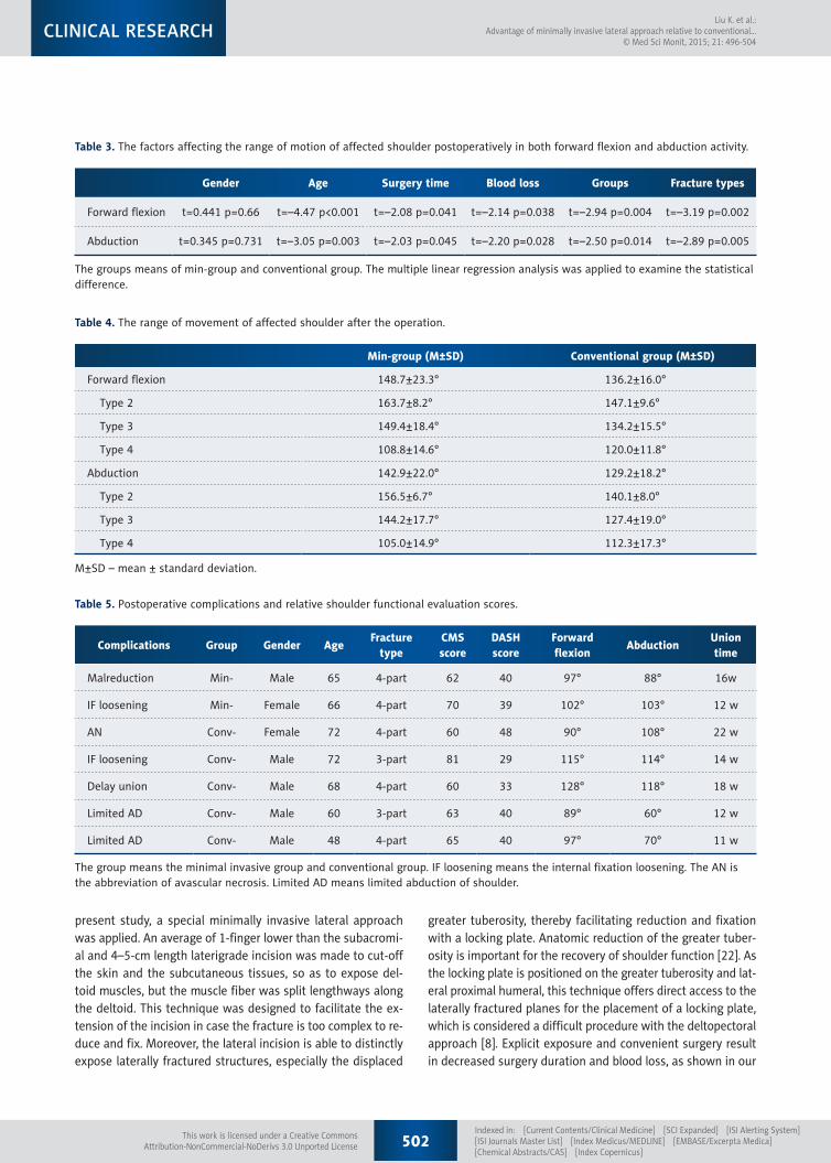
Forward flexion t=0.441 p=0.66 t=–4.47 p<0.001 t=–2.08 p=0.041 t=–2.14 p=0.038 t=–2.94 p=0.004 t=–3.19 p=0.002
Abduction t=0.345 p=0.731 t=–3.05 p=0.003 t=–2.03 p=0.045 t=–2.20 p=0.028 t=–2.50 p=0.014 t=–2.89 p=0.005
The groups means of min-group and conventional group. The multiple linear regression analysis was applied to examine the statistical difference.
Table 3. The factors affecting the range of motion of affected shoulder postoperatively in both forward flexion and abduction activity.
Min-group (M±SD) Conventional group (M±SD)
Forward flexion 148.7±23.3° 136.2±16.0°
Type 2 163.7±8.2° 147.1±9.6°
Type 3 149.4±18.4° 134.2±15.5°
Type 4 108.8±14.6° 120.0±11.8°
Abduction 142.9±22.0° 129.2±18.2°
Type 2 156.5±6.7° 140.1±8.0°
Type 3 144.2±17.7° 127.4±19.0°
Type 4 105.0±14.9° 112.3±17.3°
Table 4. The range of movement of affected shoulder after the operation.
M±SD – mean ± standard deviation.
Complications Group Gender AgeFracture
typeCMSscore
DASHscore
Forwardflexion
AbductionUniontime
Malreduction Min- Male 65 4-part 62 40 97° 88° 16w
IF loosening Min- Female 66 4-part 70 39 102° 103° 12 w
AN Conv- Female 72 4-part 60 48 90° 108° 22 w
IF loosening Conv- Male 72 3-part 81 29 115° 114° 14 w
Delay union Conv- Male 68 4-part 60 33 128° 118° 18 w
Limited AD Conv- Male 60 3-part 63 40 89° 60° 12 w
Limited AD Conv- Male 48 4-part 65 40 97° 70° 11 w
Table 5. Postoperative complications and relative shoulder functional evaluation scores.
The group means the minimal invasive group and conventional group. IF loosening means the internal fixation loosening. The AN is the abbreviation of avascular necrosis. Limited AD means limited abduction of shoulder.
502Indexed in: [Current Contents/Clinical Medicine] [SCI Expanded] [ISI Alerting System] [ISI Journals Master List] [Index Medicus/MEDLINE] [EMBASE/Excerpta Medica] [Chemical Abstracts/CAS] [Index Copernicus]
Liu K. et al.: Advantage of minimally invasive lateral approach relative to conventional…
© Med Sci Monit, 2015; 21: 496-504CLINICAL RESEARCH
This work is licensed under a Creative CommonsAttribution-NonCommercial-NoDerivs 3.0 Unported License
study (Table 1). In addition, the distinct exposure of the rotator cuff is another advantage, making it easier to find the injury and repair the tears. The rotator cuff surrounding the proximal humerus is at high risk of being simultaneously injured by the fracture, which is a critical factor that leads to pain, engorge-ment, and limitation of motion of the shoulder after the oper-ation [20]. Despite the advantages described above, the most outstanding advantage of the minimally invasive approach is the limited soft tissue damage around the proximal humerus. Decreased soft tissue stripping favors the blood supply of local tissue, which is also critical for successful clinical outcome [23]. Moreover, it is more suitable for the restless patient to receive surgery with the minimally invasive approach given the finding that the min-group had a significantly higher patient satisfac-tion rating (p=0.019). In addition, decreased soft tissue injury may facilitate quicker shoulder function recovery, as proven in our study (p<0.05, Table 2). However, there is a concern that injury to the anterior branch of the axillary nerve may occur when the deltoid is split. Indeed, previous studies have shown that splitting the deltoid lengthways along the raphe can be safe as long as the splitting length is less than 6 cm [19,24,25]. In our study, 39 patients received the minimally invasive ap-proach, with no cases of intraoperative axillary nerve branch injury or atrophy of correlative muscles.
Although satisfactory operative results and low complication rate (5.1% vs. 9.6%) were achieved with the minimally inva-sive lateral approach, it should be noted that not all PHFs are suited for this incision, as indicated by the malreduction that occurred in the min-group in the present study. The displace-ment and separation of lesser tuberosities and medial cortex usually occur in the 4-part fracture according to the Neer clas-sification, which results in loss of medial cortical buttress [26–28]. The medial cortical of the proximal humerus has the high-est bone strength and provides mechanical support during the reduction [29]. The loss of medial support is a critical factor leading to bone- and fracture-related complications, such as malreduction and Varus fracture collapse, particularly in elderly patients with osteoporosis [28]. Autologous fibular or iliac graft augmentation and medial endosteal implant have been used to restore the integrity of the medial column [39–41]. However, the minimally invasive approach is unable to distinctly expose
the anteromedial region of the proximal humerus, making it in-convenient to restore the medial column. Therefore, the mini-mally invasive approach is considered unsuitable for 4-part or loss of medial support fractures, although it has a significant advantage in the treatment of patients with 2-part or 3-part PHF. In contrast, patients with 4-part PHF in the convention-al group obtained much better postoperative range of move-ment of the affected should, suggesting that the convention-al approach is more suitable for the treatment of 4-part PHF. For the patients with complex 4-part PHF or serious osteopo-rotic, other techniques or humeral head replacement should be considered. If a surgeon is not able to achieve satisfacto-ry reduction and stable internal fixation, primary arthroplasty should be considered [30].
Conclusions
Compared to the conventional deltopectoral approach in the ORIF of a PHF, the minimally invasive lateral approach has sig-nificant advantages in terms of lesser intra-operational surgery duration and blood loss, shorter hospitalization, lower post-operative complication rate, and quicker shoulder functional recovery. Our study suggests that the minimally invasive lat-eral approach may be a good alternative to the deltopectoral approach for patients with PHF and being treated with ORIF, especially for those with 2- or 3-part fractures. Close atten-tion should be paid to the related complications, including loss of medial support, internal fixation loosening, and malreduc-tion. For patients with severe, complex 4-part PHF, other inva-sive approaches or primary arthroplasty should be considered.
Acknowledgements
We express our deepest gratitude to the other supervisors of our department for their introductions and kind help.
Conflict of interest
No benefits in any form have been received or will be received from any commercial party related directly or indirectly to the subject of this article.
References:
1. Norouzi M, Naderi MN, Komasi MH et al: Clinical results of using the proxi-mal humeral internal locking system plate for internal fixation of displaced proximal humeral fractures. Am J Orthop, 2012; 41: E64–68
2. Bartonicek J, Dzupa V, Fric V et al. [Epidemiology and economic implica-tions of fractures of proximal femur, proximal humerus, distal radius and fracture-dislocation of ankle]. Der Chirurg; Zeitschrift fur alle Gebiete der operativen Medizen, 2008; 87: 213–19 [in German]
3. Khmelnitskaya E, Lamont LE, Taylor SA et al: Evaluation and management of proximal humerus fractures. Adv Orthop, 2012; 2012: 861598
4. Gautier E, Sommer C: Guidelines for the clinical application of the LCP. Injury, 2003; 34(Suppl.2): B63–76
5. Verdano MA, Lunini E, Pellegrini A et al: Can the osteosynthesis with lock-ing plates be a better treatment for unstable fractures of the proximal hu-merus? Musculoskelet Surg, 2014; 98(1): 27–33
6. Buecking B, Mohr J, Bockmann B et al: Deltoid-split or Deltopectoral Approaches for the Treatment of Displaced Proximal Humeral Fractures? Clin Orthop Relat Res, 2014; 472(5): 1576–85
503Indexed in: [Current Contents/Clinical Medicine] [SCI Expanded] [ISI Alerting System] [ISI Journals Master List] [Index Medicus/MEDLINE] [EMBASE/Excerpta Medica] [Chemical Abstracts/CAS] [Index Copernicus]
Liu K. et al.: Advantage of minimally invasive lateral approach relative to conventional…© Med Sci Monit, 2015; 21: 496-504
CLINICAL RESEARCH
This work is licensed under a Creative CommonsAttribution-NonCommercial-NoDerivs 3.0 Unported License
7. Brunner F, Sommer C, Bahrs C et al: Open reduction and internal fixation of proximal humerus fractures using a proximal humeral locked plate: a prospective multicenter analysis. J Orthop Trauma, 2009; 23: 163–72
8. Gallo RA, Zeiders GJ, Altman GT: Two-incision technique for treatment of complex proximal humerus fractures. J Orthop Trauma, 2005; 19: 734–40
9. Robinson CM, Murray IR: The extended deltoid-splitting approach to the proximal humerus: variations and extensions. J Bone J Surg Br, 2011; 93: 387–92
10. Zlotolow DA, Catalano LW III, Barron OA et al: Surgical exposures of the hu-merus. J Am Acad Orthop Surg, 2006; 14: 754–65
11. Liu Z, Wang K, Zhang K et al: Minimally invasive surgery (MIS) of anterior ring fracture combined with pubic symphysis separation. Med Sci Monit, 2014; 20: 1913–17
12. Yimin Y, Zhiwei R, Wei M et al: Current status of percutaneous vertebro-plasty and percutaneous kyphoplasty – a review. Med Sci Monit, 2013; 19: 826–36
13. Neer CS II: Displaced proximal humeral fractures. I. Classification and eval-uation. J Bone Joint Surg Am, 1970; 52: 1077–89
14. Wu CH, Ma CH, Yeh JJ et al: Locked plating for proximal humeral fractures: differences between the deltopectoral and deltoid-splitting approaches. J Trauma, 2011; 71: 1364–70
15. Constant CR, Murley AH: A clinical method of functional assessment of the shoulder. Clin Orthop Relat Res, 1987; (214): 160–64
16. Offenbaecher M, Ewert T, Sangha O et al: Validation of a German version of the disabilities of arm, shoulder, and hand questionnaire (DASH-G). J Rheumatol, 2002; 29: 401–2
17. Cornell CN, Levine D, Pagnani MJ: Internal fixation of proximal humerus fractures using the screw-tension band technique. J Orthop Trauma, 1994; 8: 23–27
18. Terry GC, Chopp TM: Functional anatomy of the shoulder. J Athl Train, 2000; 35: 248–55
19. Gardner MJ, Griffith MH, Dines JS et al: The extended anterolateral acro-mial approach allows minimally invasive access to the proximal humerus. Clin Orthop Relat Res, 2005; (434): 123–29
20. Aksu N, Aslan O, Kara AN et al: Simultaneous repair of chronic full-thick-ness rotator cuff tears during fixation of proximal humerus fractures and clinical results. Acta Orthop Traumatol Turc, 2010; 44: 173–79
21. Rispoli DM, Athwal GS, Sperling JW et al: The anatomy of the deltoid inser-tion. J Shoulder Elbow Surg, 2009; 18(3): 386–90
22. Demirhan M, Kilicoglu O, Altinel L et al: Prognostic factors in prosthetic re-placement for acute proximal humerus fractures. J Orthop Trauma, 2003; 17: 181–88; discussion 188–89
23. Gardner MJ, Griffith MH, Dines JS et al: A minimally invasive approach for plate fixation of the proximal humerus. Bulletin, 2004; 62: 18–23
24. Smith J, Berry G, Laflamme Y et al: Percutaneous insertion of a proximal humeral locking plate: an anatomic study. Injury, 2007; 38: 206–11
25. Saran N, Bergeron SG, Benoit B et al: Risk of axillary nerve injury during percutaneous proximal humerus locking plate insertion using an external aiming guide. Injury, 2010; 41: 1037–40
26. Kennedy J, Feerick E, McGarry P et al: Effect of calcium triphosphate ce-ment on proximal humeral fracture osteosynthesis: a finite element anal-ysis. J Orthop Surg (Hong Kong), 2013; 21: 167–72
27. Weeks CA, Begum F, Beaupre LA et al: Locking plate fixation of proximal humeral fractures with impaction of the fracture site to restore medial column support: a biomechanical study. J Shoulder Elbow Surg, 2013; 22: 1552–57
28. Jung WB, Moon ES, Kim SK et al: Does medial support decrease major complications of unstable proximal humerus fractures treated with lock-ing plate? BMC Musculoskelet Disord, 2013; 14: 102
29. Burke NG, Kennedy J, Green C et al: Locking plate fixation for proximal hu-merus fractures. Orthopedics, 2012; 35: e250–54
30. Krappinger D, Bizzotto N, Riedmann S et al: Predicting failure after surgi-cal fixation of proximal humerus fractures. Injury, 2011; 42: 1283–88
504Indexed in: [Current Contents/Clinical Medicine] [SCI Expanded] [ISI Alerting System] [ISI Journals Master List] [Index Medicus/MEDLINE] [EMBASE/Excerpta Medica] [Chemical Abstracts/CAS] [Index Copernicus]
Liu K. et al.: Advantage of minimally invasive lateral approach relative to conventional…
© Med Sci Monit, 2015; 21: 496-504CLINICAL RESEARCH
This work is licensed under a Creative CommonsAttribution-NonCommercial-NoDerivs 3.0 Unported License